User login
AGA Clinical Practice Guideline: Coagulation in cirrhosis
A clinical update from the American Gastroenterological Association focuses on bleeding and thrombosis-related questions in patients with cirrhosis. It provides guidance on test strategies for bleeding risk, preprocedure management of bleeding risk, venous thromboembolism (VTE) prophylaxis, screening for portal vein thrombosis (PVT), and anticoagulation therapies. It is aimed at primary care providers, gastroenterologists, and hepatologists, among other health care providers.
In cirrhosis, there are often changes to platelet (PLT) counts and prothrombin time/international normalized ratio (PT/INR), among other parameters, and historically these changes led to concerns that patients were at greater risk of bleeding or thrombosis. More recent evidence has led to a nuanced view. Neither factor necessarily suggests increased bleeding risk, and the severity of coagulopathy predicted by them does not predict the risk of bleeding complications.
Patients with cirrhosis are at greater risk of thrombosis, but clinicians may be hesitant to prescribe anticoagulants because of uncertain risk profiles, and test strategies employing PT/INR to estimate bleeding risk and track treatment endpoints in patients receiving vitamin K antagonists may not work in cirrhosis patients with alterations in procoagulant and anticoagulant measures. Recent efforts to address this led to testing of fibrin clot formation and lysis to better gauge the variety of abnormalities in cirrhosis patients.
The guideline, published in Gastroenterology, was informed by a technical review that focused on both bleeding-related and thrombosis-related questions. Bleeding-related questions included testing strategies and preprocedure prophylaxis to reduce bleeding risk. Thrombosis-related questions included whether VTE prophylaxis may be useful in hospitalized patients with cirrhosis, whether patients should be screened for PVT, potential therapies for nontumoral PVT, and whether or not anticoagulation is safe and effective when atrial fibrillation is present alongside cirrhosis.
Because of a lack of evidence, the guideline provides no recommendations on visco-elastic testing for bleeding risk in advance of common gastrointestinal procedures for patients with stable cirrhosis. It recommends against use of extensive preprocedural testing, such as repeated PT/INR or PLT count testing.
The guideline also looked at whether preprocedural efforts to correct coagulation parameters could reduce bleeding risk in patients with cirrhosis. It recommends against giving blood products ahead of the procedure for patients with stable cirrhosis without severe thrombocytopenia or severe coagulopathy. Such interventions can be considered for patients in the latter categories who are undergoing procedures with high bleeding risk after consideration of risks and benefits, and consultation with a hematologist.
Thrombopoietin receptor agonists (TPO-RAs) are also not recommended in patients with thrombocytopenia and stable cirrhosis undergoing common procedures, but they can be considered for patients who are more concerned about reduction of bleeding events and less concerned about the risk of PVT.
Patients who are hospitalized and meet the requirements should receive VTE prophylaxis. Although there is little available evidence about the effects of thromboprophylaxis in patients with cirrhosis, there is strong evidence of benefit in acutely ill hospitalized patients, and patients with cirrhosis are believed to be at a similar risk of VTE. There is evidence of increased bleed risk, but this is of very low certainty.
PVT should not be routinely tested for, but such testing can be offered to patients with a high level of concern over PVT and are not as worried about potential harms of treatment. This recommendation does not apply to patients waiting for a liver transplant.
Patients with non-umoral PVT should receive anticoagulation therapy, but patients who have high levels of concern about bleeding risk from anticoagulation and put a lower value on possible benefits of anticoagulation may choose not to receive it.
The guideline recommends anticoagulation for patients with atrial fibrillation and cirrhosis who are indicated for it. Patients with more concern about the bleeding risk of anticoagulation and place lower value on the reduction in stroke risk may choose to not receive anticoagulation. This is particularly true for those with more advanced cirrhosis (Child-Turcotte-Pugh Class C) and/or low CHA2DS2-VASC scores.
Nearly all of the recommendations in the guideline are conditional, reflecting a lack of data and a range of knowledge gaps that need filling. The authors call for additional research to identify specific patients who are at high risk for bleeding or thrombosis “to appropriately provide prophylaxis using blood product transfusion or TPO-RAs in patients at risk for clinically significant bleeding, to screen for and treat PVT, and to prevent clinically significant thromboembolic events.”
The development of the guideline was funded fully by the AGA. Members of the panel submitted conflict of interest information, and these statements are maintained at AGA headquarters.
FROM GASTROENTEROLOGY
Noninvasive ELF test identifies risk of advanced fibrosis in NAFLD
A noninvasive enhanced liver fibrosis (ELF) blood test identifies patients with nonalcoholic fatty liver disease (NAFLD) who are at increased risk of advanced fibrosis, according to a new study.
According to the study researchers, when combined with the fibrosis-4 (FIB-4) score the ELF test may be a reliable method that can assess for advanced fibrosis in clinical practice.
Despite the utility of identifying advanced fibrosis in the NAFLD population, significant barriers exist to “risk stratifying patients in clinical practice owing to the need for liver biopsy,” wrote study authors Zobair M. Younossi, MD, of the Inova Health System in Falls Church, Va., and colleagues in JAMA Network Open.
“NASH [nonalcoholic steatohepatitis] can only be diagnosed by biopsy and liver pathology yet validated noninvasive tests that accurately diagnose NASH don’t exist,” said Dr. Younossi in an interview. “Developing noninvasive tests to accurately risk stratify patients with significant fibrosis in NASH is highly desirable in clinical practice. A blood test such as ELF can open the opportunity to order this test anywhere in the country.”
The ELF test reflects extracellular matrix metabolism as opposed to tests that assess alterations in hepatic function. The study authors explain that the noninvasive ELF test is a “blood-derived panel of biomarkers consisting of three components: type III procollagen peptide, hyaluronic acid, and tissue inhibitor of metalloproteinase-1.”
To gain a further understanding of the role of ELF in predicting the risk of nonalcoholic steatohepatitis (NASH) in NAFLD, Dr. Younossi and colleagues performed a retrospective, cross-sectional analysis of outpatients within a community-based liver clinic from 2001 in 2020. The study cohort included 829 patients (mean age, 53.1 years) with NAFLD, which was characterized by steatosis greater than 5% without any other liver disease or excessive alcohol use.
In the overall study population, the mean FIB-4 score was 1.34. In the 463 patients with liver biopsy, approximately 24.4% presented with bridging fibrosis or cirrhosis. A total of 79 (17.1%) of the 462 patients with transient elastography data presented with liver stiffness results of 9.6 kPa or greater, which according to the researchers was indicative of advanced fibrosis.
Biopsy determined that those with advanced fibrosis in the study had significantly increased ELF scores versus patients without advanced fibrosis (10.1 vs. 8.6, respectively; P <.001). Moreover, patients with advanced fibrosis had significantly greater liver stiffness as determined by transient elastography (10.0 vs. 9.0; P <.001).
In the NAFLD population, the ELF test demonstrated excellent performance in identifying patients with advanced fibrosis, as reflected by an area under the receiver operating characteristic curve of 0.81 (95% confidence interval, 0.77-0.85) for those whose fibrosis was diagnosed by biopsy as well as 0.79 (95% CI, 0.75-0.82) for fibrosis diagnosed by transient elastography. Similar performances of the ELF score were reported among those with NAFLD who were aged 65 years or older (AUROC, 0.74; 95% CI, 0.58-0.87) or patients who had type 2 diabetes (AUROC, 0.78; 95% CI, 0.71-0.84).
The researchers regarded the combination of an ELF score of 7.2 or greater with a FIB-4 score of 0.74 or greater, as indicative of a negative predictive value of 95.1% (95% CI, 91.8%-98.4%) and a sensitivity of 92.5% (95% CI, 87.4%-97.5%). According to the investigators, these values “can reliably rule out advanced fibrosis.”
Additionally, the combination of an ELF score of 9.8 or greater with a FIB-4 score of 2.9 or greater, was associated with a positive predictive value of 95.0% (95% CI, 85.5%-100%) and a specificity of 99.7% (95% CI, 99.1%-100%), suggesting this combination can conversely “be used to rule in advanced fibrosis,” the researchers wrote.
Serologic approaches for predicting the risk of NASH are more widely available and “easier” to use than radiologic approaches, but the former may not be as reliable or accurate as the latter, explained Tibor Krisko, MD, a gastroenterologist at Weill Cornell Medicine and New York-Presbyterian, in an interview. “The choice of which serologic test is utilized is often guided by what is available in a region/practice, what the provider is familiar with, and what a given patient’s insurance covers,” said Dr. Krisko, who wasn’t involved in the study.
“The study by Dr. Younossi et al. perhaps confirms that ELF alone is unlikely to be the future standard of care, and the authors weave this to a conclusion and strength, highlighting that the combination had excellent accuracy,” commented Dr. Krisko. “This is an exciting and important area of research and clinical practice advancement, but all of these serological tests have limitations, such as their lack in liver specificity, their risk of being affected by clearance rate, and the fact that they are not biomarkers but rather surrogate markers.”
Therefore, added Dr. Krisko, clinicians should continue to consider each patient’s clinical picture carefully and utilize radiographic methods liberally, particularly when serologic results are deemed ambiguous.
Dr. Younossi reported conflicts of interest with several pharmaceutical companies. No funding was reported for the study. Dr. Krisko had no relevant conflicts to disclose.
FROM JAMA NETWORK OPEN
Drink up: Large study confirms coffee beneficial to liver health
Drinking more than three cups of caffeinated coffee a day is associated with less liver stiffness, according to an analysis of a nationally representative survey, which was recently published in Clinical Gastroenterology and Hepatology.

The study is likely the most rigorous look to date on the benefits of coffee on liver health in the U.S. It was based on data from the National Health and Nutrition Examination Survey (NHANES), in which participants were asked about what they eat and drink. Crucially, in 2017, NHANES began to include elastography (FibroScan), of participants’ liver stiffness, not because of suspected problems with the liver but as across-the-board evaluations of all participants.
“Because it’s an unselected population for FibroScan and because of the detail, the granularity, the richness of the information from the nutritional surveys that they do, this is the closest we’re ever going to get to a linkage between what people are eating or drinking and the health of their liver, absent a longitudinal study where we set out to follow people for many, many years,” said Elliot Tapper, MD, assistant professor of gastroenterology at the University of Michigan, Ann Arbor, and the study’s senior author.
Researchers examined data from about 4,500 patients who had participated in the NHANES study in 2017-2018. The participants were aged 20 years or older, with an average age of 48; 73% were overweight, about the national average.
The researchers found no association between coffee consumption and controlled attenuation parameter (CAP), a measure of fatty liver. But they found a link between coffee and liver stiffness.
Those who drank more than three cups of coffee daily had a liver stiffness measure (LSM) that was 0.9 kilopascals (kPa) lower than others (P = .03). Drinking more than three cups a day also was found to be protective against an LSM of 9.5 kPa or higher, the threshold for advanced liver fibrosis (OR, 0.4; P = .05). Decaffeinated coffee was not found to be associated with LSM.
Caffeine is an antagonist to adenosine receptors in the liver cell that, if blocked, stops the production of scar tissue, according to the researchers. But when they looked at estimated caffeine consumption, calculated through the detailed, trained interviews performed by nutritionists, there was no association with liver stiffness. That said, Dr. Tapper noted that this could be due to the imperfection of making those estimations.
“If we had to hypothesize about a mechanism, it would most likely be caffeine, and the reason we couldn’t see that here is because these are estimated milligrams of caffeine per coffee – but the way that we brew coffee, and the beans that we’re using, are so highly variable it just can’t be captured in this kind of database,” he said.
He said the data will be reassuring to clinicians who suggest coffee-drinking to patients.
“There are hepatologists around the world who are actively recommending coffee – they’ll feel empowered by these data,” he said. “I would still like to see more robust longitudinal data before I start spending our precious time counseling patients about coffee. There are many other data-driven interventions for the management of liver disease that we should be focusing our time on.”
Moreover, he said that the data will be important for patients who are particularly interested in natural remedies.
“For patients who are very interested in a natural supplement, to feel like they’re taking an active role in the health of their liver, I will tell them to avoid carbohydrates and increase their exercise – and that it is OK to add coffee to their daily routine.”
A study based on a UK database found that coffee was associated with protection against chronic liver disease, but the association was seen for both caffeinated and decaffeinated drinks, noted Nathan Davies, PhD, professor of biochemistry at the Institute of the Liver and Digestive Health at the University College London.
Dr. Davies, a registered nutritionist who has studied coffee’s effects on the liver, said that while including elastography in the Michigan study is interesting, it “does not necessarily by itself add greatly” to the evidence base.
The outcomes from both studies do suggest a positive effect for coffee, but he said it’s important to remember that liver disease develops over years and decades.
“Looking at a snapshot moment does not necessarily reflect an individual’s behavior during the onset and development of their condition,” he said. “As such, there are a number of behavioral and nutritional factors that could be contributing to the observed effect over a period of years.”
He pointed out that while different coffee and brewing types affect the amount of caffeine in a cup, all cups of coffee in this study were treated the same way. He noted there was no apparent dose-dependent effect, which would have been expected if there is an active ingredient that affects liver stiffness.
“In general, my advice is to improve diet, take more exercise, and reduce alcohol consumption, which is likely to be more effective in preventing liver disease – and its progression – than drinking an extra cup of coffee,” Dr. Davies said. “That being said, for patients at increased risk for liver disease who currently drink three cups or more of coffee daily, it may be prudent for them to continue because this level of consumption might be actively lowering their chances of developing more serious disease.”
Dr. Tapper has done consulting for Novartis, Axcella and Allergan, has served on advisory boards for Mallinckrodt, Bausch Health, Kaleido, and Novo Nordisk, and has unrestricted research grants from Gilead and Valeant. The remaining authors disclose no conflicts. Dr. Davies reported no relevant disclosures.
Drinking more than three cups of caffeinated coffee a day is associated with less liver stiffness, according to an analysis of a nationally representative survey, which was recently published in Clinical Gastroenterology and Hepatology.
The study is likely the most rigorous look to date on the benefits of coffee on liver health in the U.S. It was based on data from the National Health and Nutrition Examination Survey (NHANES), in which participants were asked about what they eat and drink. Crucially, in 2017, NHANES began to include elastography (FibroScan), of participants’ liver stiffness, not because of suspected problems with the liver but as across-the-board evaluations of all participants.
“Because it’s an unselected population for FibroScan and because of the detail, the granularity, the richness of the information from the nutritional surveys that they do, this is the closest we’re ever going to get to a linkage between what people are eating or drinking and the health of their liver, absent a longitudinal study where we set out to follow people for many, many years,” said Elliot Tapper, MD, assistant professor of gastroenterology at the University of Michigan, Ann Arbor, and the study’s senior author.
Researchers examined data from about 4,500 patients who had participated in the NHANES study in 2017-2018. The participants were aged 20 years or older, with an average age of 48; 73% were overweight, about the national average.
The researchers found no association between coffee consumption and controlled attenuation parameter (CAP), a measure of fatty liver. But they found a link between coffee and liver stiffness.
Those who drank more than three cups of coffee daily had a liver stiffness measure (LSM) that was 0.9 kilopascals (kPa) lower than others (P = .03). Drinking more than three cups a day also was found to be protective against an LSM of 9.5 kPa or higher, the threshold for advanced liver fibrosis (OR, 0.4; P = .05). Decaffeinated coffee was not found to be associated with LSM.
Caffeine is an antagonist to adenosine receptors in the liver cell that, if blocked, stops the production of scar tissue, according to the researchers. But when they looked at estimated caffeine consumption, calculated through the detailed, trained interviews performed by nutritionists, there was no association with liver stiffness. That said, Dr. Tapper noted that this could be due to the imperfection of making those estimations.
“If we had to hypothesize about a mechanism, it would most likely be caffeine, and the reason we couldn’t see that here is because these are estimated milligrams of caffeine per coffee – but the way that we brew coffee, and the beans that we’re using, are so highly variable it just can’t be captured in this kind of database,” he said.
He said the data will be reassuring to clinicians who suggest coffee-drinking to patients.
“There are hepatologists around the world who are actively recommending coffee – they’ll feel empowered by these data,” he said. “I would still like to see more robust longitudinal data before I start spending our precious time counseling patients about coffee. There are many other data-driven interventions for the management of liver disease that we should be focusing our time on.”
Moreover, he said that the data will be important for patients who are particularly interested in natural remedies.
“For patients who are very interested in a natural supplement, to feel like they’re taking an active role in the health of their liver, I will tell them to avoid carbohydrates and increase their exercise – and that it is OK to add coffee to their daily routine.”
A study based on a UK database found that coffee was associated with protection against chronic liver disease, but the association was seen for both caffeinated and decaffeinated drinks, noted Nathan Davies, PhD, professor of biochemistry at the Institute of the Liver and Digestive Health at the University College London.
Dr. Davies, a registered nutritionist who has studied coffee’s effects on the liver, said that while including elastography in the Michigan study is interesting, it “does not necessarily by itself add greatly” to the evidence base.
The outcomes from both studies do suggest a positive effect for coffee, but he said it’s important to remember that liver disease develops over years and decades.
“Looking at a snapshot moment does not necessarily reflect an individual’s behavior during the onset and development of their condition,” he said. “As such, there are a number of behavioral and nutritional factors that could be contributing to the observed effect over a period of years.”
He pointed out that while different coffee and brewing types affect the amount of caffeine in a cup, all cups of coffee in this study were treated the same way. He noted there was no apparent dose-dependent effect, which would have been expected if there is an active ingredient that affects liver stiffness.
“In general, my advice is to improve diet, take more exercise, and reduce alcohol consumption, which is likely to be more effective in preventing liver disease – and its progression – than drinking an extra cup of coffee,” Dr. Davies said. “That being said, for patients at increased risk for liver disease who currently drink three cups or more of coffee daily, it may be prudent for them to continue because this level of consumption might be actively lowering their chances of developing more serious disease.”
Dr. Tapper has done consulting for Novartis, Axcella and Allergan, has served on advisory boards for Mallinckrodt, Bausch Health, Kaleido, and Novo Nordisk, and has unrestricted research grants from Gilead and Valeant. The remaining authors disclose no conflicts. Dr. Davies reported no relevant disclosures.
FROM CLINICAL GASTROENTEROLOGY AND HEPATOLOGY
AGA Section: Gastroenterology and hepatology training milestones
Updated milestones for professional development aim to help specialists in gastroenterology and transplant hepatology achieve knowledge, skills, and attitudes that will help them establish their own practices.
The new version, Milestones 2.0, represents the latest milestones created by the Accreditation Council for Graduate Medical Education, including six core competencies developed initially in 1999: Patient care (PC), medical knowledge (MK), interpersonal and communication skills (ICS), professionalism (PROF), systems-based practice (SBP), and practice-based learning and improvement (PBLI).
In 2013, the Oversight Working Network, working with gastroenterology societies, developed a companion document of 13 entrustable professional activities (EPAs) aimed at gastroenterologists: These include management of various individual disorders such as liver or pancreatic diseases, performance of specific diagnostic procedures, and managing patient adverse events and nutritional status.
Milestones 1.0 encountered some resistance from the graduate education community. Too many of the milestones were deemed to be too vague or were described using language that was too complex. Some viewed the milestones as burdensome, and a review suggested hundreds of different ways to describe ICS and PROF, leading to confusion.
In an effort to improve matters, the ACGME made some changes. The first involved standardizing milestones used for ICS, PROF, SBP, and PBLI so that they could be used across disciplines. They also developed PC and MK milestones tailored to each specialty.
In the latest article on the topic, appearing in Gastroenterology, the authors led by Brijen J. Shah, MD, of the Icahn School of Medicine at Mount Sinai, New York, outlined a second group of changes, which included development of specialty-specific milestones aimed at gastroenterology and transplant hepatology.
Development
The new set of milestones includes 17 for gastroenterology and 16 for transplant hepatology.
There are four PC milestones, which include taking a history and conducting patient examinations, patient management, and two more related to cognitive and technical components of procedures. The MK milestones include competency in gastrointestinal and liver diseases (MK1) and medical reasoning (MK2). These milestones are different from the internal medicine milestones met by graduating residents. MK1 includes specialty-specific disorders and diagnostic, therapeutic, and pharmacologic options for treatment or prevention. MK2 encompasses differential diagnoses and how cognitive bias can influence decision-making, a new concept introduced in Milestones 2.0.
Because the skills represented in the four other core milestones (ICS, PROF, SBP, and PBLI) are “common across specialties,” the authors drafted subcompetencies for these four areas with “harmonized” language for use by every specialty. These harmonized milestones were then tailored for each specialty. An important change occurred with SBP because transplant hepatology poses unique challenges in this domain. They ultimately split SBP into two, with SBP1 focusing on unique liver transplant regulatory requirements and SBP2 covering organ allocation and Model for End-Stage Liver Disease (MELD) score exceptions.
Public response
The researchers sought out comment on the updated milestones from program directors and coordinators, and published on the ACGME website, and members of the working group also shared it with faculty, fellows, and specialty societies. Overall, 48 respondents assessed “whether the updated milestone provided a realistic measure of knowledge, skills, and behavior; whether it discriminated between different levels of competency; whether the respondent knew how to assess the milestone effectively; and whether the Supplemental Guide was a useful resource in understanding the milestone.” They rated each on a scale of 1 (strongly disagree) to 4 (strongly agree). They could also provide free-text comments.
Respondents agreed that milestones realistically measure progression (mean, 3.49), could distinguish levels of competency (mean, 3.41), could be used accurately (mean, 3.43), and were explained well by the supplemental guide (mean, 3.42). No trends that suggested a need for additional action were found in the free-text comments.
Role of milestones
The milestones can be used to develop learning objectives, which in turn can be worked into clinical rotations and learning activities. For instance, the inpatient consult rotation could be used to address the SBP2 (organ allocation/MELD score exemptions), SBP3 (the physician’s role in the health care system), PBLI1 (evidence-based medicine), and some of the PC (patient care) milestones.
The milestones should not be used as an assessment method by supervisors, the authors cautioned, but rather should be used by the Clinical Competency Committee to assess trainees at various time points. The committee may combine milestones with direct observation, chart-simulated recall, multiple evaluations, and other factors to determine a trainee’s progress.
An institution’s program directors can use the milestones to adjust curriculum development and ensure that any gaps are filled. Milestones can be used at multiple times throughout training: When trainees repeat rotations, they can be used to determine year-to-year progress. Trainees who are not progressing adequately may be identified earlier on, then offered supplemental learning opportunities. On the other hand, trainees who exceed expectations may be offered additional opportunities.
Trainees can also use milestones in self-directed learning, though they should work with the program director and clinical faculty to identify gaps in their learning as well as any deficiencies.
The authors have no relevant financial disclosures.
FROM GASTROENTEROLOGY
The importance of education and screening for nonalcoholic fatty liver disease
For the past 18 months, we’ve all been focused on defeating the COVID-19 pandemic and preparing for the effects of cancer screenings that were delayed or put off entirely. But COVID isn’t the only epidemic we’re facing in the United States. Obesity is the second leading cause of preventable death in the United States. and its related diseases account for $480.7 billion in direct health care costs, with an additional $1.24 trillion in indirect costs from lost economic productivity.

More than two in five Americans are obese and that number is predicted to grow to more than half of the U.S. population by 2030. Obesity is a risk factor for nonalcoholic fatty liver disease (NAFLD), a buildup of fat in the liver with little or no inflammation or cell damage that affects one in three (30%-37%) of adults in the U.S.
NAFLD can progress to nonalcoholic steatohepatitis (NASH), which affects about 1 in 10 (8%-12%) of adults in the U.S. NASH is fat in the liver with inflammation and cell damage, and it can lead to fibrosis and liver failure. The number of patients we see with NALFD and NASH continues to rise and it’s taking its toll. One in five people who have NASH will have the disease progress to liver cirrhosis. NASH is expected to be the leading cause of liver transplant in the U.S. for the next 5 years.
Stemming the tide of NAFLD and NASH
In terms of diet, limiting sugar and eating a diet rich in vegetables, whole grains, and healthy fats can prevent the factors that lead to liver disease.
If this were easy, we wouldn’t be facing the obesity epidemic that is plaguing the United States. One of the issues is that medicine has only recognized obesity as a disease for less than 10 years. We aren’t trained in medical school, residencies, or fellowships in managing obesity, beyond advising people to exercise and eat right. We know this doesn’t work.
That’s why many independent GI groups are exploring comprehensive weight management programs that take a holistic approach to weight management involving a team of health care providers and educators helping patients gradually exercise more and eat healthy while providing a social support system to lose weight and keep it off.
The best way to educate is to listen first
As gastroenterologists, we see many obesity-related issues and have an opportunity to intervene before other more serious issues show up – like cancer, hypertension, and stroke. And educating the public and primary care physicians is key to ensuring that patients who are high risk are screened for liver disease.
Some GI practices leverage awareness events such as International NASH Day in June, or National Liver Cancer Awareness Month in October, to provide primary care physicians and patients with educational materials about making healthier choices and what options are available to screen for NAFLD and NASH.
While the awareness events offer a ready-made context for outreach, the physicians in my practice work year-round to provide information on liver disease. When patients are brought in for issues that may indicate future problems, we look for signs of chronic liver disease and educate them and their family members about liver disease and cirrhosis.
Discussions of weight are very personal, and it’s important to approach the conversation with sensitivity. It’s also good to understand as best as possible any cultural implications of discussing a person’s weight to ensure that the patient or their family members are not embarrassed by the discussion. I find that oftentimes the best approach is to listen to the patient and hear what factors are influencing their ability to exercise and eat healthy foods so that you can work together to find the best solution.
It’s also important to recognize that racial disparities exist in many aspects of NAFLD, including prevalence, severity, genetic predisposition, and overall chance of recovery. For instance, Hispanics and Asian Americans have a higher prevalence of NAFLD, compared with other ethnic and racial groups.
Early detection is key
Screenings have become a lot simpler and more convenient. There are alternatives to the painful, expensive liver biopsy. There are blood biomarker tests designed to assess liver fibrosis in patients. Specialized vibration-controlled transient elastography, such as Fibroscan, can measure scarring and fat buildup in the liver. And because it’s noninvasive, it doesn’t come with the same risks as a traditional liver biopsy. It also costs about four or five times less, which is important in this era of value-based care.
These simple tests can be reassuring, or they can lead down another path of treating the disease, but not being screened at all can come at a steep price. Severe fibrosis can lead to cirrhosis, a dangerous condition where the liver can no longer function correctly. NAFLD and NASH can also lead to liver cancer.
There are some medications that are in phase 2 and some in phase 3 clinical trials that aim to reduce fatty liver by cutting down fibrosis and steatosis, and there are other medications that can be used to help with weight loss. But the reality is that lifestyle changes are currently the best way to reverse NAFLD or stop it from progressing to NASH or cirrhosis.
Join an innovative practice
For the next 20 years, the obesity epidemic will be the biggest issue facing our society and a major focus of our cancer prevention efforts. Early-career physicians who are looking to join an independent GI practice should ask questions to determine whether the partners in the practice are taking a comprehensive approach to treating issues of obesity, NAFLD, NASH, and liver disease. Discuss what steps the practice takes to educate primary care physicians and their patients about the dangers of NAFLD and NASH.
We’re looking for early-career physicians who are entrepreneurial, not just for the sake of the practice, but because the future is in digital technologies and chronic care management, such as Chronwell, that help people maintain health through remote care and coaching. We want people who are thinking about fixing the problems of today and tomorrow with new technologies and scalable solutions. Through education and new screening and treatment options, we can ensure that fewer people develop serious liver disease or cancer.
Dr. Sanjay Sandhir is a practicing gastroenterologist at Dayton Gastroenterology, One GI in Ohio and is an executive committee member of the Digestive Health Physicians Association. He has no conflicts to declare.
For the past 18 months, we’ve all been focused on defeating the COVID-19 pandemic and preparing for the effects of cancer screenings that were delayed or put off entirely. But COVID isn’t the only epidemic we’re facing in the United States. Obesity is the second leading cause of preventable death in the United States. and its related diseases account for $480.7 billion in direct health care costs, with an additional $1.24 trillion in indirect costs from lost economic productivity.
More than two in five Americans are obese and that number is predicted to grow to more than half of the U.S. population by 2030. Obesity is a risk factor for nonalcoholic fatty liver disease (NAFLD), a buildup of fat in the liver with little or no inflammation or cell damage that affects one in three (30%-37%) of adults in the U.S.
NAFLD can progress to nonalcoholic steatohepatitis (NASH), which affects about 1 in 10 (8%-12%) of adults in the U.S. NASH is fat in the liver with inflammation and cell damage, and it can lead to fibrosis and liver failure. The number of patients we see with NALFD and NASH continues to rise and it’s taking its toll. One in five people who have NASH will have the disease progress to liver cirrhosis. NASH is expected to be the leading cause of liver transplant in the U.S. for the next 5 years.
Stemming the tide of NAFLD and NASH
In terms of diet, limiting sugar and eating a diet rich in vegetables, whole grains, and healthy fats can prevent the factors that lead to liver disease.
If this were easy, we wouldn’t be facing the obesity epidemic that is plaguing the United States. One of the issues is that medicine has only recognized obesity as a disease for less than 10 years. We aren’t trained in medical school, residencies, or fellowships in managing obesity, beyond advising people to exercise and eat right. We know this doesn’t work.
That’s why many independent GI groups are exploring comprehensive weight management programs that take a holistic approach to weight management involving a team of health care providers and educators helping patients gradually exercise more and eat healthy while providing a social support system to lose weight and keep it off.
The best way to educate is to listen first
As gastroenterologists, we see many obesity-related issues and have an opportunity to intervene before other more serious issues show up – like cancer, hypertension, and stroke. And educating the public and primary care physicians is key to ensuring that patients who are high risk are screened for liver disease.
Some GI practices leverage awareness events such as International NASH Day in June, or National Liver Cancer Awareness Month in October, to provide primary care physicians and patients with educational materials about making healthier choices and what options are available to screen for NAFLD and NASH.
While the awareness events offer a ready-made context for outreach, the physicians in my practice work year-round to provide information on liver disease. When patients are brought in for issues that may indicate future problems, we look for signs of chronic liver disease and educate them and their family members about liver disease and cirrhosis.
Discussions of weight are very personal, and it’s important to approach the conversation with sensitivity. It’s also good to understand as best as possible any cultural implications of discussing a person’s weight to ensure that the patient or their family members are not embarrassed by the discussion. I find that oftentimes the best approach is to listen to the patient and hear what factors are influencing their ability to exercise and eat healthy foods so that you can work together to find the best solution.
It’s also important to recognize that racial disparities exist in many aspects of NAFLD, including prevalence, severity, genetic predisposition, and overall chance of recovery. For instance, Hispanics and Asian Americans have a higher prevalence of NAFLD, compared with other ethnic and racial groups.
Early detection is key
Screenings have become a lot simpler and more convenient. There are alternatives to the painful, expensive liver biopsy. There are blood biomarker tests designed to assess liver fibrosis in patients. Specialized vibration-controlled transient elastography, such as Fibroscan, can measure scarring and fat buildup in the liver. And because it’s noninvasive, it doesn’t come with the same risks as a traditional liver biopsy. It also costs about four or five times less, which is important in this era of value-based care.
These simple tests can be reassuring, or they can lead down another path of treating the disease, but not being screened at all can come at a steep price. Severe fibrosis can lead to cirrhosis, a dangerous condition where the liver can no longer function correctly. NAFLD and NASH can also lead to liver cancer.
There are some medications that are in phase 2 and some in phase 3 clinical trials that aim to reduce fatty liver by cutting down fibrosis and steatosis, and there are other medications that can be used to help with weight loss. But the reality is that lifestyle changes are currently the best way to reverse NAFLD or stop it from progressing to NASH or cirrhosis.
Join an innovative practice
For the next 20 years, the obesity epidemic will be the biggest issue facing our society and a major focus of our cancer prevention efforts. Early-career physicians who are looking to join an independent GI practice should ask questions to determine whether the partners in the practice are taking a comprehensive approach to treating issues of obesity, NAFLD, NASH, and liver disease. Discuss what steps the practice takes to educate primary care physicians and their patients about the dangers of NAFLD and NASH.
We’re looking for early-career physicians who are entrepreneurial, not just for the sake of the practice, but because the future is in digital technologies and chronic care management, such as Chronwell, that help people maintain health through remote care and coaching. We want people who are thinking about fixing the problems of today and tomorrow with new technologies and scalable solutions. Through education and new screening and treatment options, we can ensure that fewer people develop serious liver disease or cancer.
Dr. Sanjay Sandhir is a practicing gastroenterologist at Dayton Gastroenterology, One GI in Ohio and is an executive committee member of the Digestive Health Physicians Association. He has no conflicts to declare.
MRE plus FIB-4 beats FAST for detecting NASH-related fibrosis
A combination of magnetic resonance elastography and blood levels of fibrosis-4 index (MEFIB) outperformed FibroScan-AST (FAST) in determining the presence of significant fibrosis among patients with nonalcoholic fatty liver disease (NAFLD), according to a new prospective cohort analysis.
Liver fibrosis is the most important prognostic factor for NAFLD, but the invasiveness, propensity for sampling error, and interoperator variability of biopsy have prompted efforts to develop alternatives. FAST, which uses vibration-controlled transient elastography (VCTE), controlled attenuation parameter (CAP), and aspartate aminotransferase levels, and MEFIB have been developed as candidates, but they had not been directly compared in screening. The findings suggest that MEFIB may be a better tool for identifying NAFLD patients at heightened risk of nonalcoholic steatohepatitis (NASH), as well as which patients could be candidates for pharmacotherapy interventions and clinical trials.
Although there are no drugs currently approved for high-risk NAFLD patients, many clinical trials are underway. Patients with stage 2 or higher fibrosis are candidates for clinical trials, but many trials experience a high screening failure rate. A noninvasive method that can identify clinical trial candidates while avoiding liver biopsy would be a welcome addition, Nobuharu Tamaki, MD, PhD, of the NAFLD Research Center, division of gastroenterology and hepatology, department of medicine, at the University of California, San Diego, and colleagues explained in Hepatology.
“We suspect that these are the patients; if there is going to be a drug approved, it will be for this patient population. So it’s important for prognosis, but it’s also important potentially for future treatment with new drugs,” said Zobair Younossi, MD, who was asked to comment on the study.
The researchers examined a cohort of 234 consecutive adults at UCSD and 314 consecutive adults at Yokohama (Japan) City University who underwent liver biopsy, magnetic resonance elastography (MRE), VCTE, and CAP assessment.
Significant fibrosis was found in 29.5% of the UCSD cohort and 66.2% of the Yokohama cohort.* MEFIB had a higher area under the receiver operating characteristic curve than FAST in the UCSD cohort (0.860 vs. 0.757; P = .005) and the Yokohama cohort (0.899 vs. 0.724; P < .001).
When the researchers employed MEFIB as a rule-in criteria (MRE value ≥3.3 kPa and FIB-4 ≥1.6), MEFIB had a positive predictive value of 91.2% in the UCSD cohort and 96.0% in the Yokohama cohort, versus 74.2% and 89.2% for FAST (≥0.67), respectively. Rule-out criteria included MRE less than 3.3 kPa and Fib-4 less than1.6 for MEFIB, as well as FAST of 0.35 or less; with those parameters, negative predictive value for significant fibrosis was 92.8% in the UCSD group and 85.6% in the Yokohama group for MEFIB, and 88.3% and 57.8% for FAST, respectively.
Most existing noninvasive tests do a pretty good job of excluding advanced fibrosis, but they don’t perform as well at identifying those with cirrhosis, according to Dr. Younossi. He added that MEFIB isn’t suitable for general population screening, but rather for case finding, in which it can be used to identify patients who are likely to have high risk for fibrosis. “Nevertheless, it seems like the combination of FIB-4 and MRE has very good performance for identifying and excluding NAFLD patients with moderate to advance fibrosis, at least in the two cohorts that were looked at,” said Dr. Younossi.
However, Dr. Younossi noted some potential limitations to the study. Both cohorts were from referral centers, making it likely that the included patients have higher prevalences of fibrosis than a typical practice patient population, making it important to validate the findings in a real-world setting. The approach also relies on magnetic resonance technology, which is costly and may not be readily available. “We need to potentially find other, simpler noninvasive test combinations that are easier to do than MRE,” said Dr. Younossi.
Several authors disclosed ties with numerous pharmaceutical and device companies, including Pfizer, AstraZeneca, and Siemens. Dr. Younossi has no relevant financial disclosures.
Correction, 1/18/22: An earlier version of this article misstated the percentage of each cohort that had significant fibrosis.
FROM HEPATOLOGY
How do alcohol, obesity impact cirrhosis?
Alcohol intake and obesity are independent risk factors for morbidity among patients with cirrhosis, but the two factors do not appear to combine for a stronger effect (supra-additive), according to conclusions from a new analysis of participants in the UK Biobank study published in Hepatology.

The researchers analyzed data from the records of 489,285 individuals in the UK Biobank from May 2006 to July 2010. Researchers defined morbidity as first-time hospitalization for cirrhosis and calculated the cumulative incidence at 10 years among included individuals. The researchers defined obesity as body mass index of at least 30 kg/m2 and healthy BMI as 20-25. Safe drinking was defined as having fewer than 22 units per week for males or fewer than 15 units for females, harmful drinking was defined as more than 50 units per week for males or more than 35 for females, and hazardous drinking was defined as 22-49 units per week for males and 15-35 for females. The researchers assumed 2 units in a pint of beer or cider, 1.5 units in a glass of wine and “other” drinks, and 1 unit per measure of spirits.
The mean age was 57.0 years, and 45.4% were male. Overall, 24.3% of subjects were obese, 76.5% had safe levels of alcohol consumption, 19.7% had hazardous alcohol consumption, and 3.8% were classified as harmful drinkers.
Overall, harmful drinking was associated with 5.0 times the 10-year cumulative incidence of cirrhosis morbidity among harmful versus safe drinkers (1.51% vs. 0.30%). However, among those with a healthy BMI, harmful was associated with an 8.6-fold increase of cirrhosis morbidity, compared with safe drinkers (1.38% vs. 0.16%). On the other hand, obese patients with harmful drinking habits had a 3.6-fold increase over obese safe drinkers (1.99% vs. 0.56%).
When looked at according to BMI, 10-year cumulative incidence was 3.1 times higher in patients who with obesity versus those who with healthy BMI (0.65% vs. 0.21%). This varied strongly with drinking: Safe drinkers who with obesity had 3.7 times the incidence, compared with safe drinkers with healthy BMI (0.56% vs. 0.15%), and harmful drinkers who were obese had a 1.4-fold increased incidence, compared with harmful drinkers of a healthy weight (1.99% vs. 1.38%).
“In contrast to some previous studies, we found little evidence that [obesity and drinking] interacted supra-additively to modulate the risk of cirrhosis morbidity,” the authors wrote. “On the contrary, through a relative risk lens, the association between alcohol intake and cirrhosis morbidity was actually weaker for individuals with obesity than for individuals with a healthy BMI (indicating a sub-additive relationship).”
Fine-Gray regression modelling seemed to confirm that the relationship was sub-additive. After controlling for various factors, researchers found that harmful drinkers had a 6.84-fold increased risk at a healthy BMI, while the risk was only 3.14 times higher in obese patients (P interaction = 3.53 x 10⁻⁶).
The findings contradict previous studies, which suggested that high BMI and harmful drinking combined may produce much higher risk than either factor alone, possibly because obesity might “prime” the liver to be vulnerable to the effects of alcohol.
The authors suggest that the differences in findings may be caused by methodological limitations of the earlier studies, such as reliance on self-reported BMI data; small sample sizes and a relatively small number of liver events among those with obesity and harmful alcohol consumption; and the failure to use a competing risk perspective. The latter is relevant because alcohol and obesity are risk factors for other potentially fatal health conditions.
But the current study is not without its own limitations, according to Nancy Reau, MD, who is a professor of medicine and chair of hepatology at Rush University Medical Center in Chicago, who was asked to comment on the findings. Dr. Reau pointed out that the authors found the highest frequency of complications was observed in people with harmful alcohol intake whose BMI was under 20. That group may be composed of subjects with sarcopenia and end-stage liver disease from alcohol use. “Until you can separate these from the truly healthy BMI but [with harmful alcohol use], you can’t interpret this arm,” said Dr. Reau.
Beyond that, the researchers found increased risks of harm among individuals regardless of BMI, but the risks were highest among those with BMI over 30. Dr. Reau posited that the frequency might have been significantly greater at BMI higher than 35 and 40, but the researchers didn’t report results among these subcategories.
“In no way does this suggest that we need to ignore alcohol use in our patients with NAFLD [nonalcoholic fatty liver disease] or [nonalcoholic steatohepatitis],” said Dr. Reau.
In fact, she pointed to a figure in the paper that showed the highest increase in frequency among those with harmful alcohol use and obesity. “It’s clear that both conditions are much more serious than just obesity alone. It is incredibly important to council our NAFLD patients on appropriate alcohol use, [since] problematic drinking increases their risk. Problematic drinking remains a serious problem and increased awareness and linking to addiction services is important,” she said.
The authors reported no conflicts of interest. Dr. Reau has no relevant financial disclosures.
Alcohol intake and obesity are independent risk factors for morbidity among patients with cirrhosis, but the two factors do not appear to combine for a stronger effect (supra-additive), according to conclusions from a new analysis of participants in the UK Biobank study published in Hepatology.
The researchers analyzed data from the records of 489,285 individuals in the UK Biobank from May 2006 to July 2010. Researchers defined morbidity as first-time hospitalization for cirrhosis and calculated the cumulative incidence at 10 years among included individuals. The researchers defined obesity as body mass index of at least 30 kg/m2 and healthy BMI as 20-25. Safe drinking was defined as having fewer than 22 units per week for males or fewer than 15 units for females, harmful drinking was defined as more than 50 units per week for males or more than 35 for females, and hazardous drinking was defined as 22-49 units per week for males and 15-35 for females. The researchers assumed 2 units in a pint of beer or cider, 1.5 units in a glass of wine and “other” drinks, and 1 unit per measure of spirits.
The mean age was 57.0 years, and 45.4% were male. Overall, 24.3% of subjects were obese, 76.5% had safe levels of alcohol consumption, 19.7% had hazardous alcohol consumption, and 3.8% were classified as harmful drinkers.
Overall, harmful drinking was associated with 5.0 times the 10-year cumulative incidence of cirrhosis morbidity among harmful versus safe drinkers (1.51% vs. 0.30%). However, among those with a healthy BMI, harmful was associated with an 8.6-fold increase of cirrhosis morbidity, compared with safe drinkers (1.38% vs. 0.16%). On the other hand, obese patients with harmful drinking habits had a 3.6-fold increase over obese safe drinkers (1.99% vs. 0.56%).
When looked at according to BMI, 10-year cumulative incidence was 3.1 times higher in patients who with obesity versus those who with healthy BMI (0.65% vs. 0.21%). This varied strongly with drinking: Safe drinkers who with obesity had 3.7 times the incidence, compared with safe drinkers with healthy BMI (0.56% vs. 0.15%), and harmful drinkers who were obese had a 1.4-fold increased incidence, compared with harmful drinkers of a healthy weight (1.99% vs. 1.38%).
“In contrast to some previous studies, we found little evidence that [obesity and drinking] interacted supra-additively to modulate the risk of cirrhosis morbidity,” the authors wrote. “On the contrary, through a relative risk lens, the association between alcohol intake and cirrhosis morbidity was actually weaker for individuals with obesity than for individuals with a healthy BMI (indicating a sub-additive relationship).”
Fine-Gray regression modelling seemed to confirm that the relationship was sub-additive. After controlling for various factors, researchers found that harmful drinkers had a 6.84-fold increased risk at a healthy BMI, while the risk was only 3.14 times higher in obese patients (P interaction = 3.53 x 10⁻⁶).
The findings contradict previous studies, which suggested that high BMI and harmful drinking combined may produce much higher risk than either factor alone, possibly because obesity might “prime” the liver to be vulnerable to the effects of alcohol.
The authors suggest that the differences in findings may be caused by methodological limitations of the earlier studies, such as reliance on self-reported BMI data; small sample sizes and a relatively small number of liver events among those with obesity and harmful alcohol consumption; and the failure to use a competing risk perspective. The latter is relevant because alcohol and obesity are risk factors for other potentially fatal health conditions.
But the current study is not without its own limitations, according to Nancy Reau, MD, who is a professor of medicine and chair of hepatology at Rush University Medical Center in Chicago, who was asked to comment on the findings. Dr. Reau pointed out that the authors found the highest frequency of complications was observed in people with harmful alcohol intake whose BMI was under 20. That group may be composed of subjects with sarcopenia and end-stage liver disease from alcohol use. “Until you can separate these from the truly healthy BMI but [with harmful alcohol use], you can’t interpret this arm,” said Dr. Reau.
Beyond that, the researchers found increased risks of harm among individuals regardless of BMI, but the risks were highest among those with BMI over 30. Dr. Reau posited that the frequency might have been significantly greater at BMI higher than 35 and 40, but the researchers didn’t report results among these subcategories.
“In no way does this suggest that we need to ignore alcohol use in our patients with NAFLD [nonalcoholic fatty liver disease] or [nonalcoholic steatohepatitis],” said Dr. Reau.
In fact, she pointed to a figure in the paper that showed the highest increase in frequency among those with harmful alcohol use and obesity. “It’s clear that both conditions are much more serious than just obesity alone. It is incredibly important to council our NAFLD patients on appropriate alcohol use, [since] problematic drinking increases their risk. Problematic drinking remains a serious problem and increased awareness and linking to addiction services is important,” she said.
The authors reported no conflicts of interest. Dr. Reau has no relevant financial disclosures.
FROM HEPATOLOGY
COVID-19 and liver disease: Answering the key questions
For those of us treating patients with liver disease throughout the pandemic, we have anticipated evidence-based guidance regarding the contribution of specific liver disease phenotypes and immune suppression/transplantation on COVID-19 susceptibility and outcome. Now, data are emerging to help answer some of the major questions surrounding COVID-19 and the liver.
Does the virus itself cause liver disease?
The answer to this question is still a bit unclear. Multiple early reports1-11 stated that hospitalized patients with SARS-CoV-2 infection frequently had elevated values on liver biochemistry tests. For example, the reported incidence of elevated serum aspartate aminotransferase (AST) or alanine aminotransferase (ALT) levels ranged from 14% to 83%, yet the magnitude of enzyme elevation was generally reported to be mild and normalized as COVID-19 symptoms improved.
Unsurprisingly, patients with severe liver injury (defined as AST and ALT levels more than five times the upper limit of normal) were more likely to have a complicated clinical course, including having elevated inflammatory markers and requiring intensive care unit admission, renal replacement therapy, and/or intubation. Currier and colleagues reported that patients with COVID-19 who had elevated AST and ALT levels had significantly higher odds of these same adverse outcomes and death.
This reflects the multifactorial pathogenesis of enzyme elevation, including a direct injurious effect of the virus on hepatocytes, cytokine or immune-mediated liver damage, drug hepatoxicity, or hypoxia and systemic inflammation.
Pellegrini and colleagues reported that 7% of patients infected with SARS-CoV-2 developed acute liver failure during their hospitalization, with a resulting mortality rate of 74%. Wagner and colleagues suggested that the pattern of peak elevated enzyme elevation was prognostic of severe clinical outcomes in hospitalized patients with COVID-19. Patients with a predominantly mixed pattern (AST/ALT and alkaline phosphatase elevations) had worse outcomes than those with a hepatocellular phenotype (isolated AST and/or ALT elevation).
Severe liver injury associated with SARS-CoV-2 infection is uncommon in children. However, elevated AST and ALT levels may be seen in association with multisystem inflammatory syndrome.12-15
Are patients with preexisting chronic liver disease more susceptible to adverse outcomes?
Early observations suggested that patients with chronic liver disease, such as cirrhosis, who acquire SARS-CoV-2 have high rates of hospitalization and mortality. However, it is unclear whether all such patients are affected or whether certain subgroups are at higher risk.
In results that they hoped would allow for better risk stratification and personalization of care, Kim and colleagues reported that patients with alcohol-related liver disease, decompensated cirrhosis, and hepatocellular carcinoma have the highest risk for all-cause mortality from COVID-19. Separate presentations at Digestive Disease Week 2021 confirmed that patients with preexisting liver disease had a threefold higher rate of mortality, thromboembolism, acute respiratory distress syndrome, and a severe COVID-19 disease course, and that patients with both COVID-19 and cirrhosis had significantly higher rates of mortality (18% vs. 13%), ICU admission (46% vs. 34%), and longer lengths of stay than those without cirrhosis.
Nonalcoholic fatty liver disease (NAFLD) is currently the most common chronic liver disease, and its impact on the course of SARS-CoV-2 infection (and vice versa) is controversial. However, metabolic risk factors, such as obesity, diabetes mellitus, and hypertension, are known to be associated with severe illness from COVID-19. It was also reported that hepatic steatosis was associated with worse outcomes in patients with liver injury and SARS-CoV-2 infection, and that a higher proportion of patients with NAFLD required mechanical ventilation during their hospital course (47% vs. 17%) and had increased mortality (41% vs. 17%).
Do immunosuppressed patients face unique risks from infection?
Data from a limited case series, patient registries, and multicenter international studies have indicated that the clinical outcome of SARS-CoV-2 infection in adults with autoimmune hepatitis (AIH) was comparable to that noted in nonimmunosuppressed persons. However, it has also been suggested that a more complicated relationship exists between this virus and autoimmunity because immunosuppression may actually protect against the inappropriate immune response, or cytokine storm, engendered during severe SARS-CoV-2 infection.
The complexity of this relationship is further illustrated by a report from Bril and colleagues that described a case of AIH that developed after a patient had received a COVID-19 vaccine. The authors were careful to state that a causal relationship between receipt of the vaccine and the onset of AIH cannot be proven.
What’s the impact on liver transplant recipients?
Findings are limited regarding clinical outcomes and disease severity of SARS-CoV-2 infection in liver transplant recipients, but initial reports raised concern for high rates of adverse outcomes.16-25
Tien and colleagues reported an increased risk for COVID-related death among liver transplant recipients. Separate international multicenter studies published in 2020 and 2021 found that liver transplant patients with COVID-19 had a significantly higher risk for hospitalization but no higher risk for mortality, thrombosis, or ICU requirement, compared with patients with COVID-19 who had not undergone liver transplantation. Increased age and the presence of comorbidities were determinants of the severity of SARS-CoV-2 infection and of mortality among liver transplant recipients.
Clearly, more data are needed to address the influence of liver transplantation in patients with COVID-19; however, some risk/protective factors have been cited. For example, Belli and colleagues reported that the use of tacrolimus was associated with a better outcome. Conversely, baseline immunosuppression containing mycophenolate mofetil was an independent predictor of severe COVID-19 in liver transplant recipients.
Do COVID-19 vaccines work differently in patients with liver disease?
Unfortunately, we haven’t been able to address many of our patients’ questions related to vaccine efficacy, safety, and durability. Data are limited because immunocompromised patients were excluded from the phase 3 trials of the COVID-19 vaccines.
We also need greater clarity on the robustness of the response to these vaccines in liver transplant recipients. Rabinowich and colleagues evaluated humoral antibody responses after vaccination with the mRNA-based vaccine BNT162b2 (BioNTech/Pfizer) and confirmed lower immunogenicity in liver transplant recipients. Antibodies were detectable in only 48% of patients, compared with 100% of healthy controls; in addition, antibody titers were significantly lower. Unfortunately, there are no data on the correlation of protection from SARS-CoV-2 with antibody titers.
Additional data will be required to assess vaccine effectiveness in protecting against severe COVID-19 as well as to determine the magnitude of humoral vaccine responses in recipients treated with high-dose steroids and mycophenolate mofetil. In addition, we eagerly await studies that determine whether booster doses are required.
What’s the bottom line?
In the face of the COVID-19 pandemic, our understanding of the impact on our patients remains a work in progress.
As we await more clarity, there are a few practical points of clinical relevance we take away from the literature, the recently released joint Statement on COVID-19 Vaccination in Solid Organ Transplant Recipients, and the American Association for the Study of Liver Diseases (AASLD) consensus statement. These suggest clinicians take the following steps:
- When assessing patients with SARS-CoV-2 infection and elevated AST and ALT levels, the first objective is to rule out etiologies unrelated to COVID-19, specifically other viruses and drug-induced injury, as well as nonhepatic causes (e.g., myositis, cardiac injury, ischemia).
- Reduction in immunosuppression in SARS-CoV-2–infected patients with AIH should be considered carefully and generally undertaken only in those with severe illness.
- Pretransplant SARS-CoV-2 vaccination is recommended for all liver transplant candidates and liver transplant recipients as well as their household members and caregivers, to reduce exposure for these patients, along with continued adherence to protective measures (masking and social distancing).
- Continuation of a stable posttransplant immunosuppression regimen at the time of vaccination is recommended to avoid the risk for organ rejection until more comprehensive data are available.
For updated responses to the evolving guidelines, visit the AASLD’s resource center.
William F. Balistreri, MD, is the Dorothy M.M. Kersten Professor of Pediatrics; director emeritus, pediatric liver care center; medical director emeritus, liver transplantation; and professor, University of Cincinnati College of Medicine, department of pediatrics, Cincinnati Children’s Hospital Medical Center. He has served as director of the division of gastroenterology, hepatology and nutrition at Cincinnati Children’s for 25 years and frequently covers gastroenterology, liver, and nutrition-related topics for this news organization. Dr Balistreri is currently editor-in-chief of the Journal of Pediatrics, having previously served as editor-in-chief of several journals and textbooks. He also became the first pediatrician to act as president of the American Association for the Study of Liver Diseases. He has disclosed no relevant financial relationships.
References
1. Bloom PB et al. Hepatology. 2021 Mar;73:890-900.
2. Guan WJ et al. N Engl J Med. 2020 Apr;382:1708-20.
3. Chen N et al. Lancet. 2020 Feb;395:507-13.
4. Fan Z et al. Clin Gastroenterol Hepatol. 2020 Jun;18:1561-6.
5. Huang C et al. Lancet. 2020 Feb;395:497-506.
6. Xu L et al. Liver Int. 2020 May;40:998-1004.
7. Zhang C et al. Lancet Gastroenterol Hepatol. 2020 May;5:428-30.
8. Richardson S et al. JAMA. 2020 May;323:2052-9.
9. Phipps MM et al. Hepatology. 2020 Sep;72:807-17.
10. Ferm S et al. Clin Gastroenterol Hepatol. 2020 Sep;18:2378-9.
11. Hundt MA et al. Hepatology. 2020 Oct;72:1169-76.
12. Zhou YH et al. Pediatr Obes. 2020 Dec;15:e12723.
13. Kehar M et al. J Pediatr Gastroenterol Nutr. 2021 Jun;72:807-814.
14. Lu X et al. N Engl J Med. 2020 Apr;382:1663-5.
15. Cantor A et al. Hepatology. 2020 Nov;72:1522-7.
16. Kim D et al. Clin Gastroenterol Hepatol. 2021 Jul;19:1469-79.
17. Colmenero J et al. J Hepatol. 2021 Jan;74:148-155.
18. Lee BT et al. Gastroenterology. 2020 Sep;159:1176-8.e2.
19. Becchetti C et al. Gut. 2020 Oct;69:1832-40.
20. Belli LS et al. Lancet Gastroenterol Hepatol. 2020 Aug;5:724-5.
21. Bhoori S et al. Lancet Gastroenterol Hepatol. 2020 Jun;5:532-3.
22. Rabiee A et al; COLD Consortium. Hepatology. 2020 Dec;72:1900-11.
23. Belli LS et al. Gastroenterology. 2021 Mar;160:1151-63.e3.
24. Webb GJ et al. Lancet Gastroenterol Hepatol. 2020 Nov;5:1008-16.
25. Marjot T et al. J Hepatol. 2021 Mar;74:567-77.
A version of this article first appeared on Medscape.com.
WTC early responders have higher prevalence of liver disease
Emergency responders to the World Trade Center (WTC) attack in 2001 paid a significant physical cost for their service in the form of exposure to chemicals, dust, and airborne particulates causally linked to hepatotoxicity. As we near the 20th anniversary of these attacks, researchers have determined that those responders who arrived at the WTC site earlier have a significantly higher prevalence of hepatic steatosis compared with those who arrived in the days that followed.
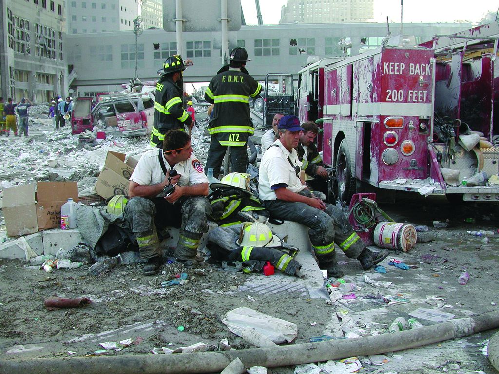
“This research is some of the first to suggest that there may be a link between the amount of exposure experienced by responders to the WTC site and the higher likelihood of excessive accumulation of fat in their livers,” study author Artit Jirapatnakul, PhD, of Icahn School of Medicine at Mount Sinai, New York, said in an interview. These findings were published in the American Journal of Industrial Medicine.
The excessive accumulation of liver fat is an indicator of liver injury, which can also predict subsequent future disease, such as cirrhosis, liver failure, and liver cancer.
Dr. Jirapatnakul said that arrival time to the WTC disaster may prove an important factor for predicting the risk of liver disease in this population and directing treatment to them accordingly.
“By identifying individuals with markers of liver injury, such as excess fat, we can offer referral to liver specialists and thereby open the door to early treatment,” he said.
“Our most important message is that many liver diseases can be treated if caught early,” Dr. Jirapatnakul added. “Early detection requires proactive monitoring because most liver diseases have few, if any, symptoms during the early stages.”
More than 20,000 men and women who responded to the WTC site on Sept. 11, 2001, were exposed to particulate matter and chemicals known to cause liver damage and increase the risk of toxicant‐associated fatty liver disease. These responders have been offered screening and treatment of different conditions associated with the attack, including CT lung cancer screening for those meeting age and smoking status criteria.
Measuring the impact of response time on the liver
To investigate the dose-response association between WTC site exposure intensity and the risk of hepatic steatosis, Dr. Jirapatnakul and colleagues reviewed low-dose CT chest scans of all participants in the WTC General Responders Cohort (GRC) who had available laboratory data within a 12-month period from their first scan following the Sept. 11, 2001, attack. Only CT chest scans performed between Sept. 11, 2001, and Dec. 31, 2018, were collected and reviewed in the study.
A total of 1,788 WTC responders were included (83.7% were male; mean age at time of attack, 42.5 years). Up to 56% of WTC responders in the study were White, and 20.4% of responders were current smokers. The mean body mass index of the group was 30.1 kg/m2.
The investigators stratified dust exposure into five groups according to when the responders arrived at the WTC site: Sept. 11, 2001, in the dust cloud; Sept. 11, no dust cloud (same-day arrival); Sept. 12 or 13 (second‐ and third‐day arrival); Sept. 14 to the end of September (fourth‐day arrival); and October and beyond.
The median duration between Sept. 11, 2001, and the earliest available CT scan was 11.3 years. Liver density was measured via Statistics‐based Liver Density Estimation from Imaging, a previously validated algorithm, with a slice thickness of 1.25 mm or below. On their earliest CT, approximately 14.4% (n = 258) of responders had liver attenuation < 40 Hounsfield units (HU). The prevalence of liver attenuation < 40 HU was 17% for responders who arrived on the day of the attack, 16% for responders who arrived at the site on Sept. 12 or 13, 10.9% for responders who arrived Sept. 14 through 30, and 9% for responders who arrived at the WTC site on Oct. 1, 2001, or later (P =.0015).
There was a statistically significant trend of increasing liver steatosis with earlier times of arrival (P <.0001). The WTC arrival time retained its status as a significant independent factor for decreased liver attenuation in an analysis adjusted for sex, age, race, smoking status, alcohol use, body mass index, diabetes, gastroesophageal reflux disease, and forced expiratory volume in 1 second.
Dr. Jirapatnakul said that the next step will be to determine whether WTC responders with excessive liver fat also have increased liver scarring. In addition, he and his colleagues are working to establish a registry to collect information on the impact of liver disease as it relates to quality of life in members of the WTC GRC.
Importance of disease severity
Another direction of future research will be to differentiate between those with only hepatic steatosis, those with inflammation from hepatic steatosis (steatohepatitis), and those with hepatic fibrosis which is the most concerning outcome from fatty liver diseases, according to Albert Do, MD, clinical director of the fatty liver disease program at Yale University, New Haven, Conn.
“It is the latter group of patients which we are most concerned about, given this is the group at highest risk for harm from liver disease,” added Dr. Do, who wasn’t involved in the research study. “The degree of steatosis is not closely linked with subsequent inflammation nor hepatic fibrosis, and so linkage of disease severity to specific occupational exposures and timing is needed to determine the allocation of support for patients who had suffered harm from fatty liver disease.”
Dr. Do noted that additional research will also need to identify the specific exposure that may be causing hepatic steatosis in early WTC responders. “Currently, only a small number of medications are known to cause this,” he explained, “and thus such knowledge will help us further understand occupational exposures and their associated risks.”
The researchers received study funding from the National Institute for Occupational Safety and Health. They disclosed conflicts of interest with Genentech, AstraZeneca, Pfizer, Bayer Healthcare, Gilead Sciences, and Boehringer Ingelheim. Dr. Do had no conflicts to declare.
Emergency responders to the World Trade Center (WTC) attack in 2001 paid a significant physical cost for their service in the form of exposure to chemicals, dust, and airborne particulates causally linked to hepatotoxicity. As we near the 20th anniversary of these attacks, researchers have determined that those responders who arrived at the WTC site earlier have a significantly higher prevalence of hepatic steatosis compared with those who arrived in the days that followed.
“This research is some of the first to suggest that there may be a link between the amount of exposure experienced by responders to the WTC site and the higher likelihood of excessive accumulation of fat in their livers,” study author Artit Jirapatnakul, PhD, of Icahn School of Medicine at Mount Sinai, New York, said in an interview. These findings were published in the American Journal of Industrial Medicine.
The excessive accumulation of liver fat is an indicator of liver injury, which can also predict subsequent future disease, such as cirrhosis, liver failure, and liver cancer.
Dr. Jirapatnakul said that arrival time to the WTC disaster may prove an important factor for predicting the risk of liver disease in this population and directing treatment to them accordingly.
“By identifying individuals with markers of liver injury, such as excess fat, we can offer referral to liver specialists and thereby open the door to early treatment,” he said.
“Our most important message is that many liver diseases can be treated if caught early,” Dr. Jirapatnakul added. “Early detection requires proactive monitoring because most liver diseases have few, if any, symptoms during the early stages.”
More than 20,000 men and women who responded to the WTC site on Sept. 11, 2001, were exposed to particulate matter and chemicals known to cause liver damage and increase the risk of toxicant‐associated fatty liver disease. These responders have been offered screening and treatment of different conditions associated with the attack, including CT lung cancer screening for those meeting age and smoking status criteria.
Measuring the impact of response time on the liver
To investigate the dose-response association between WTC site exposure intensity and the risk of hepatic steatosis, Dr. Jirapatnakul and colleagues reviewed low-dose CT chest scans of all participants in the WTC General Responders Cohort (GRC) who had available laboratory data within a 12-month period from their first scan following the Sept. 11, 2001, attack. Only CT chest scans performed between Sept. 11, 2001, and Dec. 31, 2018, were collected and reviewed in the study.
A total of 1,788 WTC responders were included (83.7% were male; mean age at time of attack, 42.5 years). Up to 56% of WTC responders in the study were White, and 20.4% of responders were current smokers. The mean body mass index of the group was 30.1 kg/m2.
The investigators stratified dust exposure into five groups according to when the responders arrived at the WTC site: Sept. 11, 2001, in the dust cloud; Sept. 11, no dust cloud (same-day arrival); Sept. 12 or 13 (second‐ and third‐day arrival); Sept. 14 to the end of September (fourth‐day arrival); and October and beyond.
The median duration between Sept. 11, 2001, and the earliest available CT scan was 11.3 years. Liver density was measured via Statistics‐based Liver Density Estimation from Imaging, a previously validated algorithm, with a slice thickness of 1.25 mm or below. On their earliest CT, approximately 14.4% (n = 258) of responders had liver attenuation < 40 Hounsfield units (HU). The prevalence of liver attenuation < 40 HU was 17% for responders who arrived on the day of the attack, 16% for responders who arrived at the site on Sept. 12 or 13, 10.9% for responders who arrived Sept. 14 through 30, and 9% for responders who arrived at the WTC site on Oct. 1, 2001, or later (P =.0015).
There was a statistically significant trend of increasing liver steatosis with earlier times of arrival (P <.0001). The WTC arrival time retained its status as a significant independent factor for decreased liver attenuation in an analysis adjusted for sex, age, race, smoking status, alcohol use, body mass index, diabetes, gastroesophageal reflux disease, and forced expiratory volume in 1 second.
Dr. Jirapatnakul said that the next step will be to determine whether WTC responders with excessive liver fat also have increased liver scarring. In addition, he and his colleagues are working to establish a registry to collect information on the impact of liver disease as it relates to quality of life in members of the WTC GRC.
Importance of disease severity
Another direction of future research will be to differentiate between those with only hepatic steatosis, those with inflammation from hepatic steatosis (steatohepatitis), and those with hepatic fibrosis which is the most concerning outcome from fatty liver diseases, according to Albert Do, MD, clinical director of the fatty liver disease program at Yale University, New Haven, Conn.
“It is the latter group of patients which we are most concerned about, given this is the group at highest risk for harm from liver disease,” added Dr. Do, who wasn’t involved in the research study. “The degree of steatosis is not closely linked with subsequent inflammation nor hepatic fibrosis, and so linkage of disease severity to specific occupational exposures and timing is needed to determine the allocation of support for patients who had suffered harm from fatty liver disease.”
Dr. Do noted that additional research will also need to identify the specific exposure that may be causing hepatic steatosis in early WTC responders. “Currently, only a small number of medications are known to cause this,” he explained, “and thus such knowledge will help us further understand occupational exposures and their associated risks.”
The researchers received study funding from the National Institute for Occupational Safety and Health. They disclosed conflicts of interest with Genentech, AstraZeneca, Pfizer, Bayer Healthcare, Gilead Sciences, and Boehringer Ingelheim. Dr. Do had no conflicts to declare.
FROM THE AMERICAN JOURNAL OF INDUSTRIAL MEDICINE
MR elastography could predict cirrhosis in NAFLD
Liver stiffness measurement with magnetic resonance elastography (MRE) may prove predictive of future cirrhosis risk in patients with nonalcoholic fatty liver disease (NAFLD), according to researchers from the Mayo Clinic in Rochester, Minn.
“These data expand the role of MRE from an accurate diagnostic method to a prognostic noninvasive imaging biomarker that can risk-stratify patients with NAFLD and guide the timing of surveillance and further refine their clinical management,” wrote Tolga Gidener, MD, and colleagues. The study authors added that the research further expands “the role of MRE beyond liver fibrosis estimation by adding a predictive feature to improve individualized disease monitoring and patient counseling.” Their study was published in Clinical Gastroenterology and Hepatology.
Currently, there are no established noninvasive strategies that can effectively identify patients with NAFLD who are at high risk of progression to cirrhosis and liver-related complications. While fibrosis stage on histology may predict liver-associated outcomes in these patients, this approach is invasive, time consuming, and is generally not well tolerated by patients.
Although the technique has been noted for its high success rate and excellent levels of reliability and reproducibility, a possible limitation of MRE is its cost. That said, standalone MRE is reimbursed under Medicare Category I Current Procedural Terminology code 76391 with a cost of $240.02. However, there is also a lack of data on whether baseline liver stiffness measurement by MRE can predict progression of NAFLD to cirrhosis.
To gauge the role of baseline liver stiffness measurement by MRE, Dr. Gidener and colleagues performed a retrospective cohort study that evaluated hard liver–related outcomes in 829 adult patients with NAFLD with or without cirrhosis (median age, 58 years; 54% female) who underwent MRE during 2007-2019.
Patients in the study were followed from the first MRE until death, last clinical encounter, or the end of the study. Clinical outcomes assessed in individual chart review included cirrhosis, hepatic decompensation, and death.
At baseline, the median liver stiffness measurement was 2.8 kPa in 639 patients with NAFLD but without cirrhosis. Over a median 4-year follow-up period, a total of 20 patients developed cirrhosis, with an overall annual incidence rate of 1%.
Baseline liver stiffness measurement by MRE was significantly predictive of subsequent cirrhosis (hazard ratio, 2.93; 95% confidence interval, 1.86-4.62; P < .0001) per 1-kPa difference in liver stiffness measurement at baseline.
According to the researchers, the probability of future cirrhosis development can be ascertained using current liver stiffness measurement. As such, a greater than 1% probability threshold can be reached in 5 years in patients with a measurement of 2 kPa, 3 years in patients with a measurement of 3 kPA, and 1 year in patients with 4-5 kPa. “These time frames inform about estimated time to progression to hard outcomes and provide guidance for subsequent noninvasive monitoring for disease progression,” wrote the researchers.
The baseline liver stiffness measurement by MRE was also significantly predictive of future hepatic decompensation or death (HR, 1.32; 95% CI, 1.13-1.56; P = .0007) per 1-kPa increment in the liver stiffness measurement. Likewise, the 1-year probability of subsequent hepatic decompensation or death in patients with cirrhosis and baseline liver stiffness measurement of 5 kPa versus 8 kPa was 9% versus 20%, respectively. In terms of covariates, age was the only factor that increased the risk of hepatic decompensation or death.
While the current study offers a glimpse into the potential clinical implications of liver stiffness measurement by MRE in NAFLD, the researchers suggest the applicability of the findings are limited by the study’s small sample size, relatively short follow-up duration, and the small number of cirrhosis events.
The researchers received study funding from the National Institute of Diabetes and Digestive and Kidney Diseases, American College of Gastroenterology, National Institutes of Health, and the Department of Defense. The researchers disclosed no other relevant conflicts of interest.
NAFLD is rapidly becoming one of the most common causes of liver disease. While most patients have a benign course, approximately 20% of patients develop nonalcoholic steatohepatitis, the progressive form of the disease. Given the high prevalence (30% of the U.S. population), it is vital to determine which patients are at risk for progression, cirrhosis, and decompensation. Although liver biopsy is the preferred method, this procedure is invasive and carries substantial risks, including severe bleeding. Noninvasive tests that measure liver stiffness have emerged: Examples are controlled elastography (VCTE), such as Fibroscan, and magnetic resonance elastography (MRE). Data support the use of liver stiffness as a surrogate measure of fibrosis; MRE has demonstrated higher fidelity and accuracy, compared with VCTE, while being limited because of cost and availability. However, there is a paucity of data regarding the use of liver stiffness to predict progression to cirrhosis or liver-related events.

This study by Dr. Gidener and colleagues highlights the use of MRE to evaluate liver stiffness measurements as a predictor for cirrhosis and decompensation. Baseline measurements more than 4-5 kPa should alert clinicians regarding increased risk of progression to cirrhosis. Patients with cirrhosis and baseline measurements of 8 kPa or higher have a high risk of decompensation/death, suggesting that they should be followed more closely. Given the burgeoning number of patients with NAFLD and NASH, this study demonstrates the importance of identifying high-risk patients in order to optimize use of resources and improve clinical outcomes.
Yamini Natarajan, MD, is an investigator at the Center for Innovations in Quality, Effectiveness and Safety at the Michael E. DeBakey VA Medical Center, Houston, and an assistant professor, department of medicine, section of gastroenterology and hepatology, Baylor College of Medicine, Houston. She has no conflicts.
NAFLD is rapidly becoming one of the most common causes of liver disease. While most patients have a benign course, approximately 20% of patients develop nonalcoholic steatohepatitis, the progressive form of the disease. Given the high prevalence (30% of the U.S. population), it is vital to determine which patients are at risk for progression, cirrhosis, and decompensation. Although liver biopsy is the preferred method, this procedure is invasive and carries substantial risks, including severe bleeding. Noninvasive tests that measure liver stiffness have emerged: Examples are controlled elastography (VCTE), such as Fibroscan, and magnetic resonance elastography (MRE). Data support the use of liver stiffness as a surrogate measure of fibrosis; MRE has demonstrated higher fidelity and accuracy, compared with VCTE, while being limited because of cost and availability. However, there is a paucity of data regarding the use of liver stiffness to predict progression to cirrhosis or liver-related events.
This study by Dr. Gidener and colleagues highlights the use of MRE to evaluate liver stiffness measurements as a predictor for cirrhosis and decompensation. Baseline measurements more than 4-5 kPa should alert clinicians regarding increased risk of progression to cirrhosis. Patients with cirrhosis and baseline measurements of 8 kPa or higher have a high risk of decompensation/death, suggesting that they should be followed more closely. Given the burgeoning number of patients with NAFLD and NASH, this study demonstrates the importance of identifying high-risk patients in order to optimize use of resources and improve clinical outcomes.
Yamini Natarajan, MD, is an investigator at the Center for Innovations in Quality, Effectiveness and Safety at the Michael E. DeBakey VA Medical Center, Houston, and an assistant professor, department of medicine, section of gastroenterology and hepatology, Baylor College of Medicine, Houston. She has no conflicts.
Liver stiffness measurement with magnetic resonance elastography (MRE) may prove predictive of future cirrhosis risk in patients with nonalcoholic fatty liver disease (NAFLD), according to researchers from the Mayo Clinic in Rochester, Minn.
“These data expand the role of MRE from an accurate diagnostic method to a prognostic noninvasive imaging biomarker that can risk-stratify patients with NAFLD and guide the timing of surveillance and further refine their clinical management,” wrote Tolga Gidener, MD, and colleagues. The study authors added that the research further expands “the role of MRE beyond liver fibrosis estimation by adding a predictive feature to improve individualized disease monitoring and patient counseling.” Their study was published in Clinical Gastroenterology and Hepatology.
Currently, there are no established noninvasive strategies that can effectively identify patients with NAFLD who are at high risk of progression to cirrhosis and liver-related complications. While fibrosis stage on histology may predict liver-associated outcomes in these patients, this approach is invasive, time consuming, and is generally not well tolerated by patients.
Although the technique has been noted for its high success rate and excellent levels of reliability and reproducibility, a possible limitation of MRE is its cost. That said, standalone MRE is reimbursed under Medicare Category I Current Procedural Terminology code 76391 with a cost of $240.02. However, there is also a lack of data on whether baseline liver stiffness measurement by MRE can predict progression of NAFLD to cirrhosis.
To gauge the role of baseline liver stiffness measurement by MRE, Dr. Gidener and colleagues performed a retrospective cohort study that evaluated hard liver–related outcomes in 829 adult patients with NAFLD with or without cirrhosis (median age, 58 years; 54% female) who underwent MRE during 2007-2019.
Patients in the study were followed from the first MRE until death, last clinical encounter, or the end of the study. Clinical outcomes assessed in individual chart review included cirrhosis, hepatic decompensation, and death.
At baseline, the median liver stiffness measurement was 2.8 kPa in 639 patients with NAFLD but without cirrhosis. Over a median 4-year follow-up period, a total of 20 patients developed cirrhosis, with an overall annual incidence rate of 1%.
Baseline liver stiffness measurement by MRE was significantly predictive of subsequent cirrhosis (hazard ratio, 2.93; 95% confidence interval, 1.86-4.62; P < .0001) per 1-kPa difference in liver stiffness measurement at baseline.
According to the researchers, the probability of future cirrhosis development can be ascertained using current liver stiffness measurement. As such, a greater than 1% probability threshold can be reached in 5 years in patients with a measurement of 2 kPa, 3 years in patients with a measurement of 3 kPA, and 1 year in patients with 4-5 kPa. “These time frames inform about estimated time to progression to hard outcomes and provide guidance for subsequent noninvasive monitoring for disease progression,” wrote the researchers.
The baseline liver stiffness measurement by MRE was also significantly predictive of future hepatic decompensation or death (HR, 1.32; 95% CI, 1.13-1.56; P = .0007) per 1-kPa increment in the liver stiffness measurement. Likewise, the 1-year probability of subsequent hepatic decompensation or death in patients with cirrhosis and baseline liver stiffness measurement of 5 kPa versus 8 kPa was 9% versus 20%, respectively. In terms of covariates, age was the only factor that increased the risk of hepatic decompensation or death.
While the current study offers a glimpse into the potential clinical implications of liver stiffness measurement by MRE in NAFLD, the researchers suggest the applicability of the findings are limited by the study’s small sample size, relatively short follow-up duration, and the small number of cirrhosis events.
The researchers received study funding from the National Institute of Diabetes and Digestive and Kidney Diseases, American College of Gastroenterology, National Institutes of Health, and the Department of Defense. The researchers disclosed no other relevant conflicts of interest.
FROM CLINICAL GASTROENTEROLOGY AND HEPATOLOGY