User login
USPSTF punts on sleep apnea screening
because the current evidence is inadequate to assess the benefits and harms of doing so, according to a Recommendation Statement published online Jan. 23 in JAMA.
The USPSTF makes recommendations about the effectiveness of specific health care services for patients who don’t have related signs or symptoms. In this case, the Recommendation Statement addresses adults who don’t snore excessively; gasp or choke while sleeping; or report the daytime sleepiness, impaired cognition, or mood changes typically associated with obstructive sleep apnea, said Kirsten Bobbins-Domingo, PhD, MD, chair of the organization and lead author of the Recommendation Statement, and her associates (JAMA 2017 Jan 23. doi: 10.1001/jama.2016.20325).
The USPSTF commissioned a comprehensive review of the literature to examine whether screening such patients by primary caregivers would effectively identify those who have obstructive sleep apnea and lead to treatment that would prevent the elevated rates of death, cognitive impairment, motor vehicle crashes, cardiovascular events, and cerebrovascular events related to the disorder. Daniel E. Jonas, MD, of the University of North Carolina at Chapel Hill and his associates reviewed 110 relevant studies involving 46,188 participants.
They found that the accuracy and clinical utility of numerous OSA screening tools was uncertain. In particular, the Epworth Sleepiness Scale, the STOP (Snoring, Tiredness, Observed Apnea, and High Blood Pressure) questionnaire, the STOP-BANG (STOP plus BMI, Age, Neck Circumference, and Gender) questionnaire, the Berlin Questionnaire, the Wisconsin Sleep Questionnaire, and the Multivariable Apnea Prediction (MVAP) tool have not been adequately validated in primary care settings.
Moreover, no studies directly assessed whether screening had an impact on actual health outcomes. Several treatments, notably CPAP and mandibular advancement devices, did improve intermediate outcomes such as scores on the apnea-hypopnea index, scores on the Epworth Sleepiness Scale, and blood pressure levels, but the evidence did not show that this in turn improved mortality, cardiovascular events, or the other “hard” outcomes of interest, Dr. Jonas and his associates said in their Evidence Report (JAMA 2017 Jan 23. doi: 10.1001/jama.2016.19635).
Dr. Bobbins-Domingo and her associates on the task force noted that this Recommendation Statement is consistent with that of the American Academy of Family Physicians, which also concluded that the current evidence is insufficient to assess the balance of benefits and harms of screening asymptomatic adults for obstructive sleep apnea.
The American College of Physicians offers a “weak” recommendation based on low-quality evidence that patients with unexplained daytime sleepiness and patients suspected of having apnea undergo a sleep study, said Dr. Bobbins-Domingo, professor of medicine at the University of California, San Francisco, and her associates.
In contrast, the American Academy of Sleep Medicine recommends that routine health visits should include questions about OSA and evaluation of risk factors such as obesity, retrognathia, and treatment-refractory hypertension. If there are positive findings, a comprehensive sleep evaluation should follow, according to the AASM.
The USPSTF is an independent voluntary group supported by the Agency for Healthcare Research and Quality as mandated by the U.S. Congress. The authors’ conflict of interest disclosures are available at www.uspreventiveservicestaskforce.org.
This recommendation must not be misinterpreted. If clinicians are discouraged from directly questioning patients about apnea signs and symptoms or from using short screening questionnaires to identify those at high risk for the disorder, it would negatively influence public health.

Susan Redline, MD, is at the Sleep Health Institute and in the Division of Sleep and Circadian Disorders at Brigham and Women’s Hospital and Harvard Medical School and Beth Israel Deaconess Medical Center, all in Boston. She reported ties to Jazz Pharmaceuticals, RosMed Inc., and the Beckman Company, as well as serving on the American Academy of Sleep Medicine’s board of directors. Dr. Redline made these remarks in an editorial accompanying the USPSTF reports (JAMA 2017;317:368-70).
This recommendation must not be misinterpreted. If clinicians are discouraged from directly questioning patients about apnea signs and symptoms or from using short screening questionnaires to identify those at high risk for the disorder, it would negatively influence public health.
Susan Redline, MD, is at the Sleep Health Institute and in the Division of Sleep and Circadian Disorders at Brigham and Women’s Hospital and Harvard Medical School and Beth Israel Deaconess Medical Center, all in Boston. She reported ties to Jazz Pharmaceuticals, RosMed Inc., and the Beckman Company, as well as serving on the American Academy of Sleep Medicine’s board of directors. Dr. Redline made these remarks in an editorial accompanying the USPSTF reports (JAMA 2017;317:368-70).
because the current evidence is inadequate to assess the benefits and harms of doing so, according to a Recommendation Statement published online Jan. 23 in JAMA.
The USPSTF makes recommendations about the effectiveness of specific health care services for patients who don’t have related signs or symptoms. In this case, the Recommendation Statement addresses adults who don’t snore excessively; gasp or choke while sleeping; or report the daytime sleepiness, impaired cognition, or mood changes typically associated with obstructive sleep apnea, said Kirsten Bobbins-Domingo, PhD, MD, chair of the organization and lead author of the Recommendation Statement, and her associates (JAMA 2017 Jan 23. doi: 10.1001/jama.2016.20325).
The USPSTF commissioned a comprehensive review of the literature to examine whether screening such patients by primary caregivers would effectively identify those who have obstructive sleep apnea and lead to treatment that would prevent the elevated rates of death, cognitive impairment, motor vehicle crashes, cardiovascular events, and cerebrovascular events related to the disorder. Daniel E. Jonas, MD, of the University of North Carolina at Chapel Hill and his associates reviewed 110 relevant studies involving 46,188 participants.
They found that the accuracy and clinical utility of numerous OSA screening tools was uncertain. In particular, the Epworth Sleepiness Scale, the STOP (Snoring, Tiredness, Observed Apnea, and High Blood Pressure) questionnaire, the STOP-BANG (STOP plus BMI, Age, Neck Circumference, and Gender) questionnaire, the Berlin Questionnaire, the Wisconsin Sleep Questionnaire, and the Multivariable Apnea Prediction (MVAP) tool have not been adequately validated in primary care settings.
Moreover, no studies directly assessed whether screening had an impact on actual health outcomes. Several treatments, notably CPAP and mandibular advancement devices, did improve intermediate outcomes such as scores on the apnea-hypopnea index, scores on the Epworth Sleepiness Scale, and blood pressure levels, but the evidence did not show that this in turn improved mortality, cardiovascular events, or the other “hard” outcomes of interest, Dr. Jonas and his associates said in their Evidence Report (JAMA 2017 Jan 23. doi: 10.1001/jama.2016.19635).
Dr. Bobbins-Domingo and her associates on the task force noted that this Recommendation Statement is consistent with that of the American Academy of Family Physicians, which also concluded that the current evidence is insufficient to assess the balance of benefits and harms of screening asymptomatic adults for obstructive sleep apnea.
The American College of Physicians offers a “weak” recommendation based on low-quality evidence that patients with unexplained daytime sleepiness and patients suspected of having apnea undergo a sleep study, said Dr. Bobbins-Domingo, professor of medicine at the University of California, San Francisco, and her associates.
In contrast, the American Academy of Sleep Medicine recommends that routine health visits should include questions about OSA and evaluation of risk factors such as obesity, retrognathia, and treatment-refractory hypertension. If there are positive findings, a comprehensive sleep evaluation should follow, according to the AASM.
The USPSTF is an independent voluntary group supported by the Agency for Healthcare Research and Quality as mandated by the U.S. Congress. The authors’ conflict of interest disclosures are available at www.uspreventiveservicestaskforce.org.
FROM JAMA
Curb AF recurrences through risk factor modification
SNOWMASS, COLO. – Overlooking the common modifiable risk factors in patients with atrial fibrillation is missing out on an excellent opportunity to help curb the growing global pandemic of the arrhythmia, Patrick T. O’Gara, MD, said at the Annual Cardiovascular Conference at Snowmass.
“My purpose here is a wake up call to improve screening for and treatment of modifiable risk factors in patients with atrial fibrillation,” declared Dr. O’Gara, professor of medicine at Harvard Medical School, Boston.
Overweight/obesity: Investigators at the University of Adelaide (Australia) demonstrated in the LEGACY trial that patients with atrial fibrillation (AF) and a BMI of 27 kg/m2 or more reduced their AF symptom burden in a dose-response fashion as they shed excess pounds as part of an intensive weight management program. Those who shed at least 10% of their baseline body weight had a 46% rate of 5-year freedom from AF without resort to rhythm control medications or ablation procedures of 46%. With 3%-9% weight loss, the rate was 22%. And with 3% weight loss, it was 13%.
The best results came from sustained linear weight loss. Weight fluctuations of greater than 5% – the classic yoyo dieting pattern – partially offset the overall benefit of weight loss with respect to recurrent AF (J Am Coll Cardiol. 2015 May 26;65(20):2159-69).
In a separate study, the same team of Australian investigators offered an opportunity to participate in a risk factor management program to patients with AF and a BMI of 27 kg/m2 or more who were undergoing radiofrequency ablation for their arrhythmia. Participants had significantly fewer repeat ablation procedures during followup and were also less likely to be on antiarrhythmic drugs than the patients who opted for usual care (J Am Coll Cardiol. 2014 Dec 2;64(21):2222-31).
Alcohol: The ‘holiday heart’ syndrome is well known, but alcohol consumption beyond binging can increase risk for AF. Dr. O’Gara noted that in a recent review article entitled “Alcohol and Atrial Fibrillation: A Sobering Review,” investigators at the University of Melbourne showed that while the relationship between the number of standard drinks per week and risk of cardiovascular mortality is J-shaped, with a nadir at 14-21 drinks per week in men and fewer in women, the risk of developing AF is linear over time and appears to increase incrementally with every additional drink per week (J Am Coll Cardiol. 2016 Dec 13;68(23):2567-76).
Also, a prospective study of nearly 80,000 Swedes free from AF at baseline, coupled with a meta-analysis of seven prospective studies found that for each additional drink per day consumed the risk of developing AF rose over time by roughly a further 10% compared to that of teetotalers (J Am Coll Cardiol. 2014; Jul 22;64(3):281-9).
Physical inactivity: In the prospective Tromso Study, in which more than 20,000 Norwegian adults were followed for 20 years, leisure time physical activity displayed a J-shaped relationship with the risk of developing AF. Moderately active subjects were an adjusted 19% less likely to develop AF than those with low physical activity, while the risk in subjects who regularly engaged in vigorous physical activity was 37% higher than in the low-activity group (Eur Heart J. 2016 Aug 1;37(29):2307-13).
“This effect of moderate exercise might be due to the associated weight loss, improved endothelial function, better sleep, perhaps a better balance between the sympathetic and parasympathetic nervous systems,” Dr. O’Gara observed.
How much physical activity is right for patients with AF? Dr. O’Gara said one of the best reviews he’s seen came from the University of Adelaide group (Circulation. 2016 Feb 2;133(5):457-9). They recommended a total of 120-200 minutes of exercise per week spread over three to five sessions. While the research base is strongest for moderate-intensity exercise, the Australians also noted the effectiveness and safety of a novel program of repeated 4-minute intervals of high-intensity exercise at 85%-95% of peak heart rate as demonstrated in a randomized controlled trial by investigators at the Norwegian University of Science and Technology in Trondheim. They showed this approach resulted in reduced time in AF and decreased AF symptoms coupled with improved quality of life and left atrial and ventricular function (Circulation. 2016 Feb 2;133(5):466-73).
“I think you could look at this review and feel very confident that there is some evidence base to substantiate your strong recommendation that patients actively engage in exercise as treatment for their atrial fibrillation,” the cardiologist said.
Sleep apnea: Investigators at Brigham and Women’s Hospital in Boston have demonstrated that effective treatment of sleep apnea with continuous positive airway pressure in patients with atrial fibrillation is associated with smaller atrial size and ventricular mass, lower blood pressure, and a significantly reduced risk of recurrent AF following an AF ablation procedure (J Am Heart Assoc. 2013 Nov 25;2(6):e000421).
“Sleep hygiene is one of the least attended aspects of cardiovascular health,” according to Dr. O’Gara. “We need to ask the partner or spouse, ‘How well does your partner sleep? Do you hear thrashing about, snoring, gagging, or notice restless legs?’ Heart failure folks are really tuned into this, but in the practice of seeing patients come into the emergency room with new-onset atrial fibrillation, it’s tenth on the list of five questions one would ask.”
Dr. O’Gara reported having no financial conflicts.
EXPERT ANALYSIS FROM THE CARDIOVASCULAR CONFERENCE AT SNOWMASS
Conference News Roundup—Radiological Society of North America
Studies Provide More Insight Into Zika Effects
Three studies reported on the effects of the Zika virus outbreak in Brazil. The first study examined CT findings of the CNS in 16 newborn babies with congenital Zika virus infection confirmed by tests in CSF. The researchers identified a pattern of CT brain findings in the babies, including decreased brain volume, simplified gyral pattern, calcifications, ventricular dilatation, and prominent occipital bone.
"We live in Pernambuco, a state in northeastern Brazil, which had the highest number of patients with microcephaly during the Zika outbreak in our country," said Natacha Calheiros de Lima Petribu, MD, of the Department of Radiology at Barão de Lucena Hospital. "Our study proves that Zika virus infection can cause congenital brain damage in babies with and without microcephaly."
Another study analyzed the imaging results of three target groups affected by Zika: adults who developed acute neurologic syndrome, newborns with vertical infection with neurologic disorders, and pregnant women with rash outbreaks suggestive of Zika. Many of the adults had symptoms of Guillain-Barré syndrome. A few showed inflammation of the brain and spinal cord (ie, Bickerstaff's encephalitis) or brainstem and spinal cord lesions. Common MRI findings included enhancement of certain spinal and facial nerves. In the newborns, MRI showed orbital injuries and anatomical changes in brain tissue.
"It was alarming to find so many cases of neurologic syndromes in adults, some very serious, related to Zika virus infection," said study author Emerson de Melo Casagrande, MD, of the Department of Radiology at Antonio Pedro University Hospital--Federal Fluminense University. "We have also noticed a difference between these syndromes, even though the trigger was the same."
In a third study, ultrasound and fetal MRI were performed on pregnant patients with Zika virus infection at different gestational ages. Once the babies were born, they underwent ultrasound, CT, and MRI. The researchers then created 3-D virtual and physical models of the skulls. More than half of the babies had microcephaly, brain calcifications, and loss of brain tissue volume, along with other structural changes.
"The emergence of Zika virus in the Americas has coincided with increased reports of babies born with microcephaly," said study author Heron Werner Jr, MD, PhD, of the Department of Radiology at Clínica de Diagnóstico por Imagem. "An early diagnosis may help in treating these babies after birth. Moreover, the knowledge of abnormalities present in the CNS may give hints about the pathophysiology of the disease."
Head Impacts Lead to Brain Changes in High School Football Players
Brain imaging exams performed on high school football players after a single season reveal changes in the gray and white matter that correlated with exposure to head impacts, according to researchers.
"It is important to understand the potential changes occurring in the brain related to youth contact sports," said Elizabeth Moody Davenport, PhD, a postdoctoral researcher at UT Southwestern Medical Center in Dallas. "We know that some professional football players suffer from a serious condition called chronic traumatic encephalopathy or CTE. We are attempting to find out when and how that process starts, so that we can keep sports a healthy activity for millions of children and adolescents."
The study included 24 players from a high school football team in North Carolina, each of whom wore a helmet outfitted with the Head Impact Telemetry System (HITS) during all practices and games. The helmets are lined with six accelerometers that measure the magnitude, location, and direction of a hit. Data from the helmets can be uploaded to a computer for analysis.
"We saw changes in these young players' brains on both structural and functional imaging after a single season of football," said Dr. Davenport.
In the study, each player underwent pre- and post-season imaging, including a specialized MRI scan, from which diffusion tensor imaging and diffusion kurtosis imaging data were extracted to measure the brain's white matter integrity, and a magnetoencephalography (MEG) scan, which records and analyzes the magnetic fields produced by brain waves. Diffusion imaging can measure the structural white matter changes in the brain, and MEG assesses changes in function.
"MEG can be used to measure delta waves in the brain, which are a type of distress signal," said Dr. Davenport. "Delta waves represent slow wave activity that increases after brain injuries. The delta waves we saw came from the surface of the brain, while diffusion imaging is a measure of the white matter deeper in the brain."
The research team calculated the change in imaging metrics between the pre- and post-season imaging exams. They measured abnormalities observed on diffusion imaging and abnormally increased delta-wave activity on MEG. The imaging results were then combined with player-specific impact data from the HITS. None of the 24 players were diagnosed with a concussion during the study.
Players with greater head impact exposure had the greatest change in diffusion imaging and MEG metrics. "Change in diffusion imaging metrics correlated most to linear acceleration, similar to the impact of a car crash," said Dr. Davenport. "MEG changes correlated most to rotational impact, similar to a boxer's punch. These results demonstrate that you need both imaging metrics to assess impact exposure, because they correlate with different biomechanical processes."
Similar studies are being conducted this fall, and a consortium has been formed to continue the brain imaging research in youth contact sports across the country, said Dr. Davenport. "Without a larger population that is closely followed in a longitudinal study, it is difficult to know the long-term effects of these changes," she said. "We do not know if the brain's developmental trajectory is altered, or if the off-season time allows for the brain to return to normal."
Depression in Soldiers Linked to Brain Disruption From Injury
Using multiple brain imaging techniques, researchers have found that a disruption of the circuitry in the brain's cognitive-emotional pathways may provide a physical foundation for depression symptoms in some service members who have had mild traumatic brain injury (mTBI) in combat. "We can link these connectivity changes in the brain to poor top-down emotional processing and greater maladaptive rumination, or worrying, in symptomatic depressed soldiers after mTBI," said Ping-Hong Yeh, PhD, scientist and physicist at the National Intrepid Center of Excellence, Walter Reed National Military Medical Center in Bethesda, Maryland.
According to the Defense and Veterans Brain Injury Center, 352,619 service members worldwide have been diagnosed with TBI since 2000, the majority of these cases being mTBI. In addition, psychiatric disorders, such as anxiety and major depressive disorders, are becoming common in military personnel with brain injuries.
"With the increased survival of soldiers due to improvements in body armor and advanced medical care, there has been an increase in the number of soldiers surviving major trauma. Consequently, a large number of soldiers are returning from war with mTBI," said Dr. Yeh. "Mood disorders are common in military-related mTBI patients. This is an ongoing problem facing a large number of warriors in current areas of conflict, and it is likely to be a persistent problem for the foreseeable future."
For the study, researchers used diffusion-weighted imaging (DWI) and resting-state functional MRI (fMRI) to examine 130 active male service members diagnosed with mTBI and a control group of 52 men without mTBI. Depression symptoms were rated based on the Beck Depression Inventory (BDI), a 21-item, self-reporting assessment that measures characteristic attitudes and symptoms of depression. Patients with a BDI score greater than 20 are considered to have moderate to severe depression symptoms.
BDI scores showed that 75 of the patients with mTBI had moderate to severe depression symptoms. Imaging results showed that white matter tracts—the circuits that connect brain regions critical for cognitive and emotional control—were disrupted in the patients with moderate to severe depression symptoms. Researchers also saw changes in the gray matter cognitive-emotional networks in these patients.
"We found consistencies in the locations of disrupted neurocircuitry, as revealed by DWI and resting-state fMRI, that are unique to the clinical symptoms of mTBI patients," said Dr. Yeh. "We have related the brain structural and functional changes in cognitive-emotional networks to depressive symptoms in mTBI patients."
This research can possibly lead to treatment strategies in the future, he added. "Though the results of this study were not applied directly to patient care, the neuroimaging changes we found might be incorporated into treatment plans for personalized medicine in the future."
Short-Term Sleep Deprivation Affects Heart Function
Getting too little sleep takes a toll on your heart, researchers reported. People who work in fire and emergency medical services, medical residencies, and other high-stress jobs are often called upon to work 24-hour shifts with little opportunity for sleep. While it is known that extreme fatigue can affect many physical, cognitive, and emotional processes, this is the first study to examine how working a 24-hour shift specifically affects cardiac function.
"For the first time, we have shown that short-term sleep deprivation in the context of 24-hour shifts can lead to a significant increase in cardiac contractility, blood pressure, and heart rate," said study author Daniel Kuetting, MD, of the Department of Diagnostic and Interventional Radiology at the University of Bonn in Germany.
For the study, Dr. Kuetting and colleagues recruited 20 healthy radiologists (19 men) with a mean age of 31.6. Each of the study participants underwent cardiovascular magnetic resonance (CMR) imaging with strain analysis before and after a 24-hour shift with an average of three hours of sleep.
"Cardiac function in the context of sleep deprivation has not previously been investigated with CMR strain analysis, the most sensitive parameter of cardiac contractility," said Dr. Kuetting. The researchers also collected blood and urine samples from the participants and measured blood pressure and heart rate.
Following short-term sleep deprivation, the participants showed significant increases in mean peak systolic strain (-23.4 vs -21.9), systolic (118.5 mmHg vs 112.8 mmHg) and diastolic (69.2 mmHg vs 62.9 mmHg) blood pressure and heart rate (68.9 bpm vs 63.0 bpm). In addition, the participants had significant increases in levels of thyroid stimulating hormone (TSH), thyroid hormones FT3 and FT4, and cortisol.
Although the researchers were able to perform follow-up examinations of half of the participants after regular sleep, further study in a larger cohort is needed to determine possible long-term effects of sleep loss, said Dr. Kuetting.
"The study was designed to investigate real-life work-related sleep deprivation," said Dr. Kuetting. "While the participants were not permitted to consume caffeine or food and beverages containing theobromine, such as chocolate, nuts, or tea, we did not take into account factors like individual stress level or environmental stimuli."
As people continue to work longer hours or work at more than one job to make ends meet, it is critical to investigate the detrimental effects of too much work and not enough sleep. The results of this pilot study are transferable to other professions in which long periods of uninterrupted labor are common, said Dr. Kuetting. "These findings may help us better understand how workload and shift duration affect public health."
Aerobic Exercise Preserves Brain Volume and Improves Cognitive Function
Using a new MRI technique, researchers have found that adults with mild cognitive impairment (MCI) who exercised four times per week during a six-month period experienced an increase in brain volume in specific areas of the brain, but adults who participated in aerobic exercise experienced greater gains than those who just stretched.
"Even over a short period of time, we saw aerobic exercise lead to a remarkable change in the brain," said Laura D. Baker, PhD, Associate Professor of Gerontology and Geriatric Medicine at Wake Forest School of Medicine (WFSM) in Winston-Salem, North Carolina.
The study included 35 adults with MCI participating in a randomized, controlled trial of exercise intervention. The participants were separated into two groups. Sixteen adults (average age, 63) engaged in aerobic activity, including treadmill, stationary bike, or elliptical training, four times per week for six months. A control group of 19 adults (average age, 67) participated in stretching exercises with the same frequency. High-resolution brain MR images were acquired from all participants before and after the six-month activity period. The MRI results were compared using conventional and biomechanical metrics to measure the change in brain volume and shape.
"We used high-resolution MR images to measure anatomical changes within areas of the brain to obtain volumetric data and directional information," said Jeongchul Kim, PhD, a coinvestigator at WFSM.
The analysis revealed that for both the aerobic and stretching groups, brain volume increased in most gray matter regions, including the temporal lobe, which supports short-term memory.
"Compared to the stretching group, the aerobic activity group had greater preservation of total brain volume, increased local gray matter volume, and increased directional stretch of brain tissue," said Dr. Kim.
Among participants of the stretching group, the analysis revealed a local contraction, or atrophy, within the white matter connecting fibers. Such directional deformation, or shape change, is partially related to volume loss, but not always, according to Dr. Kim.
"Directional changes in the brain without local volume changes could be a novel biomarker for neurologic disease," he said. "It may be a more sensitive marker for the tiny changes that occur in a specific brain region before volumetric changes are detectable on MRI."
Both MRI measures are important to the treatment of MCI and Alzheimer's disease, which require the careful tracking of changes in the brain while patients engage in interventions, including diet and exercise, to slow the progression of the disease.
Study participants were tested to determine the effect of exercise intervention on cognitive performance. Participants in the aerobic exercise group showed statistically significant improvement in executive function after six months, whereas the stretching group did not improve.
"Any type of exercise can be beneficial," said Dr. Kim. "If possible, aerobic activity may create potential benefits for higher cognitive functioning."
What Are Safe and Efficacious Therapies for Restless Legs Syndrome in Adults?
The American Academy of Neurology (AAN) has published evidence-based recommendations for management of restless legs syndrome (RLS) in adults. The practice guideline was published online ahead of print November 16, 2016, in Neurology. The practice guideline addresses the question: What are safe and effective therapies, including both pharmacologic and nonpharmacologic approaches, for the symptoms and clinical consequences (eg, disturbed sleep, periodic limb movements in sleep, depression/anxiety, and decreased quality of life) of RLS in adults.
“When addressing RLS, clinicians and patients must first determine whether symptoms require treatment, the setting in which this practice guideline is relevant,” said John W. Winkelman, MD, PhD, and colleagues. Dr. Winkelman is an Associate Professor of Psychiatry at Harvard Medical School and Medical Director of the Sleep Health Center of Brigham and Women’s Hospital in Boston. 
“Treatment should be considered if RLS symptoms interfere with sleep or daytime function to an important degree,” the guideline authors said. “Before determining the best treatment, it is important to first ensure there are no contributing factors to RLS symptoms (eg, iron deficiency or serotonergic antidepressants). The guidelines advise clinicians to consider prescribing a pharmacologic agent to reduce RLS symptoms in patients with moderate to severe primary RLS. There is strong (Level A) evidence for use of pramipexole, rotigotine, cabergoline, and gabapentin enacarbil; moderate evidence (Level B) supports ropinirole, pregabalin, and IV ferric carboxymaltose; and weak evidence (Level C) supports levodopa. When considering efficacy alone, clinicians may prefer cabergoline. It is rarely used in clinical practice for RLS, however, because it is associated with a risk of cardiac valvulopathy. Clinicians are also advised to consider the augmentation risks associated with dopaminergic agents.
For patients with periodic limb movement disorder, there is strong (Level A) evidence supporting ropinirole. Moderate evidence (Level B) supports pramipexole, rotigotine, cabergoline, and pregabalin; and weak evidence (Level C) supporting levodopa. The authors note insufficient evidence (Level U) for gabapentin enacarbil and ferric carboxymaltose. With regard to objective sleep measures (eg, total sleep time, sleep efficiency, sleep latency, wake after sleep onset) there is moderate evidence (Level B) supporting ropinirole, gabapentin, encarbil, and pregabalin. However, there is insufficient evidence (Level U) supporting pamipexole, rotigotine, cabergoline, or levodopa.
For subjective sleep measures, cabergoline and gabapentin enacarbil have Level A evidence; ropinirole, pramipexole, rotigotine, and pregabalin have Level B evidence; and levodopa and prolonged-release oxycodone/naloxone, and vibratory stimulation have Level C evidence. Insufficient evidence (Level U) exists for ferric carboxymaltose and iron sucrose.When patients with RLS fail to respond to other treatments, clinicians are advised to consider prescribing prolonged-release oxycodone/naloxone (Level C) or to consider nonpharmacologic options, including pneumatic compression (Level B), infrared spectroscopy or transcranial magnetic stimulation (Level C), and vibrating pads (Level C).
For iron-deficient patients with RLS (ferritin levels ≤ 75 µg/L), clinicians are advised to prescribe ferrous sulfate with vitamin C. In patients on hemodialysis with secondary RLS, clinicians are advised to prescribe vitamin C and E supplementation (Level B), ropinirole, levodopa, or exercise (Level C).
—Erica Tricarico
Suggested Reading
Winkelman JW, Armstrong MJ, Allen RP, et al. Practice guideline summary: treatment of restless legs syndrome in adults: report of the Guideline Development, Dissemination, and Implementation Subcommittee of the American Academy of Neurology. Neurology. 2016 Nov 16 [Epub ahead of print].
The American Academy of Neurology (AAN) has published evidence-based recommendations for management of restless legs syndrome (RLS) in adults. The practice guideline was published online ahead of print November 16, 2016, in Neurology. The practice guideline addresses the question: What are safe and effective therapies, including both pharmacologic and nonpharmacologic approaches, for the symptoms and clinical consequences (eg, disturbed sleep, periodic limb movements in sleep, depression/anxiety, and decreased quality of life) of RLS in adults.
“When addressing RLS, clinicians and patients must first determine whether symptoms require treatment, the setting in which this practice guideline is relevant,” said John W. Winkelman, MD, PhD, and colleagues. Dr. Winkelman is an Associate Professor of Psychiatry at Harvard Medical School and Medical Director of the Sleep Health Center of Brigham and Women’s Hospital in Boston.
“Treatment should be considered if RLS symptoms interfere with sleep or daytime function to an important degree,” the guideline authors said. “Before determining the best treatment, it is important to first ensure there are no contributing factors to RLS symptoms (eg, iron deficiency or serotonergic antidepressants). The guidelines advise clinicians to consider prescribing a pharmacologic agent to reduce RLS symptoms in patients with moderate to severe primary RLS. There is strong (Level A) evidence for use of pramipexole, rotigotine, cabergoline, and gabapentin enacarbil; moderate evidence (Level B) supports ropinirole, pregabalin, and IV ferric carboxymaltose; and weak evidence (Level C) supports levodopa. When considering efficacy alone, clinicians may prefer cabergoline. It is rarely used in clinical practice for RLS, however, because it is associated with a risk of cardiac valvulopathy. Clinicians are also advised to consider the augmentation risks associated with dopaminergic agents.
For patients with periodic limb movement disorder, there is strong (Level A) evidence supporting ropinirole. Moderate evidence (Level B) supports pramipexole, rotigotine, cabergoline, and pregabalin; and weak evidence (Level C) supporting levodopa. The authors note insufficient evidence (Level U) for gabapentin enacarbil and ferric carboxymaltose. With regard to objective sleep measures (eg, total sleep time, sleep efficiency, sleep latency, wake after sleep onset) there is moderate evidence (Level B) supporting ropinirole, gabapentin, encarbil, and pregabalin. However, there is insufficient evidence (Level U) supporting pamipexole, rotigotine, cabergoline, or levodopa.
For subjective sleep measures, cabergoline and gabapentin enacarbil have Level A evidence; ropinirole, pramipexole, rotigotine, and pregabalin have Level B evidence; and levodopa and prolonged-release oxycodone/naloxone, and vibratory stimulation have Level C evidence. Insufficient evidence (Level U) exists for ferric carboxymaltose and iron sucrose.When patients with RLS fail to respond to other treatments, clinicians are advised to consider prescribing prolonged-release oxycodone/naloxone (Level C) or to consider nonpharmacologic options, including pneumatic compression (Level B), infrared spectroscopy or transcranial magnetic stimulation (Level C), and vibrating pads (Level C).
For iron-deficient patients with RLS (ferritin levels ≤ 75 µg/L), clinicians are advised to prescribe ferrous sulfate with vitamin C. In patients on hemodialysis with secondary RLS, clinicians are advised to prescribe vitamin C and E supplementation (Level B), ropinirole, levodopa, or exercise (Level C).
—Erica Tricarico
Suggested Reading
Winkelman JW, Armstrong MJ, Allen RP, et al. Practice guideline summary: treatment of restless legs syndrome in adults: report of the Guideline Development, Dissemination, and Implementation Subcommittee of the American Academy of Neurology. Neurology. 2016 Nov 16 [Epub ahead of print].

Recovery From TBI and Better Sleep Go Hand in Hand
After a traumatic brain injury (TBI), people also experience major sleep problems, including changes in their sleep–wake cycle. A new study published online ahead of print December 21, 2016, in Neurology showed that recovering from these two conditions occurs in parallel.
“These results suggest that monitoring a person’s sleep–wake cycle may be a useful tool for assessing their recovery after TBI,” said study author Nadia Gosselin, PhD, an Assistant Professor in the Department of Psychology at the University of Montréal in Québec. “We found that when someone sustained a brain injury and had not recovered a certain level of consciousness to keep them awake and aware of their surroundings, they were not able to generate a good sleep–wake cycle. But as they recovered, their quality of sleep improved.” 
The study involved 30 people, ages 17 to 58, who had been hospitalized for moderate to severe TBI. Most of the patients were in a coma when they were admitted to the hospital, and all initially received care in an ICU. The injuries were caused by motor vehicle accidents for 20 people, falls for seven people, recreational or sports activities for two people and a blow to the head for one person. They were hospitalized for an average of 45 days, with monitoring for the study beginning an average of 21 days into the patient’s stay.
Each person was monitored daily for an average of 11 days for level of consciousness and thinking abilities using the Rancho Los Amigos scale, which ranges from 1 to 8. Each person also wore an activity monitor on the wrist so researchers could measure their sleep.
Researchers found that consciousness and thinking abilities improved hand in hand with measures of quality of sleep, showing a linear relationship.
One measure, the daytime activity ratio, reflects the percentage of activity that occurs during the day. Immediately after the injury, activity occurs throughout the day and night. The study showed that participants reached an acceptable sleep–wake cycle, with a daytime activity ratio of at least 80%, at the same point when they emerged from a minimally conscious state.
The participants still had inadequate sleep–wake cycles, at a score of 5 on the Rancho Los Amigos scale, where people are confused and give inappropriate responses to stimuli, but are able to follow simple commands. Sleep–wake cycles reached adequate levels at the same time that people reached a score of 6 on the Rancho Los Amigos scale, which is when people can give appropriate responses while still depending on outside input for direction. At that level, they can remember relearned tasks, but cannot remember new tasks.
The results were the same when researchers adjusted for the amount of time that had passed since the injury and the amount of medications they had received while they were in the ICU.
“It is possible that there are common underlying brain mechanisms involved in both recovery from TBI and improvement in sleep,” said Dr. Gosselin. “Still, more study needs to be done, and future research may want to examine how hospital lighting and noise also affect quality of sleep for those with TBI.”
Suggested Reading
Duclos C, Dumont M, Arbour C, et al. Parallel recovery of consciousness and sleep in acute traumatic brain injury. Neurology. 2016 Dec 21 [Epub ahead of print].
Soddu A, Bassetti CL. A good sleep for a fresh mind in patients with acute tramatic brain injury. Neurology. 2016 Dec 21 [Epub ahead of print].
After a traumatic brain injury (TBI), people also experience major sleep problems, including changes in their sleep–wake cycle. A new study published online ahead of print December 21, 2016, in Neurology showed that recovering from these two conditions occurs in parallel.
“These results suggest that monitoring a person’s sleep–wake cycle may be a useful tool for assessing their recovery after TBI,” said study author Nadia Gosselin, PhD, an Assistant Professor in the Department of Psychology at the University of Montréal in Québec. “We found that when someone sustained a brain injury and had not recovered a certain level of consciousness to keep them awake and aware of their surroundings, they were not able to generate a good sleep–wake cycle. But as they recovered, their quality of sleep improved.”
The study involved 30 people, ages 17 to 58, who had been hospitalized for moderate to severe TBI. Most of the patients were in a coma when they were admitted to the hospital, and all initially received care in an ICU. The injuries were caused by motor vehicle accidents for 20 people, falls for seven people, recreational or sports activities for two people and a blow to the head for one person. They were hospitalized for an average of 45 days, with monitoring for the study beginning an average of 21 days into the patient’s stay.
Each person was monitored daily for an average of 11 days for level of consciousness and thinking abilities using the Rancho Los Amigos scale, which ranges from 1 to 8. Each person also wore an activity monitor on the wrist so researchers could measure their sleep.
Researchers found that consciousness and thinking abilities improved hand in hand with measures of quality of sleep, showing a linear relationship.
One measure, the daytime activity ratio, reflects the percentage of activity that occurs during the day. Immediately after the injury, activity occurs throughout the day and night. The study showed that participants reached an acceptable sleep–wake cycle, with a daytime activity ratio of at least 80%, at the same point when they emerged from a minimally conscious state.
The participants still had inadequate sleep–wake cycles, at a score of 5 on the Rancho Los Amigos scale, where people are confused and give inappropriate responses to stimuli, but are able to follow simple commands. Sleep–wake cycles reached adequate levels at the same time that people reached a score of 6 on the Rancho Los Amigos scale, which is when people can give appropriate responses while still depending on outside input for direction. At that level, they can remember relearned tasks, but cannot remember new tasks.
The results were the same when researchers adjusted for the amount of time that had passed since the injury and the amount of medications they had received while they were in the ICU.
“It is possible that there are common underlying brain mechanisms involved in both recovery from TBI and improvement in sleep,” said Dr. Gosselin. “Still, more study needs to be done, and future research may want to examine how hospital lighting and noise also affect quality of sleep for those with TBI.”
Suggested Reading
Duclos C, Dumont M, Arbour C, et al. Parallel recovery of consciousness and sleep in acute traumatic brain injury. Neurology. 2016 Dec 21 [Epub ahead of print].
Soddu A, Bassetti CL. A good sleep for a fresh mind in patients with acute tramatic brain injury. Neurology. 2016 Dec 21 [Epub ahead of print].
Potential Weaknesses in Major Trial Explored in Reevaluating CPAP Benefits
LAS VEGAS—Although continuous positive airway pressure (CPAP) failed to protect patients with obstructive sleep apnea from cardiovascular events in a recently completed large randomized trial, the results are not definitive, according to Alan Z. Segal, MD, Associate Professor of Clinical Neurology at Weill Cornell Medical College in New York City. In an update on sleep disorders at the American Academy of Neurology’s Fall 2016 Conference, Dr. Segal explained that the average adherence to CPAP in this trial, called SAVE, was less than that which has been widely accepted as adequate. 
“Medicare has a requirement that CPAP be used for more than four hours on 70% of nights in a 30-day period for patients to keep their device,” observed Dr. Segal, providing one example of a way in which four hours or more of CPAP has been defined as a minimum duration. In the SAVE study by McEvoy et al, the mean duration of CPAP was 3.3 hours. Only 42% of patients met the four-hour definition of adherence, which was the duration the authors themselves had defined as good adherence.
“Our field was very much affected by this study,” Dr. Segal acknowledged. “It created quite a lot of controversy and criticisms over methodologic flaws. Like any study that does not produce the results we want, we start to pick at it to identify where it might have been inadequate, and the biggest inadequacy of this particular study is that patients in the CPAP arm may not have been adequately treated.”
Adherence Versus Effectiveness
In this study, 2,717 patients with moderate-to-severe obstructive sleep apnea and a history of coronary or cerebrovascular disease were randomized to CPAP plus usual care or usual care alone. The primary composite end point of the trial was death from cardiovascular causes, myocardial infarction, stroke, or hospitalization for unstable angina, heart failure, or transient ischemic attack.
After a mean follow-up of 3.7 years, an event had occurred in 17% of those in the CPAP group versus 15.4% of those in the usual-care group, producing a nonsignificantly increased hazard ratio (HR 1.10) for the primary end point in the CPAP arm. CPAP did reduce snoring and daytime sleepiness as well as improve quality of life and mood, all of which were secondary end points.
“The natural next step would be to look at the subgroup of patients who did have good adherence to CPAP, but the study was not powered to look at that,” Dr. Segal said. Although he reported that a post hoc analysis did show “a trend for benefit in those patients who actually adhered to the therapy,” these data are not sufficient to challenge the primary conclusion.
There was a reasonable expectation that CPAP, which has been an effective treatment in this and other trials for the symptoms of obstructive sleep apnea, would reduce cardiovascular events, according to the authors of the SAVE study. They cited strong evidence that obstructive sleep apnea increases the risk of stroke and other cardiovascular events, so control of the disorder had the potential to be protective. Moreover, CPAP has been specifically associated with improved endothelial function, improved insulin sensitivity, and reduced systolic blood pressure in patients with hypertension.
Study Flaw or Real-World Experience?
Yet judging CPAP on an intention-to-treat basis may be appropriate, because several other trials cited by the authors of SAVE, all of which were also negative for a cardiovascular benefit, have also found median adherence to be less than four hours. In SAVE, the mean adherence was 4.4 hours per night during the first 12 months of the trial, but it fell subsequently. As a reflection of what can be expected from a prescription of CPAP, the lack of protection from cardiovascular events in SAVE may not reflect a fundamental inability of CPAP to protect against vascular events, but the negative result is relevant if typical use means low adherence.
“CPAP adherence is a challenge,” Dr. Segal acknowledged. “In a dedicated sleep center such as ours, we work closely with patients to help them with adherence. One of the ways we do that is through mask fittings to find the style with which the patient is most comfortable.”
Mask styles range from minimalist devices that fit the nostrils to full “fighter-pilot” devices fitting over the entire face, according to Dr. Segal. He estimated that there are about 30 mask styles now available, and added that considerable time is devoted at his center to helping patients make the best choice.
Adherence may also be improved using modern devices, according to Dr. Segal. One example is software that ramps up pressure slowly so that the target pressure is not reached until the patient is already asleep, making use more acceptable. Another is technology that reduces pressure at each exhalation, facilitating a more natural breathing cycle and also increasing patient acceptance. Newer machines equipped with automated titration may also add convenience and ease of use.
Significant Benefit to Be Gained
The rewards of CPAP in patients who become comfortable with this therapy can be significant. The impairments in quality of sleep in patients with obstructive sleep apnea are often the impetus to seek care. “It is very important that patients get that subjective experience of feeling better the next day, and I would say about one-third of my patients get that next-day eureka feeling from initiating CPAP after sleeping so poorly for many years,” Dr. Segal reported.
These quality-of-life benefits were reflected in the SAVE study. For those in the CPAP arm, relative to those receiving usual care alone, there were highly significant improvements in objective anxiety and depression scales, as well as the physical and mental components of the 36-item Short Form (SF-36) quality of life tool. A reduction in daytime symptoms of sleep deprivation was also significant for CPAP on the Epworth Sleepiness Scale.
Relative to other tools to treat obstructive sleep apnea, CPAP is typically the most attractive option, according to Dr. Segal. For example, he is not an advocate of oral devices that advance the mandible, which he said have never been shown to be effective in moderate-to-severe apnea. He also suggested that surgery, which can be painful, does not uniformly provide benefit. However, he acknowledged that engaging patients in CPAP who do not experience a symptomatic benefit may now be more difficult after the results of the SAVE study. Although “the jury is still out” in regard to the ability of CPAP to reduce cardiovascular events in adherent patients, the evidence has now raised doubts for those already prescribed CPAP for the goal of reducing cardiovascular risk.“Those patients who are dedicated CPAP users will keep using this, but those who are perhaps looking for an excuse not to use CPAP may look at this data in a different way,” Dr. Segal said.
—Theodore Bosworth
Suggested Reading
McEvoy RD, Antic NA, Heeley E, et al. CPAP for prevention of cardiovascular events in obstructive sleep apnea. N Engl J Med. 2016;375(10):919-931.
LAS VEGAS—Although continuous positive airway pressure (CPAP) failed to protect patients with obstructive sleep apnea from cardiovascular events in a recently completed large randomized trial, the results are not definitive, according to Alan Z. Segal, MD, Associate Professor of Clinical Neurology at Weill Cornell Medical College in New York City. In an update on sleep disorders at the American Academy of Neurology’s Fall 2016 Conference, Dr. Segal explained that the average adherence to CPAP in this trial, called SAVE, was less than that which has been widely accepted as adequate.
“Medicare has a requirement that CPAP be used for more than four hours on 70% of nights in a 30-day period for patients to keep their device,” observed Dr. Segal, providing one example of a way in which four hours or more of CPAP has been defined as a minimum duration. In the SAVE study by McEvoy et al, the mean duration of CPAP was 3.3 hours. Only 42% of patients met the four-hour definition of adherence, which was the duration the authors themselves had defined as good adherence.
“Our field was very much affected by this study,” Dr. Segal acknowledged. “It created quite a lot of controversy and criticisms over methodologic flaws. Like any study that does not produce the results we want, we start to pick at it to identify where it might have been inadequate, and the biggest inadequacy of this particular study is that patients in the CPAP arm may not have been adequately treated.”
Adherence Versus Effectiveness
In this study, 2,717 patients with moderate-to-severe obstructive sleep apnea and a history of coronary or cerebrovascular disease were randomized to CPAP plus usual care or usual care alone. The primary composite end point of the trial was death from cardiovascular causes, myocardial infarction, stroke, or hospitalization for unstable angina, heart failure, or transient ischemic attack.
After a mean follow-up of 3.7 years, an event had occurred in 17% of those in the CPAP group versus 15.4% of those in the usual-care group, producing a nonsignificantly increased hazard ratio (HR 1.10) for the primary end point in the CPAP arm. CPAP did reduce snoring and daytime sleepiness as well as improve quality of life and mood, all of which were secondary end points.
“The natural next step would be to look at the subgroup of patients who did have good adherence to CPAP, but the study was not powered to look at that,” Dr. Segal said. Although he reported that a post hoc analysis did show “a trend for benefit in those patients who actually adhered to the therapy,” these data are not sufficient to challenge the primary conclusion.
There was a reasonable expectation that CPAP, which has been an effective treatment in this and other trials for the symptoms of obstructive sleep apnea, would reduce cardiovascular events, according to the authors of the SAVE study. They cited strong evidence that obstructive sleep apnea increases the risk of stroke and other cardiovascular events, so control of the disorder had the potential to be protective. Moreover, CPAP has been specifically associated with improved endothelial function, improved insulin sensitivity, and reduced systolic blood pressure in patients with hypertension.
Study Flaw or Real-World Experience?
Yet judging CPAP on an intention-to-treat basis may be appropriate, because several other trials cited by the authors of SAVE, all of which were also negative for a cardiovascular benefit, have also found median adherence to be less than four hours. In SAVE, the mean adherence was 4.4 hours per night during the first 12 months of the trial, but it fell subsequently. As a reflection of what can be expected from a prescription of CPAP, the lack of protection from cardiovascular events in SAVE may not reflect a fundamental inability of CPAP to protect against vascular events, but the negative result is relevant if typical use means low adherence.
“CPAP adherence is a challenge,” Dr. Segal acknowledged. “In a dedicated sleep center such as ours, we work closely with patients to help them with adherence. One of the ways we do that is through mask fittings to find the style with which the patient is most comfortable.”
Mask styles range from minimalist devices that fit the nostrils to full “fighter-pilot” devices fitting over the entire face, according to Dr. Segal. He estimated that there are about 30 mask styles now available, and added that considerable time is devoted at his center to helping patients make the best choice.
Adherence may also be improved using modern devices, according to Dr. Segal. One example is software that ramps up pressure slowly so that the target pressure is not reached until the patient is already asleep, making use more acceptable. Another is technology that reduces pressure at each exhalation, facilitating a more natural breathing cycle and also increasing patient acceptance. Newer machines equipped with automated titration may also add convenience and ease of use.
Significant Benefit to Be Gained
The rewards of CPAP in patients who become comfortable with this therapy can be significant. The impairments in quality of sleep in patients with obstructive sleep apnea are often the impetus to seek care. “It is very important that patients get that subjective experience of feeling better the next day, and I would say about one-third of my patients get that next-day eureka feeling from initiating CPAP after sleeping so poorly for many years,” Dr. Segal reported.
These quality-of-life benefits were reflected in the SAVE study. For those in the CPAP arm, relative to those receiving usual care alone, there were highly significant improvements in objective anxiety and depression scales, as well as the physical and mental components of the 36-item Short Form (SF-36) quality of life tool. A reduction in daytime symptoms of sleep deprivation was also significant for CPAP on the Epworth Sleepiness Scale.
Relative to other tools to treat obstructive sleep apnea, CPAP is typically the most attractive option, according to Dr. Segal. For example, he is not an advocate of oral devices that advance the mandible, which he said have never been shown to be effective in moderate-to-severe apnea. He also suggested that surgery, which can be painful, does not uniformly provide benefit. However, he acknowledged that engaging patients in CPAP who do not experience a symptomatic benefit may now be more difficult after the results of the SAVE study. Although “the jury is still out” in regard to the ability of CPAP to reduce cardiovascular events in adherent patients, the evidence has now raised doubts for those already prescribed CPAP for the goal of reducing cardiovascular risk.“Those patients who are dedicated CPAP users will keep using this, but those who are perhaps looking for an excuse not to use CPAP may look at this data in a different way,” Dr. Segal said.
—Theodore Bosworth
Suggested Reading
McEvoy RD, Antic NA, Heeley E, et al. CPAP for prevention of cardiovascular events in obstructive sleep apnea. N Engl J Med. 2016;375(10):919-931.

Sleep Disorders
The video associated with this article is no longer available on this site. Please view all of our videos on the MDedge YouTube channel
The video associated with this article is no longer available on this site. Please view all of our videos on the MDedge YouTube channel
The video associated with this article is no longer available on this site. Please view all of our videos on the MDedge YouTube channel

Blood pressure rise follows halting CPAP
Continuous positive airway pressure (CPAP) therapy for obstructive sleep apnea (OSA) has a significant beneficial effect on blood pressure, according to an analysis of participants in three randomized controlled trials.
Previous meta-analyses suggested that CPAP treatment led to an average of improvement of 2-3 mm Hg, but the estimates relied on heterogeneous trials that often had low levels of CPAP adherence, and those factors might have led to an underestimation of the treatment effect. The new analysis showed that halting CPAP increases blood pressure between 5.0 and 9.0 mm Hg, compared with patients who continued using CPAP (Chest. 2016;150[6]:1202-10).
To get around the problem of adherence, researchers led by Malcolm Kohler, MD, at University Hospital of Zürich analyzed the results of three previous studies looking at the effects of CPAP withdrawal. The analysis included 153 OSA patients on CPAP therapy, who had been randomized to continue therapy or to withdraw from therapy for 2 weeks. Eighty-seven of these patients discontinued CPAP, and the remaining 66 patients continued the therapy. Blood pressure was measured at home and in hospital.
On average, those who discontinued CPAP had an increase in office systolic blood pressure of 5.4 mm Hg (95% confidence interval, 1.8-8.9 mm Hg; P = .003) and an increase in home systolic blood pressure of 9.0 mm Hg (95% CI, 5.7-12.3 mm Hg; P less than .001), compared with patients who continued CPAP. The effects of stopping CPAP, instead of continuing the therapy, on office diastolic blood pressure and home diastolic pressure were increases of 5.0 mm Hg (95% CI, 2.7-7.3 mm Hg; P less than .001) and 7.8 mm Hg (95% CI, 5.6-10.0 mm Hg; P less than .001), respectively.
Patients who discontinued CPAP also experienced a significant increase in apnea-hypopnea index, from 2.8/h to 33.2/h, while those who continued using CPAP, on average, experienced only a 0.3/h increase in apnea-hypopnea index from baseline.
“One clinical implication is that if you do not need to stop CPAP for obstructive sleep apnea, do not stop it. This study also suggests the importance of monitoring your blood pressure in a home setting, under usual conditions,” summed up Robert Kloner, MD, PhD, director of the Huntington Medical Research Institutes Cardiovascular Research Lab, Pasadena, Calif., who was not involved in the study.
Previous studies of CPAP, such as the SAVE study published in the New England Journal of Medicine in September (N Engl J Med. 2016;375:919-31), often find little or no connection between CPAP therapy and cardiovascular outcomes. That is probably because of inadequate adherence to CPAP therapy. “That’s always been the bane of sleep apnea studies,” said Krishna M. Sundar, MD, FCCP, who also did not participate in the study.
The current work got around the problem by looking at patients who had already established use of CPAP. “This is a very good study,” said Dr. Sundar, who is the medical director of the Sleep-Wake Center at the University of Utah, Salt Lake City.
The study was funded by the Swiss National Science Foundation and the University of Zürich. The analysis’ authors and the outside experts quoted in this story reported no financial disclosures.
FROM CHEST
Key clinical point: Interrupting CPAP therapy leads to a rise in blood pressure.
Major finding: Stopping CPAP was associated with 5.0-9.0 mm Hg blood pressure increase.
Data source: Analysis of 153 patients with moderate to severe OSA, who had participated in three randomized controlled trials.
Disclosures: The study was funded by the Swiss National Science Foundation and the University of Zürich. The authors of the analysis and the outside experts quoted in this story reported no financial disclosures.
Dr. Sundar and Dr. Kloner reported having no financial disclosures.
Internet-based CBT may be effective treatment for chronic insomnia
Internet-delivered cognitive-behavioral therapy is effective at overcoming chronic insomnia, a randomized controlled trial of 303 adults showed.
In the trial, patients who were randomized to receive cognitive-behavioral therapy for insomnia (CBT-I) used a Web-based program called Sleep Healthy Using the Internet (SHUTi), which “incorporates the primary tenets of face-to-face CBT-I, including sleep restriction, stimulus control, cognitive restructuring, sleep hygiene, and relapse prevention,” wrote Lee M. Ritterband, PhD, and his associates. Meanwhile, patients in the control group received online, nonspecific patient education.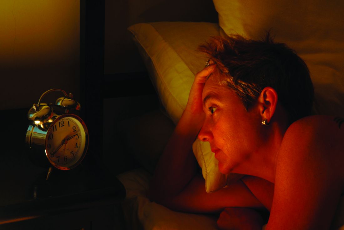
After a 9-week study period, the investigators found that sleep-onset latency, wake after sleep onset, and ratings on the Insomnia Severity Index all were significantly reduced in the SHUTi group, compared with the control group. The effect remained after the 6-month and 1-year follow-ups, reported Dr. Ritterband of the Center for Behavioral Health and Technology at the University of Virginia, Charlottesville, and his associates.
No significant difference was found in total sleep time between the SHUTi group and the control group, but sleep efficiency and sleep quality did improve. In addition, the number of awakenings dropped in the SHUTi group. Both groups reported a decrease in the use of sleep aids, such as medication. After completing the post assessment and 6-month follow-up, participants received online gift certificates of $50 each, and after completing the assessment at the 1-year follow-up, they received $100 gift certificates.
“Internet-delivered CBT-I provides a less expensive, scalable treatment option that could reach previously unimaginable numbers of people,” the investigators said. “Future studies are necessary to determine who may be best served by this type of intervention and how the next steps of dissemination should occur. Ensuring that these interventions work with different patient populations, whether tailored or not for those groups, should also be examined,” they noted.
Find the full study in JAMA Psychiatry (2016 Nov 30. doi: 10.1001/jamapsychiatry.2016.3249)
FROM JAMA PSYCHIATRY
Pediatric OSA improved with oral montelukast
The majority of children with obstructive sleep apnea (OSA) who took oral montelukast showed reductions in their apnea-hypopnea index (AHI) scores, according to the results of a randomized, double-blind placebo-controlled study. Typically, OSA in children is treated with adenotonsillectomy, explained Leila Kheirandish-Gozal, MD, director of clinical sleep research at the University of Chicago, and her colleagues. However, in this study, children were given either montelukast or placebo for 16 weeks and then participated in an overnight polysomnographic study. Seventy-one percent of the patients who took montelukast had fewer AHI events per hour of total sleep time at the end of the study. To learn more, see Family Practice News: http://www.mdedge.com/familypracticenews/article/116479/pulmonology/pediatric-osa-improved-oral-montelukast.
The majority of children with obstructive sleep apnea (OSA) who took oral montelukast showed reductions in their apnea-hypopnea index (AHI) scores, according to the results of a randomized, double-blind placebo-controlled study. Typically, OSA in children is treated with adenotonsillectomy, explained Leila Kheirandish-Gozal, MD, director of clinical sleep research at the University of Chicago, and her colleagues. However, in this study, children were given either montelukast or placebo for 16 weeks and then participated in an overnight polysomnographic study. Seventy-one percent of the patients who took montelukast had fewer AHI events per hour of total sleep time at the end of the study. To learn more, see Family Practice News: http://www.mdedge.com/familypracticenews/article/116479/pulmonology/pediatric-osa-improved-oral-montelukast.
The majority of children with obstructive sleep apnea (OSA) who took oral montelukast showed reductions in their apnea-hypopnea index (AHI) scores, according to the results of a randomized, double-blind placebo-controlled study. Typically, OSA in children is treated with adenotonsillectomy, explained Leila Kheirandish-Gozal, MD, director of clinical sleep research at the University of Chicago, and her colleagues. However, in this study, children were given either montelukast or placebo for 16 weeks and then participated in an overnight polysomnographic study. Seventy-one percent of the patients who took montelukast had fewer AHI events per hour of total sleep time at the end of the study. To learn more, see Family Practice News: http://www.mdedge.com/familypracticenews/article/116479/pulmonology/pediatric-osa-improved-oral-montelukast.