User login
My Fitness Journey
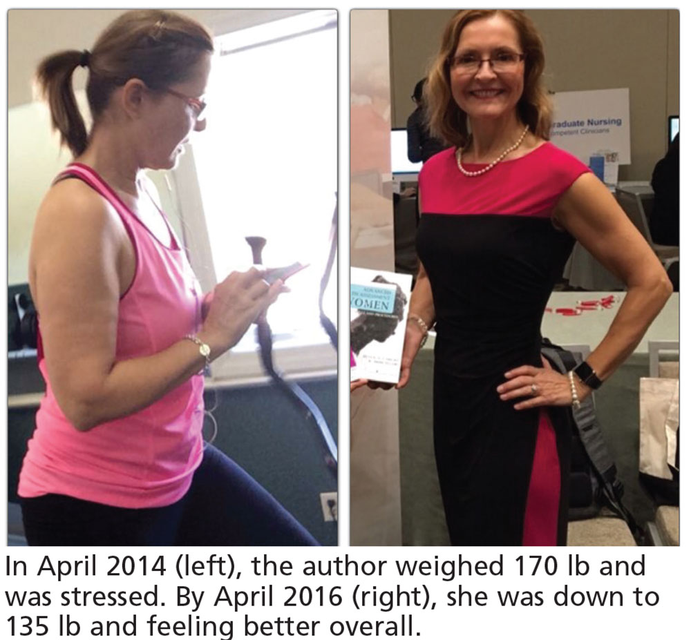
One month into my doctoral program, I was stressed out, anxious, not sleeping well, and gaining weight. I expressed these concerns to my daughter, who is a professional bodybuilder and fitness coach; she offered to help me get healthy and fit while earning my doctorate from Rocky Mountain University of Health Professions in Provo, Utah. Over the next two years, I lost 35 pounds and 12 inches of fat from my belly and became more muscular than ever before! I even decided to participate in my first bodybuilding show, “Debut at 62,” on November 19th in Providence, Rhode Island.
I was honored to be the student commencement speaker and recipient of the Student Service Award for my contributions to the NP profession—but what I’m most proud of is adopting a healthy and fit lifestyle in the midst of the most stressful two years of my life. In fact, I believe learning to cope with extreme stress helped me perform more effectively in my doctoral program. I had more energy and concentration, better sleep, and very few physical complaints.
Certainly, I realize that it can be difficult for us to walk our talk and be role models for our patients. When faced with extreme stress we often “crash and burn.” We gain weight, get depressed, sleep poorly, stop having sex, drink excessively, you name it! We know what we should do but lack the energy, time, and motivation to implement and maintain healthy habits. In order to effectively deal with the stress in our lives, we must practice self-care. This approach may seem counterintuitive, as our natural reaction to stress is usually to abandon healthy habits and resort to eating junk food, drinking alcohol, watching TV, and not exercising. It requires conscious effort, development of new habits (and breaking of old ones!), practice, consistency, and a lot of support to cope with stress in a positive way. Mind control, meditation, and paced breathing are other cognitive-based techniques that can be used to help combat the negative effects of stress and anxiety.
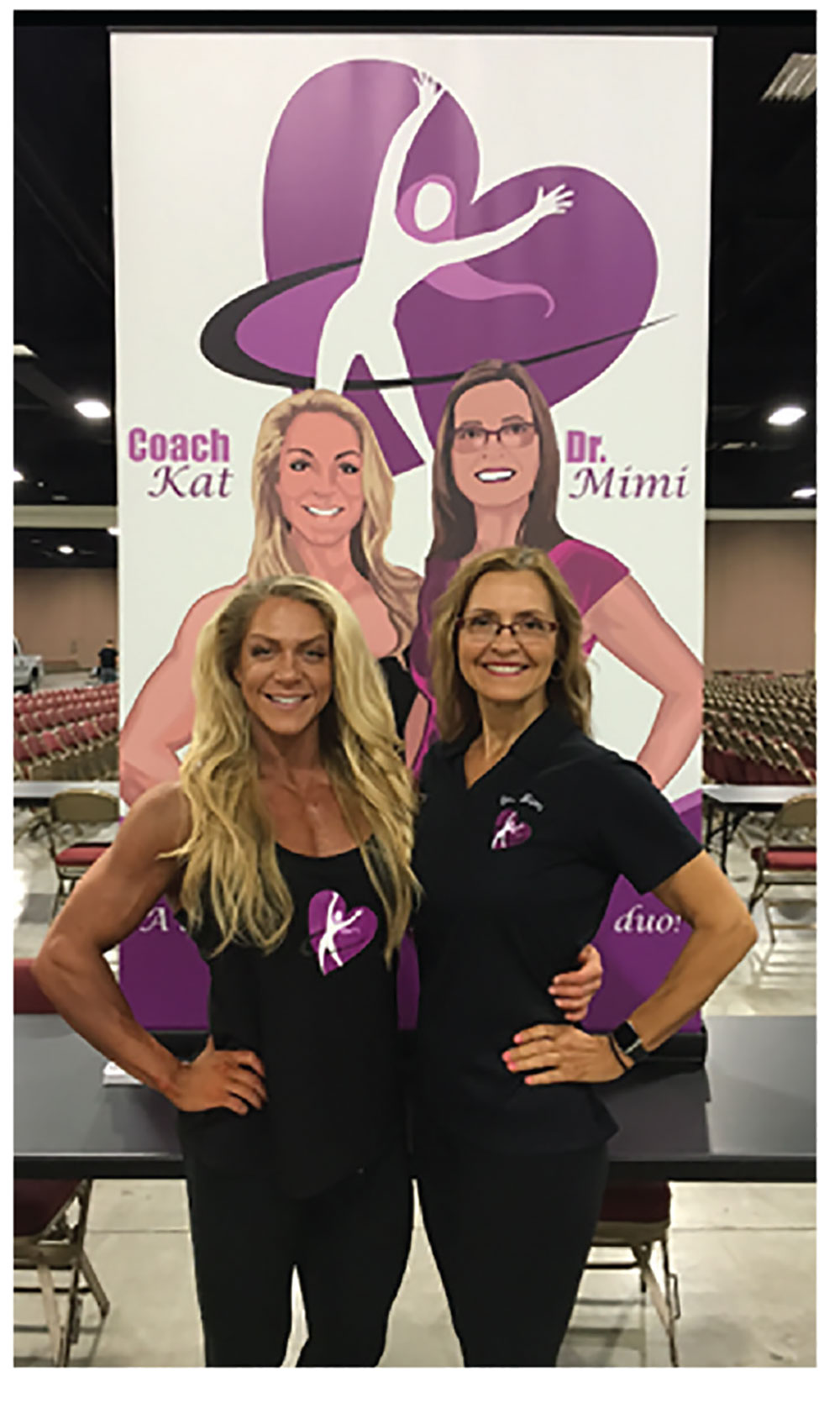
As a result of my experience and transformation, my daughter and I decided to team up to help other NPs and PAs make positive changes in their lives. Under the names Coach Kat and Doctor Mimi, we developed The Secor Initiative—an intensive, one-year, online program for NPs or PAs who are seriously committed to becoming healthy, happy, and fit. Upon completion of the program, participants achieve the esteemed title of “Top NP” or “Top PA,” enabling them to be role models for their peers and patients.
The Secor Initiative helps NPs and PAs gain insight into all the sources of stress in their lives and then go on to learn how to cope with these stressors. Our program includes five (10-week) courses, with topics such as nutrition, exercise, money/wealth, stress management, and advanced women’s health; for more information, visit www.MimiSecor.com and click on The Secor Initiative or check out our Facebook page, Coach Kat and Dr. Mimi.
Let’s step up to the plate and become healthy (and happy) role models for our patients.
One month into my doctoral program, I was stressed out, anxious, not sleeping well, and gaining weight. I expressed these concerns to my daughter, who is a professional bodybuilder and fitness coach; she offered to help me get healthy and fit while earning my doctorate from Rocky Mountain University of Health Professions in Provo, Utah. Over the next two years, I lost 35 pounds and 12 inches of fat from my belly and became more muscular than ever before! I even decided to participate in my first bodybuilding show, “Debut at 62,” on November 19th in Providence, Rhode Island.
I was honored to be the student commencement speaker and recipient of the Student Service Award for my contributions to the NP profession—but what I’m most proud of is adopting a healthy and fit lifestyle in the midst of the most stressful two years of my life. In fact, I believe learning to cope with extreme stress helped me perform more effectively in my doctoral program. I had more energy and concentration, better sleep, and very few physical complaints.
Certainly, I realize that it can be difficult for us to walk our talk and be role models for our patients. When faced with extreme stress we often “crash and burn.” We gain weight, get depressed, sleep poorly, stop having sex, drink excessively, you name it! We know what we should do but lack the energy, time, and motivation to implement and maintain healthy habits. In order to effectively deal with the stress in our lives, we must practice self-care. This approach may seem counterintuitive, as our natural reaction to stress is usually to abandon healthy habits and resort to eating junk food, drinking alcohol, watching TV, and not exercising. It requires conscious effort, development of new habits (and breaking of old ones!), practice, consistency, and a lot of support to cope with stress in a positive way. Mind control, meditation, and paced breathing are other cognitive-based techniques that can be used to help combat the negative effects of stress and anxiety.
As a result of my experience and transformation, my daughter and I decided to team up to help other NPs and PAs make positive changes in their lives. Under the names Coach Kat and Doctor Mimi, we developed The Secor Initiative—an intensive, one-year, online program for NPs or PAs who are seriously committed to becoming healthy, happy, and fit. Upon completion of the program, participants achieve the esteemed title of “Top NP” or “Top PA,” enabling them to be role models for their peers and patients.
The Secor Initiative helps NPs and PAs gain insight into all the sources of stress in their lives and then go on to learn how to cope with these stressors. Our program includes five (10-week) courses, with topics such as nutrition, exercise, money/wealth, stress management, and advanced women’s health; for more information, visit www.MimiSecor.com and click on The Secor Initiative or check out our Facebook page, Coach Kat and Dr. Mimi.
Let’s step up to the plate and become healthy (and happy) role models for our patients.
Neonatal Sleep Measures Predict Neurodevelopmental Outcomes
VANCOUVER—Among newborns at risk of neurologic dysfunction, measures of neonatal sleep help predict 18-month neurodevelopmental outcomes, according to research presented at the 45th Annual Meeting of the Child Neurology Society.
Studies suggest that abnormal sleep has neurocognitive consequences for older infants and children and that polysomnogram data are associated with brain function in newborns who require neonatal intensive care. “Although sleep is a highly sophisticated brain function, it is not typically included in the newborn clinical neurological assessment,” said Renée A. Shellhaas, MD, MS, Assistant Professor of Pediatrics and Communicable Diseases at the University of Michigan in Ann Arbor, and colleagues. 
To evaluate how polysomnography measures may add to standard predictors of neurodevelopmental outcome for newborns who require intensive care and are at risk for neurologic dysfunction, Dr. Shellhaas and colleagues conducted a longitudinal study of 29 newborns. Patients had a gestational age of 35 weeks or more, were cared for in a neonatal intensive care unit, and were clinically determined to be at risk of seizures. Researchers excluded patients with congenital anomalies or syndromes known to affect neurodevelopmental outcome or predispose patients to sleep-disordered breathing. They also excluded patients who had severely abnormal EEG without sleep–wake cycling.
Once a newborn was medically stable, researchers conducted a 12-hour attended, bedside polysomnogram. Polysomnograms were scored by a polysomnography technologist and reviewed by a sleep-medicine physician. Researchers calculated the proportion of each sleep–wake stage, entropy of the sequence of sleep–wake state transitions, and power spectra of the EEG portion of the polysomnogram.
Researchers evaluated neurodevelopmental outcome at 18 months to 22 months using the third edition of the Bayley Scales of Infant Development (BSID). They assessed associations between polysomnogram results and neurodevelopmental outcomes using regression techniques that de-emphasized outliers. Patients’ mean gestational age was 39.6 weeks. Seventeen of the 29 patients were male. Mean birth weight was 3.42 kg, and median five-minute Apgar score was 8.
In univariate analysis, increased time in quiet sleep predicted lower 18-month cognitive, language, and motor BSID scores. Higher entropy of sleep–wake transitions predicted lower motor scores. Increased low-frequency EEG power during quiet sleep predicted higher motor and language BSID scores. Gestational age and illness severity were not predictive of BSID results. A more abnormal neonatal neurologic exam score (ie, Thompson score) predicted lower cognitive and motor BSID scores.
In analyses adjusted for Thompson score, higher EEG power during neonatal quiet sleep was associated with better 18-month motor and language scores. In addition, increased time in neonatal quiet sleep was associated with lower 18-month cognitive and motor scores. “Notably, Thompson score was not an independent predictor of outcome when the sleep data were included in the bivariate models,” Dr. Shellhaas and colleagues said.
“Our results suggest that inefficient neonatal quiet sleep—more time in quiet sleep and lower delta frequency power during that stage—predicts lower 18-month neurodevelopmental outcome scores,” the researchers concluded. “Importantly, these novel measures of brain functional integrity were robust predictors even after adjusting for the neonatal neurologic examination score.”
—Jake Remaly
Suggested Reading
Shellhaas RA, Burns JW, Barks JD, Chervin RD. Quantitative sleep stage analyses as a window to neonatal neurologic function. Neurology. 2014;82(5):390-395.
Shellhaas RA, Burns JW, Wiggins SA, et al. Sleep-wake cycling and cerebral oxygen metabolism among critically ill neonates. J Child Neurol. 2014;29(4):530-533.
VANCOUVER—Among newborns at risk of neurologic dysfunction, measures of neonatal sleep help predict 18-month neurodevelopmental outcomes, according to research presented at the 45th Annual Meeting of the Child Neurology Society.
Studies suggest that abnormal sleep has neurocognitive consequences for older infants and children and that polysomnogram data are associated with brain function in newborns who require neonatal intensive care. “Although sleep is a highly sophisticated brain function, it is not typically included in the newborn clinical neurological assessment,” said Renée A. Shellhaas, MD, MS, Assistant Professor of Pediatrics and Communicable Diseases at the University of Michigan in Ann Arbor, and colleagues.
To evaluate how polysomnography measures may add to standard predictors of neurodevelopmental outcome for newborns who require intensive care and are at risk for neurologic dysfunction, Dr. Shellhaas and colleagues conducted a longitudinal study of 29 newborns. Patients had a gestational age of 35 weeks or more, were cared for in a neonatal intensive care unit, and were clinically determined to be at risk of seizures. Researchers excluded patients with congenital anomalies or syndromes known to affect neurodevelopmental outcome or predispose patients to sleep-disordered breathing. They also excluded patients who had severely abnormal EEG without sleep–wake cycling.
Once a newborn was medically stable, researchers conducted a 12-hour attended, bedside polysomnogram. Polysomnograms were scored by a polysomnography technologist and reviewed by a sleep-medicine physician. Researchers calculated the proportion of each sleep–wake stage, entropy of the sequence of sleep–wake state transitions, and power spectra of the EEG portion of the polysomnogram.
Researchers evaluated neurodevelopmental outcome at 18 months to 22 months using the third edition of the Bayley Scales of Infant Development (BSID). They assessed associations between polysomnogram results and neurodevelopmental outcomes using regression techniques that de-emphasized outliers. Patients’ mean gestational age was 39.6 weeks. Seventeen of the 29 patients were male. Mean birth weight was 3.42 kg, and median five-minute Apgar score was 8.
In univariate analysis, increased time in quiet sleep predicted lower 18-month cognitive, language, and motor BSID scores. Higher entropy of sleep–wake transitions predicted lower motor scores. Increased low-frequency EEG power during quiet sleep predicted higher motor and language BSID scores. Gestational age and illness severity were not predictive of BSID results. A more abnormal neonatal neurologic exam score (ie, Thompson score) predicted lower cognitive and motor BSID scores.
In analyses adjusted for Thompson score, higher EEG power during neonatal quiet sleep was associated with better 18-month motor and language scores. In addition, increased time in neonatal quiet sleep was associated with lower 18-month cognitive and motor scores. “Notably, Thompson score was not an independent predictor of outcome when the sleep data were included in the bivariate models,” Dr. Shellhaas and colleagues said.
“Our results suggest that inefficient neonatal quiet sleep—more time in quiet sleep and lower delta frequency power during that stage—predicts lower 18-month neurodevelopmental outcome scores,” the researchers concluded. “Importantly, these novel measures of brain functional integrity were robust predictors even after adjusting for the neonatal neurologic examination score.”
—Jake Remaly
Suggested Reading
Shellhaas RA, Burns JW, Barks JD, Chervin RD. Quantitative sleep stage analyses as a window to neonatal neurologic function. Neurology. 2014;82(5):390-395.
Shellhaas RA, Burns JW, Wiggins SA, et al. Sleep-wake cycling and cerebral oxygen metabolism among critically ill neonates. J Child Neurol. 2014;29(4):530-533.

Roommates
The American Academy of Pediatrics has recently released a new policy for parents on safe sleep practices that in addition to the previous warnings about bed sharing and positioning includes the recommendation that an infant sleep in the same room as her parent for at least the first 6 months (Pediatrics. 2016 Oct;138[5]:e20162938). Apparently what prompted this new set of recommendations is the observation that deaths from sudden unexpected infant deaths (SUIDS) and sudden infant deaths (SIDS) has plateaued since the dramatic decline we witnessed in the 1990s following the Back-to-Sleep campaign.
Although the policy statement refers to “new research” that has become available since the last policy statement was released in 2011, I have had trouble finding convincing evidence in the references I reviewed to support the room sharing recommendation. In some studies, room sharing was the cultural norm, making it difficult to establish a control group. In one of the most frequently cited papers from New Zealand, the authors could not sort out the effects of prone sleeping and sleeping alone, and wonder whether both factors may be affecting risk “through a common mechanism” (Lancet. 1996 Jan 6;347[8993]:7-12).
For some, parents attempting to follow this recommendation may not be without its negative consequences. Sleeping like a baby is not the same as sleeping quietly. Infants often breathe in a pattern that includes long, anxiety-provoking pauses. The implication of this policy recommendation is that parents can prevent crib death by being more vigilant at night. Do we have enough evidence that this is indeed the case?
Most parents are already anxious, and none of them are getting enough sleep. I can envision that trying to follow this recommendation could aggravate both conditions for some parents. Sleep-deprived parents often are not as capable parents as they could be. And they certainly aren’t as happy as they could be. Postpartum depression compounded by sleep deprivation continues to be an underreported and inadequately managed condition that can have negative effects for the health of the child.
For some parents, room sharing is something they gravitate toward naturally, and it can help them deal with the anxiety of new parenthood. They may sleep better with their infant close by. But for others, the better solution to their own sleep deprivation lies in sleep training, a strategy that is very difficult, if not impossible, for parents who are sharing their bedroom with their infant.
As the authors of one of the most frequently quoted papers that supports room sharing have written, “the traditional habit of labeling one sleep arrangement as being superior to another without awareness of the family context is not only wrong but potentially harmful” (Paediatric Resp Review. 2005, Jun;6[2]:134-52).
I think the academy has gone too far or at least moved prematurely with its room sharing recommendation. For some families, room sharing is a better arrangement, for others it is not. It may well be that the plateau in crib deaths is telling us that we have reached the limits of our abilities to effect any further decline with our recommendations about sleep environments. But more research needs to be done.
On a more positive note, the new recommendation may force parents to reevaluate their habit of having a television in their bedroom. Will it be baby or TV in the bedroom? Unfortunately, I fear too many will opt to have both.
Dr. Wilkoff practiced primary care pediatrics in Brunswick, Maine, for nearly 40 years. He has authored several books on behavioral pediatrics including “How to Say No to Your Toddler.” Email him at pdnews@frontlinemedcom.com.
Tips for sleep hygiene: A handout for patients
Are you in search of materials that can reinforce what you’ve told patients about how to get a good night’s sleep? Then download this handout, which includes 8 tips that cover the wake-promoting agents to avoid, the proper environment in which to go to sleep, and the dos and don’ts of before-bedtime activities. It also discusses when patients should seek professional help for a possible sleep disorder. This PDF from Neurology Reviews is available at: http://www.mdedge.com/neurologyreviews/article/115138/sleep-medicine/tips-sleep-hygiene/pdf.
Are you in search of materials that can reinforce what you’ve told patients about how to get a good night’s sleep? Then download this handout, which includes 8 tips that cover the wake-promoting agents to avoid, the proper environment in which to go to sleep, and the dos and don’ts of before-bedtime activities. It also discusses when patients should seek professional help for a possible sleep disorder. This PDF from Neurology Reviews is available at: http://www.mdedge.com/neurologyreviews/article/115138/sleep-medicine/tips-sleep-hygiene/pdf.
Are you in search of materials that can reinforce what you’ve told patients about how to get a good night’s sleep? Then download this handout, which includes 8 tips that cover the wake-promoting agents to avoid, the proper environment in which to go to sleep, and the dos and don’ts of before-bedtime activities. It also discusses when patients should seek professional help for a possible sleep disorder. This PDF from Neurology Reviews is available at: http://www.mdedge.com/neurologyreviews/article/115138/sleep-medicine/tips-sleep-hygiene/pdf.
Adaptive servo ventilation cuts atrial fib burden
ORLANDO – Adaptive servo ventilation produced a significant and clinically meaningful reduction in atrial fibrillation burden in patients with heart failure and sleep apnea in results from an exploratory, prospective, randomized study with 35 patients.
Adaptive servo ventilation (ASV) “may be an effective antiarrhythmic treatment producing a significant reduction in atrial fibrillation without clear evidence of being proarrhythmogenic,” Jonathan P. Piccini, MD, said at the annual scientific meeting of the Heart Failure Society of America. “Given the potential importance of this finding further studies should validate and quantify the efficacy of ASV for reducing atrial fibrillation in patients with or without heart failure.”
“A mound of data has shown that treating sleep apnea reduced arrhythmias, but until now it’s all been observational and retrospective,” Dr. Piccini, an electrophysiologist at Duke University in Durham, N.C., said in an interview. The study he reported is “the first time” the arrhythmia effects of a sleep apnea intervention, in this case ASV, was studied in a prospective, randomized way while using implanted devices to measure the antiarrhythmic effect of the treatment.
The new finding means that additional, larger studies are now needed, he said. “If patients have sleep apnea, treating the apnea may be an incredibly important way to prevent AF or reduce its burden”
The CAT-HF (Cardiovascular Improvements With Minute Ventilation-Targeted ASV Therapy in Heart Failure) trial was originally designed to randomize 215 heart failure patients with sleep disordered breathing – and who were hospitalized for heart failure – to optimal medical therapy with or without ASV at any of 15 centers in the United States and Germany. But in August 2015, results from the SERVE-HF (Treatment of Sleep-Disordered Breathing with Predominant Central Sleep Apnea by Adaptive Servo Ventilation in Patients with Heart Failure) trial, which generally had a similar design to CAT-HF, showed an unexpected danger from ASV in patients with central sleep apnea and heart failure with reduced ejection fraction (N Engl J Med. 2015 Sept 17;373[12]:1095-105). In SERVE-HF, ASV was associated with significant increases in all-cause and cardiovascular mortality. As a result, enrollment into CAT-HF stopped prematurely with just 126 patients entered, and ASV treatment of patients already enrolled came to a halt.
The primary endpoint in the underpowered and shortened CAT-HF study, survival without cardiovascular hospitalization and with improved functional capacity measured on a 6-minute walk test, showed similar outcomes in both the ASV and control arms. But in a prespecified subgroup analysis by baseline ejection fraction, the 24 patients with heart failure with preserved ejection fraction (19% of the CAT-HF enrollment) showed a statistically significant, 62% relative improvement in the primary endpoint linked with ASV treatment compared with similar patients who did not receive ASV, Christopher M. O’Connor, MD, professor of medicine at Duke University, reported in May 2016 at the European Heart Failure meeting in Florence.
Dr. Piccini’s report focused on a prespecified subgroup analysis of CAT-HF designed to examine the impact of ASV on arrhythmias. Assessment of the impact of ASV on atrial fibrillation was possible in 35 of the 126 patients in CAT-HF who had an implanted cardiac device (pacemaker, defibrillator, or cardiac resynchronization device) with an atrial lead, and assessment of ventricular arrhythmias occurred in 46 of the CAT-HF patients with an implanted high-voltage device (a defibrillator or resynchronization device) that allowed monitoring of ventricular arrhythmias.
For the atrial fibrillation analysis, the 35 patients averaged 60 years of age, and about 90% had a reduced ejection fraction. About two-thirds had an apnea-hypopnea index greater than 30.
The results showed that the 19 patients randomized to receive ASV had an average atrial fibrillation burden of 30% at baseline that dropped to 14% after 6 months of treatment. In contrast, the 16 patients in the control arm had a AF burden of 6% at baseline and 8% after 6 months. The between-group difference for change in AF burden was statistically significant, Dr. Piccini reported, with a burden that decreased by a relative 21% with ASV treatment and increased by a relative 31% in the control arm.
Analysis of the ventricular arrhythmia subgroup showed that ASV had no statistically significant impact for either lowering or raising ventricular tachyarrhythmias or fibrillations.
Trying to reconcile this AF benefit and lack of ventricular arrhythmia harm from ASV in CAT-HF with the excess in cardiovascular deaths seen with ASV in SERVE-HF, Dr. Piccini speculated that some of the SERVE-HF deaths may not have been related to arrhythmia.
“Sudden cardiac death adjudication is profoundly difficult, and does not always equal ventricular arrhythmia,” he said. “We need to consider that some of the adverse events in patients with severe central sleep apnea and low left ventricular ejection fraction [enrolled in SERVE-HF] may have been due to causes other than arrhythmias. The CAT-HF results should motivate investigations of alternative mechanisms of death in SERVE-HF.”
The CAT-HF trial was funded by ResMed, a company that markets adaptive servo ventilation equipment. Dr. Piccini has received research support from ResMed and from Janssen, Gilead, St. Jude, Spectranetics, and he has been a consultant to Janssen, Spectranetics, Medtronic, GSK and BMS-Pfizer. Dr. O’Connor has been a consultant to ResMed and to several other drug and device companies.
mzoler@frontlinemedcom.com
On Twitter @mitchelzoler
A small prespecified sub-group of patients in the CAT-HF (Cardiovascuar improvements with minute ventilation-targeted ASV therapy in heart failure) trial randomized to adaptive servo ventilation (ASV) showed a 21% relative reduction in atrial fibrillation burden as compared to the control arm which had only 31% relative reduction. While the CAT-HF study was discontinued following results of SERVE-HF trial, this subgroup analysis included 35 patients (19 ASV arm; 16 control arm), the majority of whom had a reduced ejection fraction. This report poses interesting questions about effects of ASV on atrial fibrillation burden in those with reduced EF given the finding that central sleep apnea and Cheyne-Stokes respiration are shown to be associated with incident atrial fibrillation in older men (May et al. Am J Respir Crit Care Med 2016).
A small prespecified sub-group of patients in the CAT-HF (Cardiovascuar improvements with minute ventilation-targeted ASV therapy in heart failure) trial randomized to adaptive servo ventilation (ASV) showed a 21% relative reduction in atrial fibrillation burden as compared to the control arm which had only 31% relative reduction. While the CAT-HF study was discontinued following results of SERVE-HF trial, this subgroup analysis included 35 patients (19 ASV arm; 16 control arm), the majority of whom had a reduced ejection fraction. This report poses interesting questions about effects of ASV on atrial fibrillation burden in those with reduced EF given the finding that central sleep apnea and Cheyne-Stokes respiration are shown to be associated with incident atrial fibrillation in older men (May et al. Am J Respir Crit Care Med 2016).
A small prespecified sub-group of patients in the CAT-HF (Cardiovascuar improvements with minute ventilation-targeted ASV therapy in heart failure) trial randomized to adaptive servo ventilation (ASV) showed a 21% relative reduction in atrial fibrillation burden as compared to the control arm which had only 31% relative reduction. While the CAT-HF study was discontinued following results of SERVE-HF trial, this subgroup analysis included 35 patients (19 ASV arm; 16 control arm), the majority of whom had a reduced ejection fraction. This report poses interesting questions about effects of ASV on atrial fibrillation burden in those with reduced EF given the finding that central sleep apnea and Cheyne-Stokes respiration are shown to be associated with incident atrial fibrillation in older men (May et al. Am J Respir Crit Care Med 2016).
ORLANDO – Adaptive servo ventilation produced a significant and clinically meaningful reduction in atrial fibrillation burden in patients with heart failure and sleep apnea in results from an exploratory, prospective, randomized study with 35 patients.
Adaptive servo ventilation (ASV) “may be an effective antiarrhythmic treatment producing a significant reduction in atrial fibrillation without clear evidence of being proarrhythmogenic,” Jonathan P. Piccini, MD, said at the annual scientific meeting of the Heart Failure Society of America. “Given the potential importance of this finding further studies should validate and quantify the efficacy of ASV for reducing atrial fibrillation in patients with or without heart failure.”
“A mound of data has shown that treating sleep apnea reduced arrhythmias, but until now it’s all been observational and retrospective,” Dr. Piccini, an electrophysiologist at Duke University in Durham, N.C., said in an interview. The study he reported is “the first time” the arrhythmia effects of a sleep apnea intervention, in this case ASV, was studied in a prospective, randomized way while using implanted devices to measure the antiarrhythmic effect of the treatment.
The new finding means that additional, larger studies are now needed, he said. “If patients have sleep apnea, treating the apnea may be an incredibly important way to prevent AF or reduce its burden”
The CAT-HF (Cardiovascular Improvements With Minute Ventilation-Targeted ASV Therapy in Heart Failure) trial was originally designed to randomize 215 heart failure patients with sleep disordered breathing – and who were hospitalized for heart failure – to optimal medical therapy with or without ASV at any of 15 centers in the United States and Germany. But in August 2015, results from the SERVE-HF (Treatment of Sleep-Disordered Breathing with Predominant Central Sleep Apnea by Adaptive Servo Ventilation in Patients with Heart Failure) trial, which generally had a similar design to CAT-HF, showed an unexpected danger from ASV in patients with central sleep apnea and heart failure with reduced ejection fraction (N Engl J Med. 2015 Sept 17;373[12]:1095-105). In SERVE-HF, ASV was associated with significant increases in all-cause and cardiovascular mortality. As a result, enrollment into CAT-HF stopped prematurely with just 126 patients entered, and ASV treatment of patients already enrolled came to a halt.
The primary endpoint in the underpowered and shortened CAT-HF study, survival without cardiovascular hospitalization and with improved functional capacity measured on a 6-minute walk test, showed similar outcomes in both the ASV and control arms. But in a prespecified subgroup analysis by baseline ejection fraction, the 24 patients with heart failure with preserved ejection fraction (19% of the CAT-HF enrollment) showed a statistically significant, 62% relative improvement in the primary endpoint linked with ASV treatment compared with similar patients who did not receive ASV, Christopher M. O’Connor, MD, professor of medicine at Duke University, reported in May 2016 at the European Heart Failure meeting in Florence.
Dr. Piccini’s report focused on a prespecified subgroup analysis of CAT-HF designed to examine the impact of ASV on arrhythmias. Assessment of the impact of ASV on atrial fibrillation was possible in 35 of the 126 patients in CAT-HF who had an implanted cardiac device (pacemaker, defibrillator, or cardiac resynchronization device) with an atrial lead, and assessment of ventricular arrhythmias occurred in 46 of the CAT-HF patients with an implanted high-voltage device (a defibrillator or resynchronization device) that allowed monitoring of ventricular arrhythmias.
For the atrial fibrillation analysis, the 35 patients averaged 60 years of age, and about 90% had a reduced ejection fraction. About two-thirds had an apnea-hypopnea index greater than 30.
The results showed that the 19 patients randomized to receive ASV had an average atrial fibrillation burden of 30% at baseline that dropped to 14% after 6 months of treatment. In contrast, the 16 patients in the control arm had a AF burden of 6% at baseline and 8% after 6 months. The between-group difference for change in AF burden was statistically significant, Dr. Piccini reported, with a burden that decreased by a relative 21% with ASV treatment and increased by a relative 31% in the control arm.
Analysis of the ventricular arrhythmia subgroup showed that ASV had no statistically significant impact for either lowering or raising ventricular tachyarrhythmias or fibrillations.
Trying to reconcile this AF benefit and lack of ventricular arrhythmia harm from ASV in CAT-HF with the excess in cardiovascular deaths seen with ASV in SERVE-HF, Dr. Piccini speculated that some of the SERVE-HF deaths may not have been related to arrhythmia.
“Sudden cardiac death adjudication is profoundly difficult, and does not always equal ventricular arrhythmia,” he said. “We need to consider that some of the adverse events in patients with severe central sleep apnea and low left ventricular ejection fraction [enrolled in SERVE-HF] may have been due to causes other than arrhythmias. The CAT-HF results should motivate investigations of alternative mechanisms of death in SERVE-HF.”
The CAT-HF trial was funded by ResMed, a company that markets adaptive servo ventilation equipment. Dr. Piccini has received research support from ResMed and from Janssen, Gilead, St. Jude, Spectranetics, and he has been a consultant to Janssen, Spectranetics, Medtronic, GSK and BMS-Pfizer. Dr. O’Connor has been a consultant to ResMed and to several other drug and device companies.
mzoler@frontlinemedcom.com
On Twitter @mitchelzoler
Key clinical point:
Major finding: After 6 months, ASV produced a relative 21% drop in atrial fibrillation burden, compared with increased burden in control patients.
Data source: CAT-HF, a multicenter randomized trial that enrolled 126 heart failure patients with sleep apnea.
Disclosures: The CAT-HF trial was funded by ResMed, a company that markets adaptive servo ventilation equipment. Dr. Piccini has received research support and/or consultant fees from ResMed, Janssen, Gilead, St. Jude, Spectranetics, Medtronic, GSK and BMS-Pfizer.
Choosing Wisely Initiative Helps Physicians Provide Appropriate Care
HILTON HEAD, SC—Physicians sometimes order unnecessary medical tests and procedures for their patients, which results in wasteful spending and inappropriate care. Following medical associations’ practice recommendations, which have been collected by the Choosing Wisely initiative, can help physicians reduce waste in the health care system and provide appropriate treatment for patients, according to an overview presented at the 39th Annual Contemporary Clinical Neurology Symposium.
Avoiding Unnecessary Treatments and Tests
The American Board of Internal Medicine Foundation created the Choosing Wisely website to encourage dialogue between physicians and patients about the overuse of treatments and tests. An additional goal was to empower patients to make informed treatment decisions. More than 70 societies, including the American Academy of Neurology (AAN) and the American Headache Society, submitted recommendations to advise patients and clinicians about proper healthcare. “You can find a list of all the organizations that contributed on the Choosing Wisely website, and each one was asked to contribute five different topics for Choosing Wisely,” said Peter Donofrio, MD, Professor of Neurology at Vanderbilt University in Nashville. 
The AAN recommends that clinicians not perform an EEG for headaches. In addition, the organization recommends that physicians not perform imaging of the carotid arteries for simple syncope without other neurologic symptoms. For patients with migraine, opioids or butalbital treatment should be a last resort. The AAN also recommends that doctors not prescribe interferon-beta or glatiramer acetate for patients with disability resulting from progressive, nonrelapsing forms of multiple sclerosis, because the drugs are ineffective. Finally, it advises doctors not to recommend carotid endarterectomy for asymptomatic carotid stenosis unless the complication rate from surgery is less than 3%.
The American Association of Neuromuscular and Electrodiagnostic Medicine recommends that physicians not perform MRI scans of the brain or spine for patients with peripheral neuropathy without signs of cerebral or spinal cord disease. In addition, the association discourages physicians from performing nerve conduction studies (NCSs) without a needle EMG for radiculopathy assessment. It also recommends that physicians not order or perform four-limb EMG/NCS testing for neck or back pain after trauma.
Treating Acute Low Back Pain and Headache
Other medical associations have made recommendations regarding the assessment and treatment of acute low back pain and headache. The North American Spine Society does not recommend advanced imaging of the spine within the first six weeks in patients with nonspecific acute low back pain in the absence of red flags.
The American Headache Society (AHS) recommends that physicians avoid advising prolonged or frequent use of over-the-counter pain medications for headache. The organization also discourages physicians from prescribing opioid or butalbital-containing medications as first-line treatment for recurrent headache disorders. Furthermore, it does not recommend surgical deactivation of migraine trigger points outside of a clinical trial. In addition, the society advises physicians not to perform CT imaging for headache when an MRI is available, except in emergency settings. The society also recommends that physicians should not perform neuroimaging studies for patients with stable headaches that meet migraine criteria.
When CT Scans Are Unnecessary in Children
The American Academy of Pediatrics (AAP) advises that CT scans and MRI scans are not necessary in a child with simple febrile seizure. The AAP also does not recommend CT scans for the immediate evaluation of minor head injuries; clinical observation and Pediatric Emergency Care Applied Research Network (PECARN) criteria should be used to determine whether imaging is indicated.
Treating Insomnia and Sleep Disorders
The American Academy of Sleep Medicine advises doctors not to prescribe medication for childhood insomnia, which usually arises from parent–child interactions and responds to behavioral intervention. In addition, the academy does no
—Erica Tricarico
Suggested Reading
Callaghan BC, De Lott LB, Kerber KA, et al. Neurology Choosing Wisely recommendations: 74 and growing. Neurol Clin Pract. 2015;5(5):439-447.
Langer-Gould AM, Anderson WE, Armstrong MJ, et al. The American Academy of Neurology’s top five choosing wisely recommendations. Neurology. 2013;81(11):1004-1011.
Loder E, Weisenbaum E, Fishberg B, et al. Choosing wisely in headache medicine: the American Headache Society’s list of five things physicians and patients should question. Headache. 2013;53(10):1651-1659.
HILTON HEAD, SC—Physicians sometimes order unnecessary medical tests and procedures for their patients, which results in wasteful spending and inappropriate care. Following medical associations’ practice recommendations, which have been collected by the Choosing Wisely initiative, can help physicians reduce waste in the health care system and provide appropriate treatment for patients, according to an overview presented at the 39th Annual Contemporary Clinical Neurology Symposium.
Avoiding Unnecessary Treatments and Tests
The American Board of Internal Medicine Foundation created the Choosing Wisely website to encourage dialogue between physicians and patients about the overuse of treatments and tests. An additional goal was to empower patients to make informed treatment decisions. More than 70 societies, including the American Academy of Neurology (AAN) and the American Headache Society, submitted recommendations to advise patients and clinicians about proper healthcare. “You can find a list of all the organizations that contributed on the Choosing Wisely website, and each one was asked to contribute five different topics for Choosing Wisely,” said Peter Donofrio, MD, Professor of Neurology at Vanderbilt University in Nashville.
The AAN recommends that clinicians not perform an EEG for headaches. In addition, the organization recommends that physicians not perform imaging of the carotid arteries for simple syncope without other neurologic symptoms. For patients with migraine, opioids or butalbital treatment should be a last resort. The AAN also recommends that doctors not prescribe interferon-beta or glatiramer acetate for patients with disability resulting from progressive, nonrelapsing forms of multiple sclerosis, because the drugs are ineffective. Finally, it advises doctors not to recommend carotid endarterectomy for asymptomatic carotid stenosis unless the complication rate from surgery is less than 3%.
The American Association of Neuromuscular and Electrodiagnostic Medicine recommends that physicians not perform MRI scans of the brain or spine for patients with peripheral neuropathy without signs of cerebral or spinal cord disease. In addition, the association discourages physicians from performing nerve conduction studies (NCSs) without a needle EMG for radiculopathy assessment. It also recommends that physicians not order or perform four-limb EMG/NCS testing for neck or back pain after trauma.
Treating Acute Low Back Pain and Headache
Other medical associations have made recommendations regarding the assessment and treatment of acute low back pain and headache. The North American Spine Society does not recommend advanced imaging of the spine within the first six weeks in patients with nonspecific acute low back pain in the absence of red flags.
The American Headache Society (AHS) recommends that physicians avoid advising prolonged or frequent use of over-the-counter pain medications for headache. The organization also discourages physicians from prescribing opioid or butalbital-containing medications as first-line treatment for recurrent headache disorders. Furthermore, it does not recommend surgical deactivation of migraine trigger points outside of a clinical trial. In addition, the society advises physicians not to perform CT imaging for headache when an MRI is available, except in emergency settings. The society also recommends that physicians should not perform neuroimaging studies for patients with stable headaches that meet migraine criteria.
When CT Scans Are Unnecessary in Children
The American Academy of Pediatrics (AAP) advises that CT scans and MRI scans are not necessary in a child with simple febrile seizure. The AAP also does not recommend CT scans for the immediate evaluation of minor head injuries; clinical observation and Pediatric Emergency Care Applied Research Network (PECARN) criteria should be used to determine whether imaging is indicated.
Treating Insomnia and Sleep Disorders
The American Academy of Sleep Medicine advises doctors not to prescribe medication for childhood insomnia, which usually arises from parent–child interactions and responds to behavioral intervention. In addition, the academy does no
—Erica Tricarico
Suggested Reading
Callaghan BC, De Lott LB, Kerber KA, et al. Neurology Choosing Wisely recommendations: 74 and growing. Neurol Clin Pract. 2015;5(5):439-447.
Langer-Gould AM, Anderson WE, Armstrong MJ, et al. The American Academy of Neurology’s top five choosing wisely recommendations. Neurology. 2013;81(11):1004-1011.
Loder E, Weisenbaum E, Fishberg B, et al. Choosing wisely in headache medicine: the American Headache Society’s list of five things physicians and patients should question. Headache. 2013;53(10):1651-1659.

Treatment plan addresses circadian rhythm disorders from nighttime screen use
SAN FRANCISCO – Among the most significant concerns associated with youth’s increasing use of screen media is the impact on their sleep, according to two policy statements of the American Academy of Pediatrics on children and media use.
To help pediatricians better understand how media might affect sleep, Sujay Kansagra, MD, a pediatric neurologist at Duke University Medical Center in Durham, N.C., presented an overview of circadian rhythm disorders and how to address them during a program on electronic media at the AAP annual meeting.
Approximately 3 hours before waking, a person experiences a nadir in body temperature that designates “the point at which the light exposure flips from delaying your rhythm to advancing your rhythm,” Dr. Kansagra said. “Five minutes before this point, it delays your phase; 5 minutes after, it advances your phase.”
A significantly advanced or delayed sleep phase can become a circadian rhythm disorder, in which a person gets the normal amount and quality of sleep he or she needs – but not at the right times. “Those with circadian rhythm disorders have failed to entrain to their environmental cues,” such as light, food, and activity levels, he said.
In youth, particularly adolescents, the most common circadian rhythm disorder is delayed sleep-wake phase syndrome, defined in the International Classification of Sleep Disorders – Third Edition (ICSD-3) with four criteria:
• A significant delay in major sleep episode in relation to desired or required sleep time and waking time (often involving a sleep time around 4 a.m.).
• The symptoms are present for more than 3 months.
• When allowed to choose a schedule, the person will exhibit improved sleep quality/duration and maintain delayed phase.
• A sleep log and/or actigraphy demonstrates a delay in timing of the sleep period for at least 7 days.
Because nearly all screen media emit light, use of such media in the evenings may contribute to this disorder. “When you combine light exposure with someone who has that later chronotype, you’re setting yourself up for disaster,” Dr. Kansagra said.
Delayed sleep-wake phase disorder can greatly interfere with school, work, and normal daily activities, and Dr. Kansagra outlined the major steps in preventing and/or treating it, starting with avoiding light exposure at night, whether from the TV, tablets, laptops, or cell phones.
“If they can’t avoid light completely, the brightness is also important,” he said. “We know that the brighter the light, the more likely you are to suppress your brain’s melatonin.” Therefore, reducing the brightness on devices that must be used can mitigate the problem, as can using red- or yellow-tinted light, provided as a “night mode” on some devices, instead of the blue light emitted by the majority of devices.
Next, he recommended that individuals maintain a set bedtime and wake time each day, including on the weekends. Although he acknowledged the challenge this schedule might present, particularly in teenagers, he described how detrimental it can be to stay up late and sleep in late on the weekends. If teens stay up until 11 p.m. throughout the week, then a little later on Friday night, and then up to 2 a.m. on Saturday night, they will likely sleep in until around 11 a.m. on Sunday. But if they need to get up at 6 a.m. for school Monday morning, that’s the equivalent of flying from Hawaii to New York in terms of jet lag effects, he explained.
“They will spend the rest of the school week slowly advancing their clock until the weekend and do it all over again,” Dr. Kansagra said. “They are perpetually jet lagged. No wonder they’re so angry all the time,” he joked. “It’s social jet lag.”
Such social jet lag leads to sleepiness throughout the week, often mistaken for laziness by frustrated parents, he said.
“Sleepiness is not laziness,” he emphasized. “It’s a problem with the quality or quantity of sleep. It’s really important to get parents’ buy in on this because it’s a contentious topic in a lot of families.”
After getting the child or teen on a regular schedule, the next important step in realigning a circadian rhythm and then maintaining it is to expose the person to light early in the morning – but after that temperature nadir that occurs 3 hours before waking. Meanwhile, 2-6 hours before their sleep time, youth trying to adjust their clocks can take a low dose of melatonin, around 0.5-1 mg. But he pointed out a common misconception about how melatonin works.
“Melatonin plays no role in fixing insomnia; melatonin doesn’t make you sleepy,” Dr. Kansagra said. “Melatonin just tells your brain what to do when it’s dark. Melatonin is good for shifting your circadian rhythm.”
But all of these steps can be successful only if the pediatrician and/or parent can convince the child or teen that it’s important to adjust their circadian rhythm. This can include discussions that lead them to realize or conclude that they are unpleasant, angry, or irritable when they don’t get enough sleep. Perhaps they have been told they are rude by a classmate on days they don’t get enough sleep, or perhaps they realize they do not perform as well while playing sports when they don’t have the rest they need. Children who can make those connections can help get the buy in needed to follow all the previous steps.
Some individuals, however, can be particularly resistant to adjusting the circadian rhythm, which calls for a much more dramatic and difficult treatment called chronotherapy. This treatment begins very counterintuitively by flipping the script: The youth should now actually try to stay up later than their bedtime while playing video games, watching TV, using a computer, or engaging in similar activities. Ideally, they should stay up until 6 a.m. and then sleep in as late as they wish.
The next evening, they should stay up even later – until 8 a.m. – and again sleep in as late as they need to. Each successive day, they should go to bed 2 hours later – 10 a.m., 12 p.m., 2 p.m., and so forth – and sleep the adequate amount anyone would need, until they eventually are going to bed at the time they should be, such as 8 p.m. or 10 p.m. Although this is a dramatic treatment, it can be very effective at resetting a person’s clock when other methods have not succeeded, he said.
The key practice-altering elements of Dr. Kansagra’s talk focused on using melatonin as a “clock-shifting” medication instead of a “sleep-inducing one” and dosing children at the appropriate time, 2-6 hours before bed. If nighttime use of light cannot be eliminated, have patients reduce the brightness and duration, and change the color, of the light to lessen its effect on the brain’s melatonin release. Finally, help families understand the concept of “social jet lag” so they grasp the importance of regular sleep times and do not mistake sleepiness for laziness.
Dr. Kansagra reported no relevant financial disclosures or external funding.
EXPERT ANALYSIS FROM AAP 16
Pediatric OSA improved with oral montelukast
The majority of children with obstructive sleep apnea (OSA) who took oral montelukast showed reductions in their apnea-hypopnea index (AHI) scores, in a randomized, double-blind placebo-controlled study.
Typically, OSA in children is treated by adenotonsillectomy, according to Leila Kheirandish-Gozal, MD, director of clinical sleep research at the University of Chicago, and her colleagues. Prior to this study, only one randomized controlled trial had showed that children with mild OSA “responded favorably” to the leukotriene modifier montelukast (Pediatrics. 2012 Aug 31. doi: 10.1542/peds.2012-0310).
Twenty (71%) of the children who received montelukast had fewer AHI events per hour of total sleep time at the end of the study. The average number of such events for these patients was 4.2 plus or minus 2.8 after taking the drug, compared with 9.2 plus or minus 4.1 at the beginning of the study (P less than .0001). Only two (6.9%) of the patients who took the placebo had lower AHI scores at the end of the study, with the average AHI score for the placebo group having been 8.7 plus or minus 4.9 events per hour of total sleep time. At baseline, the average score for patients in the placebo group was 8.2 plus or minus 5.0 AHI events per hour of total sleep time at baseline.
Another improvement seen by patients who received the drug was a decrease in the number of 3% reductions in arterial oxygen saturation per hour of sleep. At the beginning of the study, these patients had 7.2 plus or minus 3.1 of these events; by the end of the study, the number of these events was down to 2.8 plus or minus 1.8 (P less than .001). No significant decrease in the number of these events was seen among patients in the placebo group.
In this study, “montelukast emerges as favorably reducing the severity of OSA short term in children 2-10 years of age. These findings add to the existing evidence supporting a therapeutic role for anti-inflammatory approaches in the management of this highly prevalent condition in children, and clearly justify future studies targeting the long-term benefits of these approaches in children with OSA,” the researchers wrote.
All patients participated in overnight sleep studies following a referral to one of two sleep clinics by their primary care pediatrician or pediatric otolaryngologist, at the beginning of the study. Children who had been diagnosed with symptomatic snoring and had an AHI score of greater than 2 events per hour of total sleep time, and for whom adenotonsillectomy was contemplated, were included in the study.
Central, obstructive, mixed apneic events were counted and hypopneas were assessed. OSA was defined “as the absence of airflow with continued chest wall and abdominal movement for a duration of at least two breaths,” the investigators said. Hypopneas were defined “as a decrease in oronasal flow greater than 50% on either the thermistor or nasal pressure transducer signal. with a corresponding decrease in arterial oxygen saturation greater than 3% or arousal,” Dr. Kheirandish-Gozal and her coauthors said.
Patients were excluded from the study for a variety of reasons, including having severe OSA requiring early surgical intervention.
Adverse events included headache in two children, one from the experimental group and one from the placebo group, and nausea in two subjects from the placebo group and in one from the montelukast group.
Merck provided tablets used in this study. Dr. Kheirandish-Gozal reported grants from Merck and the National Institutes of Health during the conduct of the study. David Gozal, MD, is supported by the Herbert T. Abelson Chair in Pediatrics at the University of Chicago.
Key clinical point:
Major finding: 71% of patients who took montelukast had a significant reduction in AHI events per hour of total sleep time (P less than .0001).
Data source: A prospective, randomized, double-blind placebo-controlled study of 57 children with obstructive sleep apnea.
Disclosures: Merck provided tablets used in this study. Dr. Kheirandish-Gozal reported grants from Merck and the National Institutes of Health during the conduct of the study. David Gozal, MD, is supported by the Herbert T. Abelson Chair in Pediatrics at the University of Chicago.
MRI measurements reveal effects of sleep deprivation
Lack of sleep significantly impacts the brain’s response to an attention task, and circadian rhythms play a role, according to functional magnetic resonance imaging data from 33 healthy adults that was published in Science. Study participants stayed awake for 42 hours and periodically performed a psychomotor vigilance task (PVT)—a visual reaction time task designed to measure attention—and an auditory n-back task. Overall, PVT performance was stable during the first day, but decreased significantly after sleep deprivation. For more on the study, see this article from Family Practice News: http://www.familypracticenews.com/specialty-focus/pulmonary-sleep-medicine/single-article-page/mri-measurements-reveal-effects-of-sleep-deprivation/e4929289d9671662155a4344b4429149.html.
Lack of sleep significantly impacts the brain’s response to an attention task, and circadian rhythms play a role, according to functional magnetic resonance imaging data from 33 healthy adults that was published in Science. Study participants stayed awake for 42 hours and periodically performed a psychomotor vigilance task (PVT)—a visual reaction time task designed to measure attention—and an auditory n-back task. Overall, PVT performance was stable during the first day, but decreased significantly after sleep deprivation. For more on the study, see this article from Family Practice News: http://www.familypracticenews.com/specialty-focus/pulmonary-sleep-medicine/single-article-page/mri-measurements-reveal-effects-of-sleep-deprivation/e4929289d9671662155a4344b4429149.html.
Lack of sleep significantly impacts the brain’s response to an attention task, and circadian rhythms play a role, according to functional magnetic resonance imaging data from 33 healthy adults that was published in Science. Study participants stayed awake for 42 hours and periodically performed a psychomotor vigilance task (PVT)—a visual reaction time task designed to measure attention—and an auditory n-back task. Overall, PVT performance was stable during the first day, but decreased significantly after sleep deprivation. For more on the study, see this article from Family Practice News: http://www.familypracticenews.com/specialty-focus/pulmonary-sleep-medicine/single-article-page/mri-measurements-reveal-effects-of-sleep-deprivation/e4929289d9671662155a4344b4429149.html.
Acoustic pharyngometry no additional benefit in OSA diagnosis
Assessment of upper airway cross-sectional area using acoustic pharyngometry is no better than the use of clinical variables to diagnose obstructive sleep apnea (OSA), according to a study in the Annals of the American Thoracic Society.
Tetyana Kendzerska, PhD, of the Institute for Clinical Evaluative Sciences, Toronto, and her colleagues found that the median upper airway cross-sectional area at functional residual capacity when sitting was significantly reduced in individuals with OSA, compared with those without the condition (3.3 cm2 vs. 3.7 cm2).
The researchers found that, at a cut-off value of 3.75 cm2, which struck the best balance of sensitivity and specificity, upper airway cross-sectional area had a sensitivity of 73% and specificity of 46%. Varying the apnea-hypopnea index to define OSA or varying the analysis of upper airway cross-sectional area did not improve its predictive or discriminative ability, nor was there any benefit to measuring upper airway cross-sectional area when an individual was supine, compared with sitting.
Dr. Kendzerska and her colleagues had hypothesized that acoustic pharyngometry could play a role in screening for OSA, based on previous studies suggesting significant differences in upper airway cross-sectional area measures in individuals with and without the condition. Their cross-sectional study included 576 subjects with suspected OSA who underwent acoustic pharyngometry within 35 days of standard diagnostic polysomnography (Ann Am Thorac Soc. 2016 Aug 16. doi: 10.1513/AnnalsATS.201601-056OC).
“Although the mean [upper airway cross-sectional area] at [functional residual capacity] when sitting was a significant predictor of OSA controlling for important confounders, it had only fair discriminant validity for identifying those with OSA in a clinic population and had no significantly greater discriminant value than the use of clinical variables,” the researchers reported. “Therefore, it is probably of no clinical utility in this setting.”
The investigators said that they had no conflicts of interest.
Key clinical point: .
Major finding: The addition of upper airway cross-sectional area to the clinical variables of age; sex; BMI; and heart, kidney, and lung disease only led to a very small and nonsignificant increase in predictive ability for obstructive sleep apnea.
Data source: Cross-sectional study in 576 subjects with suspected obstructive sleep apnea.
Disclosures: No conflicts of interest were declared.