Acquired Acrodermatitis Enteropathica in an Infant

Article Type
Changed
Tue, 05/02/2023 - 13:45
Display Headline
Acquired Acrodermatitis Enteropathica in an Infant

Acrodermatitis enteropathica (AE) is a rare disorder of zinc metabolism that typically presents in infancy.1 Although it is clinically characterized by acral and periorificial dermatitis, alopecia, and diarrhea, only 20% of cases present with this triad.2 Zinc deficiency in AE can either be acquired or inborn (congenital). Acquired forms can occur from dietary inadequacy or malabsorption, whereas genetic causes are related to an autosomal-recessive disorder affecting zinc transporters.1 We report a case of a 3-month-old female infant with acquired AE who was successfully treated with zinc supplementation over the course of 3 weeks.

Case Report

A 3-month-old female infant presented to the emergency department with a rash of 2 weeks’ duration. She was born full term with no birth complications. The patient’s mother reported that the rash started on the cheeks, then enlarged and spread to the neck, back, and perineum. The patient also had been having diarrhea during this time. She previously had received mupirocin and cephalexin with no response to treatment. Maternal history was negative for lupus, and the mother’s diet consisted of a variety of foods but not many vegetables. The patient was exclusively breastfed, and there was no pertinent history of similar rashes occurring in other family members.

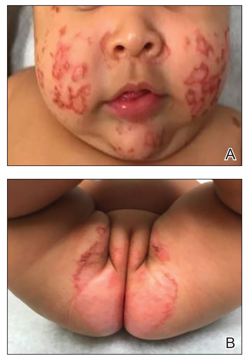
Physical examination revealed the patient had annular and polycyclic, hyperkeratotic, crusted papules and plaques on the cheeks, neck, back, and axillae, as well as the perineum/groin and perianal regions (Figure 1). The differential diagnosis at the time included neonatal lupus, zinc deficiency, and syphilis. Relevant laboratory testing and a shave biopsy of the left axilla were obtained.
CT110005281_Fig1_AB.jpg
%3Cp%3E%3Cstrong%3EFIGURE%201.%3C%2Fstrong%3E%20A%2C%20Annular%20and%20polycyclic%2C%20hyperkeratotic%2C%20crusted%20papules%20and%20plaques%20on%20the%20cheeks.%20B%2C%20Similar%20lesions%20were%20present%20in%20the%20perineum%2Fgroin%20and%20perianal%20regions.%3C%2Fp%3E


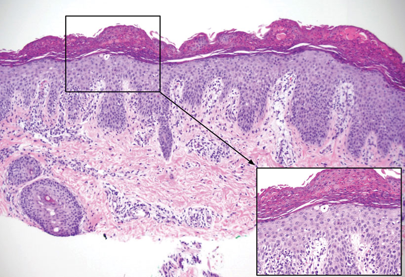
Pertinent laboratory findings included a low zinc level (23 μg/dL [reference range, 26–141 μg/dL]), low alkaline phosphatase level (74 U/L [reference range, 94–486 U/L]), and thrombocytosis (826×109/L [reference range, 150–400×109/L). Results for antinuclear antibody and anti–Sjögren syndrome–related antigen A and B antibody testing were negative. A rapid plasma reagin test was nonreactive. Histologic examination revealed psoriasiform hyperplasia with overlying confluent parakeratosis, focal spongiosis, multiple dyskeratotic keratinocytes, and mitotic figures (Figure 2). Ballooning was evident in focal cells in the subcorneal region in addition to an accompanying lymphocytic infiltrate and occasional neutrophils.
Gillooly_2.jpg
%3Cp%3E%3Cstrong%3EFIGURE%202.%3C%2Fstrong%3E%20Biopsy%20of%20the%20left%20axilla%20showed%20psoriasiform%20hyperplasia%20with%20overlying%20confluent%20parakeratosis%2C%20focal%20spongiosis%2C%20multiple%20dyskeratotic%20keratinocytes%2C%20and%20mitotic%20figures%20(H%26amp%3BE%2C%20original%20magnification%20%C3%9710).%20Focal%20cells%20in%20the%20subcorneal%20region%20showed%20ballooning%20with%20a%20lymphocytic%20infiltrate%20and%20neutrophils%20(inset%3A%20H%26amp%3BE%2C%20original%20magnification%20%C3%9740).%3C%2Fp%3E


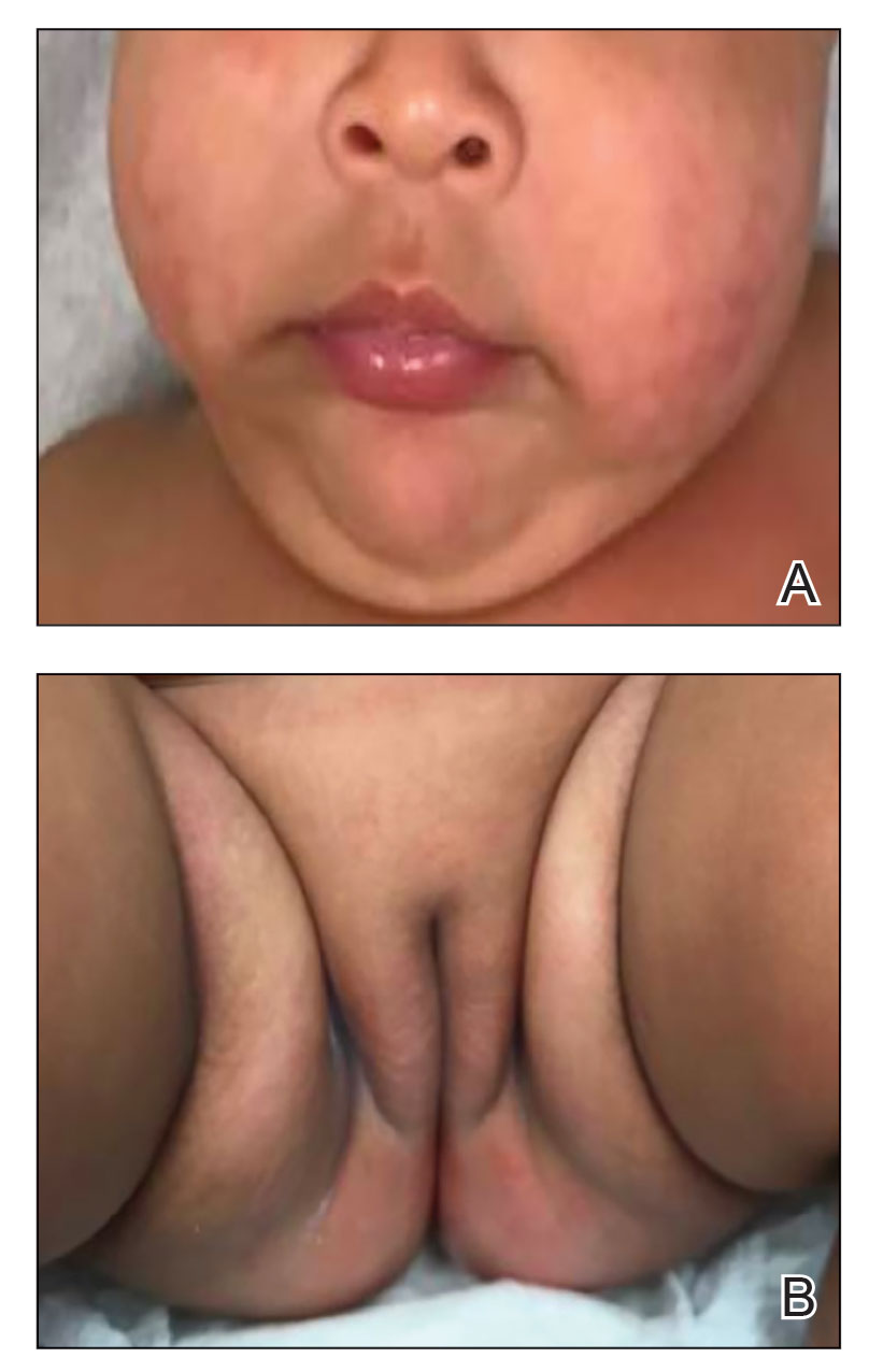
The patient was given a 10-mg/mL suspension of elemental zinc and was advised to take 1 mL (10 mg) by mouth twice daily with food. This dosage equated to 3 mg/kg/d. On follow-up 3 weeks later, the skin began to clear (Figure 3). Follow-up laboratory testing showed an increase in zinc (114 μg/dL) and alkaline phosphatase levels (313 U/L). The patient was able to discontinue the zinc supplementation, and follow-up during the next year revealed no recurrence.
CT110005281_Fig3_AB.jpg
%3Cp%3E%3Cstrong%3EFIGURE%203.%3C%2Fstrong%3E%20A%2C%20Three%20weeks%20after%20treatment%20with%20zinc%20supplementation%2C%20the%20annular%20crusted%20papules%20and%20plaques%20were%20no%20longer%20evident%20on%20the%20cheeks.%20B%2C%20The%20perineum%2Fgroin%20and%20perianal%20regions%20showed%20similar%20clearance.%3C%2Fp%3E

Comment

Etiology of AE—Acrodermatitis enteropathica was first identified in 1942 as an acral rash associated with diarrhea3; in 1973, Barnes and Moynahan4 discovered zinc deficiency as a causal agent for these findings. The causes of AE are further subclassified as either an acquired or inborn etiology. Congenital causes commonly are seen in infants within the first few months of life, whereas acquired forms are seen at any age. Acquired forms in infants can occur from failure of the mother to secrete zinc in breast milk, low maternal serum zinc levels, or other reasons causing low nutritional intake. A single mutation in the SLC30A2 gene has been found to markedly reduce zinc concentrations in breast milk, thus causing zinc deficiency in breastfed infants.5 Other acquired forms can be caused by malabsorption, sometimes after surgery such as intestinal bypass or from intravenous nutrition without sufficient zinc.1 The congenital form of AE is an autosomal-recessive disorder occurring from mutations in the SLC39A4 gene located on band 8q24.3. Affected individuals have a decreased ability to absorb zinc in the small intestine because of defects in zinc transporters ZIP and ZnT.6 Based on our patient’s laboratory findings and history, it is believed that the zinc deficiency was acquired, as the condition normalized with repletion and has not required any supplementation in the year of follow-up. In addition, the absence of a pertinent family history supported an acquired diagnosis, which has various etiologies, whereas the congenital form primarily is a genetic disease.

Diagnosis of AE—The characteristic clinical features of AE include erythematous, dry, scaly papules and plaques that may evolve into crusted, erosive, pustular lesions. These lesions typically are distributed in a periorificial and acral pattern.1,2 Although AE includes the clinical triad of acral and periorificial dermatitis, alopecia, and diarrhea, most cases present with only partial features of this syndrome, as seen in our patient, who presented with only 2 symptoms—dermatitis and diarrhea. The diagnosis of AE is based on clinical and laboratory abnormalities, especially a low serum zinc level. Low levels of zinc-dependent enzymes, such as alkaline phosphatase, may support the diagnosis, as seen in our patient. Histologic evaluation is characteristic but is not diagnostic, as the same findings can be seen in other nutritional disorders. Such findings include confluent parakeratosis associated with a reduced granular layer in early lesions and subsequent ballooning of subcorneal keratinocytes, upper epidermal pallor, and intraepidermal clefts. Late lesions exhibit psoriasiform hyperplasia of the epidermis with less epidermal pallor.7


Management—Treatment of AE includes supplementation with oral elemental zinc; however, there are scant evidence-based recommendations on the exact dose of zinc to be given. Generally, the recommended amount is 3 mg/kg/d.8 For individuals with the congenital form of AE, lifelong zinc supplementation is additionally recommended.9 It is important to recognize this presentation because the patient can develop worsening irritability, severe diarrhea, nail dystrophy, hair loss, immune dysfunction, and numerous ophthalmic disorders if left untreated. Acute zinc toxicity due to excess administration is rare, with symptoms of nausea and vomiting occurring with dosages of 50 to 100 mg/d. Additionally, dosages of up to 70 mg twice weekly have been provided without any toxic effect.10 In our case, 3 mg/kg/d of oral zinc supplementation proved to be effective in resolving the patient’s symptoms of acquired zinc deficiency.

Differential Diagnosis—It is important to note that deficiencies of other nutrients may present as an AE-like eruption called acrodermatitis dysmetabolica (AD). Both diseases may present with the triad of dermatitis, alopecia, and diarrhea; however, AD is associated with inborn errors of metabolism. There have been cases that describe AD in patients with a zinc deficiency in conjunction with a deficiency of branched-chain amino acids.11,12 It is important to consider AD in the differential diagnosis of an AE eruption, especially in the context of a metabolic disorder, as it may affect the treatment plan. One case described the dermatitis of AD as not responding to zinc supplementation alone, while another described improvement after increasing an isoleucine supplementation dose.11,12

Other considerations in the differential diagnoses include AE-like conditions such as biotinidase deficiency, multiple carboxylase deficiency, and essential fatty acid deficiency. An AE-like condition may present with the triad of dermatitis, alopecia, and diarrhea. However, unlike in true AE, zinc and alkaline phosphatase levels tend to be normal in these conditions. Other features seen in AE-like conditions depend on the underlying cause but often include failure to thrive, neurologic defects, ophthalmic abnormalities, and metabolic abnormalities.13
References
  1. Acrodermatitis enteropathica. National Organization for Rare Disorders. Accessed October 16, 2022. https://rarediseases.org/rare-diseases/acrodermatitis-enteropathica/
  2. Perafán-Riveros C, França LFS, Alves ACF, et al. Acrodermatitis enteropathica: case report and review of the literature. Pediatr Dermatol. 2002;19:426-431.
  3. Danbolt N. Acrodermatitis enteropathica. Br J Dermatol. 1979;100:37-40.
  4. Barnes PM, Moynahan EJ. Zinc deficiency in acrodermatitis enteropathica: multiple dietary intolerance treated with synthetic diet. Proc R Soc Med. 1973;66:327-329.
  5. Lee S, Zhou Y, Gill DL, et al. A genetic variant in SLC30A2 causes breast dysfunction during lactation by inducing ER stress, oxidative stress and epithelial barrier defects. Sci Rep. 2018;8:3542.
  6. Kaur S, Sangwan A, Sahu P, et al. Clinical variants of acrodermatitis enteropathica and its co-relation with genetics. Indian J Paediatr Dermatol. 2016;17:35-37.
  7. Dela Rosa KM, James WD. Acrodermatitis enteropathica workup. Medscape. Updated June 4, 2021. Accessed October 16, 2022. https://emedicine.medscape.com/article/1102575-workup#showall
  8. Ngan V, Gangakhedkar A, Oakley A. Acrodermatitis enteropathica. DermNet. Accessed October 16, 2022. https://dermnetnz.org/topics/acrodermatitis-enteropathica/
  9. Ranugha P, Sethi P, Veeranna S. Acrodermatitis enteropathica: the need for sustained high dose zinc supplementation. Dermatol Online J. 2018;24:13030/qt1w9002sr.
  10. Larson CP, Roy SK, Khan AI, et al. Zinc treatment to under-five children: applications to improve child survival and reduce burden of disease. J Health Popul Nutr. 2008;26:356-365.
  11. Samady JA, Schwartz RA, Shih LY, et al. Acrodermatitis enteropathica-like eruption in an infant with nonketotic hyperglycinemia. J Dermatol. 2000;27:604-608.
  12. Flores K, Chikowski R, Morrell DS. Acrodermatitis dysmetabolica in an infant with maple syrup urine disease. Clin Exp Dermatol. 2016;41:651-654.
  13. Jones L, Oakley A. Acrodermatitis enteropathica-like conditions. DermNet. Accessed August 30, 2022. https://dermnetnz.org/topics/acrodermatitis-enteropathica-like-conditions
Article PDF
Author and Disclosure Information

Ms. Vu and Drs. Becker and Osswald are from the University of Texas Health Science Center at San Antonio. Ms. Vu is from the Long School of Medicine, and Drs. Becker and Osswald are from the Department of Dermatology. Dr. Gillooly is from Wright-Patterson Medical Center, Wright-Patterson Air Force Base, Ohio.

The authors report no conflict of interest.

The views expressed in this article are those of the authors and do not necessarily reflect the official policy or position of the Department of the Air Force, Department of Defense, or the US government.

Correspondence: Marie Vu, BSA, University of Texas Health Science Center at San Antonio, 7979 Wurzbach Rd, Grossman, 3rd Floor, San Antonio, TX 78229 (vum1@livemail.uthscsa.edu).

Issue
Cutis - 110(5)
Publications
Topics
Page Number
281-283
Sections
Author and Disclosure Information

Ms. Vu and Drs. Becker and Osswald are from the University of Texas Health Science Center at San Antonio. Ms. Vu is from the Long School of Medicine, and Drs. Becker and Osswald are from the Department of Dermatology. Dr. Gillooly is from Wright-Patterson Medical Center, Wright-Patterson Air Force Base, Ohio.

The authors report no conflict of interest.

The views expressed in this article are those of the authors and do not necessarily reflect the official policy or position of the Department of the Air Force, Department of Defense, or the US government.

Correspondence: Marie Vu, BSA, University of Texas Health Science Center at San Antonio, 7979 Wurzbach Rd, Grossman, 3rd Floor, San Antonio, TX 78229 (vum1@livemail.uthscsa.edu).

Article PDF
Article PDF

Acrodermatitis enteropathica (AE) is a rare disorder of zinc metabolism that typically presents in infancy.1 Although it is clinically characterized by acral and periorificial dermatitis, alopecia, and diarrhea, only 20% of cases present with this triad.2 Zinc deficiency in AE can either be acquired or inborn (congenital). Acquired forms can occur from dietary inadequacy or malabsorption, whereas genetic causes are related to an autosomal-recessive disorder affecting zinc transporters.1 We report a case of a 3-month-old female infant with acquired AE who was successfully treated with zinc supplementation over the course of 3 weeks.

Case Report

A 3-month-old female infant presented to the emergency department with a rash of 2 weeks’ duration. She was born full term with no birth complications. The patient’s mother reported that the rash started on the cheeks, then enlarged and spread to the neck, back, and perineum. The patient also had been having diarrhea during this time. She previously had received mupirocin and cephalexin with no response to treatment. Maternal history was negative for lupus, and the mother’s diet consisted of a variety of foods but not many vegetables. The patient was exclusively breastfed, and there was no pertinent history of similar rashes occurring in other family members.

Physical examination revealed the patient had annular and polycyclic, hyperkeratotic, crusted papules and plaques on the cheeks, neck, back, and axillae, as well as the perineum/groin and perianal regions (Figure 1). The differential diagnosis at the time included neonatal lupus, zinc deficiency, and syphilis. Relevant laboratory testing and a shave biopsy of the left axilla were obtained.

CT110005281_Fig1_AB.jpg
%3Cp%3E%3Cstrong%3EFIGURE%201.%3C%2Fstrong%3E%20A%2C%20Annular%20and%20polycyclic%2C%20hyperkeratotic%2C%20crusted%20papules%20and%20plaques%20on%20the%20cheeks.%20B%2C%20Similar%20lesions%20were%20present%20in%20the%20perineum%2Fgroin%20and%20perianal%20regions.%3C%2Fp%3E


Pertinent laboratory findings included a low zinc level (23 μg/dL [reference range, 26–141 μg/dL]), low alkaline phosphatase level (74 U/L [reference range, 94–486 U/L]), and thrombocytosis (826×109/L [reference range, 150–400×109/L). Results for antinuclear antibody and anti–Sjögren syndrome–related antigen A and B antibody testing were negative. A rapid plasma reagin test was nonreactive. Histologic examination revealed psoriasiform hyperplasia with overlying confluent parakeratosis, focal spongiosis, multiple dyskeratotic keratinocytes, and mitotic figures (Figure 2). Ballooning was evident in focal cells in the subcorneal region in addition to an accompanying lymphocytic infiltrate and occasional neutrophils.

Gillooly_2.jpg
%3Cp%3E%3Cstrong%3EFIGURE%202.%3C%2Fstrong%3E%20Biopsy%20of%20the%20left%20axilla%20showed%20psoriasiform%20hyperplasia%20with%20overlying%20confluent%20parakeratosis%2C%20focal%20spongiosis%2C%20multiple%20dyskeratotic%20keratinocytes%2C%20and%20mitotic%20figures%20(H%26amp%3BE%2C%20original%20magnification%20%C3%9710).%20Focal%20cells%20in%20the%20subcorneal%20region%20showed%20ballooning%20with%20a%20lymphocytic%20infiltrate%20and%20neutrophils%20(inset%3A%20H%26amp%3BE%2C%20original%20magnification%20%C3%9740).%3C%2Fp%3E


The patient was given a 10-mg/mL suspension of elemental zinc and was advised to take 1 mL (10 mg) by mouth twice daily with food. This dosage equated to 3 mg/kg/d. On follow-up 3 weeks later, the skin began to clear (Figure 3). Follow-up laboratory testing showed an increase in zinc (114 μg/dL) and alkaline phosphatase levels (313 U/L). The patient was able to discontinue the zinc supplementation, and follow-up during the next year revealed no recurrence.

CT110005281_Fig3_AB.jpg
%3Cp%3E%3Cstrong%3EFIGURE%203.%3C%2Fstrong%3E%20A%2C%20Three%20weeks%20after%20treatment%20with%20zinc%20supplementation%2C%20the%20annular%20crusted%20papules%20and%20plaques%20were%20no%20longer%20evident%20on%20the%20cheeks.%20B%2C%20The%20perineum%2Fgroin%20and%20perianal%20regions%20showed%20similar%20clearance.%3C%2Fp%3E

Comment

Etiology of AE—Acrodermatitis enteropathica was first identified in 1942 as an acral rash associated with diarrhea3; in 1973, Barnes and Moynahan4 discovered zinc deficiency as a causal agent for these findings. The causes of AE are further subclassified as either an acquired or inborn etiology. Congenital causes commonly are seen in infants within the first few months of life, whereas acquired forms are seen at any age. Acquired forms in infants can occur from failure of the mother to secrete zinc in breast milk, low maternal serum zinc levels, or other reasons causing low nutritional intake. A single mutation in the SLC30A2 gene has been found to markedly reduce zinc concentrations in breast milk, thus causing zinc deficiency in breastfed infants.5 Other acquired forms can be caused by malabsorption, sometimes after surgery such as intestinal bypass or from intravenous nutrition without sufficient zinc.1 The congenital form of AE is an autosomal-recessive disorder occurring from mutations in the SLC39A4 gene located on band 8q24.3. Affected individuals have a decreased ability to absorb zinc in the small intestine because of defects in zinc transporters ZIP and ZnT.6 Based on our patient’s laboratory findings and history, it is believed that the zinc deficiency was acquired, as the condition normalized with repletion and has not required any supplementation in the year of follow-up. In addition, the absence of a pertinent family history supported an acquired diagnosis, which has various etiologies, whereas the congenital form primarily is a genetic disease.

Diagnosis of AE—The characteristic clinical features of AE include erythematous, dry, scaly papules and plaques that may evolve into crusted, erosive, pustular lesions. These lesions typically are distributed in a periorificial and acral pattern.1,2 Although AE includes the clinical triad of acral and periorificial dermatitis, alopecia, and diarrhea, most cases present with only partial features of this syndrome, as seen in our patient, who presented with only 2 symptoms—dermatitis and diarrhea. The diagnosis of AE is based on clinical and laboratory abnormalities, especially a low serum zinc level. Low levels of zinc-dependent enzymes, such as alkaline phosphatase, may support the diagnosis, as seen in our patient. Histologic evaluation is characteristic but is not diagnostic, as the same findings can be seen in other nutritional disorders. Such findings include confluent parakeratosis associated with a reduced granular layer in early lesions and subsequent ballooning of subcorneal keratinocytes, upper epidermal pallor, and intraepidermal clefts. Late lesions exhibit psoriasiform hyperplasia of the epidermis with less epidermal pallor.7


Management—Treatment of AE includes supplementation with oral elemental zinc; however, there are scant evidence-based recommendations on the exact dose of zinc to be given. Generally, the recommended amount is 3 mg/kg/d.8 For individuals with the congenital form of AE, lifelong zinc supplementation is additionally recommended.9 It is important to recognize this presentation because the patient can develop worsening irritability, severe diarrhea, nail dystrophy, hair loss, immune dysfunction, and numerous ophthalmic disorders if left untreated. Acute zinc toxicity due to excess administration is rare, with symptoms of nausea and vomiting occurring with dosages of 50 to 100 mg/d. Additionally, dosages of up to 70 mg twice weekly have been provided without any toxic effect.10 In our case, 3 mg/kg/d of oral zinc supplementation proved to be effective in resolving the patient’s symptoms of acquired zinc deficiency.

Differential Diagnosis—It is important to note that deficiencies of other nutrients may present as an AE-like eruption called acrodermatitis dysmetabolica (AD). Both diseases may present with the triad of dermatitis, alopecia, and diarrhea; however, AD is associated with inborn errors of metabolism. There have been cases that describe AD in patients with a zinc deficiency in conjunction with a deficiency of branched-chain amino acids.11,12 It is important to consider AD in the differential diagnosis of an AE eruption, especially in the context of a metabolic disorder, as it may affect the treatment plan. One case described the dermatitis of AD as not responding to zinc supplementation alone, while another described improvement after increasing an isoleucine supplementation dose.11,12

Other considerations in the differential diagnoses include AE-like conditions such as biotinidase deficiency, multiple carboxylase deficiency, and essential fatty acid deficiency. An AE-like condition may present with the triad of dermatitis, alopecia, and diarrhea. However, unlike in true AE, zinc and alkaline phosphatase levels tend to be normal in these conditions. Other features seen in AE-like conditions depend on the underlying cause but often include failure to thrive, neurologic defects, ophthalmic abnormalities, and metabolic abnormalities.13

Acrodermatitis enteropathica (AE) is a rare disorder of zinc metabolism that typically presents in infancy.1 Although it is clinically characterized by acral and periorificial dermatitis, alopecia, and diarrhea, only 20% of cases present with this triad.2 Zinc deficiency in AE can either be acquired or inborn (congenital). Acquired forms can occur from dietary inadequacy or malabsorption, whereas genetic causes are related to an autosomal-recessive disorder affecting zinc transporters.1 We report a case of a 3-month-old female infant with acquired AE who was successfully treated with zinc supplementation over the course of 3 weeks.

Case Report

A 3-month-old female infant presented to the emergency department with a rash of 2 weeks’ duration. She was born full term with no birth complications. The patient’s mother reported that the rash started on the cheeks, then enlarged and spread to the neck, back, and perineum. The patient also had been having diarrhea during this time. She previously had received mupirocin and cephalexin with no response to treatment. Maternal history was negative for lupus, and the mother’s diet consisted of a variety of foods but not many vegetables. The patient was exclusively breastfed, and there was no pertinent history of similar rashes occurring in other family members.

Physical examination revealed the patient had annular and polycyclic, hyperkeratotic, crusted papules and plaques on the cheeks, neck, back, and axillae, as well as the perineum/groin and perianal regions (Figure 1). The differential diagnosis at the time included neonatal lupus, zinc deficiency, and syphilis. Relevant laboratory testing and a shave biopsy of the left axilla were obtained.

CT110005281_Fig1_AB.jpg
%3Cp%3E%3Cstrong%3EFIGURE%201.%3C%2Fstrong%3E%20A%2C%20Annular%20and%20polycyclic%2C%20hyperkeratotic%2C%20crusted%20papules%20and%20plaques%20on%20the%20cheeks.%20B%2C%20Similar%20lesions%20were%20present%20in%20the%20perineum%2Fgroin%20and%20perianal%20regions.%3C%2Fp%3E


Pertinent laboratory findings included a low zinc level (23 μg/dL [reference range, 26–141 μg/dL]), low alkaline phosphatase level (74 U/L [reference range, 94–486 U/L]), and thrombocytosis (826×109/L [reference range, 150–400×109/L). Results for antinuclear antibody and anti–Sjögren syndrome–related antigen A and B antibody testing were negative. A rapid plasma reagin test was nonreactive. Histologic examination revealed psoriasiform hyperplasia with overlying confluent parakeratosis, focal spongiosis, multiple dyskeratotic keratinocytes, and mitotic figures (Figure 2). Ballooning was evident in focal cells in the subcorneal region in addition to an accompanying lymphocytic infiltrate and occasional neutrophils.

Gillooly_2.jpg
%3Cp%3E%3Cstrong%3EFIGURE%202.%3C%2Fstrong%3E%20Biopsy%20of%20the%20left%20axilla%20showed%20psoriasiform%20hyperplasia%20with%20overlying%20confluent%20parakeratosis%2C%20focal%20spongiosis%2C%20multiple%20dyskeratotic%20keratinocytes%2C%20and%20mitotic%20figures%20(H%26amp%3BE%2C%20original%20magnification%20%C3%9710).%20Focal%20cells%20in%20the%20subcorneal%20region%20showed%20ballooning%20with%20a%20lymphocytic%20infiltrate%20and%20neutrophils%20(inset%3A%20H%26amp%3BE%2C%20original%20magnification%20%C3%9740).%3C%2Fp%3E


The patient was given a 10-mg/mL suspension of elemental zinc and was advised to take 1 mL (10 mg) by mouth twice daily with food. This dosage equated to 3 mg/kg/d. On follow-up 3 weeks later, the skin began to clear (Figure 3). Follow-up laboratory testing showed an increase in zinc (114 μg/dL) and alkaline phosphatase levels (313 U/L). The patient was able to discontinue the zinc supplementation, and follow-up during the next year revealed no recurrence.

CT110005281_Fig3_AB.jpg
%3Cp%3E%3Cstrong%3EFIGURE%203.%3C%2Fstrong%3E%20A%2C%20Three%20weeks%20after%20treatment%20with%20zinc%20supplementation%2C%20the%20annular%20crusted%20papules%20and%20plaques%20were%20no%20longer%20evident%20on%20the%20cheeks.%20B%2C%20The%20perineum%2Fgroin%20and%20perianal%20regions%20showed%20similar%20clearance.%3C%2Fp%3E

Comment

Etiology of AE—Acrodermatitis enteropathica was first identified in 1942 as an acral rash associated with diarrhea3; in 1973, Barnes and Moynahan4 discovered zinc deficiency as a causal agent for these findings. The causes of AE are further subclassified as either an acquired or inborn etiology. Congenital causes commonly are seen in infants within the first few months of life, whereas acquired forms are seen at any age. Acquired forms in infants can occur from failure of the mother to secrete zinc in breast milk, low maternal serum zinc levels, or other reasons causing low nutritional intake. A single mutation in the SLC30A2 gene has been found to markedly reduce zinc concentrations in breast milk, thus causing zinc deficiency in breastfed infants.5 Other acquired forms can be caused by malabsorption, sometimes after surgery such as intestinal bypass or from intravenous nutrition without sufficient zinc.1 The congenital form of AE is an autosomal-recessive disorder occurring from mutations in the SLC39A4 gene located on band 8q24.3. Affected individuals have a decreased ability to absorb zinc in the small intestine because of defects in zinc transporters ZIP and ZnT.6 Based on our patient’s laboratory findings and history, it is believed that the zinc deficiency was acquired, as the condition normalized with repletion and has not required any supplementation in the year of follow-up. In addition, the absence of a pertinent family history supported an acquired diagnosis, which has various etiologies, whereas the congenital form primarily is a genetic disease.

Diagnosis of AE—The characteristic clinical features of AE include erythematous, dry, scaly papules and plaques that may evolve into crusted, erosive, pustular lesions. These lesions typically are distributed in a periorificial and acral pattern.1,2 Although AE includes the clinical triad of acral and periorificial dermatitis, alopecia, and diarrhea, most cases present with only partial features of this syndrome, as seen in our patient, who presented with only 2 symptoms—dermatitis and diarrhea. The diagnosis of AE is based on clinical and laboratory abnormalities, especially a low serum zinc level. Low levels of zinc-dependent enzymes, such as alkaline phosphatase, may support the diagnosis, as seen in our patient. Histologic evaluation is characteristic but is not diagnostic, as the same findings can be seen in other nutritional disorders. Such findings include confluent parakeratosis associated with a reduced granular layer in early lesions and subsequent ballooning of subcorneal keratinocytes, upper epidermal pallor, and intraepidermal clefts. Late lesions exhibit psoriasiform hyperplasia of the epidermis with less epidermal pallor.7


Management—Treatment of AE includes supplementation with oral elemental zinc; however, there are scant evidence-based recommendations on the exact dose of zinc to be given. Generally, the recommended amount is 3 mg/kg/d.8 For individuals with the congenital form of AE, lifelong zinc supplementation is additionally recommended.9 It is important to recognize this presentation because the patient can develop worsening irritability, severe diarrhea, nail dystrophy, hair loss, immune dysfunction, and numerous ophthalmic disorders if left untreated. Acute zinc toxicity due to excess administration is rare, with symptoms of nausea and vomiting occurring with dosages of 50 to 100 mg/d. Additionally, dosages of up to 70 mg twice weekly have been provided without any toxic effect.10 In our case, 3 mg/kg/d of oral zinc supplementation proved to be effective in resolving the patient’s symptoms of acquired zinc deficiency.

Differential Diagnosis—It is important to note that deficiencies of other nutrients may present as an AE-like eruption called acrodermatitis dysmetabolica (AD). Both diseases may present with the triad of dermatitis, alopecia, and diarrhea; however, AD is associated with inborn errors of metabolism. There have been cases that describe AD in patients with a zinc deficiency in conjunction with a deficiency of branched-chain amino acids.11,12 It is important to consider AD in the differential diagnosis of an AE eruption, especially in the context of a metabolic disorder, as it may affect the treatment plan. One case described the dermatitis of AD as not responding to zinc supplementation alone, while another described improvement after increasing an isoleucine supplementation dose.11,12

Other considerations in the differential diagnoses include AE-like conditions such as biotinidase deficiency, multiple carboxylase deficiency, and essential fatty acid deficiency. An AE-like condition may present with the triad of dermatitis, alopecia, and diarrhea. However, unlike in true AE, zinc and alkaline phosphatase levels tend to be normal in these conditions. Other features seen in AE-like conditions depend on the underlying cause but often include failure to thrive, neurologic defects, ophthalmic abnormalities, and metabolic abnormalities.13
References
  1. Acrodermatitis enteropathica. National Organization for Rare Disorders. Accessed October 16, 2022. https://rarediseases.org/rare-diseases/acrodermatitis-enteropathica/
  2. Perafán-Riveros C, França LFS, Alves ACF, et al. Acrodermatitis enteropathica: case report and review of the literature. Pediatr Dermatol. 2002;19:426-431.
  3. Danbolt N. Acrodermatitis enteropathica. Br J Dermatol. 1979;100:37-40.
  4. Barnes PM, Moynahan EJ. Zinc deficiency in acrodermatitis enteropathica: multiple dietary intolerance treated with synthetic diet. Proc R Soc Med. 1973;66:327-329.
  5. Lee S, Zhou Y, Gill DL, et al. A genetic variant in SLC30A2 causes breast dysfunction during lactation by inducing ER stress, oxidative stress and epithelial barrier defects. Sci Rep. 2018;8:3542.
  6. Kaur S, Sangwan A, Sahu P, et al. Clinical variants of acrodermatitis enteropathica and its co-relation with genetics. Indian J Paediatr Dermatol. 2016;17:35-37.
  7. Dela Rosa KM, James WD. Acrodermatitis enteropathica workup. Medscape. Updated June 4, 2021. Accessed October 16, 2022. https://emedicine.medscape.com/article/1102575-workup#showall
  8. Ngan V, Gangakhedkar A, Oakley A. Acrodermatitis enteropathica. DermNet. Accessed October 16, 2022. https://dermnetnz.org/topics/acrodermatitis-enteropathica/
  9. Ranugha P, Sethi P, Veeranna S. Acrodermatitis enteropathica: the need for sustained high dose zinc supplementation. Dermatol Online J. 2018;24:13030/qt1w9002sr.
  10. Larson CP, Roy SK, Khan AI, et al. Zinc treatment to under-five children: applications to improve child survival and reduce burden of disease. J Health Popul Nutr. 2008;26:356-365.
  11. Samady JA, Schwartz RA, Shih LY, et al. Acrodermatitis enteropathica-like eruption in an infant with nonketotic hyperglycinemia. J Dermatol. 2000;27:604-608.
  12. Flores K, Chikowski R, Morrell DS. Acrodermatitis dysmetabolica in an infant with maple syrup urine disease. Clin Exp Dermatol. 2016;41:651-654.
  13. Jones L, Oakley A. Acrodermatitis enteropathica-like conditions. DermNet. Accessed August 30, 2022. https://dermnetnz.org/topics/acrodermatitis-enteropathica-like-conditions
Issue
Cutis - 110(5)
Issue
Cutis - 110(5)
Page Number
281-283
Page Number
281-283
Publications
Publications
Topics
Article Type
Display Headline
Acquired Acrodermatitis Enteropathica in an Infant
Display Headline
Acquired Acrodermatitis Enteropathica in an Infant
Sections
Teambase XML
<?xml version="1.0" encoding="UTF-8"?>
<!--$RCSfile: InCopy_agile.xsl,v $ $Revision: 1.35 $-->
<!--$RCSfile: drupal.xsl,v $ $Revision: 1.7 $-->
<root generator="drupal.xsl" gversion="1.7"> <header> <fileName>Gillooly</fileName> <TBEID>0C02B1C7.SIG</TBEID> <TBUniqueIdentifier>NJ_0C02B1C7</TBUniqueIdentifier> <newsOrJournal>Journal</newsOrJournal> <publisherName>Frontline Medical Communications Inc.</publisherName> <storyname>Gillooly</storyname> <articleType>1</articleType> <TBLocation>Copyfitting-CT</TBLocation> <QCDate/> <firstPublished>20221104T141027</firstPublished> <LastPublished>20221104T141027</LastPublished> <pubStatus qcode="stat:"/> <embargoDate/> <killDate/> <CMSDate>20221104T141027</CMSDate> <articleSource/> <facebookInfo/> <meetingNumber/> <byline>Marie Vu, BSA; Zachary Gillooly, MD; Emily Becker, MD</byline> <bylineText>Marie Vu, BSA; Zachary Gillooly, MD; Emily Becker, MD; Sandra Osswald, MD</bylineText> <bylineFull>Marie Vu, BSA; Zachary Gillooly, MD; Emily Becker, MD</bylineFull> <bylineTitleText/> <USOrGlobal/> <wireDocType/> <newsDocType/> <journalDocType/> <linkLabel/> <pageRange>281-283</pageRange> <citation/> <quizID/> <indexIssueDate/> <itemClass qcode="ninat:text"/> <provider qcode="provider:"> <name/> <rightsInfo> <copyrightHolder> <name/> </copyrightHolder> <copyrightNotice/> </rightsInfo> </provider> <abstract/> <metaDescription>Acrodermatitis enteropathica (AE) is a rare disorder of zinc metabolism that typically presents in infancy.1 Although it is clinically characterized by acral an</metaDescription> <articlePDF>290696</articlePDF> <teaserImage/> <title>Acquired Acrodermatitis Enteropathica in an Infant</title> <deck/> <disclaimer/> <AuthorList/> <articleURL/> <doi/> <pubMedID/> <publishXMLStatus/> <publishXMLVersion>1</publishXMLVersion> <useEISSN>0</useEISSN> <urgency/> <pubPubdateYear>2022</pubPubdateYear> <pubPubdateMonth>November</pubPubdateMonth> <pubPubdateDay/> <pubVolume>110</pubVolume> <pubNumber>5</pubNumber> <wireChannels/> <primaryCMSID/> <CMSIDs> <CMSID>2161</CMSID> </CMSIDs> <keywords> <keyword>dermatopathology</keyword> <keyword> pediatrics</keyword> <keyword> rare disease</keyword> <keyword> acquiried acrodermatitis enteropathica</keyword> <keyword> acrodermatitis enteropathica</keyword> <keyword> AE</keyword> </keywords> <seeAlsos/> <publications_g> <publicationData> <publicationCode>CT</publicationCode> <pubIssueName>November 2022</pubIssueName> <pubArticleType>Original Articles | 2161</pubArticleType> <pubTopics/> <pubCategories/> <pubSections/> <journalTitle>Cutis</journalTitle> <journalFullTitle>Cutis</journalFullTitle> <copyrightStatement>Copyright 2015 Frontline Medical Communications Inc., Parsippany, NJ, USA. All rights reserved.</copyrightStatement> </publicationData> </publications_g> <publications> <term canonical="true">12</term> </publications> <sections> <term canonical="true">45</term> </sections> <topics> <term canonical="true">271</term> <term>204</term> <term>285</term> </topics> <links> <link> <itemClass qcode="ninat:composite"/> <altRep contenttype="application/pdf">images/18002287.pdf</altRep> <description role="drol:caption"/> <description role="drol:credit"/> </link> </links> </header> <itemSet> <newsItem> <itemMeta> <itemRole>Main</itemRole> <itemClass>text</itemClass> <title>Acquired Acrodermatitis Enteropathica in an Infant</title> <deck/> </itemMeta> <itemContent> <p class="abstract">Acrodermatitis enteropathica (AE) is an acquired or inborn (congenital) disorder of zinc metabolism that leads to zinc deficiency. The congenital form typically presents in infants during the first few months of life when they are weaned from breast milk, presenting even earlier in those who are formula fed. Acquired deficiency may be seen at any age. The characteristic clinical features of AE include erythematous, dry, scaly papules and plaques that may evolve into crusted, erosive, pustular lesions. These lesions typically are distributed in an acral and periorificial pattern and are associated with alopecia and diarrhea. Evidence-based recommendations are sparse but generally indicate 3 mg/kg/d of oral zinc supplementation for both congenital and acquired AE. Appropriate dosing helps to avoid acute zinc toxicity involving nausea and vomiting. We report a case of a 3-month-old female infant with acquired AE who was successfully treated with zinc supplementation over the course of 3 weeks. </p> <p> <em><em>Cutis.</em> 2022;110:281-283.</em> </p> <p>Acrodermatitis enteropathica (AE) is a rare disorder of zinc metabolism that typically presents in infancy.<sup>1</sup> Although it is clinically characterized by acral and periorificial dermatitis, alopecia, and diarrhea, only 20% of cases present with this triad.<sup>2</sup> Zinc deficiency in AE can either be acquired or inborn (congenital). Acquired forms can occur from dietary inadequacy or malabsorption, whereas genetic causes are related to an autosomal-recessive disorder affecting zinc transporters.<sup>1</sup> We report a case of a 3-month-old female infant with acquired AE who was successfully treated with zinc supplementation over the course of 3 weeks.</p> <h3>Case Report</h3> <p>A 3-month-old female infant presented to the emergency department with a rash of 2 weeks’ duration. She was born full term with no birth complications. The patient’s mother reported that the rash started on the cheeks, then enlarged and spread to the neck, back, and perineum. The patient also had been having diarrhea during this time. She previously had received mupirocin and cephalexin with no response to treatment. Maternal history was negative for lupus, and the mother’s diet consisted of a variety of foods but not many vegetables. The patient was exclusively breastfed, and there was no pertinent history of similar rashes occurring in other family members. </p> <p>Physical examination revealed the patient had annular and polycyclic, hyperkeratotic, crusted papules and plaques on the cheeks, neck, back, and axillae, as well as the perineum/groin and perianal regions (Figure 1). The differential diagnosis at the time included neonatal lupus, zinc deficiency, and syphilis. Relevant laboratory testing and a shave biopsy of the left axilla were obtained. <br/><br/>Pertinent laboratory findings included a low zinc level (23 <span class="body">μ</span>g/dL [reference range, 26–141 <span class="body">μ</span>g/dL]), low alkaline phosphatase level (74 U/L [reference range, 94–486 U/L]), and thrombocytosis (826<span class="body">×</span>10<sup>9</sup>/L [reference range, 150–400<span class="body">×</span>10<sup>9</sup>/L). Results for antinuclear antibody and anti–Sjögren syndrome–related antigen A and B antibody testing were negative. A rapid plasma reagin test was nonreactive. Histologic examination revealed psoriasiform hyperplasia with overlying confluent parakeratosis, focal spongiosis, multiple dyskeratotic keratinocytes, and mitotic figures (Figure 2). Ballooning was evident in focal cells in the subcorneal region in addition to an accompanying lymphocytic infiltrate and occasional neutrophils. <br/><br/>The patient was given a 10-mg/mL suspension of elemental zinc and was advised to take 1 mL (10 mg) by mouth twice daily with food. This dosage equated to 3 mg/kg/d. On follow-up 3 weeks later, the skin began to clear (Figure 3). Follow-up laboratory testing showed an increase in zinc (114 <span class="body">μ</span>g/dL) and alkaline phosphatase levels (313 U/L). The patient was able to discontinue the zinc supplementation, and follow-up during the next year revealed no recurrence. </p> <h3>Comment</h3> <p><i>Etiology of AE</i>—Acrodermatitis enteropathica was first identified in 1942 as an acral rash associated with diarrhea<sup>3</sup>; in 1973, Barnes and Moynahan<sup>4</sup> discovered zinc deficiency as a causal agent for these findings. The causes of AE are further subclassified as either an acquired or inborn etiology. Congenital causes commonly are seen in infants within the first few months of life, whereas acquired forms are seen at any age. Acquired forms in infants can occur from failure of the mother to secrete zinc in breast milk, low maternal serum zinc levels, or other reasons causing low nutritional intake. A single mutation in the <i>SLC30A2</i> gene has been found to markedly reduce zinc concentrations in breast milk, thus causing zinc deficiency in breastfed infants.<sup>5</sup> Other acquired forms can be caused by malabsorption, sometimes after surgery such as intestinal bypass or from intravenous nutrition without sufficient zinc.<sup>1</sup> The congenital form of AE is an autosomal-recessive disorder occurring from mutations in the <i>SLC39A4</i> gene located on band 8q24.3. Affected individuals have a decreased ability to absorb zinc in the small intestine because of defects in zinc transporters ZIP and ZnT.<sup>6</sup> Based on our patient’s laboratory findings and history, it is believed that the zinc deficiency was acquired, as the condition normalized with repletion and has not required any supplementation in the year of follow-up. In addition, the absence of a pertinent family history supported an acquired diagnosis, which has various etiologies, whereas the congenital form primarily is a genetic disease. </p> <p><i><hl name="4"/>Diagnosis of AE</i>—The characteristic clinical features of AE include erythematous, dry, scaly papules and plaques that may evolve into crusted, erosive, pustular lesions. These lesions typically are distributed in a periorificial and acral pattern.<sup>1,2</sup> Although AE includes the clinical triad of acral and periorificial dermatitis, alopecia, and diarrhea, most cases present with only partial features of this syndrome, as seen in our patient, who presented with only 2 symptoms—dermatitis and diarrhea. The diagnosis of AE is based on clinical and laboratory abnormalities, especially a low serum zinc level. Low levels of zinc-dependent enzymes, such as alkaline phosphatase, may support the diagnosis, as seen in our patient. Histologic evaluation is characteristic but is not diagnostic, as the same findings can be seen in other nutritional disorders. Such findings include confluent parakeratosis associated with a reduced granular layer in early lesions and subsequent ballooning of subcorneal keratinocytes, upper epidermal pallor, and intraepidermal clefts. Late lesions exhibit psoriasiform hyperplasia of the epidermis with less epidermal pallor.<sup>7</sup> <br/><br/><i>Management</i>—Treatment of AE includes supplementation with oral elemental zinc; however, there are scant evidence-based recommendations on the exact dose of zinc to be given. Generally, the recommended amount is 3 mg/kg/d.<sup>8</sup> For individuals with the congenital form of AE, lifelong zinc supplementation is additionally recommended.<sup>9</sup> It is important to recognize this presentation because the patient can develop worsening irritability, severe diarrhea, nail dystrophy, hair loss, immune dysfunction, and numerous ophthalmic disorders if left untreated. Acute zinc toxicity due to excess administration is rare, with symptoms of nausea and vomiting occurring with dosages of 50 to 100 mg/d. Additionally, dosages of up to 70 mg twice weekly have been provided without any toxic effect.<sup>10</sup> In our case, 3 mg/kg/d of oral zinc supplementation proved to be effective in resolving the patient’s symptoms of acquired zinc deficiency.<br/><br/><i>Differential Diagnosis</i>—It is important to note that deficiencies of other nutrients may present as an AE-like eruption called acrodermatitis dysmetabolica (AD). Both diseases may present with the triad of dermatitis, alopecia, and diarrhea; however, AD is associated with inborn errors of metabolism. There have been cases that describe AD in patients with a zinc deficiency in conjunction with a deficiency of branched-chain amino acids.<sup>11,12</sup> It is important to consider AD in the differential diagnosis of an AE eruption, especially in the context of a metabolic disorder, as it may affect the treatment plan. One case described the dermatitis of AD as not responding to zinc supplementation alone, while another described improvement after increasing an isoleucine supplementation dose.<sup>11,12</sup> <br/><br/>Other considerations in the differential diagnoses include AE-like conditions such as biotinidase deficiency, multiple carboxylase deficiency, and essential fatty acid deficiency. An AE-like condition may present with the triad of dermatitis, alopecia, and diarrhea. However, unlike in true AE, zinc and alkaline phosphatase levels tend to be normal in these conditions. Other features seen in AE-like conditions depend on the underlying cause but often include failure to thrive, neurologic defects, ophthalmic abnormalities, and metabolic abnormalities.<sup>13</sup> </p> <h2>References </h2> <p class="reference"> 1. Acrodermatitis enteropathica. National Organization for Rare Disorders. Accessed October 16, 2022. https://rarediseases.org/rare-diseases/acrodermatitis-enteropathica/</p> <p class="reference"> 2. <a href="https://onlinelibrary.wiley.com/action/doSearch?ContribAuthorStored=Peraf%C3%A1n-Riveros%2C+Claudia">Perafán-Riveros</a> C,<a href="https://onlinelibrary.wiley.com/action/doSearch?ContribAuthorStored=Fran%C3%A7a%2C+Luis+Fernando+Sayago"> França </a>LFS, <a href="https://onlinelibrary.wiley.com/action/doSearch?ContribAuthorStored=Alves%2C+Ana+Cristina+Fortes">Alves </a>ACF, et al. Acrodermatitis enteropathica: case report and review of the literature. <i>Pediatr Dermatol</i>. 2002;19:426-431.<br/><br/> 3. Danbolt N. Acrodermatitis enteropathica. <i>Br J Dermatol</i>. 1979;100:37-40. <br/><br/> 4. Barnes PM, Moynahan EJ. Zinc deficiency in acrodermatitis enteropathica: multiple dietary intolerance treated with synthetic diet. <i>Proc R Soc Med</i>. 1973;66:327-329. <br/><br/> 5. Lee S, Zhou Y, Gill DL, et al<i>.</i> A genetic variant in <i>SLC30A2</i> causes breast dysfunction during lactation by inducing ER stress, oxidative stress and epithelial barrier defects. <i>Sci Rep.</i> 2018;8:3542. <br/><br/> 6. Kaur S, Sangwan A, Sahu P, et al. Clinical variants of acrodermatitis enteropathica and its co-relation with genetics. <i>Indian J Paediatr Dermatol</i>. 2016;17:35-37.<br/><br/> 7. Dela Rosa KM, James WD. Acrodermatitis enteropathica workup. <i>Medscape</i>. Updated June 4, 2021. Accessed October 16, 2022. https://emedicine.medscape.com/article/1102575-workup#showall<br/><br/> 8. Ngan V, Gangakhedkar A, Oakley A. Acrodermatitis enteropathica. DermNet. Accessed October 16, 2022. https://dermnetnz.org/topics/acrodermatitis-enteropathica/ <br/><br/> 9. Ranugha P, Sethi P, Veeranna S. Acrodermatitis enteropathica: the need for sustained high dose zinc supplementation. <i>Dermatol Online J</i>. 2018;24:13030/qt1w9002sr. <br/><br/>10. Larson CP, Roy SK, Khan AI, et al. Zinc treatment to under-five children: applications to improve child survival and reduce burden of disease. <i>J Health Popul Nutr</i>. 2008;26:356-365.<br/><br/>11. Samady JA, Schwartz RA, Shih LY, et al. Acrodermatitis enteropathica-like eruption in an infant with nonketotic hyperglycinemia. <i>J Dermatol</i>. 2000;27:604-608.<br/><br/>12. Flores K, Chikowski R, Morrell DS. Acrodermatitis dysmetabolica in an infant with maple syrup urine disease. <i>Clin Exp Dermatol</i>. 2016;41:651-654. <br/><br/>13. Jones L, Oakley A. Acrodermatitis enteropathica-like conditions. DermNet. Accessed August 30, 2022. https://dermnetnz.org/topics/acrodermatitis-enteropathica-like-conditions</p> </itemContent> </newsItem> <newsItem> <itemMeta> <itemRole>bio</itemRole> <itemClass>text</itemClass> <title/> <deck/> </itemMeta> <itemContent> <p class="disclosure">Ms. Vu and Drs. Becker and Osswald are from the University of Texas Health Science Center at San Antonio. Ms. Vu is from the Long School of Medicine, and Drs. Becker and Osswald are from the Department of Dermatology. Dr. Gillooly is from Wright-Patterson Medical Center, </p> <p class="disclosure">Wright-Patterson Air Force Base, Ohio. <br/><br/>The authors report no conflict of interest. <br/><br/>The views expressed in this article are those of the authors and do not necessarily reflect the official policy or position of the Department of the Air Force, Department of Defense, or the US government. <br/><br/>Correspondence: Marie Vu, BSA, University of Texas Health Science Center at San Antonio, 7979 Wurzbach Rd, Grossman, 3rd Floor, San Antonio, TX 78229 (vum1@livemail.uthscsa.edu).<br/><br/>doi:10.12788/cutis.0642</p> </itemContent> </newsItem> <newsItem> <itemMeta> <itemRole>in</itemRole> <itemClass>text</itemClass> <title/> <deck/> </itemMeta> <itemContent> <p class="insidehead">Practice <strong>Points</strong></p> <ul class="insidebody"> <li>Although clinically characterized by the triad of acral and periorificial dermatitis, alopecia, and diarrhea, most cases of acrodermatitis enteropathica (AE) present with only partial features of this syndrome.</li> <li>Low levels of zinc-dependent enzymes such as alkaline phosphatase may support the diagnosis of AE.</li> </ul> </itemContent> </newsItem> </itemSet></root>
Inside the Article

Practice Points

  • Although clinically characterized by the triad of acral and periorificial dermatitis, alopecia, and diarrhea, most cases of acrodermatitis enteropathica (AE) present with only partial features of this syndrome.
  • Low levels of zinc-dependent enzymes such as alkaline phosphatase may support the diagnosis of AE.
Disallow All Ads
Content Gating
No Gating (article Unlocked/Free)
Alternative CME
Disqus Comments
Default
Use ProPublica
Hide sidebar & use full width
render the right sidebar.
Conference Recap Checkbox
Not Conference Recap
Clinical Edge
Display the Slideshow in this Article
Medscape Article
Display survey writer
Reuters content
Disable Inline Native ads
WebMD Article
Article PDF Media
Image
Teambase ID
18002287.SIG
Disable zoom
Off