User login
Disease Education
Q) The billing consultant who came to our office said we can increase our reimbursements if we also provide education to our patients with chronic kidney disease (CKD). Is she right?
In 2010, under an omnibus bill, kidney disease education (KDE) classes were added as a Medicare benefit. These are for patients with stage 4 CKD (glomerular filtration rate, 15-30 mL/min) and are to be taught by a qualified instructor (MD, PA, NP, or CNS).
The classes can be taught on the same day as an evaluation/management visit (ie, a regular office visit) and are compensated by the hour. (Side note: Medicare defines an hour as 31 minutes—yes, 31 minutes; Medicare takes for granted that you will also need time to chart!) You can teach two classes in the same day. Thus, if you wanted to, you could have a patient arrive for an office visit, then teach two 31-minute classes, and bill all three for the same day. The entire visit could be 75 minutes (although this may be exhausting for this population).
You can conduct the classes in a number of settings, including nursing homes, hospitals, skilled nursing facilities, the office, or even the patient’s home. Many PAs and NPs have taught these classes to hospitalized patients who have lost kidney function due to an acute insult (ie, medications, dehydration, contrast).
Each Medicare recipient has a lifetime benefit of six KDE classes. The CPT billing code is G0420 for an individual class and G0421 for a group class. You must make sure you also code for the stage 4 CKD diagnosis (code: 585.4).
Congress stipulated KDE classes must include information on causes, symptoms, and treatments and comprise a posttest at a specific health literacy level. To make it simple, the National Kidney Foundation Council of Advanced Practitioners (NKF-CAP) has developed two free Power-Point slide decks for clinicians to use in KDE classes (available at www.kidney.org/professionals/CAP/sub_resources#kde). References and updated peer-reviewed guidelines are included. You can print the slides for your patients and/or share the program with your colleagues.
Many nephrology practitioners teach the two slide sets over and over, because patients only retain one-third of the info we provide them on a given day. So if you teach each slide set three times, you have six lifetime classes—and hopefully the patient will have retained everything.
One caveat: Before you initiate KDE classes for a specific patient, check with the patient’s nephrology group (we hope at stage 4 the patient has a nephrologist) to see if they are providing the education. —KZ and JD
Kim Zuber, PA-C, MSPS, DFAAPA
American Academy of Nephrology PAs
Jane S. Davis, CRNP, DNP
Division of Nephrology at the University of Alabama
National Kidney Foundation's Council of Advanced Practitioners
| Clinician Reviews in partnership with |
Renal Consult is edited by Jane S. Davis, CRNP, DNP, a member of the Clinician Reviews editorial board, who is a nurse practitioner in the Division of Nephrology at the University of Alabama at Birmingham and is the communications chairperson for the National Kidney Foundation’s Council of Advanced Practitioners (NKF-CAP); and Kim Zuber, PA-C, MSPS, DFAAPA, a retired physician assistant who works with the American Academy of Nephrology PAs and is also past chair of the NKF-CAP. This month’s responses were authored by Della Connor, PhD, RN, FNP-BC, who is an Assistant Professor at Stephen F. Austin State University in Nacogdoches, Texas, and practices at East Texas Nephrology Associates in Lufkin, and the department editors.
Q) The billing consultant who came to our office said we can increase our reimbursements if we also provide education to our patients with chronic kidney disease (CKD). Is she right?
In 2010, under an omnibus bill, kidney disease education (KDE) classes were added as a Medicare benefit. These are for patients with stage 4 CKD (glomerular filtration rate, 15-30 mL/min) and are to be taught by a qualified instructor (MD, PA, NP, or CNS).
The classes can be taught on the same day as an evaluation/management visit (ie, a regular office visit) and are compensated by the hour. (Side note: Medicare defines an hour as 31 minutes—yes, 31 minutes; Medicare takes for granted that you will also need time to chart!) You can teach two classes in the same day. Thus, if you wanted to, you could have a patient arrive for an office visit, then teach two 31-minute classes, and bill all three for the same day. The entire visit could be 75 minutes (although this may be exhausting for this population).
You can conduct the classes in a number of settings, including nursing homes, hospitals, skilled nursing facilities, the office, or even the patient’s home. Many PAs and NPs have taught these classes to hospitalized patients who have lost kidney function due to an acute insult (ie, medications, dehydration, contrast).
Each Medicare recipient has a lifetime benefit of six KDE classes. The CPT billing code is G0420 for an individual class and G0421 for a group class. You must make sure you also code for the stage 4 CKD diagnosis (code: 585.4).
Congress stipulated KDE classes must include information on causes, symptoms, and treatments and comprise a posttest at a specific health literacy level. To make it simple, the National Kidney Foundation Council of Advanced Practitioners (NKF-CAP) has developed two free Power-Point slide decks for clinicians to use in KDE classes (available at www.kidney.org/professionals/CAP/sub_resources#kde). References and updated peer-reviewed guidelines are included. You can print the slides for your patients and/or share the program with your colleagues.
Many nephrology practitioners teach the two slide sets over and over, because patients only retain one-third of the info we provide them on a given day. So if you teach each slide set three times, you have six lifetime classes—and hopefully the patient will have retained everything.
One caveat: Before you initiate KDE classes for a specific patient, check with the patient’s nephrology group (we hope at stage 4 the patient has a nephrologist) to see if they are providing the education. —KZ and JD
Kim Zuber, PA-C, MSPS, DFAAPA
American Academy of Nephrology PAs
Jane S. Davis, CRNP, DNP
Division of Nephrology at the University of Alabama
National Kidney Foundation's Council of Advanced Practitioners
Liver Transplant and HCV: The New Horizon
The question of when to treat liver transplant patients with chronic hepatitis C virus (HCV) infection is vital. HCV is the diagnosis most commonly leading to liver transplantation, and recurrence of HCV after transplant is nearly universal. An estimated 30% of HCV liver transplant recipients experience recurrence within five years of transplantation, resulting in death or loss of the allograft because of cirrhosis or graft failure.1 Thus, if we knew when and how to treat HCV in this population, we could envision a world in which liver transplants would not be required—and certainly one in which retransplant because of HCV would never be needed.
In planning to treat HCV-positive patients who need a liver transplant, the question arises: Do we treat them preoperatively, perioperatively, or postoperatively? And if we select postoperative treatment, do we wait until the graft shows HCV recurrence, or do we treat prophylactically—knowing that recurrence is likely?
Results from multiple small studies of interferon-free treatment of HCV, both prior to and following recurrence of HCV posttransplant, have been reported. The SOLAR trial, which used ribavirin plus sofosbuvir, demonstrated a sustained viral response (SVR) rate of 70%.2 The SOLAR-1 trial of ledipasvir/sofosbuvir (Harvoni®) plus ribavirin in patients with genotype 1 or 4 who were treated following posttransplant recurrence showed an average SVR rate of 96%.3 Duration of treatment varied, depending on genotype and presence of cirrhosis, and there was differing dosing and addition of ribavirin at multiple doses. A 10% mortality rate was reported, but deaths occurred mainly in patients with severe cirrhosis.
A small trial was also conducted of genotype 3 patients treated with ledipasvir/sofosbuvir, with or without ribavirin.4 The addition of ribavirin appeared to shorten treatment, but patients in both treatment groups had excellent rates of SVR. However, further study is needed before this combination of medications can be recommended and/or FDA approved. Full details are available at http://hcvguidelines.org.
Ombitasvir/paritaprevir/ ritonavir with dasabuvir (Viekira Pak™) plus ribavirin for 24 weeks was evaluated in patients with HCV recurrence following liver transplant.5 This group of patients had very minimal fibrosis and essentially normal liver function. In this study group, an SVR rate exceeding 95% was achieved.
Continue for data on treatment of patients >>
The data on treatment of patients who need a transplant or who experience HCV recurrence posttransplant are very encouraging. If the SVR rates are maintained over time, it may be possible to significantly decrease the requirement for retransplantation—or even transplant in the first place. As transplant is not without significant morbidity and mortality, especially in the recurrence group, avoiding it would allow patients to live longer and healthier lives. More work is needed to understand the optimal time to treat and the best and safest regimens. Numerous trials evaluating these important management questions are ongoing.6-9
In the not-too-distant future, the requirement for liver transplantation in patients with chronic HCV infection is likely to decrease substantially. A recent New England Journal of Medicine editorial discussing liver allocation concluded that transplant centers now treating significant numbers of patients with HCV cirrhosis may soon be idle.10
If only we could be so lucky ...
REFERENCES
1. Watt K, Veldt B, Charlton M. A practical guide to the management of HCV infection following liver transplantation. Am J Transplant. 2009;9(8):1707-1713.
2. Charlton M, Gane E, Manns MP, et al. Sofosbuvir and ribavirin for treatment of compensated recurrent hepatitis C virus infection after liver transplantation. Gastroenterology. 2015;148(1):108-117.
3. Reddy KR, Everson GT, Flamm SL, et al. Ledipasvir/sofosbuvir with ribavirin for the treatment of HCV in patients with post transplant recurrence: preliminary results of a prospective, multicenter study. Presented at: 65th Annual Meeting of the American Association for the Study of Liver Diseases; November 7-11, 2014; Boston, MA. Abstract 8.
4. Kohler JJ, Nettles JH, Amblard F, et al. Approaches to hepatitis C treatment and cure using NS5A inhibitors. Infect Drug Resist. 2014;7:41-56.
5. Curry MP, Forns X, Chung RT, et al. Sofosbuvir and ribavirin prevent recurrence of HCV infection after liver transplantation: an open-label study. Gastroenterology. 2015;148(1):100-107.
6. A Phase 2 open-label study in patients with recurrent genotype 1 hepatitis C post–orthotopic liver transplant to explore the safety and efficacy of simeprevir and sofosbuvir with and without ribavirin. https://clinicaltrials.gov/ct2/show/NCT02165189?term=%22olysio%22&rank=1. Accessed February 15, 2015.
7. Phase 2, open-label study to investigate the pharmacokinetics, efficacy, safety, and tolerability of the combination of simeprevir (TMC435), daclatasvir (BMS-790052) and ribavirin (RBV) in subjects with recurrent chronic hepatitis C genotype 1b infection after orthotopic liver transplantation. https://clinicaltrials.gov/ct2/show/NCT01938625?term=%22olysio%22&rank=4. Accessed February 15, 2015.
8. A Phase 3, multicenter, open-label, single-arm study to investigate the efficacy and safety of a 12-week regimen of simeprevir in combination with sofosbuvir in treatment-naïve or -experienced subjects with chronic genotype 4 hepatitis C virus infection. https://clinicaltrials.gov/ct2/show/NCT02250807?term=%22olysio%22&rank=7. Accessed February 15, 2015.
9. A Phase 2 open-label study in patients with recurrent genotype 1 hepatitis C post–orthotopic liver transplant to explore the safety and efficacy of simeprevir and sofosbuvir with and without ribavirin. https://clinicaltrials.gov/ct2/show/NCT02165189?term=%22sovaldi%22&rank=6. Accessed February 15, 2015.
10. Lamas D, Rosenbaum L. Very complicated math: reconfiguring organ allocation. N Engl J Med. 2014;371(26):2447-2450.
CKD and HCV: Do We Know What We’re Doing?
Oh what a tangled web it weaves, this virus called hepatitis C. HCV has evolved over several thousand years into seven genotypes and multiple subtypes, making identification and treatment extremely variable.1 The genotypes are usually geographically specific, with 70% to 75% of HCV patients in the United States classed as genotype 1.2 Genotypes 2 and 3 account for most of the rest.3
While genotype does not impact the course or severity of the disease, it does affect the response to treatment. With today’s newest treatments, success rates for sustained viral response (SVR) are as high as 90% in patients with genotype 1. However, the treatment is not without cost: About $100,000 for patients without cirrhosis and even more for those with it, due to the extended length of treatment.
Chronic kidney disease (CKD) and HCV overlap in 10% to nearly 59% of the population in some geographic areas.4 Those with CKD who have had a previous kidney transplant, been on dialysis, and/or been treated prior to the introduction of epoetin alfa (Epogen®, when blood transfusions were common) are more likely to be HCV-positive.
Unfortunately, patients often convert to HCV while on hemodialysis.4 This largely represents a nosocomial infection related to parenteral exposure. In the dialysis patient, the course of HCV is more aggressive than in others, with high morbidity—often resulting from cirrhosis and/or hepatocellular carcinoma.
With new HCV drugs available, CKD patients would appear to be a prime population to treat. In order to gain marketing approval, the FDA requires testing of new drugs in patients with varying degrees of renal impairment. However, the requirement is for “moderate” CKD, defined by the FDA as a glomerular filtration rate (GFR) of at least 30 mL/min/1.73m2. The FDA does not require testing for patients with a lower GFR, or what nephrology practices refer to as stage 4 or stage 5 CKD; thus, no data are available for this population. The available pharmacokinetic data for the recently approved regimens of sofosbuvir/ledipasvir (Harvoni®) and of ombitasvir/paritaprevir/ritonavir with dasabuvir (Viekira Pak™) suggest that no dosing modifications are necessary in the moderately impaired kidney population.
Continue for the newest HCV drugs >>
The recent approvals of Harvoni and Viekira Pak are considered game-changers in the treatment of chronic HCV infection: They demonstrate that interferon is no longer always necessary. Interferon, especially in combination with ribavirin, was especially toxic for patients in the kidney failure population, although the pegylated form of interferon was less problematic. FDA approvals for Harvoni and Viekira Pak were based on clinical data demonstrating SVR rates exceeding 90%, even in traditionally difficult-to-treat populations (ie, patients with genotype 1, African-Americans, those with high viral load levels), which include many CKD patients.
The lack of clinical data in the CKD stage 4/5 population at the time of drug approval raises significant management challenges for the HCV-positive CKD patient. It is encouraging, however, that the FDA is requiring that all new medications be tested in the renally impaired population, even if not in the severely impaired groups. In the interim, and based solely on renal impairment study results for patients with GFR at or exceeding 30 mL/min/1.73m2, HCV guidelines issued jointly by the American Association for the Study of Liver Diseases and the Infectious Diseases Society of America5 include the recommendation that these new HCV medications be considered for use in the CKD population. Furthermore, there are no dosing adjustments recommended for any of the new HCV medications in stage 4 or stage 5 CKD due to lack of data. However, until more data become available, CKD patients must be managed on a case-by-case basis after consultation with a renal expert, with close attention to changes in GFR and adverse events.
When these new HCV medications are used in the general population and those with significant CKD, postmarketing reports to the FDA will be vital. Practitioners are encouraged to report adverse outcomes to the FDA MedWatch system (https://www.accessdata.fda.gov/scripts/medwatch). As any nephrology practice will tell you, we are starting to get phone calls from our hepatology colleagues asking for guidance, and the next few years will be a fascinating time for all of us. The promise of virologic cure for our HCV-positive patients is tangible and on the horizon.
REFERENCES
1. Maeterns G, Stuyyver L. Genotypes and genetic variation of hepatitis C cirus. Hep C Review. Dec 1998;ed 23.
2. Spach DH, Kim HN. Treatment of HCV genotype 1 (2015). www.hepatitisc.uw.edu/go/treatment-infection/treatment-genotype-1/core-concept/all. Accessed February 15, 2015.
3. Hepatitis C Technical Advisory Group. Hepatitis C genotypes and quasispecies (2005). www.hepatitis.va.gov/provider/reviews/genotypes.asp. Accessed February 15, 2015.
4. Carvalho-Filho RJ, Feldner AC, Silva AE, Ferraz ML. Management of hepatitis C in patients with chronic kidney disease. World J Gastroenterol. 2015;21(2):408-422.
5. American Association for the Study of Liver Diseases, Infectious Diseases Society of America. Recommendations for testing, managing, and treating hepatitis C. http://hcvguidelines.org. Accessed February 15, 2015.
Treatment for Kidney Stones
Since the 1940s, scientists have been stymied by the cause of the “sliding stones” in California’s Death Valley. How, without human intervention, does a 700-lb boulder move from one place to another, leaving a distinctive, meandering trail? Finally, in August 2014, recorded pictures showed that the power of water, freezing and melting, actually moves these boulders.1 Those who treat kidney stones (nephrolithiasis) have always known the power of liquid to move “boulders.”
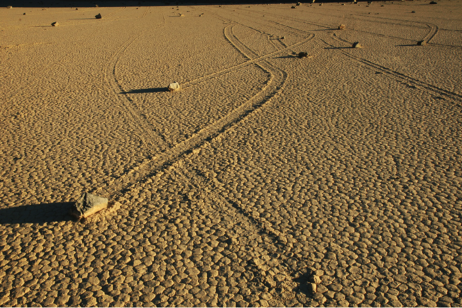
The Sliding Stones of Death Valley, California
The incidence of kidney stones in the United States has risen from 3.8% of the population in the 1970s to 8.8% of the population in the 2010s.2,3 Stones are three times more common in whites than in nonwhites and twice as common in men as in women.4 The cost of kidney stones in the US, including hospitalizations, surgery, and time lost from work, is calculated at $5 billion per year.5
Next page: History of treatment >>
Until the early 1980s, the treatment of choice for a kidney stone was “watchful waiting,” with hydration and pain management. A patient would be given a piece of cheesecloth or a basket, and instructed to urinate through it in order to “catch” the stone. When a stone finally passed, its chemical composition was analyzed. In patients with stones that were too large to pass or found in a location that made passage unlikely, surgical attempts were made to retrieve the stones. These surgeries could be open or “closed” (endoscopic), but they often caused permanent damage to the ureters and/or renal pelvis. Not surprisingly, the introduction of extracorporeal shock wave lithotripsy (ESWL) in the 1980s caused an immediate sensation.6
Stones can remain asymptomatic for some time—only to be found incidentally on radiologic exam for another condition.2 But when a patient presents with “classic” symptoms of kidney stones—colicky flank pain, hematuria, testicular pain (males only!), urinary frequency and urgency, nausea and vomiting—a helical CT is ordered to determine stone position; knowing this is vital to treatment. If the stone is non-obstructing and measures less than 10 mm, medical management is the first choice.7,8 This consists of IV or oral fluids, accompanied by narcotic and/or non-narcotic pain medications, as kidney stone pain can be excruciating. NSAIDs alone are rarely strong enough, and their use incurs a risk for intrinsic kidney damage.
If conservative care does not allow the stone to pass, alpha-adrenergic blockers and/or calcium channel blockers are added.9 In the case of cysteine stones, alkalization of the urine will help dissolve the stone.2 Only 20% of stones are found in the ureter; the vast majority (up to 70%) are lower urethral stones (LUS). Use of tamsulosin has been shown to move LUS stones at a faster rate, so long as they measure less than 10 mm.10
Before treating the stone patient with acute presentation, the urology practitioner may wait a couple of days to see whether the stone passes. The treatment choice then depends on the size of the stone and the position at presentation. If a stone measures less than 6 mm, medical management will be chosen.2 In fact, for smaller, nonobstructing stones, fluids, pain control, and alpha-blockers have been shown in the literature to produce a better outcome than other treatment options.9
For stones larger than 6 mm, or those causing an obstruction or a complication (pyelonephritis or urosepsis), removal is imperative.4 Modality choice depends on the position of the stone and the size of the patient. ESWL, the least invasive means, is the treatment of choice.2 However, as obesity becomes more prevalent (with its underlying metabolic abnormalities), the effectiveness of ESWL may be hindered by the obese patient’s body mass. That said, some manufacturers are increasing the reach of their lithotripsy machines for just this reason.11
Continue for more treatment options >>
Stenting, another option to allow stone fragments to pass, can be uncomfortable, and it requires anesthesia; however, its use is associated with minimal damage to the ureter.2 Percutaneous nephrolithotomy, with or without a basket or a nephrostomy tube, can also be used.12 This method is often needed in patients with a large “stone burden.”2 Open procedures to remove stones, though the gold standard in the early 1980s, are rarely required today.
Recurrence rates for stones can be as high as 50%.13 Depending on the type of stone, certain interventions are essential to reduce recurrence. The ROKS stone calculator can be used to identify patients at increased risk for stone recurrence.14
REFERENCES
1. Norris RD, Norris JM, Lorenz RD, et al. Sliding rocks on racetrack playa, Death Valley National Park: first observation of rocks in motion. PLoS One. 2014;9(8):e105948.
2. Curhan G. Nephrolithiasis. In: Gilbert SJ, Weiner DE, eds. National Kidney Foundation’s Primer on Kidney Diseases. 6th ed. Philadelphia, PA: Elsevier; 2013:405-411.
3. Trinchieri A. Epidemiology of urolithiasis: an update. Clin Cases Miner Bone Metab. 2008;5(2):101-106.
4. Worcester EM, Coe FL. Clinical practice: calcium kidney stones. N Engl J Med. 2010;363(10):954-963.
5. Saigal CS, Joyce G, Timilsina AR; Urologic Diseases in America Project. Direct and indirect costs of nephrolithiasis in an employed population: opportunity for disease management? Kidney Int. 2005;68(4):1808-1814.
6. Segura JW, Patterson DE, LeRoy AJ, et al. Percutaneous removal of kidney stones: review of 1,000 cases. J Urol. 1985;134(6):1077-1081.
7. Wells CG, Chandrashekar KB, Jyothirmayi GN, et al. Kidney stones: current diagnosis and management. Clinician Reviews. 2012;22(2):31-37.
8. Coll DM, Varanelli MJ, Smith RC. Relationship of spontaneous passage of ureteral calculi to stone size and location as revealed by unenhanced helical CT. AJR Am J Roentgenol. 2002;178(1):101-103.
9. Campschroer T, Zhu Y, Duijvesz D, et al. Alpha-blockers as medical expulsive therapy for ureteral stones. Cochrane Database Syst Rev. 2014;4:CD008509.
10. Erturhan S, Erbagci A, Yagci F, et al. Comparative evaluation of efficacy of use of tamsulosin and/or tolterodine for medical treatment of distal ureteral stones. Urology. 2007;69(4):633-636.
11. Mezentsev VA. Extracorporeal shock wave lithotripsy in the treatment of renal pelvicalyceal stones in morbidly obese patients. Int Braz J Urol. 2005;31(2):105-110.
12. Amer T, Ahmed K, Bultitude M, et al. Standard versus tubeless percutaneous nephrolithotomy: a systematic review. Urol Int. 2012;88(4):373-82.
13. Ljunghall S. Incidence of upper urinary tract stones. Miner Electrolyte Metab. 1987;13(4):220-227.
14. Rule AD, Lieske JC, Li X, et al. The ROKS Nomogram for Predicting a Second Symptomatic Stone Episode. J Am Soc Nephrol. 2014 Aug 7. [Epub ahead of print]
Since the 1940s, scientists have been stymied by the cause of the “sliding stones” in California’s Death Valley. How, without human intervention, does a 700-lb boulder move from one place to another, leaving a distinctive, meandering trail? Finally, in August 2014, recorded pictures showed that the power of water, freezing and melting, actually moves these boulders.1 Those who treat kidney stones (nephrolithiasis) have always known the power of liquid to move “boulders.”
The Sliding Stones of Death Valley, California
The incidence of kidney stones in the United States has risen from 3.8% of the population in the 1970s to 8.8% of the population in the 2010s.2,3 Stones are three times more common in whites than in nonwhites and twice as common in men as in women.4 The cost of kidney stones in the US, including hospitalizations, surgery, and time lost from work, is calculated at $5 billion per year.5
Next page: History of treatment >>
Until the early 1980s, the treatment of choice for a kidney stone was “watchful waiting,” with hydration and pain management. A patient would be given a piece of cheesecloth or a basket, and instructed to urinate through it in order to “catch” the stone. When a stone finally passed, its chemical composition was analyzed. In patients with stones that were too large to pass or found in a location that made passage unlikely, surgical attempts were made to retrieve the stones. These surgeries could be open or “closed” (endoscopic), but they often caused permanent damage to the ureters and/or renal pelvis. Not surprisingly, the introduction of extracorporeal shock wave lithotripsy (ESWL) in the 1980s caused an immediate sensation.6
Stones can remain asymptomatic for some time—only to be found incidentally on radiologic exam for another condition.2 But when a patient presents with “classic” symptoms of kidney stones—colicky flank pain, hematuria, testicular pain (males only!), urinary frequency and urgency, nausea and vomiting—a helical CT is ordered to determine stone position; knowing this is vital to treatment. If the stone is non-obstructing and measures less than 10 mm, medical management is the first choice.7,8 This consists of IV or oral fluids, accompanied by narcotic and/or non-narcotic pain medications, as kidney stone pain can be excruciating. NSAIDs alone are rarely strong enough, and their use incurs a risk for intrinsic kidney damage.
If conservative care does not allow the stone to pass, alpha-adrenergic blockers and/or calcium channel blockers are added.9 In the case of cysteine stones, alkalization of the urine will help dissolve the stone.2 Only 20% of stones are found in the ureter; the vast majority (up to 70%) are lower urethral stones (LUS). Use of tamsulosin has been shown to move LUS stones at a faster rate, so long as they measure less than 10 mm.10
Before treating the stone patient with acute presentation, the urology practitioner may wait a couple of days to see whether the stone passes. The treatment choice then depends on the size of the stone and the position at presentation. If a stone measures less than 6 mm, medical management will be chosen.2 In fact, for smaller, nonobstructing stones, fluids, pain control, and alpha-blockers have been shown in the literature to produce a better outcome than other treatment options.9
For stones larger than 6 mm, or those causing an obstruction or a complication (pyelonephritis or urosepsis), removal is imperative.4 Modality choice depends on the position of the stone and the size of the patient. ESWL, the least invasive means, is the treatment of choice.2 However, as obesity becomes more prevalent (with its underlying metabolic abnormalities), the effectiveness of ESWL may be hindered by the obese patient’s body mass. That said, some manufacturers are increasing the reach of their lithotripsy machines for just this reason.11
Continue for more treatment options >>
Stenting, another option to allow stone fragments to pass, can be uncomfortable, and it requires anesthesia; however, its use is associated with minimal damage to the ureter.2 Percutaneous nephrolithotomy, with or without a basket or a nephrostomy tube, can also be used.12 This method is often needed in patients with a large “stone burden.”2 Open procedures to remove stones, though the gold standard in the early 1980s, are rarely required today.
Recurrence rates for stones can be as high as 50%.13 Depending on the type of stone, certain interventions are essential to reduce recurrence. The ROKS stone calculator can be used to identify patients at increased risk for stone recurrence.14
REFERENCES
1. Norris RD, Norris JM, Lorenz RD, et al. Sliding rocks on racetrack playa, Death Valley National Park: first observation of rocks in motion. PLoS One. 2014;9(8):e105948.
2. Curhan G. Nephrolithiasis. In: Gilbert SJ, Weiner DE, eds. National Kidney Foundation’s Primer on Kidney Diseases. 6th ed. Philadelphia, PA: Elsevier; 2013:405-411.
3. Trinchieri A. Epidemiology of urolithiasis: an update. Clin Cases Miner Bone Metab. 2008;5(2):101-106.
4. Worcester EM, Coe FL. Clinical practice: calcium kidney stones. N Engl J Med. 2010;363(10):954-963.
5. Saigal CS, Joyce G, Timilsina AR; Urologic Diseases in America Project. Direct and indirect costs of nephrolithiasis in an employed population: opportunity for disease management? Kidney Int. 2005;68(4):1808-1814.
6. Segura JW, Patterson DE, LeRoy AJ, et al. Percutaneous removal of kidney stones: review of 1,000 cases. J Urol. 1985;134(6):1077-1081.
7. Wells CG, Chandrashekar KB, Jyothirmayi GN, et al. Kidney stones: current diagnosis and management. Clinician Reviews. 2012;22(2):31-37.
8. Coll DM, Varanelli MJ, Smith RC. Relationship of spontaneous passage of ureteral calculi to stone size and location as revealed by unenhanced helical CT. AJR Am J Roentgenol. 2002;178(1):101-103.
9. Campschroer T, Zhu Y, Duijvesz D, et al. Alpha-blockers as medical expulsive therapy for ureteral stones. Cochrane Database Syst Rev. 2014;4:CD008509.
10. Erturhan S, Erbagci A, Yagci F, et al. Comparative evaluation of efficacy of use of tamsulosin and/or tolterodine for medical treatment of distal ureteral stones. Urology. 2007;69(4):633-636.
11. Mezentsev VA. Extracorporeal shock wave lithotripsy in the treatment of renal pelvicalyceal stones in morbidly obese patients. Int Braz J Urol. 2005;31(2):105-110.
12. Amer T, Ahmed K, Bultitude M, et al. Standard versus tubeless percutaneous nephrolithotomy: a systematic review. Urol Int. 2012;88(4):373-82.
13. Ljunghall S. Incidence of upper urinary tract stones. Miner Electrolyte Metab. 1987;13(4):220-227.
14. Rule AD, Lieske JC, Li X, et al. The ROKS Nomogram for Predicting a Second Symptomatic Stone Episode. J Am Soc Nephrol. 2014 Aug 7. [Epub ahead of print]

Proteus mirabilis: Isolating a Cause of Kidney Stones
The incidence of kidney stones in the United States is as high as 11% among men and 6% among women.1 This translates into a 1-in-11 risk nationwide, with white men being at greater risk than any other cohort. However, for those patients with high-risk medical issues, such as metabolic syndrome, chronic indwelling urinary catheters, frequent catheterization, and/or recurrent urinary tract infections (UTIs), kidney stones are even more common.2 Often, the cause of the stones in at-risk patients is an infection. The most frequent source of UTIs is Escherichia coli, but more complicated infections are often caused by Proteus mirabilis.3 Seventy percent of the stones resulting from UTIs are attributed to P mirabilis.4
In 2008, a group of microbiology researchers led by Melanie Pearson, PhD, were able to isolate the genome sequence of P mirabilis.5Proteus is a gram-negative enteric bacterium that is often found in complicated UTIs. Proteus is more common among patients with the aforementioned high-risk medical issues and is a particularly common cause of UTIs in the nursing home population (particularly in residents with indwelling catheters).3 It is also a potential cause of kidney stones.
While calcium stones are the most common type of stones,1 infections, though uncommon, are a secondary source. Stones resulting from infection are imminently treatable; the difficulty is in isolating the source of the infection. Proteus is a particularly toxic, difficult-to treat bacterium that can become resistant to antibiotics.3Proteus produces the enzyme urease, which can reduce the acidity of the urine, allowing stones to form. Once stone formation begins, bacteria can sequester within the stone, making them less susceptible to antibiotic treatment.
Proteus often seems to occur randomly. It has been found as a cause of kidney stones, for example, in a patient who four months earlier underwent transurethral resection of the prostate.7Proteus has been reported in a nursing home patient with dementia but no known risk factors.8Proteus can cause a pyelonephritis to coalesce into a stone; this complicates what is already an insult to the urinary tract and makes treatment all the more complicated.9
Pearson’s group from the University of Michigan has spent the past 10 years sequencing the Proteus bacterium in order to try to gain a foothold in the fight against the infection and the kidney stones it can produce. In 2014 they published their findings on the fimbriae of the Proteus organism.10 Fimbriae are small pili, or adherence factors, found on the surface of a bacterium (more often on gram-negative bacteria than on gram-positive bacteria), which allow the bacteria to attach to urinary tract tissue and prevent them from being easily dislodged.11 Pearson’s group also found that the fimbriae of the Proteus bacterium are more numerous than those of other bacteria, allowing Proteus to more easily attach to tissue than, say, Salmonella enterica.5 Thus, Proteus is more likely than other uropathogenic agents to cause a kidney stone, in part because of the “stickiness” of its many fimbriae.
While stones with an infectious cause are less common than others, they are a danger to our most fragile patients. Thus, when an infectious kidney stone forms, it will require aggressive treatment and a hard-hitting plan to minimize recurrence. Proteus is an especially virulent organism that will require all our resources to overcome it.
REFERENCES
1. Scales CD Jr, Smith AC, Hanley JM, Saigal CS; Urologic Diseases in America Project. Prevalence of kidney stones in the United States. Eur Urol. 2012;62(1):160-165.
2. National Kidney Foundation. Diet and kidney stones. kidney.org/atoz/content/diet.cfm. Accessed October 1, 2104.
3. University of Michigan Health System. Bacterium that causes kidney stones and complicated urinary tract infections gives up its genetic secrets (2006). ScienceDaily. sciencedaily.com/releases/2006/05/060524125023.htm. Accessed October 1, 2014.
4. Torzewska A, Budzyńska A, Białczak-Kokot M, Różalski A. In vitro studies of epithelium-associated crystallization caused by uropathogens during urinary calculi development. Microb Pathog. 2014;71-72:25-31.
5. Pearson MM, Sebaihia M, Churcher C, et al. Complete genome sequence of uropathogenic Proteus mirabilis, a master of both adherence and motility. J Bacteriol. 2008;190(11):4027-4037.
6. Wells CG, Chandrashekar KB, Jyothirmayi GN, et al. Kidney stones: current diagnosis and management. Clinician Reviews. 2012;22(2):31-37.
7. Rowe CM, Ghei M, Adamska E. Moans, groans and renal stones: an interesting case of abdominal pain. BMJ Case Rep. 2013 Nov 4.
8. Chew R, Thomas S, Mantha ML, et al. Large urate cystolith associated with Proteus urinary tract infection. Kidney Int. 2012;81(8):802.
9. Shields J, Maxwell AP. Acute pyelonephritis can have serious complications. Practitioner. 2010;254(1728):19, 21, 23-24.
10. Kuan L, Schaffer JN, Zouzias CD, Pearson MM. Characterization of 17 chaperone-usher fimbriae encoded by Proteus mirabilis reveals strong conservation. J Med Microbiol. 2014;63(pt 7):911-922.
11. Proft T, Baker EN. Pili in Gram-negative and Gram-positive bacteria: structure, assembly and their role in disease. Cell Mol Life Sci. 2009;66(4):613-635.
Breaking News: Kidney Stones and Heart Disease
Kidney stones are found in 11% of the population and are more than twice as common in men as in women.1 Patients with underlying metabolic conditions (ie, obesity, diabetes, hypertension, hyperuricemia, hypercholesterolemia, hypertension, chronic kidney disease) are much more likely than others to have kidney stones. This is also a population at increased risk for cardiac disease. So if a patient has both kidney stones and heart disease, is kidney stone disease a cause or effect of heart disease? Or are the causes of heart disease also the causes of kidney stones?
For many years, professionals thought the population affected by kidney stones was also the group with risk factors for heart disease—that the metabolic conditions were found in both groups, but that the stones were not meaningful: The finding of kidney stones in the cardiac patient was an incidental, and slightly interesting, finding. Yet a study published recently in the American Journal of Kidney Disease has turned this concept on its head.2 Liu and associates have done a meta-analysis of studies involving more than 3.5 million patients to explain exactly how kidney stones are related to cardiac disease.
In 1973, Westlund reported that patients who had kidney stones were just as likely to have had a myocardial infarction (MI).3 He was unable to identify an increased risk for MI in stone formers in his all-male cohort. In 1976, Elmfeldt and colleagues reported twice as many MIs in study subjects with kidney stones as in patients without stones.4 However, that same year, Ljunghall and Hedstrand were unable to find a correlation between kidney stones and heart disease in middle-aged men.5 Recently, Rule et al, using a 10,800-member study cohort of Minnesota residents, did show a 31% increased incidence of MI in patients with kidney stones.6
Liu and his group decided to try to answer this question once and for all. Using a graded system that examined studies of kidney stone patients who also showed signs of cardiac disease, they performed a meta-analysis comparing 50,000 patients with kidney stones and more than 3.5 million controls (patients without kidney stones). Controlling for the standard risk factors within this group (eg, age, gender, BMI, medication use, diabetes, smoking, alcohol use), the investigators identified kidney stones as a separate identifiable risk factor for cardiac disease.
In fact, Liu’s study team was able to quantify exactly how much greater a risk for cardiac disease exists in a kidney stone patient.2 Using a multivariate outcomes data plot and defining cardiac disease using hard endpoints (fatal or nonfatal MI or coronary revascularization), the researchers found a 19% increase in cardiac disease among the kidney stone patients.7 When the endpoint used was cerebrovascular accident (CVA), patients with kidney stones had a 40% higher rate of stroke.
However, the real shock came when Liu and colleagues looked at the risk factors by gender. Quite simply, women with kidney stones had a higher rate of cardiac disease and stroke than men. In totality, the increased risk for cardiac disease and stroke for a male stone patient was not statistically significant. But in female stone patients, a 40% higher rate of stroke and a 31% higher rate of cardiac disease was discovered. This explains the huge disparity in previous studies. The increased risk for cardiac disease in the kidney stone patient is borne solely by the females of the population!2
Thus, we need to aggressively evaluate and treat our female patients for heart disease when they present with kidney stones. Just as the woman’s symptoms of MI are not “classic,” and one needs to consider gender when evaluating for MI, so do we need to consider gender in the kidney stone patient.8
Next time your patient is a woman with a kidney stone, remember to raise cardiac issues with her. The MI you prevent may be in my mother or my sister.
REFERENCES
1. Wells CG, Chandrashekar KB, Jyothirmayi GN, et al. Kidney stones: current diagnosis and management. Clinician Reviews. 2012;22(2):31-37.
2. Liu Y, Li S, Zeng Z, et al. Kidney stones and cardiovascular risk: a meta-analysis of cohort studies. Am J Kidney Dis. 2014;64(3):402-410.
3. Westlund K. Urolithiasis and coronary heart disease: a note on association. Am J Epidemiol. 1973;97(3):167-172.
4. Elmfeldt D, Vedin A, Wilhelmsson C, et al. Morbidity in representative male survivors of myocardial infarction compared to representative population samples. J Chronic Dis. 1976;29(4):221-231.
5. Ljunghall S, Hedstrand H. Renal stones and coronary heart disease. Acta Med Scand. 1976;199(6):481-485.
6. Rule AD, Roger VL, Melton LJ 3rd, et al. Kidney stones associate with increased risk for myocardial infarction. J Am Soc Nephrol. 2010;21(10):1641-1644.
7. Kidney stones linked to coronary heart disease, stroke [press release]. New York, NY: National Kidney Foundation; September 1, 2014. www.kidney.org/news/kidney-stones-linked-coronary-heart-disease-stroke. Accessed September 30, 2014.
8. McSweeney JC, Cody M, O’Sullivan P, et al. Women’s early warning symptoms of acute myocardial infarction. Circulation. 2003;108(21):2619-2623.
ROKS: The Kidney Stone Calculator
Each year in the United States alone, more than half a million patients are seen in emergency departments with kidney stones. This translates to a stone incidence of 9% of all males and 6% of all females over the lifespan, or 1 in 11 people.1 Of these, 53% will have another stone during their lifetime.2 Since kidney stones are extremely painful and can cause permanent kidney damage, the ability to predict which patients are likely to have a stone recurrence is extremely valuable. Recently, an online calculator has been developed that can help clinicians predict which first-time kidney stone patients are in the “worrisome” group and which are less likely to have a recurrence.3
Kidney stones (nephrolithiasis) are increasingly common in males, those between ages 30 and 50, and those with certain underlying medical conditions, such as hypertension, diabetes, and gout, as well as those with a history of bariatric surgery.4 A family history of stone formation is also predictive.
Since more than half of patients will have a second episode of kidney stones, the ability to identify and aggressively treat at-risk patients is vital. Using a data set of clinical characteristics from more than 2,000 first-time kidney stone patients from Olmsted County, Minnesota, and following these patients longitudinally for more than 20 years, Rule et al identified patients who had a second episode of kidney stones—as well as the characteristics associated with stone recurrence.5 They then developed a multivariate calculator, the ROKS nomogram, which can be used after a patient’s first episode of kidney stones to predict the likelihood of stone recurrence at 2, 5, and 10 years.3 The authors of the ROKS stone calculator were inspired by the World Health Organization’s FRAX calculator, which predicts 10-year fracture probability.5,6
Rule et al identified 11 risk factors that they incorporated into the ROKS calculator. These include, but are not limited to, age, sex, race, family history, hematuria, presence of uric acid stones, and characteristics of the position of the initial stone in the pelvis. Not surprisingly, Rule et al found the kidney stone recurrence rate greatest in younger males who were white and had a family history of stones.
Minnesota, it should be noted, is an area of the country where summers are cooler; since people in warmer climates are at increased risk for kidney stones (particularly during the summer months), it is possible that the ROKS calculator would be even more useful in the South or Southwest than in the original study setting.5
A free, downloadable app of the ROKS calculator is available at the QxMD app “Calculate” (iOS: http://qx.md/qx, Android: http://qx.md/android, or web-based http://qxmd.com/ROKS).
Any episode of kidney stones increases the risk for chronic kidney disease and/or kidney failure.7 The absolute risk increase, though small, is present; any preventive measures one can offer at-risk patients is valuable. These range from increasing fluids and acidifying the urine to adding medications, including thiazide diuretics and allopurinol, to reduce stone formation. Unfortunately, only 3% of all first-time kidney stone patients are currently treated with medication to prevent a second episode.3 This creates an unnecessary risk for complications in a significant number of patients. By identifying members of the stone population who are likely to have a second event, clinicians can reduce the risk for recurrence.
Close monitoring of a patient with a significant stone history is vital. The ROKS calculator will allow practitioners to identify patients at increased risk for recurrent kidney stones and treat them aggressively—reducing the associated kidney damage and pain.
References
1. Scales CD Jr, Smith AC, Hanley JM, Saigal CS. Urologic Diseases in America Project: prevalence of kidney stones in the United States. Eur Urol. 2012;62:160-165.
2. Ljunghall S. Incidence of upper urinary tract stones. Miner Electrolyte Metab. 1987;13(4):220-227.
3. Rule AD, Lieske JC, Li X, et al. The ROKS Nomogram for Predicting a Second Symptomatic Stone Episode. J Am Soc Nephrol. 2014 Aug 7. [Epub ahead of print]
4. National Kidney Foundation. Kidney stones: how common are kidney stones? www.kidney.org/atoz/content/kidneystones.cfm. Accessed September 24, 2014.
5. Eisner BH, Goldfarb DS. A Nomogram for the Prediction of Kidney Stone Recurrence. J Am Soc Nephrol. 2014 Aug 7. [Epub ahead of print]
6. Kanis JA on behalf of the World Health Organization Scientific Group (2007). Assessment of osteoporosis at the primary healthcare level: report of a WHO Scientific Group. www.iofbonehealth.org/sites/default/files/WHO_Technical_Report-2007.pdf. Accessed September 26, 2014.
7. Alexander RT, Hemmelgarn BR, Wiebe N, et al. Kidney stones and kidney function loss: a cohort study. BMJ. 2012;345:e5287.
What Are the Types of Kidney Stones?
By age 70, 11% of men and 6% of women in the United States will have had at least one occurrence of kidney stones (nephrolithiasis).1,2 This translates to a nationwide incidence rate of one in 11. More than 50% of those affected by kidney stones will experience a recurrence.3
Treatments for kidney stones, though available, are underutilized; only 3% of patients are treated after their first occurrence.3 Since treatment depends on the composition of the initial stone, identification is essential. Once the stone type is identified, treatment can be directed at the metabolic abnormality that caused the stone’s formation.
There are five types of stones: calcium-based, struvite, uric-acid, cystine, and the problematic “mixed.” The most common is the calcium-based stone, which accounts for nearly 80% of identified stones.3,4 It is not the amount of calcium in the diet that usually causes a stone but rather the calcium excreted by the kidney collecting system.
One of the first recommendations for treatment of a calcium-based stone is a low-salt diet.4 Extra urinary sodium excretion (as a result of excess consumption) will increase calcium excretion in the urine. Decreasing salt in the diet will reduce sodium in the urine and, by extension, calcium as well. If conservative dietary changes are insufficient, a thiazide diuretic may be prescribed. (At present, a randomized clinical trial assessing treatment with oral potassium vs thiazides vs allopurinol for calcium-based stones is underway. Data from this trial will direct prevention strategies for calcium-based stones in the future.)
Uric acid stones can occur if the urine contains a high level of purine as a result of acidic foods in the diet. This usually means a diet rich in meats, shellfish, and high-purine foods (the same ones that can trigger gout).5 Control of the diet, alkalization of the urine, and/or treatment of the underlying high serum uric acid levels with allopurinol are the current recommended treatments.6
Struvite stones are caused by kidney infections. Many require long-term low-dose antibiotics in order to reduce reoccurrence.6 It is vital to know if a stone is struvite, since the treatment is significantly different from that for other types of stones.
Cystine stones result from a genetic disorder (cystinuria) that affects an amino acid. Often, these types of stones are seen in younger patients, and any teen who presents with kidney stones should undergo a work-up for the genetic abnormality. (See Zuber K. Woman, 26, with kidney stones. Clinician Reviews. 2011;21(3):8-10.)
When a patient complains of severe, colicky abdominal pain, hematuria, or a sharp pain in the back or flank, the thought of kidney stones must be front and center. Evaluation incudes both serum testing and CT.5,7 Abdominal plain films and/or an intravenous pyelogram were considered state of the art in the 1980s and 1990s, but helical CT has become the scan of choice since it allows for measurement of the size, position, and level of obstruction. Helical CTs are increasingly available nationwide—and a sensitivity of 95% to 100% makes them the preferred method of evalution.5,8
REFERENCES
1. Scales CD Jr, Smith AC, Hanley JM, Saigal CS. Urologic Diseases in America Project: Prevalence of kidney stones in the United States. Eur Urol. 2012;62:160-165.
2. Worcester EM, Coe FL. Calcium kidney stones. N Engl J Med. 2010;363:954-963.
3. Rule A, Lieske JC, Li X, et al. The ROKS Nomogram for Predicting a Second Symptomatic Stone Episode. J Am Soc Nephrol. 2014 Aug 7; [Epub ahead of print].
4. National Kidney Foundation. Diet and kidney stones. www.kidney.org/atoz/content/diet.cfm. Accessed September 9, 2104.
5. Jackman SV, Potter SR, Regan F, Jarrett TW. Plain abdominal x-ray versus computerized tomography screening: sensitivity for stone localization after nonenhanced spiral computerized tomography. J Urol. 2000;164(2):308-310.
6. Lipkin ME, Preminger GM. Demystifying the medical management of nephrolithiasis. Rev Urol. 2011;13(1):34-38.
7. National Institute of Diabetes and Digestive and Kidney Diseases. Kidney stones in adults: what are the types of kidney stones? http://kidney.niddk.nih.gov/KUDiseases/pubs/stonesadults/index.aspx#types. Accessed September 9, 2014.
8. Harrington K, Torreggiani W. CT analysis of renal stone composition: a novel and noninvasive method to analyse stones. Ir Med J. 2014;107(3):69.
Hot & Bothered About Kidney Stones
The long lazy days of summer are ending: The warm evenings, the iced tea, the sounds of kids playing at the pool being overshadowed by the loud moans of the patient with kidney stones ….
Kidney stones (nephrolithiasis) are collections of crystals that coalesce into a hard ball and can lodge in any location of the urinary collecting systems. More than half a million patients seen in US emergency departments each year will receive a diagnosis of nephrolithiasis.1
But the problem is much more common in the summer, thanks to the double whammy of heat and humidity.2-4 Research indicates that it is not geographic area but instead the effects of climate that impact stone incidence.5 As climate change occurs, it is expected that the incidence of kidney stones will rise.
There is a “stone belt” that covers the southern portion of the United States (see Figure). As reported in Kidney International, this area is growing due to climate change and is expected to reach as far north as Nebraska, Illinois, Pennsylvania, and Oregon by 2095. Thus, the incidence of stone formation will increase throughout the 21st century in many parts of the US.6
Kidney stones are more common in men than in women and in white than in nonwhite persons (by three to four times). Peak incidence occurs between ages 20 and 50.1 Heat plays a greater role in the increased incidence of stone formation in men for unknown reasons.6
Stones that lodge in the ureter or the calyces of the kidney will often cause obstruction. When the flow of urine is obstructed, infection, loss of kidney function, and chronic permanent damage can result. Thus, decreasing the incidence of stones is vital at any time of the year—but most significant during the summer.
All patients with a history of stones require fluid hydration, up to 2.5 L/d, with extra intake during the heat of summer.7 Patients who travel to hot, humid regions must be encouraged to increase fluid consumption. Often, foreign travel can be problematic due to a decrease in access to clean drinking water and/or lavatory facilities. It is incumbent upon the practitioner to review risk for kidney stones with patients who plan to travel to warm areas.
As the summer season closes and school starts, this is a perfect time to review the causes, treatment, and most importantly, the methods to decrease recurrent kidney stone formation with patients. Each incident of stone formation for our patients can translate to an increased incidence of chronic kidney disease and a 50% risk for another stone during their lifetime.1
REFERENCES
1. National Kidney Foundation. Kidney stones. www.kidney.org/atoz/content/kidneystones.cfm. Accessed September 10, 2014.
2.Schade GR, Faerber GJ. Urinary tract stones. Prim Care. 2010;37(3):565-581, ix.
3. Pearle MS, Calhoun E, Curhan GC. Urolithiasis. In: Litwin MS, Saigal CS, eds. Urologic Diseases in America. National Institute of Diabetes and Digestive and Kidney Diseases. 2007:281-320. http://kidney.niddk.nih.gov/statistics/uda/Urologic_Diseases_in_America.pdf. Accessed September 10, 2014.
4. Romero V, Akpinar H, Assimos DG. Kidney stones: a global picture of prevalence, incidence and associated risk factors. Rev Urol. 2010;12(2-3):e86-e96.
5. Eisner BH, Sheth S, Herrick B, et al. The effects of ambient temperature, humidity and season of year on urine composition in patients with nephrolithiasis. BJU Int. 2012;110(11c):E1014–E1017.
6. Fakheri RJ, Goldfarb DS. Ambient temperature as a contributor to kidney stone formation: Implications of global warming. Kidney Int. 2011;79:1178–1185.
7. Lipkin ME, Preminger GM. Demystifying the medical management of nephrolithiasis. Rev Urol. 2011;13(1):34-38.
Updates on Kidney Donation
Q) A good friend was diagnosed with chronic kidney disease (CKD) and is presently undergoing workup for a transplant. He is 60 and otherwise healthy; his glomerular filtration rate (GFR) is 14, and he has no uremic symptoms. If I volunteer to give him a kidney, are there any long-term risks for me?
Kidney failure, dialysis, and kidney transplant are terms that can invoke stress and uncertainty in patients with end-stage renal disease (ESRD) and among their family members and friends. In addition to adjusting to the changes wrought by ESRD, patients may also be burdened by the prospect of a family member or friend donating a kidney to them and the concern that the donation will lead to complications for their donor. Family members or friends who volunteer may also experience stress, uncertain of their own risk for ESRD in the future.
Past research improperly compared relative risk for ESRD in donors with that in the general population (without accounting for higher propensity for complications in donors with preexisting conditions). In an effort to correct this misperception, a study recently published in JAMA compared the risk for ESRD in donors with that in a healthy group of nondonors.1 The nondonor pool was taken from the National Health and Nutrition Examination Survey (NHANES III), which assesses the health and nutritional status of adults and children in the United States.
The JAMA study included a cohort of 96,217 kidney donors in the US in a 17-year period and a cohort of 20,024 participants in a six-year period of the NHANES III trial. This data was then compared to Centers for Medicare & Medicaid Services (CMS) data to determine the development of ESRD in kidney donors. ESRD was defined by CMS as the initiation of dialysis, placement on the kidney transplant waiting list, or receipt of a living or deceased donor kidney transplant.
In addition to comparing risk for ESRD in kidney donors with that of a healthy population of nondonors, the researchers also stratified their results demographically. Thus, the lifetime rate of kidney failure in donors is 90 per 10,000, compared with 326 per 10,000 in the general population of nondonors. In healthy nondonors, the risk for kidney failure was 14 per 10,000. After 15 years, the risk for kidney failure associated with donating a kidney was 51 per 10,000 in African-American donors and 23 per 10,000 in white donors. So while the study did reveal an increased risk associated with kidney donation, the degree of risk is considered small.
These findings demonstrate the importance of understanding the facts surrounding inherent risk for ESRD in kidney donation. Overall, a donor’s lifetime risk is considered minuscule. So, to answer the question, yes, there is a slight increase in risk for kidney failure if you donate to your friend. That said, the risk is 0.014 x a standardized risk of 1. This increases at 15 years to 0.51 for African-American and 0.23 for white donors. With such tiny increases, you can safely feel good about donating a kidney to your friend.
Donna Reesman, MSN, CNP
VP Clinical & Quality Management
St Clair Specialty Physicians Detroit
REFERENCES
1. Muzaale AD, Massie AB, Wang MC, et al. Risk of end-stage renal disease following live kidney donation. JAMA. 2014;311(6):579-586.
2. CDC. HIV in the United States: at a glance (2013). www.cdc.gov/hiv/statistics/basics/ataglance.html. Accessed June 16, 2014.
3. Frassetto LA, Tan-Tam C, Stock PG. Renal transplantation in patients with HIV. Nat Rev Nephrol. 2009;5(10):582-589.
4. Malani PN. New law allows organ transplants from deceased HIV-infected donors to HIV-infected recipients. JAMA. 2013;310(23): 2492-2493.
5. Muller E, Kahn D, Mendelson M. Renal transplantation between HIV-positive donors and recipients. N Engl J Med. 2010;362(24):2336-2337.
6. Mariani LH, Berns JS. Viral nephropathies. In: Gilbert SJ, Weiner DE, eds. National Kidney Foundation’s Primer on Kidney Diseases. 6th ed. Elsevier; 2014:253-261.
| Clinician Reviews in partnership with |
Renal Consult is edited by Jane S. Davis, CRNP, DNP, a member of the Clinician Reviews editorial board, who is a nurse practitioner in the Division of Nephrology at the University of Alabama at Birmingham and is the communications chairperson for the National Kidney Foundation’s Council of Advanced Practitioners (NKF-CAP); and Kim Zuber, PA-C, MSPS, DFAAPA, who is a physician assistant with Metropolitan Nephrology in Alexandria, Virginia, and Clinton, Maryland; she is also past chair of the NKF-CAP.
Q) A good friend was diagnosed with chronic kidney disease (CKD) and is presently undergoing workup for a transplant. He is 60 and otherwise healthy; his glomerular filtration rate (GFR) is 14, and he has no uremic symptoms. If I volunteer to give him a kidney, are there any long-term risks for me?
Kidney failure, dialysis, and kidney transplant are terms that can invoke stress and uncertainty in patients with end-stage renal disease (ESRD) and among their family members and friends. In addition to adjusting to the changes wrought by ESRD, patients may also be burdened by the prospect of a family member or friend donating a kidney to them and the concern that the donation will lead to complications for their donor. Family members or friends who volunteer may also experience stress, uncertain of their own risk for ESRD in the future.
Past research improperly compared relative risk for ESRD in donors with that in the general population (without accounting for higher propensity for complications in donors with preexisting conditions). In an effort to correct this misperception, a study recently published in JAMA compared the risk for ESRD in donors with that in a healthy group of nondonors.1 The nondonor pool was taken from the National Health and Nutrition Examination Survey (NHANES III), which assesses the health and nutritional status of adults and children in the United States.
The JAMA study included a cohort of 96,217 kidney donors in the US in a 17-year period and a cohort of 20,024 participants in a six-year period of the NHANES III trial. This data was then compared to Centers for Medicare & Medicaid Services (CMS) data to determine the development of ESRD in kidney donors. ESRD was defined by CMS as the initiation of dialysis, placement on the kidney transplant waiting list, or receipt of a living or deceased donor kidney transplant.
In addition to comparing risk for ESRD in kidney donors with that of a healthy population of nondonors, the researchers also stratified their results demographically. Thus, the lifetime rate of kidney failure in donors is 90 per 10,000, compared with 326 per 10,000 in the general population of nondonors. In healthy nondonors, the risk for kidney failure was 14 per 10,000. After 15 years, the risk for kidney failure associated with donating a kidney was 51 per 10,000 in African-American donors and 23 per 10,000 in white donors. So while the study did reveal an increased risk associated with kidney donation, the degree of risk is considered small.
These findings demonstrate the importance of understanding the facts surrounding inherent risk for ESRD in kidney donation. Overall, a donor’s lifetime risk is considered minuscule. So, to answer the question, yes, there is a slight increase in risk for kidney failure if you donate to your friend. That said, the risk is 0.014 x a standardized risk of 1. This increases at 15 years to 0.51 for African-American and 0.23 for white donors. With such tiny increases, you can safely feel good about donating a kidney to your friend.
Donna Reesman, MSN, CNP
VP Clinical & Quality Management
St Clair Specialty Physicians Detroit
REFERENCES
1. Muzaale AD, Massie AB, Wang MC, et al. Risk of end-stage renal disease following live kidney donation. JAMA. 2014;311(6):579-586.
2. CDC. HIV in the United States: at a glance (2013). www.cdc.gov/hiv/statistics/basics/ataglance.html. Accessed June 16, 2014.
3. Frassetto LA, Tan-Tam C, Stock PG. Renal transplantation in patients with HIV. Nat Rev Nephrol. 2009;5(10):582-589.
4. Malani PN. New law allows organ transplants from deceased HIV-infected donors to HIV-infected recipients. JAMA. 2013;310(23): 2492-2493.
5. Muller E, Kahn D, Mendelson M. Renal transplantation between HIV-positive donors and recipients. N Engl J Med. 2010;362(24):2336-2337.
6. Mariani LH, Berns JS. Viral nephropathies. In: Gilbert SJ, Weiner DE, eds. National Kidney Foundation’s Primer on Kidney Diseases. 6th ed. Elsevier; 2014:253-261.