User login
Bringing you the latest news, research and reviews, exclusive interviews, podcasts, quizzes, and more.
div[contains(@class, 'read-next-article')]
div[contains(@class, 'nav-primary')]
nav[contains(@class, 'nav-primary')]
section[contains(@class, 'footer-nav-section-wrapper')]
nav[contains(@class, 'nav-ce-stack nav-ce-stack__large-screen')]
header[@id='header']
div[contains(@class, 'header__large-screen')]
div[contains(@class, 'read-next-article')]
div[contains(@class, 'main-prefix')]
div[contains(@class, 'nav-primary')]
nav[contains(@class, 'nav-primary')]
section[contains(@class, 'footer-nav-section-wrapper')]
footer[@id='footer']
section[contains(@class, 'nav-hidden')]
div[contains(@class, 'ce-card-content')]
nav[contains(@class, 'nav-ce-stack')]
div[contains(@class, 'view-medstat-quiz-listing-panes')]
div[contains(@class, 'pane-article-sidebar-latest-news')]
Diagnosing patients with sarcoidosis
A 40-year-old women is evaluated for liver abnormalities. She had elevated transaminases and alkaline phosphatase. A liver ultrasound showed multiple lesions. She underwent liver biopsy, which showed granulomas. What test results, if abnormal, would be most suggestive of sarcoidosis?
A. Erythrocyte sedimentation rate
B. C-reactive protein
C. Lymphocyte count
D. Antinuclear antibodies
The correct answer here is lymphocyte count. Sarcoidosis is in just about every differential diagnosis, as it can involve every organ system. I will share with you a few pearls I have learned over 30 years of taking care of patients with sarcoidosis. Lymphocyte counts drop with active sarcoidosis. Sarcoidosis should always be part of the differential when you see lymphopenia. El Jammal et al. studied 90 patients referred for possible granulomatous hepatitis.1 Seventy-three patients had a final diagnosis of granulomatous hepatitis, and 38 of those patients had sarcoidosis. Lymphopenia had a high specificity (85.7%) for the diagnosis of sarcoidosis, with a specificity of 100% in the patients under 50 years old.
Morell and colleagues looked at whether low lymphocyte counts and low lymphocyte percentage were markers of active sarcoidosis.2 Forty patients with biopsy-proven sarcoidosis were prospectively evaluated every 6 months. A low lymphocyte count and a low lymphocyte percentage (< 20%) were detected more frequently in patients with active sarcoidosis than in the patients with asymptomatic sarcoidosis (P < .02 and P < .0001).
Jones et al. looked at lymphopenia as a marker of sarcoidosis in patients presenting with uveitis.3 The study was a retrospective case-control study (112 patients with sarcoidosis-associated uveitis and 398 controls with other forms of uveitis). The mean lymphocyte count for patients with sarcoidosis was 1.43 vs. 2.04 for other causes of uveitis (P ≤ .0001).
Patients with sarcoidosis are at risk of hypercalciuria, hypercalcemia, and kidney stones. These are common in patients with sarcoidosis, with up to 50% of such patients having hypercalciuria. This is because in sarcoidosis patients 25(OH) vitamin D is converted in granulomas by activated macrophages to 1,25(OH)2 vitamin D, which is the active form of vitamin D.
Several studies have looked at the diagnostic utility of 1,25(OH)2 vitamin D levels in patients with suspected sarcoidosis. Rohmer and colleagues looked at whether 1,25(OH)2 vitamin D levels could help with the diagnosis of sarcoidosis as the cause of uveitis.4 They found that the level of 25(OH) vitamin D in sarcoidosis patients with uveitis was lower than in patients with uveitis without sarcoidosis, 34 vs. 43 nmol/mL (P < .02), whereas the 1,25(OH)2 vitamin D level was higher in patients with sarcoidosis than in those with uveitis without sarcoidosis, 132 vs. 108 pmol/L (P = .02). They looked at the 1,25(OH)2D/25(OH)D ratio; a ratio > 3.5 was strongly associated with an abnormal chest CT-scan (OR = 5.7, P = .003) and granulomas on bronchial biopsy (OR = 14.7, P = .007).
Kavathia et al. looked at whether elevated 1,25(OH)2 vitamin D levels predicted chronicity of sarcoidosis.5 A total of 59 sarcoidosis patients were recruited for the study. Higher serum 1,25(OH)2 vitamin D levels were associated with patients requiring repeated systemic immunosuppressive therapy or > 1 year of therapy. Increasing quartiles of serum 1,25(OH)2 vitamin D level were associated with increased odds of patients having chronic sarcoidosis (OR = 1.82; 95% CI, 1.11-2.99, P = .019).
Because of the higher activated vitamin D levels in sarcoidosis patients, they are at risk for problems with vitamin D supplementation. I have seen two patients develop large numbers of kidney stones after receiving high-dose vitamin D. Sodhi and Aldrich reported on a cohort of 196 sarcoidosis patients who had received vitamin D and compared them with 196 control patients with sarcoidosis who were not receiving vitamin D.6 Hypercalcemia was more frequent in the group that received vitamin D (42.3%) than in the group that did not (18.3%, P < .0001). In this study, only a minority (23%) of patients receiving vitamin D had their 1,25(OH)2 vitamin D level checked.
Pearl: Lymphocyte count and 1,25(OH)2 vitamin D levels can be helpful tests in assessing sarcoidosis activity. Patients with sarcoidosis who receive vitamin D should have their 1.25(OH)2 vitamin D levels monitored.
Dr. Paauw is professor of medicine in the division of general internal medicine at the University of Washington, Seattle, and he serves as third-year medical student clerkship director at the University of Washington. Contact Dr. Paauw at dpaauw@uw.edu.
References
1. El Jammal et al. Sarcoidosis Vasc Diffuse Lung Dis. 2023 Sep 13;40(3):e2023031.
2. Morell F et al. Chest. 2002 Apr;121(4):1239-44.
3. Jones NP et al. Br J Ophthalmol. 2016 Oct;100(10):1393-6.
4. Rohmer J et al. Ocul Immunol Inflamm. 2020 Apr 2;28(3):341-7.
5. Kavathia D et al. Respir Med. 2010 Apr;104(4):564–70.
6. Sodhi A and Aldrich T. Am J Med Sci. 2016 Sep;352(3):252-7.
New at-home test approved for chlamydia and gonorrhea
Called Simple 2, it’s the first test approved by the Food and Drug Administration that uses a sample collected at home to test for an STD, other than tests for HIV. The test can be purchased over-the-counter in stores or ordered online and delivered in discreet packaging. A vaginal swab or urine sample is collected and then sent for laboratory testing using a prepaid shipping label.
The FDA issued the final needed approval on Nov. 15, and the product is already for sale on the website of the manufacturer, LetsGetChecked. The listed price is $99 with free shipping for a single test kit, and the site offers a discounted subscription to receive a kit every 3 months for $69.30 per kit.
Gonorrhea cases have surged 28% since 2017, reaching 700,000 cases during 2021, Centers for Disease Control and Prevention data show. Chlamydia has also been on the rise, up 4% from 2020 to 2021, with 1.6 million annual infections.
Previously, tests for the two STDs required that samples be taken at a health care location such as a doctor’s office. The Simple 2 test results can be retrieved online, and a health care provider will reach out to people whose tests are positive or invalid. Results are typically received in 2-5 days, according to a press release from LetsGetChecked, which also offers treatment services.
“This authorization marks an important public health milestone, giving patients more information about their health from the privacy of their own home,” said Jeff Shuren, MD, JD, director of the FDA’s Center for Devices and Radiological Health, in a statement. “We are eager to continue supporting greater consumer access to diagnostic tests, which helps further our goal of bringing more health care into the home.”
A version of this article first appeared on WebMD.com.
Hourly air pollution exposure: A risk factor for stroke
TOPLINE:
METHODOLOGY:
- Limited studies have investigated the association between hourly exposure to air pollutants and specific stroke subtypes, especially in regions with moderate to high levels of air pollution.
- The multicenter case-crossover study evaluated the association between hourly exposure to air pollution and stroke among 86,635 emergency admissions for stroke across 10 hospitals in 3 cities.
- Of 86,635 admissions, 79,478 were admitted for ischemic stroke, 3,122 for hemorrhagic stroke, and 4,035 for undetermined type of stroke.
- Hourly levels of fine particulate matter (PM2.5), respirable PM (PM10), nitrogen dioxide (NO2), and sulfur dioxide (SO2) were collected from the China National Environmental Monitoring Center.
TAKEAWAY:
- Exposure to NO2 and SO2 increased the risk for emergency admission for stroke shortly after exposure by 3.34% (95% confidence interval, 1.41%-5.31%) and 2.81% (95% CI, 1.15%-4.51%), respectively.
- Among men, exposure to PM2.5 and PM10 increased the risk for emergency admission for stroke by 3.40% (95% CI, 1.21%-5.64%) and 4.33% (95% CI, 2.18%-6.53%), respectively.
- Among patients aged less than 65 years, exposure to PM10 and NO2 increased the risk for emergency admissions for stroke shortly after exposure by 4.88% (95% CI, 2.29%-7.54%) and 5.59% (95% CI, 2.34%-8.93%), respectively.
IN PRACTICE:
“These variations in susceptibility highlight the importance of implementing effective health protection measures to reduce exposure to air pollution and mitigate the risk of stroke in younger and male populations,” wrote the authors.
SOURCE:
The study was led by Xin Lv, MD, department of epidemiology and biostatistics, School of Public Health, Capital Medical University, Beijing. It was published online in the journal Stroke.
LIMITATIONS:
- Using data from the nearest monitoring site to the hospital address may lead to localized variations in pollution concentrations when assessing exposure.
- There may be a possibility of residual confounding resulting from time-varying lifestyle-related factors.
DISCLOSURES:
This study was supported by the Zhejiang Provincial Project for Medical Research and Health Sciences. No disclosures were reported.
A version of this article first appeared on Medscape.com.
Unexplained collapse unveils rare blood disorder
This case report was published in the New England Journal of Medicine.
Noting the patient’s confusion and aphasia, emergency medical services were alerted, and she was taken to the emergency department of Massachusetts General Hospital. Initial examination revealed aphasia and coordination difficulties. However, imaging studies, including CT angiography, showed no signs of stroke or other neurological abnormalities.
The patient’s coworkers had observed that she appeared “unwell.” Her medical history included hypertension, which was managed with amlodipine, and there was no known family history of neurologic disorders.
During the examination, her vital signs were within normal ranges.
The patient’s potassium level of 2.5 mmol/L was noteworthy, indicating hypokalemia. Additionally, the patient presented with anemia and thrombocytopenia. Additional laboratory results unveiled thrombotic thrombocytopenic purpura (TTP), a rare blood disorder characterized by microangiopathic hemolytic anemia. The microscopic examination of a peripheral blood smear confirmed the extent of thrombocytopenia and was particularly notable for the increased number of schistocytes. The patient’s peripheral blood smear revealed five or six schistocytes per high-power field, constituting approximately 5% of the red cells. This significant number of schistocytes aligned with the severity of anemia and thrombocytopenia, confirming the diagnosis of microangiopathic hemolytic anemia.
Acquired TTP is an autoimmune condition driven by antibody-mediated clearance of the plasma enzyme ADAMTS13 (a disintegrin and metalloproteinase with thrombospondin motif 13). Confirmatory laboratory testing for ADAMTS13 takes 1-3 days; therefore, therapeutic plasma exchange with glucocorticoid therapy and rituximab was initiated, which promptly improved her condition.
In this patient, the ADAMTS13 activity level was severely reduced (< 5%; reference value > 67%), and the inhibitor was present (1.4 inhibitor units; reference value ≤ 0.4).
Rectal cancer was diagnosed in this patient 2 months after the diagnosis of acquired TTP.
After undergoing four weekly infusions of rituximab and a 2-month tapering course of glucocorticoids, the patient experienced a relapse, approximately 6 months following the acquired TTP diagnosis. In response, therapeutic plasma exchange and glucocorticoid therapy were administered. There is a possibility that the underlying cancer played a role in the relapse. To minimize the risk for recurrence, the patient also received a second round of rituximab.
While establishing a clear cause is difficult, acquired TTP often appears to arise in connection with either an immune trigger, such as a viral infection, or immune dysregulation associated with another autoimmune disease or ongoing cancer. In this case, 4 weeks before the acquired TTP diagnosis, the patient had experienced COVID-19, which was likely to be the most probable trigger. However, rectal cancer was also identified in the patient, and whether these conditions are directly linked remains unclear.
A version of this article first appeared on Medscape.com.
FROM THE NEW ENGLAND JOURNAL OF MEDICINE
Low-dose aspirin reduces liver fat, inflammation markers
BOSTON – Patients with metabolic-associated steatotic liver disease (MASLD, formerly NAFLD) without cirrhosis who took daily low-dose aspirin in a double-blind randomized trial demonstrated significant reductions in liver fat content over 6 months compared with similar patients who took a placebo, study results show.
“In MASLD without cirrhosis, low-dose aspirin, 81 milligrams daily, led to decreases in liver fat and improved markers of hepatic inflammation and fibrosis,” reported Robert M. Wilechansky, MD, a transplant hepatology fellow at Massachusetts General Hospital in Boston.
“It was safe and well tolerated in this study, but we would like to see larger, longer-term clinical trials to test the efficacy of aspirin for improving histology and preventing adverse outcomes in MASLD,” he said at the annual meeting of the American Association for the Study of Liver Diseases.
“We don’t have current plans, to my knowledge, to test full-dose aspirin,” he said in an interview. “I’m encouraged by the results with low-dose aspirin, and I think that, given the risk profile, using a lower dose is preferable.”
Reduction in inflammation
Although promising therapies for MASLD are in development, none are currently approved by the Food and Drug Administration, prompting Dr. Wilechansky and colleagues to investigate aspirin, with its anti-inflammatory properties, as a potential treatment.
In preclinical studies, aspirin has been shown to have both anti-inflammatory and antitumor effects in the liver through inhibition of cycloxygenase-2 and platelet-derived growth factor signaling, as well as through modulation of bioactive lipids, Dr. Wilechansky said.
In observational studies, use of aspirin was associated with a reduction in the prevalence of hepatic steatosis and fibrosis progression in patients with MASLD, and there was a decrease in the incidence of hepatocellular carcinoma and liver-related mortality among patients with viral hepatitis, he noted.
As for the potential mechanism of action of aspirin for patients with MASLD, Dr. Wilechansky noted that there may be some reduction in steatosis, and “if there is a reduction in inflammation, we may see some reduction in steatohepatitis.”
Study details
To see whether the so-called “wonder drug” could work wonders for patients with MASLD without cirrhosis, the researchers recruited 80 adults with MASLD and randomly assigned them to receive either aspirin 81 mg once daily or placebo for 6 months.
Patients with baseline cirrhosis or other liver disease, heavy drinkers, those who had used aspirin within 6 months, or those who used other antiplatelet or anticoagulant agents were excluded, as were patients with severe renal or cardiovascular disease, active cancer, pregnancy, were breastfeeding, had thrombocytopenia, or had undergone bariatric surgery within the past 2 years.
At baseline, 36.3% of all patients had F2-F3 fibrosis, as determined by vibration-controlled transient elastography (VCTE), and of 44 patients who had previously undergone liver biopsy, 37 (84.1%) were confirmed to have steatohepatitis.
At 6 months, the absolute change in hepatic fat fraction (HFF) from baseline, the primary endpoint, was a decline of 6.1% for patients taking aspirin, compared with a 4.2% increase for patients taking placebo, which translates into a 10.3% difference in favor of aspirin (P = .009).
The relative change in HFF, a secondary endpoint, for aspirin versus placebo was –59.2% (P = .003).
In addition, the use of aspirin was associated with a relative reduction in HFF of at least 30% among 16 of the 40 patients who received it.
Aspirin was significantly better than placebo for the secondary endpoints of absolute change in hepatic fat by MRI proton-density fat fraction, with –2.9% versus placebo (P = .018), and the relative change in hepatic fat by MRI-PDFF, with a difference of –24.8% versus placebo (P = .009).
Aspirin was also associated with significantly greater reductions in liver transaminase levels and liver stiffness by VCTE.
About one-third of patients in each study arm had at least one adverse event. There was only one aspirin-related adverse event (heartburn) that led to discontinuation. There were no serious bleeding events in either arm.
“We’re going to have to consider stratifying by aspirin use now in our trials,” said Mark Hartman, MD, from Eli Lilly in Indianapolis.
Significant weight gain in placebo group
Mary E. McCarthy Rinella, MD, FAASLD, professor of medicine at the University of Chicago, commented that the 4% increase in liver fat in the control arm “is kind of a lot for a placebo, and I’m wondering how much that accounts for the [difference] that you saw.” Dr. Rinella served as a comoderator of the session.
Dr. Wilechansky said that there were a few outliers in the placebo group who experienced significant weight gain during the study, including one patient who gained 15 kg over 6 months.
A post hoc analysis suggested that most of the increase in hepatic fat among patients who took placebo could have been among that handful of patients, he added. When those patients were removed in an adjusted analysis, the difference between the aspirin and placebo groups was smaller but remained significant.
The trial was sponsored by Massachusetts General Hospital. Dr. Wilechansky, Dr. Rinella, and Dr. Hartman had no relevant disclosures.
A version of this article first appeared on Medscape.com.
AT THE LIVER MEETING 2023
Albuminuria reduction fuels finerenone’s kidney benefits
PHILADELPHIA – Reducing albuminuria is a key mediator of the way finerenone (Kerendia, Bayer) reduces adverse renal and cardiovascular events in people with type 2 diabetes and chronic kidney disease (CKD), based on findings from two novel mediation analyses run on data from more than 12,000 people included in the two finerenone pivotal trials.
Results from these analyses showed that that finerenone treatment produced in the FIDELIO-DKD and FIGARO-DKD phase 3 trials. FIDELIO-DKD, which had protection against adverse kidney outcomes as its primary endpoint, supplied the data that led to finerenone’s approval in 2021 by the U.S. Food and Drug Administration for treating people with type 2 diabetes and CKD.
The findings of the mediation analyses underscore the important role that albuminuria plays in the nephropathy and related comorbidities associated with type 2 diabetes and CKD and highlight the importance of ongoing monitoring of albuminuria to guide treatments aimed at minimizing this pathology, said Rajiv Agarwal, MD, who presented a poster on the mediation analyses at Kidney Week 2023, organized by the American Society of Nephrology.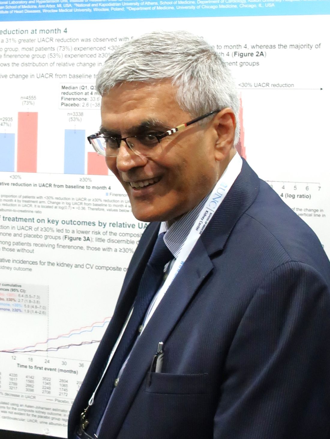
“My hope is that this [report] heightens awareness of UACR” as an important marker of both CKD and of the response by patients with CKD to their treatment, said Dr. Agarwal, a nephrologist and professor at Indiana University in Indianapolis.
“Only about half of people with type 2 diabetes get their UACR measured even though every guideline says measure UACR in people with diabetes. Our findings say that UACR is important not just for CKD diagnosis but also to give feedback” on whether management is working, Dr. Agarwal said in an interview.
Incorporate UACR into clinical decision-making
“My hope is that clinicians will look at UACR as something they should incorporate into clinical decision-making. I measure UACR in my patients [with CKD and type 2 diabetes] at every visit; it’s so inexpensive. Albuminuria is not a good sign. If it’s not reduced in a patient by at least 30% [the recommended minimum reduction by the American Diabetes Association for people who start with a UACR of at least 300 mg/g] clinicians should think of what else they could do to lower albuminuria”: Reduce salt intake, improve blood pressure control, make sure the patient is adherent to treatments, and add additional treatments, Dr. Agarwal advised.
Multiple efforts are now underway or will soon start to boost the rate at which at-risk people get their UACR measured, noted Leslie A. Inker, MD, in a separate talk during Kidney Week. These efforts include the National Kidney Foundation’s CKD Learning Collaborative, which aims to improve clinician awareness of CKD and improve routine testing for CKD. Early results during 2023 from this program in Missouri showed a nearly 8–percentage point increase in the screening rate for UACR levels in at-risk people, said Dr. Inker, professor and director of the Kidney and Blood Pressure Center at Tufts Medical Center in Boston.
A second advance was introduction in 2018 of the “kidney profile” lab order by the American College of Clinical Pathology that allows clinicians to order as a single test both an estimated glomerular filtration rate (eGFR) and a UACR.
Also, the Centers for Medicare & Medicaid Services and the National Committee for Quality Assurance have both taken steps to encourage UACR ordering. The NCQA established a new Healthcare Effectiveness Data and Information Set performance measure for U.S. physicians starting in 2023 that will track measurement of UACR and eGFR in people with diabetes. CMS also has made assessment of kidney health a measure of care quality in programs effective in 2023 and 2024, Dr. Inker noted.
Most subjects had elevated UACRs
The study run by Dr. Agarwal and his associates used data from 12,512 of the more than 13,000 people enrolled in either FIDELITY-DKD or FIGARO-DKD who had UACR measurements recorded at baseline, at 4 months into either study, or both. Their median UACR at the time they began on finerenone or placebo was 514 mg/g, with 67% having a UACR of at least 300 mg/g (macroalbuminuria) and 31% having a UACR of 30-299 mg/g (microalbuminuria). By design, virtually all patients in these two trials were on a renin-angiotensin system inhibitor (either an angiotensin-converting enzyme inhibitor or an angiotensin-receptor blocker), but given the time period when the two trials enrolled participants (during 2015-2018) only 7% of those enrolled were on a sodium-glucose cotransporter 2 inhibitor and only 7% were on a glucagonlike peptide–1 receptor agonist.
Four months after treatment began, 53% of those randomized to finerenone treatment and 27% of those in the placebo arm had their UACR reduced by at least 30% from baseline, the cutpoint chosen by Dr. Agarwal based on the American Diabetes Association guideline.
Kaplan-Meier analyses showed that the incidence of the primary kidney outcome – kidney failure, a sustained ≥ 57% decrease in eGFR from baseline, or kidney death – showed close correlation with at least a 30% reduction in UACR regardless of whether the patients in this subgroup received finerenone or placebo.
A different correlation was found in those with a less than 30% reduction in their UACR from baseline to 4 months, regardless of whether this happened on finerenone or placebo. People in the two finerenone trials who had a lesser reduction from baseline in their UACR also had a significantly higher rate of adverse kidney outcomes whether they received finerenone or placebo.
84% of finerenone’s kidney benefit linked to lowering of UACR
The causal-mediation analysis run by Dr. Agarwal quantified this observation, showing that 84% of finerenone’s effect on the kidney outcome was mediated by the reduction in UACR.
“It seems like the kidney benefit [from finerenone] travels through the level of albuminuria. This has broad implications for treatment of people with type 2 diabetes and CKD,” he said.
The link with reduction in albuminuria was weaker for the primary cardiovascular disease outcome: CV death, nonfatal myocardial infarction, nonfatal stroke, or hospitalization for heart failure. The strongest effect on this outcome was only seen in Kaplan-Meier analysis in those on finerenone who had at least a 30% reduction in their UACR. Those on placebo and with a similarly robust 4-month reduction in UACR showed a much more modest cardiovascular benefit that resembled those on either finerenone or placebo who had a smaller, less than 30% UACR reduction. The mediation analysis of these data showed that UACR reduction accounted for about 37% of the observed cardiovascular benefit seen during the trials.
“The effect of UACR is much stronger for the kidney outcomes,” summed up Dr. Agarwal. The results suggest that for cardiovascular outcomes finerenone works through factors other than lowering of UACR, but he admitted that no one currently knows what those other factors might be.
Treat aggressively to lower UACR by 30%
“I wouldn’t stop finerenone treatment in people who do not get a 30% reduction in their UACR” because these analyses suggest that a portion of the overall benefits from finerenone occurs via other mechanisms, he said. But in patients whose UACR is not reduced by at least 30% “be more aggressive on other measures to reduce UACR,” he advised.
The mediation analyses he ran are “the first time this has been done in nephrology,” producing a “groundbreaking” analysis and finding, Dr. Agarwal said. He also highlighted that the findings primarily relate to the importance of controlling UACR rather than an endorsement of finerenone as the best way to achieve this.
“All I care about is that people think about UACR as a modifiable risk factor. It doesn’t have to be treated with finerenone. It could be a renin-angiotensin system inhibitor, it could be chlorthalidone [a thiazide diuretic]. It just happened that we had a large dataset of people treated with finerenone or placebo.”
He said that future mediation analyses should look at the link between outcomes and UACR reductions produced by agents from the classes of sodium-glucose cotransporter 2 inhibitors and the glucagonlike peptide–1 receptor agonists.
FIDELIO-DKD and FIGARO-DKD were both sponsored by Bayer, the company that markets finerenone. Dr. Agarwal has received personal fees and nonfinancial support from Bayer. He has also received personal fees and nonfinancial support from Akebia Therapeutics, AstraZeneca, Boehringer Ingelheim, Eli Lilly, and Vifor Pharma, and he is a member of data safety monitoring committees for Chinook and Vertex. Dr. Inker is a consultant to Diamtrix, and her department receives research funding from Chinook, Omeros, Reata, and Tricida.
AT KIDNEY WEEK 2023
Sleeping beats sitting? What a new study means for your patients
Sit less, move more. Or stand more. Or sleep more.
Replacing 30 minutes of sitting a day with equal time standing or even sleeping could improve obesity markers like body weight and waist circumference, according to a new cross-sectional study investigating the impact of movement behavior on cardiometabolic health.
The findings suggest that, while higher-intensity activity may confer benefits sooner, adding more light activity or more standing, or even going to bed earlier, could improve heart health measures over time.
“Our study highlights that replacing sedentary behavior with any other behavior can be beneficial,” said study author Joanna M. Blodgett, PhD, a researcher at University College London’s Institute of Sport, Exercise and Health, and department of targeted intervention.
The study builds on a large and growing body of evidence that movement behaviors impact cardiometabolic health. Increasing physical activity to 150 minutes a week has been shown to reduce the risk for cardiovascular disease by 17% and type 2 diabetes by 26%. Other studies suggest that even modest increases in physical activity can be beneficial. A prospective study published in October found that even short activity bouts of a few minutes a day may lower risks for heart attack, stroke, and early death.
In the new study, researchers analyzed data from six studies and more than 15,000 participants, ranking behaviors according to their association with heart health. Moderate-to-vigorous activity was linked to the greatest benefit, followed by light activity, standing, sleeping, and finally – dead last on the list – sitting.
A thigh-worn device tracked participants’ activity throughout the day, and six measures gauged heart health: body mass index (BMI), waist circumference, HDL cholesterol, total-cholesterol-to-HDL ratio, triglycerides, and glycated hemoglobin.
Researchers modeled what would happen if people swapped various amounts of one activity for another every day for a week. Replacing just 4-13 minutes of sitting with moderate to vigorous activity improved heart health markers.
The cardiovascular demands of regular intense activity like running, cycling, dancing, or playing sports – even in small bouts – strengthen the heart and improve blood flow throughout the body, Dr. Blodgett said. “This can lower cholesterol, blood pressure, and resting heart rate.”
Even if adding moderate to vigorous activity is not an option, the findings suggest that people can still see benefits by replacing sitting with virtually any other activity – walking, standing, even sleeping.
Limitations
Because the study was observational, results can’t be used to infer causality.
“We cannot directly lean on the study results to guide prescriptions for particular exercise or lifestyle changes,” said Matthew Tomey, MD, a cardiologist with the Mount Sinai Health System, New York, who was not involved with the study. An interventional trial would be needed to confirm the findings.
The finding that sleep was better for participants than sitting is a good example. The benefits of replacing sitting with sleep were “clear” for adiposity measures like BMI and waist circumference, but negligible for blood markers such as cholesterol, triglycerides, and blood glucose, Dr. Blodgett said.
One explanation: “The negative impact of sitting on these obesity measures is likely due to related unhealthy behaviors like snacking rather than the physiological benefits of sleep itself,” Dr. Blodgett said.
What’s more, study participants were relatively young, healthy, and active. The average age was 54, and they averaged nearly 8 hours of sleep, 10 hours of sitting, 3 hours of standing, 1.5 hours of light activity, and more than an hour of moderate to vigorous activity per day. So it’s difficult to draw conclusions about patients who don’t fit those metrics.
Impact on patient care
That said, the results could help tailor recommendations for patients, Dr. Blodgett said.
If a patient is struggling to exercise or is unable to exercise because of health or ability restrictions, you could help them find ways to add a lighter activity to their day, such as taking the stairs or walking briskly to catch the bus. Even swapping a regular desk for a standing one, or going to bed 30 minutes earlier, could be a more practical and effective suggestion.
More than that: The research could be used to educate patients on the power of small changes. It shows that shifting daily habits even in small ways can make a difference, and people who are the least active stand to benefit the most.
You can also remind patients that moderate or vigorous activity doesn’t need to happen at the gym. It could be lawn work, taking a walk, or moving heavy boxes. In fact, many activities can be “moderate” or even “vigorous” depending on the effort put into them.
Share this rule of thumb: “An activity is classified as moderate intensity if you can talk but not sing while doing it, and an activity is generally considered vigorous intensity if you can’t say more than a few words without stopping to breathe,” Dr. Blodgett said.
The study also has implications for the potential of wearable activity trackers to monitor progress. Combining objective activity data with results from studies like this, and longer prospective studies, could help inform more helpful advice.
“Ultimately, this research helps move us closer to more personalized guidance of how changing behaviors can improve your health,” Dr. Blodgett said.
A version of this article first appeared on Medscape.com.
FROM THE EUROPEAN HEART JOURNAL
T2D: Real benefits of new oral antidiabetic drugs
Cardiovascular disease is the most common cause of death in people living with type 2 diabetes (T2D). It is true that patient prognoses have improved with the use of metformin and by addressing cardiovascular risk factors. But the new oral antidiabetic drugs, SGLT2 (sodium glucose cotransporter-2) inhibitors (SGLT2i), and glucagon-like peptide-1 receptor agonists (GLP-1Ra) offer fresh therapeutic approaches.
A cohort of more than 2 million patients with T2D
What about in the real world, far away from the ideal conditions of randomized trials? Could combining SGLT2 inhibitors with GLP-1R agonists be even more effective?
These are the questions answered by a large retrospective cohort study in which 2.2 million patients with T2D receiving insulin were initially enrolled and monitored at 85 specialist centers spread throughout three countries (Denmark, the United Kingdom, and the United States).
Three groups were formed from this cohort according to whether they received monotherapy or combination treatments: SGLT2i (n = 143,600), GLP-1Ra (n = 186,841), and SGLT2i + GLP-1Ra (n = 108,5040). A control group received none of these treatments.
Propensity score matching took into account the following relevant variables: age, sex, ischemic heart disease, hypertension, chronic kidney disease, heart failure, and glycated hemoglobin. The data was analyzed using the Cox’s proportional hazards model, with follow-up at 5 years.
Real-world benefits – Even better when combined
The inter-group comparison suggests that oral antidiabetic agents are effective when taking into account three major events:
All-cause mortality: SGLT2i (hazard ratio, 0.49; confidence interval 95% 0.48-0.50); GLP-1Ra (HR, 0.47; CI 95% 0.46-0.48); SGLT2i + GLP-1Ra (HR, 0.25; CI 95% 0.24-0.26).
Admissions rate: respectively HR: 0.73 (0.72-0.74); 0.69 (0.68-0.69); 0.60 (0.59-0.61).
Myocardial infarction rate: respectively HR: 0.75 (0.72-0.78); 0.70 (0.68-0.73); 0.63 (0.60-0.66).
A complementary sub-analysis also revealed a more significant reduction in all-cause mortality in the event of exposure to the combination of two antidiabetic drugs versus SGLT2i alone (HR, 0.53 [0.50-0.55]) and GLP-1Ra as monotherapy (HR, 0.56 [0.54-0.59]).
This real-world retrospective cohort study involves a large sample size: more than 400,000 patients with T2D treated with new oral antidiabetic drugs and as many control patients. It suggests that SGLT2 inhibitors and GLP-1R agonists have a significant effect on overall mortality, as well as on the risk of myocardial infarction and the admissions rate. Yes, it is retrospective, but its findings are in line with those from the most recent and conclusive randomized trials that suggest a cardio- and nephroprotective effect, at least with regard to SGLT2 inhibitors.
This article was translated from JIM and a version appeared on Medscape.com.
Does laughter offer better blood glucose control?
David S. Greene, PhD, associate professor in the department of rehabilitation and human services at the University of Northern Colorado, Greeley, has pioneered research into the field, one previously overlooked when it comes to diabetes management.
This news organization caught up with Dr. Greene to ask about his research along with the implications for diabetes care and for patient well-being.
Question: What prompted you to research the link between humor and diabetes control?
Answer: I was diagnosed with type 1 diabetes in 1966 and consequently have lived with, and studied, various aspects of diabetes for the past 57 years.
For a time, I maintained a small private practice counseling people with diabetes. There I noticed, anecdotally, that my clients’ ability to laugh at, and see the humor in, their diabetes correlated with their emotional adjustment to living with their condition.
While I could find research confirming the physical and psychological benefits of humor in general, I was unable to find any research specifically relating to humor and diabetes.
My new research agenda was born.
Q: What did your research reveal?
A: My first study, published in 2020, found that people living with diabetes displayed the same level of both types of positive, or affiliative and self-enhancing, humor as the norm group used in developing the Humor Styles Questionnaire.
This was a surprising finding given that individuals with diabetes are dealing with a life-altering, chronic illness, with higher rates of depression and anxiety. Moreover, positive humor scores are known to be negatively correlated with depression and anxiety.
While levels of aggressive humor were not altered in my study, people with diabetes did have higher levels of self-defeating humor in my study, which is correlated with psychiatric and somatic concerns, and symptomatology, and so is to be expected.
My second study, published in 2021, examined whether there was a difference between people with diabetes who had an hemoglobin A1c level of 6.99% or less versus those with levels of at least 7% on four disparate types of humor.
The A1c of 6.99% or less group scored significantly higher for both affiliative humor and self-enhancing humor. This implies that better diabetes control is associated with positive humor. This was expected, as was the finding that negative humor was not associated with better control.
On the other hand, there was no significant difference between participants on either type of negative humor, whether aggressive or self-defeating.
Finally, my third study, published in 2023 [with coauthor Nancy D. King, PhD], found that offering humor training to people with type 1 diabetes can strengthen both their affiliative and self-enhancing sense of humor, while leaving their aggressive or self-defeating humor unaffected.
Q: What is ‘positive humor,’ and why do you think it is associated with diabetes control?
A: Both affiliative and self-enhancing humor enhance a person’s sense of self and their relationships with others.
Affiliative humor has been shown to be positively correlated with self-esteem, psychological well-being, social intimacy, and emotional stability. It is also negatively correlated with depression and anxiety.
Self-enhancing humor involves the ability to maintain a humorous outlook on life and to use humor to regulate emotions and as a coping strategy. Like affiliative humor, it is correlated with cheerfulness, self-esteem, optimism, psychological well-being, and life satisfaction. It is negatively related to depression and anxiety.
The preponderance of literature suggests positive humor specifically is associated with buffering many of the risk factors and complications associated with diabetes.
Q: What could underlie the associations between humor and diabetes control?
A: Unfortunately, none of my studies have been able to determine cause and effect, although the most recent one came the closest.
While not reaching statistical significance, the mean reduction in A1c levels from 7.12% at baseline to 6.75% at the post–humor training assessment may very well imply a practical and psychological significance to patients.
I believe, with a larger sample size, significance will be achieved, and that the relationship between positive humor and diabetes control will be shown to be bidirectional, with positive humor improving diabetes control, and improved control improving positive humor.
I hypothesize this will also bring psychological and physiological benefits. After all, humor has already been associated with reduced blood glucose levels, reduced microvascular complications, improved heart health, reduced blood pressure, decreased levels of depression and anxiety.
Humor also helps us deal with stress and trauma, so a cause-and-effect relationship makes sense.
Q: Can a positive sense of humor be taught?
A: Absolutely. There is evidence that humor can be developed and strengthened. Paul McGhee, PhD, developed a seven-step humor training program that has been effective in elevating measures of humor across a number of studies.
Others have successfully developed their own protocols, and of course my third study demonstrated a significant increase in both affiliative and self-enhancing humor with training.
Q: Do you think humor training could be incorporated into diabetes care?
A: Humor training programs are easily accessible, incur low to no cost, and are easy to implement. Furthermore, once a person is trained, access to ongoing humor is free, readily available, and fun.
Several diabetes educators have also reported that humor can promote connections, encourage and support diabetes management, galvanize effectiveness, and increase an audience’s attention during education programs.
A version of this article appeared on Medscape.com.
Low-salt diet cut BP by 6 mm Hg in 1 week: CARDIA-SSBP
The CARDIA-SSBP trial involved 213 individuals aged 50-75 years, including those with and those without hypertension, and showed that the decline in blood pressure brought about by a low-salt diet was independent of hypertension status and antihypertensive medication use. It was also generally consistent across subgroups and did not result in excess adverse events.
“The blood pressure reduction we see here is meaningful, and comparable to that produced by one antihypertensive medication,” lead investigator Deepak Gupta, MD, Vanderbilt University Medical Center, Nashville, Tenn., said in an interview.
Dr. Gupta presented the CARDIA-SSBP study on Nov. 11 at the American Heart Association scientific sessions, held in Philadelphia. The study was published online in JAMA. The exact menus used in the study are available in a supplement to the JAMA paper.
“In order to live a healthy lifestyle, understanding what we eat has important health effects. Raised blood pressure contributes to one out of every eight deaths worldwide,” Dr. Gupta noted. “If people want to lower their blood pressure, attention to dietary sodium is one part of that. If individuals can stick with a low sodium diet, they may be able to stop taking one of their antihypertensive medications, and those who are normotensive will be less likely to develop hypertension.”
Commentators said the study had significant implications for public health, but they pointed out that maintaining a low-sodium diet over the long term is challenging, given the high salt content of generally available foods.
Dr. Gupta noted that the study did use commercially available products in the low-sodium diets and the menus are available for people to follow, making it more accessible than some diets used in previous studies.
“What may also be attractive to people is that you don’t have to wait for months to see an effect. If you start to consume a low-sodium diet, you can see results on blood pressure rapidly, within a week,” he said.
The diet in this study brought about a large reduction in dietary sodium, but Dr. Gupta says any reduction in dietary sodium is likely to be beneficial.
“If you go to the level that we got to, you could expect to see a reduction of around 6 mm Hg. But it’s like walking – you don’t necessarily need to get to 10,000 steps every day. Any amount of walking or physical activity is of benefit. The same is probably true for salt: Any reduction that you can make is probably of benefit.”
For the study, participants had their blood pressure measured by 24-hour ambulatory monitoring while on their usual diets. They were then randomly assigned to either a high-sodium diet or a low-sodium diet for 1 week. Participants then crossed over to the opposite diet for 1 week, with blood pressure measured over a 24-hour period on the last day of each diet.
As assessed by 24-hour urine excretion, the usual diet of participants was found to already be high in sodium (median, 4.45 g/d). This increased to a median of 5.00 g/d when on the high-sodium diet in the study and decreased to 1.27 g/d while on the low-sodium diet.
Results found participants had a median systolic blood pressure of 125 mm Hg on their usual diets. This was raised to 126 mm Hg on the high-sodium diet and lowered to 119 mm Hg on the low-sodium diet.
The researchers also reported that 75% of individuals showed a blood pressure reduction on the low-sodium diet and are thus defined as “salt-sensitive.” This is a higher percentage than found in previous studies.
“Of those that didn’t show a blood pressure reduction with a low-sodium diet in this study, it appears that they may not have been so adherent to the diet as those who did show a blood pressure reduction,” Dr. Gupta said.
He noted that hypertension is the most common chronic disease condition worldwide, with about 1.3 billion people affected, and although it has been known for some time that dietary sodium affects blood pressure, there have been some gaps in previous studies.
For example, many studies have excluded individuals who were already taking antihypertensive medications and people with diabetes, and they have generally not included many older individuals. The current study found that all of these groups showed significant blood pressure reductions by reducing dietary sodium.
Large effect in people with diabetes
Subgroup analysis largely showed consistent results across the population, regardless of age, sex, race, and body mass index and whether participants were taking antihypertensive medication or not, but there were a couple of exceptions. Individuals with higher blood pressure at baseline seemed to have a greater effect of lowering dietary sodium, although those who were normotensive at baseline still showed significant blood pressure reduction, Dr. Gupta reported.
The researchers found a particularly large reduction in blood pressure from lowering sodium intake in people with diabetes, who made up about 21% of the overall cohort. Their average reduction in systolic blood pressure between the high and low sodium diet was close to 17 mm Hg rather than the 7-8 mm Hg in the whole cohort.
Dr. Gupta said that the results are applicable to most of the population.
“The people who will be most motivated to follow a low-sodium diet are those with hypertension. But even in normotensive individuals, there is likely to be benefit.”
To help people follow a low-sodium diet, Dr. Gupta says education campaigns are needed “to show people that they can do it and make it work.” But there are bigger structural issues that need to be addressed at policy and governmental levels.
“Most of our food available in grocery stores and restaurants is high in salt. We now have a preponderance of evidence showing us that we need to change what’s available in the food supply,” he said. “There is a push going on for this now, and the U.S. has introduced some guidelines for the food industry on sodium content of foods. These are voluntary at this point, but it’s a start.”
Difficult to maintain long term
Commenting on the study, Paul Whelton, MD, chair in global public health at Tulane University, New Orleans, noted that sodium reduction is known to reduce blood pressure, with greater sodium reductions giving greater blood pressure decreases, and that some people are more sensitive to the effects of sodium than others.
He described CARDIA-SSBP as a “well-done study.”
“They managed to get a very low sodium intake and a large difference between the two groups, which translated into a big reduction in systolic blood pressure,” Dr. Whelton said. “However, the problem with these sorts of trials where the diets are provided to the participants is that although they show proof of concept, it is difficult to generalize because we can’t normally provide patients with their meals. In this type of ‘feeding’ study, we find it difficult to maintain people on their behavioral intervention over the long term.”
Dr. Whelton said that he was more excited about this trial knowing that the food given was commercially available. “That makes it more practical, but you still have to be quite motivated to follow a diet like this. Buying low-sodium products in the supermarket does require quite a lot of work to read the labels, and sometimes the low-sodium foods are specialty products and are more expensive.”
He pointed out that older people in higher socioeconomic classes are more likely to attempt this and do better from behavioral interventions in general. “Unfortunately, people who don’t do well from behavioral interventions like this are those from lower socioeconomic groups, who are ones at most at risk for cardiovascular disease.”
Dr. Whelton noted that the food industry has been reluctant to lower sodium content because high-salt foods sell better. “Unfortunately, foods high in saturated fat and salt taste good to most people. We are generally attuned to a high salt intake. But when people have been following a low-salt diet for a while, they generally don’t like high-salt foods anymore. They become attuned to lower-sodium diet,” he added.
New U.S. sodium reduction guidelines
Discussant of the CARDIA-SSBP study at the AHA meeting, Cheryl Anderson, MD, University of California, San Diego, said that the findings were important and consistent with prior studies.
“These studies have global implications because salt is ubiquitous in the food supply in much of the world,” she noted, adding that, “Americans consume almost 50% more sodium than recommended, and there has been a persistent lack of adherence to healthy diet recommendations for reductions in salt, sugar, and fats.”
Dr. Anderson pointed out that in 2021, the Food and Drug Administration issued guidance for voluntary sodium reduction, which uses a gradual approach, with targets to reach a population goal of 3,000 mg/d of sodium by 2023 and 2,300 mg/d by 2031.
“These targets apply to 150 categories of food that are sales-weighted to focus on dominant sellers in each category. They apply to food manufacturers, restaurants and food service operations,” she concluded. “These targets serve as a basis for continued dialogue. The research community eagerly awaits the review of population-based data to help refine this approach and goals.”
This study was supported by grants from the National Heart, Lung, and Blood Institute, the National Institutes of Health, the American Heart Association, and the National Center for Advancing Translational Sciences. The authors report no disclosures.
A version of this article appeared on Medscape.com.
FROM AHA 2023