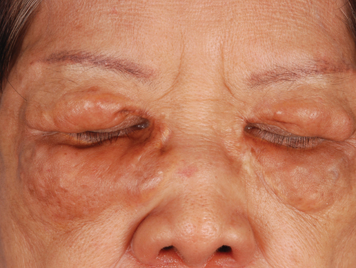
Because our patient’s lesions were longstanding and disfiguring, we opted for active intervention with intralesional triamcinolone, which resulted in only a slight reduction in size of the lesions. The lesions remain largely unchanged in 2 years of follow-up.
Photo Challenge
Yellowish Papulonodular Periorbital Eruption
Cutis. 2014 September;94(3):E7-E10
Author and Disclosure Information
Brian Chia, MBBS, MRCP; Hong Liang Tey, MBBS, MRCP, MRCPS
Dr. Chia is from the Department of General Medicine, Tan Tock Seng Hospital, Singapore. Dr. Tey is from the National Skin Centre, Singapore.
The authors report no conflict of interest.
Correspondence: Hong Liang Tey, MBBS, MRCP, MRCPS, 1 Mandalay Rd, Singapore 308205 (teyhongliang111@yahoo.com).

A 66-year-old woman with a history of type 2 diabetes mellitus and mild dyslipidemia presented with persistent lesions over the eyelids and cheeks of 10 years’ duration. Systemic review was unremarkable. There was no family or personal history of atopy, asthma, or other dermatologic disorders. Physical examination revealed confluent yellowish plaques and nodules over the periorbital regions as well as yellowish plaques over the neck and back. The lesions were firm to palpation and the epidermis appeared unaffected. The ophthalmic examination was normal and other mucosal surfaces were unaffected.

