User login
Evolving nanotechnology reveals potential treatment advantages
DENVER – As the nanotechnology field evolves, there are at least three reasons a clinician might prefer to use nanoparticles over conventional drugs, according to Dr. Jack L. Arbiser. The first is to minimize the effects of a systemic drug.
"What if steroids could be directed to the site of the pathologic lesion but not cause side effects such as glucose intolerance, bone fractures, or gastric ulcers?" he asked during a session on nanotechnology at the annual meeting of the American Academy of Dermatology. "What if propranolol could be given to patients with hemangiomas without having to worry about cardiac side effects?"
A second reason to turn to nanoparticles would be to use highly toxic substances in a safe way, continued Dr. Arbiser, a professor of dermatology at Emory University, Atlanta. "We already use Botox by injection, which is one of the most toxic substances known to man, but what if other highly toxic substances could be specifically delivered to cancers, infections, or inflammation?"
A third reason to use nanoparticles is that they may offer a way for compounds "that are not highly active be delivered to sites of disease in high enough concentration so that they have therapeutic activity and provide a high measure of safety," he said.
Dr. Arbiser discussed his nanotechnology experience as part of a research team developing a derivative of gentian violet for cancer treatment. He and his associates observed that gentian violet has potential antitumor activity through inhibition of NADPH (nicotinamide adenine dinucleotide phosphate) oxidase. However, the researchers faced early challenges, because gentian violet is not patentable by itself, and, because it is hydrophilic, it may not pass through the blood-brain barrier.
"Our solution was to synthesize and patent a more lipophilic derivative of gentian violet called imipramine blue," Dr. Arbiser explained. "We packaged it in liposomes so it has a much longer half-life and is directed to tumors because liposomes preferentially localize to leaky blood vessels."
Imipramine blue can be given weekly and is efficacious against cancer tumors. Dr. Arbiser and his associates have demonstrated that systemic delivery of imipramine blue significantly inhibited migration of glioblastoma cells into the brain parenchyma (Sci. Transl. Med. 2012;4:127ra36).
"We also see this [inhibition of spread] in prostate, breast, and melanoma cells," he said. "So it seems to be hitting the same pathway in multiple different tumor types." Such an approach targets delivery of therapeutic and diagnostic imaging agents to the site of cancer and chronic inflammation/infection, potentially reducing side effects and increasing efficacy.
"My own personal bias is that we should start using metal-based nanotechnology for severe illnesses, such as cancers, allowing the effects of these materials to be studied in humans, before widespread introduction into cosmetics," he concluded.
Dr. Arbiser disclosed that he is the cofounder of companies ABBY Therapeutics and Accuitis.
EXPERT ANALYSIS FROM THE AAD ANNUAL MEETING
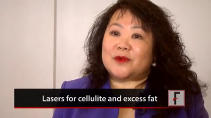
VIDEO: Concise guide to lasers: cellulite, tattoo removal, and skin tightening
DENVER – In 15 minutes, Dr. M. Christine Lee brings you a definitive guide to lasers for various patient needs, including tattoo removal, treatment of cellulite, skin tightening, and skin resurfacing. Learn more about lasers that you might want to add to your practice and get ideas on how to make the most of devices you already have.
Dr. Lee is with the department of dermatologic surgery at the University of California, San Francisco, and director of East Bay Laser & Skin Care Center in Walnut Creek, Calif. In this video, she also shares clinical pearls about different types of laser procedures.
The video associated with this article is no longer available on this site. Please view all of our videos on the MDedge YouTube channel
On Twitter @naseemmiller
EXPERT ANALYSIS FROM THE AAD ANNUAL MEETING
Monoclonal antibody pinpoints BRAF status in melanoma
DENVER - A murine monoclonal antibody had very high sensitivity and specificity for melanomas with the V600E BRAF mutation, and exhibited perfect concordance between the primary and metastatic tumors in individual patients.
In addition to being a valuable screening tool, VE1 (anti-BRAF V600E) could be an extremely useful adjunct to DNA analysis, Michelle Vernali said at the annual meeting of the American Academy of Dermatology.
"I think that the sequential use of immunohistochemistry and molecular analysis will dramatically improve sensitivity and specificity for the detection of BRAF mutations, which is essential for the effective use of BRAF inhibitors," said Ms. Vernali, a fourth-year medical student at the University of North Carolina, Chapel Hill.
According to Roche Diagnostics, the VE1 antibody has demonstrated 100% sensitivity and 99% specificity for BRAF mutations in colon cancer. In addition, it has shown high efficacy in detecting those mutations in thyroid cancer and hairy cell leukemia, and "shows promise" in non–small cell lung cancer and serous ovarian tumors.
According to the company, "The ... antibody has also been said to be a promising tool for patient stratification among individuals presenting with brain metastases."
Ms. Vernali and her colleagues examined the benefit of VE1 staining in 93 patients with metastatic melanoma. Of these, 76 had DNA pyrosequencing of either the primary (19) or metastatic lesion (57). Both primary and metastatic tumor samples were available for 17 patients.
Of the 76 patients with either primary or metastatic lesion samples, DNA pyrosequencing identified 26 that were positive for V600E and 40 that were negative. VE1 staining identified 22 positive samples and 44 negative samples, for a specificity of 100% and a sensitivity of 85%.
Sequencing also identified eight samples positive for V600K, and one each for V600R and V600Q. VE1 did not stain any of these samples.
Among the 17 patients with both primary and metastatic samples, VE1 was in 100% concordance with DNA sequencing, identifying three positive samples and 14 negative samples.
"There was little variability of strength or intensity of the staining, and very little intra-interpreter variance," Ms. Vernali said.
She proposed an algorithm for BRAF testing using VE1 with and without DNA sequencing.
· Insufficient tissue for initial DNA pyrosequencing:
– Stain with VE1.
– Identify BRAF V600E-positive or -negative patients.
· Sufficient tissue for DNA pyrosequencing:
– Stain with VE1.
– Stratify as VE1 positive or negative.
– If VE1 positive, conclude the patient is BRAF V600E positive.
– If VE1 negative, send sample for molecular sequencing to stratify into V600E positive, positive for another BRAF mutation, or BRAF negative.
This algorithm would identify V600E status in patients with tissue samples that would otherwise be insufficient for BRAF testing, she said. "If they had insufficient tissue for DNA sequencing, they could be stratified by immunohistochemistry and if positive, could be treated. Otherwise this is a population that now goes without BRAF-inhibiting therapy."
The algorithm is being tested in some sites already, she added, but needs additional validation before it can be broadly adopted.
Ms. Vernali had no financial disclosures.
AT THE AAD ANNUAL MEETING
Major finding: A monoclonal antibody showed 100% specificity and 85% sensitivity for identifying V600E BRAF mutations in metastatic melanoma.
Data source: The prospective study comprised 93 patients.
Disclosures: Ms. Vernali had no financial disclosures.

Try Tackling Cysts or Lipomas With Punch Tool Instead of Scalpel
DENVER – A little lidocaine and a punch biopsy tool allow physicians to tackle most cysts and lipomas in the office, providing effective treatment without interrupting patient flow.
The punch technique easily eliminates the contents of these annoying, sometimes painful, lesions, Dr. Robert T. Brodell said at the annual meeting of the American Academy of Dermatology. But more importantly, it allows the complete removal of the cyst wall, which is key to preventing recurrence, he said.
Despite dogma, it’s not really necessary deliver the entire cyst unbroken, a technique that requires a much larger incision, explained Dr Brodell, professor and chairman of dermatology and a professor of pathology at the University of Mississippi Medical Center, Jackson.

"When I was training, it was common for surgeons to excise cysts and hold them up ... to demonstrate that the entire cyst had been removed intact," he said. "But it doesn’t really matter if it is removed this way. What does matter is that the entire wall is removed. If you don’t, the remainder will keep making keratin and set up a foreign body reaction," or set the stage for cyst recurrence.
Pilar cysts and multiple painful lipomas on the forearms or legs seem to be the easiest to remove this way, Dr. Brodell said in an interview. Lesions on the back where the skin is quite thick are more difficult. But his early study on the technique found a recurrence rate of only 14% in these more difficult areas and an overall recurrence rate of 8% for all cysts.
Dr. Brodell’s preferred technique starts with an intradermal injection of 1% lidocaine plus epinephrine, enough to raise and blanch the overlying skin. "You don’t need any deep anesthesia," he said. "This seems to be enough to take care of the innervation of these structures."
A punch appropriate to the cyst size – usually 4-6 mm – is used to incise the lesion. A forceps is used to free the wall from the surrounding dermis. Dr. Brodell then manually expresses the contents with lateral finger pressure. Portions of the cyst wall will also be extruded. Looking through the punch excision defect, any remaining fragments of cyst wall are removed.
It most cases, it takes only a stitch or two to close the small wound.
Lipomas are slightly more complicated. After incising the lesion, Dr. Brodell again applies lateral pressure, and the contents will start to deliver. "If you grab the lipoma with forceps, a portion of the lipoma is incised and removed. Then more fat can be delivered through the punch incision hole and removed. The process continues until the entire contents are removed. "If I’m not getting all of it out, I’m getting at least 90%-95%, which seems to minimize the chance for recurrence," Dr. Brodell said.
He said that he finishes by making a bolster with gauze to place over the wound and wrapping it with a pressure bandage. This strategy prevents a hematoma from developing in the potential space where the lipoma was removed.
The technique also is appropriate for infected cysts, Dr. Brodell said, and in such cases it is accompanied by a standard course of systemic antibiotics.
"For most cysts, and for lipomas smaller than 2 cm, the process takes less than 5 minutes," Dr. Brodell said. "For a really big lipoma, maybe 15 minutes."
Punch excision carries benefits for both the provider and the patient. "You can do it in the office, without interrupting patient flow," said Dr. Brodell. "In some cases it saves the patient a trip to the surgery center. In this case it is certainly less expensive."
The charge is calculated according to lesion size, typically ranging from $130 to $350. "A trip to the surgery center is going to be at least $1,200," Dr. Brodell added.
Dr. Brodell began performing this procedure in the late 1980s. In 2002, he published a retrospective review of 299 patients who had undergone pilar or keratinous cyst removal via punch technique and who had complete follow-up data (Dermatol. Surg. 2002;28:673-7). Follow-up averaged 80 weeks.
Most of the patients (267) reported no recurrence. The recurrence rate was 9% for keratinous cysts, 5% for pilar cysts, and 8% overall. Recurrence was higher among back and ear lobe cysts (14% and 13%, respectively).
Most cysts (54%) recurred within 1 year of surgery; 24% recurred 1-2 years later.
Given the ease and convenience of this technique, and the excellent clinical results, Dr. Brodell encouraged physicians to reconsider their anti-cyst weaponry – and try putting down the scalpels and reaching for a punch.
Dr. Brodell had no financial disclosures relevant to this presentation.
On Twitter @alz_gal
DENVER – A little lidocaine and a punch biopsy tool allow physicians to tackle most cysts and lipomas in the office, providing effective treatment without interrupting patient flow.
The punch technique easily eliminates the contents of these annoying, sometimes painful, lesions, Dr. Robert T. Brodell said at the annual meeting of the American Academy of Dermatology. But more importantly, it allows the complete removal of the cyst wall, which is key to preventing recurrence, he said.
Despite dogma, it’s not really necessary deliver the entire cyst unbroken, a technique that requires a much larger incision, explained Dr Brodell, professor and chairman of dermatology and a professor of pathology at the University of Mississippi Medical Center, Jackson.
"When I was training, it was common for surgeons to excise cysts and hold them up ... to demonstrate that the entire cyst had been removed intact," he said. "But it doesn’t really matter if it is removed this way. What does matter is that the entire wall is removed. If you don’t, the remainder will keep making keratin and set up a foreign body reaction," or set the stage for cyst recurrence.
Pilar cysts and multiple painful lipomas on the forearms or legs seem to be the easiest to remove this way, Dr. Brodell said in an interview. Lesions on the back where the skin is quite thick are more difficult. But his early study on the technique found a recurrence rate of only 14% in these more difficult areas and an overall recurrence rate of 8% for all cysts.
Dr. Brodell’s preferred technique starts with an intradermal injection of 1% lidocaine plus epinephrine, enough to raise and blanch the overlying skin. "You don’t need any deep anesthesia," he said. "This seems to be enough to take care of the innervation of these structures."
A punch appropriate to the cyst size – usually 4-6 mm – is used to incise the lesion. A forceps is used to free the wall from the surrounding dermis. Dr. Brodell then manually expresses the contents with lateral finger pressure. Portions of the cyst wall will also be extruded. Looking through the punch excision defect, any remaining fragments of cyst wall are removed.
It most cases, it takes only a stitch or two to close the small wound.
Lipomas are slightly more complicated. After incising the lesion, Dr. Brodell again applies lateral pressure, and the contents will start to deliver. "If you grab the lipoma with forceps, a portion of the lipoma is incised and removed. Then more fat can be delivered through the punch incision hole and removed. The process continues until the entire contents are removed. "If I’m not getting all of it out, I’m getting at least 90%-95%, which seems to minimize the chance for recurrence," Dr. Brodell said.
He said that he finishes by making a bolster with gauze to place over the wound and wrapping it with a pressure bandage. This strategy prevents a hematoma from developing in the potential space where the lipoma was removed.
The technique also is appropriate for infected cysts, Dr. Brodell said, and in such cases it is accompanied by a standard course of systemic antibiotics.
"For most cysts, and for lipomas smaller than 2 cm, the process takes less than 5 minutes," Dr. Brodell said. "For a really big lipoma, maybe 15 minutes."
Punch excision carries benefits for both the provider and the patient. "You can do it in the office, without interrupting patient flow," said Dr. Brodell. "In some cases it saves the patient a trip to the surgery center. In this case it is certainly less expensive."
The charge is calculated according to lesion size, typically ranging from $130 to $350. "A trip to the surgery center is going to be at least $1,200," Dr. Brodell added.
Dr. Brodell began performing this procedure in the late 1980s. In 2002, he published a retrospective review of 299 patients who had undergone pilar or keratinous cyst removal via punch technique and who had complete follow-up data (Dermatol. Surg. 2002;28:673-7). Follow-up averaged 80 weeks.
Most of the patients (267) reported no recurrence. The recurrence rate was 9% for keratinous cysts, 5% for pilar cysts, and 8% overall. Recurrence was higher among back and ear lobe cysts (14% and 13%, respectively).
Most cysts (54%) recurred within 1 year of surgery; 24% recurred 1-2 years later.
Given the ease and convenience of this technique, and the excellent clinical results, Dr. Brodell encouraged physicians to reconsider their anti-cyst weaponry – and try putting down the scalpels and reaching for a punch.
Dr. Brodell had no financial disclosures relevant to this presentation.
On Twitter @alz_gal
EXPERT ANALYSIS FROM AAD 2014

Try tackling cysts or lipomas with punch tool instead of scalpel
DENVER – A little lidocaine and a punch biopsy tool allow physicians to tackle most cysts and lipomas in the office, providing effective treatment without interrupting patient flow.
The punch technique easily eliminates the contents of these annoying, sometimes painful, lesions, Dr. Robert T. Brodell said at the annual meeting of the American Academy of Dermatology. But more importantly, it allows the complete removal of the cyst wall, which is key to preventing recurrence, he said.
Despite dogma, it’s not really necessary deliver the entire cyst unbroken, a technique that requires a much larger incision, explained Dr Brodell, professor and chairman of dermatology and a professor of pathology at the University of Mississippi Medical Center, Jackson.

"When I was training, it was common for surgeons to excise cysts and hold them up ... to demonstrate that the entire cyst had been removed intact," he said. "But it doesn’t really matter if it is removed this way. What does matter is that the entire wall is removed. If you don’t, the remainder will keep making keratin and set up a foreign body reaction," or set the stage for cyst recurrence.
Pilar cysts and multiple painful lipomas on the forearms or legs seem to be the easiest to remove this way, Dr. Brodell said in an interview. Lesions on the back where the skin is quite thick are more difficult. But his early study on the technique found a recurrence rate of only 14% in these more difficult areas and an overall recurrence rate of 8% for all cysts.
Dr. Brodell’s preferred technique starts with an intradermal injection of 1% lidocaine plus epinephrine, enough to raise and blanch the overlying skin. "You don’t need any deep anesthesia," he said. "This seems to be enough to take care of the innervation of these structures."
A punch appropriate to the cyst size – usually 4-6 mm – is used to incise the lesion. A forceps is used to free the wall from the surrounding dermis. Dr. Brodell then manually expresses the contents with lateral finger pressure. Portions of the cyst wall will also be extruded. Looking through the punch excision defect, any remaining fragments of cyst wall are removed.
It most cases, it takes only a stitch or two to close the small wound.
Lipomas are slightly more complicated. After incising the lesion, Dr. Brodell again applies lateral pressure, and the contents will start to deliver. "If you grab the lipoma with forceps, a portion of the lipoma is incised and removed. Then more fat can be delivered through the punch incision hole and removed. The process continues until the entire contents are removed. "If I’m not getting all of it out, I’m getting at least 90%-95%, which seems to minimize the chance for recurrence," Dr. Brodell said.
He said that he finishes by making a bolster with gauze to place over the wound and wrapping it with a pressure bandage. This strategy prevents a hematoma from developing in the potential space where the lipoma was removed.
The technique also is appropriate for infected cysts, Dr. Brodell said, and in such cases it is accompanied by a standard course of systemic antibiotics.
"For most cysts, and for lipomas smaller than 2 cm, the process takes less than 5 minutes," Dr. Brodell said. "For a really big lipoma, maybe 15 minutes."
Punch excision carries benefits for both the provider and the patient. "You can do it in the office, without interrupting patient flow," said Dr. Brodell. "In some cases it saves the patient a trip to the surgery center. In this case it is certainly less expensive."
The charge is calculated according to lesion size, typically ranging from $130 to $350. "A trip to the surgery center is going to be at least $1,200," Dr. Brodell added.
Dr. Brodell began performing this procedure in the late 1980s. In 2002, he published a retrospective review of 299 patients who had undergone pilar or keratinous cyst removal via punch technique and who had complete follow-up data (Dermatol. Surg. 2002;28:673-7). Follow-up averaged 80 weeks.
Most of the patients (267) reported no recurrence. The recurrence rate was 9% for keratinous cysts, 5% for pilar cysts, and 8% overall. Recurrence was higher among back and ear lobe cysts (14% and 13%, respectively).
Most cysts (54%) recurred within 1 year of surgery; 24% recurred 1-2 years later.
Given the ease and convenience of this technique, and the excellent clinical results, Dr. Brodell encouraged physicians to reconsider their anti-cyst weaponry – and try putting down the scalpels and reaching for a punch.
Dr. Brodell had no financial disclosures relevant to this presentation.
On Twitter @alz_gal
DENVER – A little lidocaine and a punch biopsy tool allow physicians to tackle most cysts and lipomas in the office, providing effective treatment without interrupting patient flow.
The punch technique easily eliminates the contents of these annoying, sometimes painful, lesions, Dr. Robert T. Brodell said at the annual meeting of the American Academy of Dermatology. But more importantly, it allows the complete removal of the cyst wall, which is key to preventing recurrence, he said.
Despite dogma, it’s not really necessary deliver the entire cyst unbroken, a technique that requires a much larger incision, explained Dr Brodell, professor and chairman of dermatology and a professor of pathology at the University of Mississippi Medical Center, Jackson.
"When I was training, it was common for surgeons to excise cysts and hold them up ... to demonstrate that the entire cyst had been removed intact," he said. "But it doesn’t really matter if it is removed this way. What does matter is that the entire wall is removed. If you don’t, the remainder will keep making keratin and set up a foreign body reaction," or set the stage for cyst recurrence.
Pilar cysts and multiple painful lipomas on the forearms or legs seem to be the easiest to remove this way, Dr. Brodell said in an interview. Lesions on the back where the skin is quite thick are more difficult. But his early study on the technique found a recurrence rate of only 14% in these more difficult areas and an overall recurrence rate of 8% for all cysts.
Dr. Brodell’s preferred technique starts with an intradermal injection of 1% lidocaine plus epinephrine, enough to raise and blanch the overlying skin. "You don’t need any deep anesthesia," he said. "This seems to be enough to take care of the innervation of these structures."
A punch appropriate to the cyst size – usually 4-6 mm – is used to incise the lesion. A forceps is used to free the wall from the surrounding dermis. Dr. Brodell then manually expresses the contents with lateral finger pressure. Portions of the cyst wall will also be extruded. Looking through the punch excision defect, any remaining fragments of cyst wall are removed.
It most cases, it takes only a stitch or two to close the small wound.
Lipomas are slightly more complicated. After incising the lesion, Dr. Brodell again applies lateral pressure, and the contents will start to deliver. "If you grab the lipoma with forceps, a portion of the lipoma is incised and removed. Then more fat can be delivered through the punch incision hole and removed. The process continues until the entire contents are removed. "If I’m not getting all of it out, I’m getting at least 90%-95%, which seems to minimize the chance for recurrence," Dr. Brodell said.
He said that he finishes by making a bolster with gauze to place over the wound and wrapping it with a pressure bandage. This strategy prevents a hematoma from developing in the potential space where the lipoma was removed.
The technique also is appropriate for infected cysts, Dr. Brodell said, and in such cases it is accompanied by a standard course of systemic antibiotics.
"For most cysts, and for lipomas smaller than 2 cm, the process takes less than 5 minutes," Dr. Brodell said. "For a really big lipoma, maybe 15 minutes."
Punch excision carries benefits for both the provider and the patient. "You can do it in the office, without interrupting patient flow," said Dr. Brodell. "In some cases it saves the patient a trip to the surgery center. In this case it is certainly less expensive."
The charge is calculated according to lesion size, typically ranging from $130 to $350. "A trip to the surgery center is going to be at least $1,200," Dr. Brodell added.
Dr. Brodell began performing this procedure in the late 1980s. In 2002, he published a retrospective review of 299 patients who had undergone pilar or keratinous cyst removal via punch technique and who had complete follow-up data (Dermatol. Surg. 2002;28:673-7). Follow-up averaged 80 weeks.
Most of the patients (267) reported no recurrence. The recurrence rate was 9% for keratinous cysts, 5% for pilar cysts, and 8% overall. Recurrence was higher among back and ear lobe cysts (14% and 13%, respectively).
Most cysts (54%) recurred within 1 year of surgery; 24% recurred 1-2 years later.
Given the ease and convenience of this technique, and the excellent clinical results, Dr. Brodell encouraged physicians to reconsider their anti-cyst weaponry – and try putting down the scalpels and reaching for a punch.
Dr. Brodell had no financial disclosures relevant to this presentation.
On Twitter @alz_gal
EXPERT ANALYSIS FROM AAD 2014
Investigational acne treatment targets the sebaceous gland
DENVER – In the not-so-distant future, a treatment option for patients with moderate to severe acne may involve destruction of the sebaceous gland, according to Dr. R. Rox Anderson, director of the Wellman Center for Photomedicine at Massachusetts General Hospital, Boston.
"If you look at the existing devices that are available for treating acne, none of them are really targeting the sebaceous gland itself," said Dr. Anderson. "They’re having secondary effects by eating skin or changing inflammatory reactions."
At the annual meeting of the American Academy of Dermatology, Dr. Anderson presented findings from a randomized phase I study of an investigational technique to target the sebaceous gland that consists of externally delivered plasmonic microparticles followed by pulsed laser irradiation, much like laser hair removal.

From a physiologic standpoint, Dr. Anderson likened the sebaceous gland to a small version of a lung. "It has an outflow tract that puts sebum out, but it doesn’t know how to inhale; it only exhales sebum," he explained. "The question is, can we make [the sebaceous gland] inhale and drive material into the gland itself? The strategy is to come up with a light-absorbing material, force it into the gland, then once the gland is made to absorb light, do selective photothermolysis – the same principles that were developed for hair removal."
Dr. Anderson and his associates randomized 48 adult patients with moderate to severe acne to treatment using a technology being developed by Duluth, Ga.–based company Sebacia, or to a control treatment. The 23 patients in the treatment arm received massage of 3 mL of a topical gold nanoparticle suspension and 800-nm laser irradiation without anesthesia for 12 weeks. In the control group, 25 patients used an over-the-counter face wash that contained salicylic acid, followed by laser treatment for 12 weeks, and then were crossed over to treatment with gold nanoparticles. The laser treatments occurred three times, two weeks apart, and consisted of two passes on a 9-mm square spot delivered at a fluence of 25-35 J/cm3 with a 30-second pulse duration. The study was conducted at two sites in Poland.
At 12 weeks, patients in the nanoparticle group had a 34% reduction in inflammatory lesion counts, compared with 16% in the salicylic acid group, a difference that reached significance (P = .02). At 7 months, patients in the treatment group had a 61% reduction in inflammatory lesion counts, compared with a 50% reduction among patients in the crossover group. "What was interesting to me is the lack of side effects," Dr. Anderson said. The treatment "was very well tolerated, very much like laser hair removal. You don’t have the pustulation and side effects of photodynamic therapy."
The gold nanoparticles used in the study were 150-300 nanometers in diameter and "are basically like a tattoo," Dr. Anderson said. "Once they get into the glands, do they stay there? Or do they come out?" To answer this question, the researchers measured the number of nanograms of material inserted, and observed that the sebaceus gland "constantly spits these particles out," even after laser exposure, he said.
Dr. Anderson characterized selective photothermolysis of the sebaceous gland as a technology "at the beginning, but we’ll see how this goes. It’s not available clinically yet, but maybe we’ll see it in our hands someday."
Dr. Anderson disclosed that he is a consultant for numerous pharmaceutical and device companies, including Sebacia.
DENVER – In the not-so-distant future, a treatment option for patients with moderate to severe acne may involve destruction of the sebaceous gland, according to Dr. R. Rox Anderson, director of the Wellman Center for Photomedicine at Massachusetts General Hospital, Boston.
"If you look at the existing devices that are available for treating acne, none of them are really targeting the sebaceous gland itself," said Dr. Anderson. "They’re having secondary effects by eating skin or changing inflammatory reactions."
At the annual meeting of the American Academy of Dermatology, Dr. Anderson presented findings from a randomized phase I study of an investigational technique to target the sebaceous gland that consists of externally delivered plasmonic microparticles followed by pulsed laser irradiation, much like laser hair removal.
From a physiologic standpoint, Dr. Anderson likened the sebaceous gland to a small version of a lung. "It has an outflow tract that puts sebum out, but it doesn’t know how to inhale; it only exhales sebum," he explained. "The question is, can we make [the sebaceous gland] inhale and drive material into the gland itself? The strategy is to come up with a light-absorbing material, force it into the gland, then once the gland is made to absorb light, do selective photothermolysis – the same principles that were developed for hair removal."
Dr. Anderson and his associates randomized 48 adult patients with moderate to severe acne to treatment using a technology being developed by Duluth, Ga.–based company Sebacia, or to a control treatment. The 23 patients in the treatment arm received massage of 3 mL of a topical gold nanoparticle suspension and 800-nm laser irradiation without anesthesia for 12 weeks. In the control group, 25 patients used an over-the-counter face wash that contained salicylic acid, followed by laser treatment for 12 weeks, and then were crossed over to treatment with gold nanoparticles. The laser treatments occurred three times, two weeks apart, and consisted of two passes on a 9-mm square spot delivered at a fluence of 25-35 J/cm3 with a 30-second pulse duration. The study was conducted at two sites in Poland.
At 12 weeks, patients in the nanoparticle group had a 34% reduction in inflammatory lesion counts, compared with 16% in the salicylic acid group, a difference that reached significance (P = .02). At 7 months, patients in the treatment group had a 61% reduction in inflammatory lesion counts, compared with a 50% reduction among patients in the crossover group. "What was interesting to me is the lack of side effects," Dr. Anderson said. The treatment "was very well tolerated, very much like laser hair removal. You don’t have the pustulation and side effects of photodynamic therapy."
The gold nanoparticles used in the study were 150-300 nanometers in diameter and "are basically like a tattoo," Dr. Anderson said. "Once they get into the glands, do they stay there? Or do they come out?" To answer this question, the researchers measured the number of nanograms of material inserted, and observed that the sebaceus gland "constantly spits these particles out," even after laser exposure, he said.
Dr. Anderson characterized selective photothermolysis of the sebaceous gland as a technology "at the beginning, but we’ll see how this goes. It’s not available clinically yet, but maybe we’ll see it in our hands someday."
Dr. Anderson disclosed that he is a consultant for numerous pharmaceutical and device companies, including Sebacia.
EXPERT ANALYSIS FROM THE AAD ANNUAL MEETING
Major Finding: At 12 weeks, patients in the treatment group had a 34% reduction in inflammatory lesion counts, compared with 16% in the salicylic acid group, a significant difference.
Data Source: A randomized study of 48 patients with moderate to severe acne.
Disclosures: Dr. Anderson disclosed that he is a consultant for numerous pharmaceutical and device companies, including Sebacia.

What’s Old is New Again for Actinic Keratoses Treatment
DENVER – Changes in health insurance coverage are prompting a resurgence in the use of 5-fluorouracil to treat actinic keratoses, according to Dr. Linda Susan Marcus.
Not only is 5-fluorouracil (5-FU) effective, "but it has become increasingly difficult to get some of the newer topical agents covered by health insurance plans, especially Medicare. This is the reality now," Dr. Marcus said at the annual meeting of the American Academy of Dermatology.
Dr. Marcus, a dermatologist in Wyckoff, N.J., noted that 5-FU blocks methylation of deoxyuridylic acid to thymidylic acid in DNA, altering only fast-dividing cancerous cells. The agent is available in 1%, 2%, and 5% solutions, and in 1% and 5% creams. "We don’t really use the solutions much anymore; they’re very irritating," Dr. Marcus said. "The 1% cream is less irritating, but 5% cream is really the gold standard." Her approach is to have patients apply the 5% 5-FU cream to the affected area twice a day for 3 weeks. Another option is a 0.5% 5-FU cream with a microsphere delivery system "that traps the active ingredients in the skin surface to increase efficacy and decrease irritancy," she said. "Some people use this for maintenance or cycle therapy prior to cryosurgery."
As for side effects, 5-FU elicits erythema, scaliness, and crusting (which can be avoided with the milder preparations); but these conditions are self-limited, Dr. Marcus noted. Some dermatologists use topical steroids or hyaluronic acid gels "to make the erythema go away faster," she added. "There are studies that say if you use these topical steroids, it curtails efficacy and you might lose some efficacy. That might be true. However, you have to make it user-friendly for the patient. Use your clinical judgment."
Other topical preparations for actinic keratoses on the market include:
• Diclofenac sodium 3% in 2.5% hyaluronic acid gel. This colorless agent is designed to be applied twice a day for 2-3 months. "That can pose a compliance issue for some patients," Dr. Marcus said. "The mechanism is unknown, but it probably functions as an NSAID that may involve prostaglandin levels in UV exposed skin and upregulation of COX-2, which may promote proliferation. Cyclooxygenase is the rate-limiting enzyme step in prostaglandin synthesis."
Dr. Marcus said that that diclofenac sodium 3% in 2.5% hyaluronic acid gel may be best suited for patients with mild lesions and for pre- or post cryosurgery.
• Imiquimod. A 5% formulation of imiquimod "is becoming the new gold standard of topical therapies, but it can be irritating," Dr. Marcus said. A 3.75% formulation is available that is designed to be used for 2 weeks, followed by a 2-week break, and then the patient repeats the cycle, Dr. Marcus said, adding that she uses the 3.75% formulation most often for her patients with actinic keratoses.
Dr. Marcus described imiquimod as an immune response modifier that induces mRNA encoding cytokines like alpha-interferon, TNF, and interleukin-12 for a cytotoxic T-lymphocyte response.
"There’s a direct proapoptotic effect in changing cancerous cells as a result of bypassing transduction paths activating caspase-3 downstream of membrane-bound death receptor activation," she said. "You can get a severe reaction, but there shouldn’t be a lot of pain. You get excellent cosmetic results upon healing."
Dermatologists often tweak the frequency of application, she added, and results from some studies suggest that outcomes with imiquimod are similar to those obtained with 5-FU, while others hint that imiquimod may provide longer-lasting results. "Field-directed therapy is the advantage since it brings out subclinical lesions, but you need a lot of hand holding to encourage patients with this phenomenon," Dr. Marcus said.
• Ingenol mebutate (PEP005). Approved as a gel in January of 2012, ingenol mebutate is a natural diterpene from the Euphorbia peplus flowering plant in Southeast Asia. The agent is believed to augment neutrophil-killing ability on abnormal cells via damaging mitochondria, and its antiangiogenic properties promote healing and skin regeneration. "It’s proven histologically in superficial basal cell epithelioma, which is interesting, because this drug is approved only in the United States and the indication is only for actinic keratosis and not for superficial basal cells," Dr. Marcus said.
In early studies, a 0.0025% preparation resulted in 38% clearance and was tolerable, while a 0.125% preparation gave 100% clearance but was too irritating. The approved form of ingenol mebutate gel is a 0.015% formulation applied daily for 3 days to head areas and ingenol mebutate gel 0.05% applied daily for 2 days to body areas. "You apply it for 3 days, but the reaction actually peaks on the 4th day, so when the patients are not applying it, they are still going to get a bit of redness before they’re into the healing phase," Dr. Marcus said. "The advantage is that you’re only applying it for 2-3 days, and then you’re done, so it increases compliance."
Photodynamic therapy is useful for field therapy, and it is done in one office visit, so compliance is not an issue, Dr. Marcus said. Photodynamic therapy also is covered by Medicare. "It may illicit some burning and require hand holding, but is effective," she said. "The key is combination therapy."
Dr. Marcus disclosed that she has received honoraria, grants, and research support from numerous pharmaceutical companies.
EXPERT ANALYSIS FROM THE AAD ANNUAL MEETING
What’s old is new again for actinic keratoses treatment
DENVER – Changes in health insurance coverage are prompting a resurgence in the use of 5-fluorouracil to treat actinic keratoses, according to Dr. Linda Susan Marcus.
Not only is 5-fluorouracil (5-FU) effective, "but it has become increasingly difficult to get some of the newer topical agents covered by health insurance plans, especially Medicare. This is the reality now," Dr. Marcus said at the annual meeting of the American Academy of Dermatology.
Dr. Marcus, a dermatologist in Wyckoff, N.J., noted that 5-FU blocks methylation of deoxyuridylic acid to thymidylic acid in DNA, altering only fast-dividing cancerous cells. The agent is available in 1%, 2%, and 5% solutions, and in 1% and 5% creams. "We don’t really use the solutions much anymore; they’re very irritating," Dr. Marcus said. "The 1% cream is less irritating, but 5% cream is really the gold standard." Her approach is to have patients apply the 5% 5-FU cream to the affected area twice a day for 3 weeks. Another option is a 0.5% 5-FU cream with a microsphere delivery system "that traps the active ingredients in the skin surface to increase efficacy and decrease irritancy," she said. "Some people use this for maintenance or cycle therapy prior to cryosurgery."
As for side effects, 5-FU elicits erythema, scaliness, and crusting (which can be avoided with the milder preparations); but these conditions are self-limited, Dr. Marcus noted. Some dermatologists use topical steroids or hyaluronic acid gels "to make the erythema go away faster," she added. "There are studies that say if you use these topical steroids, it curtails efficacy and you might lose some efficacy. That might be true. However, you have to make it user-friendly for the patient. Use your clinical judgment."
Other topical preparations for actinic keratoses on the market include:
• Diclofenac sodium 3% in 2.5% hyaluronic acid gel. This colorless agent is designed to be applied twice a day for 2-3 months. "That can pose a compliance issue for some patients," Dr. Marcus said. "The mechanism is unknown, but it probably functions as an NSAID that may involve prostaglandin levels in UV exposed skin and upregulation of COX-2, which may promote proliferation. Cyclooxygenase is the rate-limiting enzyme step in prostaglandin synthesis."
Dr. Marcus said that that diclofenac sodium 3% in 2.5% hyaluronic acid gel may be best suited for patients with mild lesions and for pre- or post cryosurgery.
• Imiquimod. A 5% formulation of imiquimod "is becoming the new gold standard of topical therapies, but it can be irritating," Dr. Marcus said. A 3.75% formulation is available that is designed to be used for 2 weeks, followed by a 2-week break, and then the patient repeats the cycle, Dr. Marcus said, adding that she uses the 3.75% formulation most often for her patients with actinic keratoses.
Dr. Marcus described imiquimod as an immune response modifier that induces mRNA encoding cytokines like alpha-interferon, TNF, and interleukin-12 for a cytotoxic T-lymphocyte response.
"There’s a direct proapoptotic effect in changing cancerous cells as a result of bypassing transduction paths activating caspase-3 downstream of membrane-bound death receptor activation," she said. "You can get a severe reaction, but there shouldn’t be a lot of pain. You get excellent cosmetic results upon healing."
Dermatologists often tweak the frequency of application, she added, and results from some studies suggest that outcomes with imiquimod are similar to those obtained with 5-FU, while others hint that imiquimod may provide longer-lasting results. "Field-directed therapy is the advantage since it brings out subclinical lesions, but you need a lot of hand holding to encourage patients with this phenomenon," Dr. Marcus said.
• Ingenol mebutate (PEP005). Approved as a gel in January of 2012, ingenol mebutate is a natural diterpene from the Euphorbia peplus flowering plant in Southeast Asia. The agent is believed to augment neutrophil-killing ability on abnormal cells via damaging mitochondria, and its antiangiogenic properties promote healing and skin regeneration. "It’s proven histologically in superficial basal cell epithelioma, which is interesting, because this drug is approved only in the United States and the indication is only for actinic keratosis and not for superficial basal cells," Dr. Marcus said.
In early studies, a 0.0025% preparation resulted in 38% clearance and was tolerable, while a 0.125% preparation gave 100% clearance but was too irritating. The approved form of ingenol mebutate gel is a 0.015% formulation applied daily for 3 days to head areas and ingenol mebutate gel 0.05% applied daily for 2 days to body areas. "You apply it for 3 days, but the reaction actually peaks on the 4th day, so when the patients are not applying it, they are still going to get a bit of redness before they’re into the healing phase," Dr. Marcus said. "The advantage is that you’re only applying it for 2-3 days, and then you’re done, so it increases compliance."
Photodynamic therapy is useful for field therapy, and it is done in one office visit, so compliance is not an issue, Dr. Marcus said. Photodynamic therapy also is covered by Medicare. "It may illicit some burning and require hand holding, but is effective," she said. "The key is combination therapy."
Dr. Marcus disclosed that she has received honoraria, grants, and research support from numerous pharmaceutical companies.
EXPERT ANALYSIS FROM THE AAD ANNUAL MEETING

VIDEO: Battlefield lessons improve treatment of traumatic scars
DENVER – Military dermatologists have played an active role in helping wounded warriors with their scars, and those physicians’ practices, including the use of ablative fractional resurfacing, are now entering the civilian world to treat injuries from fire, car crashes, or blasts.
Earlier this year, a group of dermatologists published a consensus report to highlight best practices for laser treatment of traumatic scars. The groups, which included several military dermatologists, concluded that "laser treatment, particularly ablative fractional resurfacing, deserves a prominent role in future scar treatment paradigms, with the possible inclusion of early intervention for contracture avoidance and assistance with wound healing."
In a video interview at the American Academy of Dermatology’s annual meeting, Lt. Col. Chad M. Hivnor, USAF, MC, FS, USA, a staff dermatologist for the San Antonio (Tex.) Military Health System, discussed the use of lasers in treating traumatic scars, talked about the psychology of scars, and shared a few of his own practice pearls.
The video associated with this article is no longer available on this site. Please view all of our videos on the MDedge YouTube channel
On Twitter @naseemmiller
AT THE AAD ANNUAL MEETING
Tofacitinib found noninferior to etanercept for treating psoriasis
DENVER – Key results from a phase III trial found that an investigational Janus kinase inhibitor was noninferior to a biologic agent for treating psoriasis.
During a late-breaking session at the annual meeting of the American Academy of Dermatology, Dr. Fernando Valenzuela presented findings from a multicenter, double-dummy 12-week comparison of tofacitinib vs. etanercept or placebo for the treatment of patients with moderate to severe psoriasis. Tofacitinib is an investigational Janus kinase inhibitor being developed by Pfizer.
Twice-daily tofacitinib at 5 mg and 10 mg was effective in patients with moderate to severe psoriasis, and the efficacy of twice-daily tofacitinib at 10 mg was similar to twice-weekly etanercept at 50 mg and was superior to placebo.
"Plaque psoriasis is a chronic, immune-mediated systemic disease [that] can lead to major quality of life impairment in its moderate to severe forms," said Dr. Valenzuela of the department of dermatology at the University of Chile Clinical Hospital, Santiago. "The JAK/STAT [Janus kinase/signal transducers and activators of transcription] signaling pathway is implicated in the pathogenesis of chronic immune-mediated diseases including psoriasis. Tofacitinib has previously demonstrated significant efficacy in a placebo-controlled, phase II study in patients with moderate to severe plaque psoriasis."
In the 12-week multicenter trial known as OPT Compare, which was conducted in 23 countries outside of North America, he and his associates assessed the noninferiority/superiority of two doses of oral tofacitinib vs. high-dose etanercept or placebo. To be eligible for the trial, patients had to be at least 18 years of age; have moderate to severe chronic plaque psoriasis; be candidates for systemic therapy or phototherapy; have a Psoriasis Area and Severity Index (PASI) of 12 or greater; have a Physician’s Global Assessment (PGA) of moderate or severe; have psoriasis involvement of 10% or greater body surface area; and had failed to respond, tolerate, or have a contraindication to at least one systemic therapy. Patients previously treated with etanercept or who previously experienced failure to treatment with a tumor necrosis factor therapy were excluded from the trial.
Patients were randomized 3:3:3:1 in the following fashion over a 12-week period: 329 received tofacitinib b.i.d.; 330 received tofacitinib 10 mg b.i.d.; 335 received etanercept 50 mg subcutaneously twice weekly; and 107 received placebo. The coprimary efficacy endpoints were the proportion of patients achieving at least a 75% reduction in PASI score at week 12 from baseline and a PGA score of "clear" or "almost clear" at week 12. The researchers used a fixed sequence procedure to assess noninferiority/superiority sequentially in order to control overall type 1 error. The mean age of patients was 44 years and about 82% of patients had moderate disease.
Dr. Valenzuela reported that at 12 weeks, 39.5%, 63.6%, 58.8%, and 5.6% of patients achieved PASI 75 responses and 47.1%, 68.2%, 66.3%, and 15.0% achieved PGA responses with tofacitinib 5 mg twice daily, tofacitinib 10 mg twice daily, etanercept 50 mg twice weekly, and placebo, respectively. Tofacitinib 10 mg twice a day was noninferior to etanercept and superior to placebo (P less than .0001), but tofacitinib 5 mg twice a day did not meet the noninferiority criterion vs. etanercept.
Over 12 weeks, the safety and tolerability of either dose of tofacitinib was similar to that of etanercept. "The most frequent adverse events were infections, most commonly nasopharyngitis and upper respiratory tract infection," Dr. Valenzuela said. "Hypercholesterolemia and dyslipidemia were more common in tofacitinib recipients, as was an increase in creatine phosphokinase*." Injection site reactions were most frequent among etanercept recipients.
The study was sponsored by Pfizer. Dr. Valenzuela disclosed that he has been a principal investigator, member of a scientific advisory board, or speaker for Pfizer, Janssen, Eli Lilly, Merck, and Novartis.
*Correction, 3/27/2014: An earlier version of this article misstated the term creatine phosphokinase.
AT THE AAD ANNUAL MEETING
Major finding: Twice-daily tofacitinib at 5 mg and 10 mg was effective in patients with moderate to severe psoriasis, and the efficacy of twice-daily tofacitinib at 10 mg was similar to twice-weekly etanercept at 50 mg and was superior to placebo.
Data source: A phase III, multicenter, double dummy trial in which patients with moderate to severe psoriasis were randomized 3:3:3:1 in the following fashion over a 12-week period: 329 received tofacitinib twice a day; 330 received tofacitinib 10 mg twice a day; 335 received etanercept 50 mg subcutaneously twice weekly; and 107 received placebo.
Disclosures: The study was sponsored by Pfizer. Dr. Valenzuela disclosed that he has been a principal investigator, member of a scientific advisory board, or speaker for Pfizer, Janssen, Eli Lilly, Merck, and Novartis.