User login
Society of Thoracic Surgeons (STS): Annual Meeting
VIDEO: Shorter gap from heart attack to CABG shown safe
PHOENIX – Patients who are stable following a myocardial infarction and need isolated coronary artery bypass surgery (CABG) don’t need to wait 5 or so days for their surgery, a delay that many surgeons and cardiologists often impose.
The operation can safely occur after just a 1- or 2-day gap following either an ST-elevation MI or a non–ST-elevation MI, based on real-world outcomes seen in more than 3,000 patients treated at any of seven U.S. medical centers.
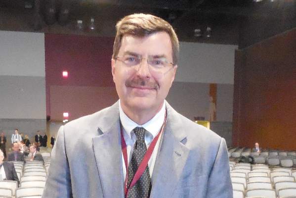
“Waiting an arbitrary 5 days is not important,” Elizabeth L. Nichols said during a video interview and during her report at the annual meeting of the Society of Thoracic Surgeons.
Ms. Nichols and her associates analyzed the in-hospital mortality rates among 3,060 patients who underwent isolated CABG during 2008-2014 at any of the seven medical centers that participate in the Northern New England Cardiovascular Disease Study Group and offer CABG. They included patients who had their surgery within 21 days of their MI, and excluded patients who had their CABG within 6 hours of their MI, had emergency surgery, or those with shock or incomplete data. The study group included 529 patients who had a ST-elevation MI and 2,531 patients with a non-ST-elevation MI.
The analysis divided patients into four groups based on timing of their CABG: 99 patients (3%) had surgery within the first 24 hours, 369 patients (12%) had their surgery 1-2 days after their MI, 1,966 (64%) had their operation 3-7 days following their MI, and 626 (21%) had their surgery 8-21 days after the MI.
The unadjusted mortality rates for these four subgroups were 5.1%, 1.6%, 1.6%, and 2.7%, respectively, reported Ms. Nichols, a health services researcher at the Dartmouth Institute for Health Policy & Clinical Practice, Lebanon, N.H.
After researchers adjusted for several demographic and clinical variables, the mortality rates remained identical for patients who underwent CABG 1 or 2 days following their MI, compared with patients whose surgery was deferred until 3-7 days after the MI. Patients with surgery 8-21 days following the MI had a small but not statistically significant higher rate of in-hospital death.
Patients who had their surgery 7-23 hours following an MI had a statistically significant increased hospital mortality following surgery that ran more than threefold greater than patients who underwent CABG 3-7 days after their MI.
The main message from the analysis is that for the typical, stable MI patient who requires CABG to treat multivessel coronary disease, no need exists to wait several days following an MI to do the surgery, Ms. Nichols explained. A delay of just 1 or 2 days is safe and sufficient, as long as it provides adequate time for any acutely administered antiplatelet or antithrombotic drugs to clear.

The findings “provide a degree of comfort for not waiting the 3-5 days that had previously been thought necessary,” said Dr. Jock N. McCullough, chief of cardiac surgery at Dartmouth-Hitchcock Medical Center in Lebanon and a collaborator on the study.
The findings are not meant to supersede clinical judgment, both Dr. McCullough and Ms. Nichols emphasized. Individual patients might have good reasons to either undergo faster surgery or to wait at least 8 days following their MI.
“The patients who waited 8-21 days had a lot of comorbidities and were sicker patients, and their delay is often warranted” to make sure the patient is stable enough for surgery, Ms. Nichols explained. Other patients might be worsening following their MI and need to undergo their surgery within 24 hours of their MI.
“Clinical judgment is always the trump card,” Ms. Nichols said.
Ms. Nichols and Dr. McCullough had no disclosures.
The video associated with this article is no longer available on this site. Please view all of our videos on the MDedge YouTube channel
On Twitter @mitchelzoler
PHOENIX – Patients who are stable following a myocardial infarction and need isolated coronary artery bypass surgery (CABG) don’t need to wait 5 or so days for their surgery, a delay that many surgeons and cardiologists often impose.
The operation can safely occur after just a 1- or 2-day gap following either an ST-elevation MI or a non–ST-elevation MI, based on real-world outcomes seen in more than 3,000 patients treated at any of seven U.S. medical centers.
“Waiting an arbitrary 5 days is not important,” Elizabeth L. Nichols said during a video interview and during her report at the annual meeting of the Society of Thoracic Surgeons.
Ms. Nichols and her associates analyzed the in-hospital mortality rates among 3,060 patients who underwent isolated CABG during 2008-2014 at any of the seven medical centers that participate in the Northern New England Cardiovascular Disease Study Group and offer CABG. They included patients who had their surgery within 21 days of their MI, and excluded patients who had their CABG within 6 hours of their MI, had emergency surgery, or those with shock or incomplete data. The study group included 529 patients who had a ST-elevation MI and 2,531 patients with a non-ST-elevation MI.
The analysis divided patients into four groups based on timing of their CABG: 99 patients (3%) had surgery within the first 24 hours, 369 patients (12%) had their surgery 1-2 days after their MI, 1,966 (64%) had their operation 3-7 days following their MI, and 626 (21%) had their surgery 8-21 days after the MI.
The unadjusted mortality rates for these four subgroups were 5.1%, 1.6%, 1.6%, and 2.7%, respectively, reported Ms. Nichols, a health services researcher at the Dartmouth Institute for Health Policy & Clinical Practice, Lebanon, N.H.
After researchers adjusted for several demographic and clinical variables, the mortality rates remained identical for patients who underwent CABG 1 or 2 days following their MI, compared with patients whose surgery was deferred until 3-7 days after the MI. Patients with surgery 8-21 days following the MI had a small but not statistically significant higher rate of in-hospital death.
Patients who had their surgery 7-23 hours following an MI had a statistically significant increased hospital mortality following surgery that ran more than threefold greater than patients who underwent CABG 3-7 days after their MI.
The main message from the analysis is that for the typical, stable MI patient who requires CABG to treat multivessel coronary disease, no need exists to wait several days following an MI to do the surgery, Ms. Nichols explained. A delay of just 1 or 2 days is safe and sufficient, as long as it provides adequate time for any acutely administered antiplatelet or antithrombotic drugs to clear.
The findings “provide a degree of comfort for not waiting the 3-5 days that had previously been thought necessary,” said Dr. Jock N. McCullough, chief of cardiac surgery at Dartmouth-Hitchcock Medical Center in Lebanon and a collaborator on the study.
The findings are not meant to supersede clinical judgment, both Dr. McCullough and Ms. Nichols emphasized. Individual patients might have good reasons to either undergo faster surgery or to wait at least 8 days following their MI.
“The patients who waited 8-21 days had a lot of comorbidities and were sicker patients, and their delay is often warranted” to make sure the patient is stable enough for surgery, Ms. Nichols explained. Other patients might be worsening following their MI and need to undergo their surgery within 24 hours of their MI.
“Clinical judgment is always the trump card,” Ms. Nichols said.
Ms. Nichols and Dr. McCullough had no disclosures.
The video associated with this article is no longer available on this site. Please view all of our videos on the MDedge YouTube channel
On Twitter @mitchelzoler
AT THE STS ANNUAL MEETING
Key clinical point: Performing coronary artery bypass grafting 1-2 days following an MI was as safe as when surgery was delayed 3-7 days.
Major finding: In-hospital mortality after CABG was identical in patients operated on 1-2 days or 3-7 days following an MI.
Data source: Retrospective analysis of 3,060 patients who underwent CABG within 21 days following an MI at any of seven U.S. centers.
Disclosures: Ms. Nichols and Dr. McCullough had no disclosures.
Point/Counterpoint: Does surgery play a role in N2 disease treatment following induction therapy?
POINT: Surgery has its uses for some
BY DR. STEPHEN G. SWISHER
When talking about the role of surgery after induction therapy with persistent N2 disease, one must acknowledge that this is such a heterogeneous disease. You can have single-station N2; resectable, bulky multistation N2; and so on. Then there’s unresectable stage IIIA, but let’s focus mainly on resectable stage IIIA disease.
I can’t tell you how many audiences I’ve faced that absolutely believe the myth that surgery plays no role in Stage IIIA non–small cell lung cancer based on data from stage IIIA disease patients randomized to chemoradiation followed by surgery. The problem with these study results is the high mortality in the pneumonectomy subset. There’s no difference in the overall survival of the two groups, but that doesn’t mean that everyone in that group wouldn’t benefit from surgery.

The curve showed that pneumonectomy did not benefit after chemoradiation in a non–high-volume center. You can see a steep drop in the mortality early on, but it catches up again at the end. If you look at the overall 5-year survival rate, even in the pneumonectomy subset, you’re looking at 22% vs. 24%.
But in the lobectomy set, you see something completely different. You’ve got a doubling of survivors and no mortality early on, and a doubling of 5-year survival from 18% to 36%.
And yet, people continue saying that there’s no role for surgery. Well, I think there is a role for surgery, and there are subsets of N2 for which surgery can be particularly beneficial. We have to move more toward what the medical oncologists do, which is personalize therapy and look at subsets of N2 disease so that we know which patients we can benefit and which ones we can’t.
Moving on to the second myth: Surgery plays no role in N2 residual disease after induction therapy. This myth is based on the results of a couple of prospective studies in the 1980s and ’90s that showed residual N2 disease after chemo- and radiotherapy leads to survival of 16-35 months in most cases. I’d say that those results are true, but it’s not to say there aren’t subsets within these populations that benefited. With preoperative chemo and radiation, it’s basically the same thing – poor prognoses in patients with N2 or N3 disease, so the standard becomes never to operate on these individuals.
A European study prospectively took 30 patients and treated them with induction chemotherapy. They saw a 5-year survival rate of 62% if a patient downgrades to lymph node–negative disease and the positron-emission tomographic (PET) findings were good. But they also saw a subset with a small amount of disease within the lymph nodes at the N2 stage and a poor response on PET; Their 5-year survival rate was around 19%. So I’d argue that PET response and the number of lymph nodes involved are the key criteria, and you shouldn’t routinely deny surgery to these patients.
Our experience at MD Anderson Cancer Center over the last 10 years has been to treat N2 and N3 admissions, with surgery, followed by postoperative radiation of 50 Gy. We’re able to achieve very-low morbidity with this regimen, and no mortality after 30 and 90 days. Just to show the heterogeneity: Single-station, microscopic N2 disease should really be resected.
You just can’t lump together everyone with residual N2 after induction therapy, since PET-CTs and most other diagnostic procedures have high false-negative rates. And like I’ve said, it doesn’t matter because N2 disease is really a subset disease. Microscopic N2 disease behaves in a completely different way than does macroscopic, multiple-level N2 disease. And even more important is how the patient’s primary tumor responds to the chemotherapy or chemoradiation; that will tell you how well they’re going to do even if they have a small amount of residual disease in the lymph nodes.
Dr. Swisher is at the University of Texas MD Anderson Cancer Center in Houston. He disclosed that he is a consultant/advisory board member for GlaxoSmithKline.
COUNTERPOINT: Surgery seems to have little value, adds risk.
BY DR. SCOTT J. SWANSON
Dr. Swisher and I probably agree more than we disagree, but I’m going to start by saying that N2 disease is bad, and most of these populations are heterogeneous. But if you feel that a curve toward the bottom of a graph is good, then you should think twice. Anywhere from a 15%-30% survival rate is not great and shows that we have a long way to go. The overall impression among oncologists in several countries is that it’s not really clear whether surgery adds value. Even in very good centers like MD Anderson where there is minimal risk, surgery inherently still involves some risk.
So then, what do we do with persistent N2 disease? I’d say that most of the time, it should be treated with chemotherapy or chemotherapy and radiation. In some cases, N2 disease can be treated with a creative, mutation-driven immunotherapy approach. Most of the time though, surgery is just not a good idea.
Interestingly, about one-third of lung cancer patients present with stage IIIA disease, so it’s important that we as a medical community sort out these treatment options. I think we’re all in agreement that single-station, microscopic, PET-negative/CT-negative disease is not the same as extranodal or multistation PET-positive disease, so we’ve got to begin to substratify N2 disease.
In an intergroup trial of about 200 patients per arm published in the Lancet, patients went to surgery if they didn’t regress after evaluation. The progression-free survival rate did seem to favor surgery; and, if you look at the lobectomy subset, the results are certainly strong. But again, we’re dealing with curves that are pretty low. The pneumonectomy subset drops off and then starts to catch up, but clearly pneumonectomy was a problem in this multicenter trial. The most important graph to this debate shows subjects that persistently had nodal disease had very poor survival. It’s hard to argue for surgery when results show that only about 24% of them are going to be alive down the road. If oncologists across most of the United States say they believe that surgery isn’t a good idea, we’re not going to use this graph to change their minds; we need to change our way of thinking.
So the conclusion to take away from that presentation is that N0 status at surgery significantly predicts greater 5-year survival. So, conversely, surgery is not helpful for the patient with node-positive disease. Surgical resection should only be considered if lobectomy is the operation in question. Pneumonectomy carries risk, and surgery has no beneficial value, compared with chemotherapy unless the patient has been downstaged.
Our experience at Brigham and Women’s Hospital in Boston is similar to Dr. Swisher’s at MD Anderson. During the first 8 years of our thoracic division, we looked at 103 patients who had surgery after induction therapy for N2 disease. The induction plan in those patients was chemotherapy only for 75, radiation only for 18, and chemoradiation for 10. Almost 40% of patients had pneumonectomies, and the rest had lobectomies.
Mortality was 3.9%, major morbidity was 7%, and about 30% were downstaged. Those 5-year survival rates were about 36%. Persistent nodal disease, either N1 or N2, was seen in about 75%, and most of them were N2; the 5-year survival rate there was about 9% with a median of about 15.9 months. Beauty is in the eye of the beholder, so you may look at that and say that 15.9 months isn’t bad, but there’s still a huge subgroup that’s node positive, so here I’d say that pushing surgery is not the best strategy. We also found in this group that adenocarcinomas were much harder to clear.
We’re on very-safe ground to push surgery in the node-negative group, but you’ve got to be careful in the node-positive group.
Survival is relatively limited in N2 disease in general. Surgery may be of value if you downstage the patient, if you’re doing a lobectomy or if you see squamous cell carcinoma. Going forward, we really ought to focus our attention on identifying responders more reliably and improving downstaging – with different or individualized chemotherapy, or perhaps even immunologic therapy.
For the present, we can talk about radiation dosing. High-dose radiation is clearly a viable option for some patients. In addition, we can improve identification of N2 subgroups. Not all N2 disease is the same, so it should not be treated the same across the board.
Dr. Swanson is at Brigham and Women’s Hospital in Boston. He disclosed that he is a consultant/advisory board member for Covidien and Ethicon Endo-Surgery.
This article grew out of the debate by Dr. Swisher and Dr. Swanson at the annual meeting of the Society of Thoracic Surgeons.
POINT: Surgery has its uses for some
BY DR. STEPHEN G. SWISHER
When talking about the role of surgery after induction therapy with persistent N2 disease, one must acknowledge that this is such a heterogeneous disease. You can have single-station N2; resectable, bulky multistation N2; and so on. Then there’s unresectable stage IIIA, but let’s focus mainly on resectable stage IIIA disease.
I can’t tell you how many audiences I’ve faced that absolutely believe the myth that surgery plays no role in Stage IIIA non–small cell lung cancer based on data from stage IIIA disease patients randomized to chemoradiation followed by surgery. The problem with these study results is the high mortality in the pneumonectomy subset. There’s no difference in the overall survival of the two groups, but that doesn’t mean that everyone in that group wouldn’t benefit from surgery.
The curve showed that pneumonectomy did not benefit after chemoradiation in a non–high-volume center. You can see a steep drop in the mortality early on, but it catches up again at the end. If you look at the overall 5-year survival rate, even in the pneumonectomy subset, you’re looking at 22% vs. 24%.
But in the lobectomy set, you see something completely different. You’ve got a doubling of survivors and no mortality early on, and a doubling of 5-year survival from 18% to 36%.
And yet, people continue saying that there’s no role for surgery. Well, I think there is a role for surgery, and there are subsets of N2 for which surgery can be particularly beneficial. We have to move more toward what the medical oncologists do, which is personalize therapy and look at subsets of N2 disease so that we know which patients we can benefit and which ones we can’t.
Moving on to the second myth: Surgery plays no role in N2 residual disease after induction therapy. This myth is based on the results of a couple of prospective studies in the 1980s and ’90s that showed residual N2 disease after chemo- and radiotherapy leads to survival of 16-35 months in most cases. I’d say that those results are true, but it’s not to say there aren’t subsets within these populations that benefited. With preoperative chemo and radiation, it’s basically the same thing – poor prognoses in patients with N2 or N3 disease, so the standard becomes never to operate on these individuals.
A European study prospectively took 30 patients and treated them with induction chemotherapy. They saw a 5-year survival rate of 62% if a patient downgrades to lymph node–negative disease and the positron-emission tomographic (PET) findings were good. But they also saw a subset with a small amount of disease within the lymph nodes at the N2 stage and a poor response on PET; Their 5-year survival rate was around 19%. So I’d argue that PET response and the number of lymph nodes involved are the key criteria, and you shouldn’t routinely deny surgery to these patients.
Our experience at MD Anderson Cancer Center over the last 10 years has been to treat N2 and N3 admissions, with surgery, followed by postoperative radiation of 50 Gy. We’re able to achieve very-low morbidity with this regimen, and no mortality after 30 and 90 days. Just to show the heterogeneity: Single-station, microscopic N2 disease should really be resected.
You just can’t lump together everyone with residual N2 after induction therapy, since PET-CTs and most other diagnostic procedures have high false-negative rates. And like I’ve said, it doesn’t matter because N2 disease is really a subset disease. Microscopic N2 disease behaves in a completely different way than does macroscopic, multiple-level N2 disease. And even more important is how the patient’s primary tumor responds to the chemotherapy or chemoradiation; that will tell you how well they’re going to do even if they have a small amount of residual disease in the lymph nodes.
Dr. Swisher is at the University of Texas MD Anderson Cancer Center in Houston. He disclosed that he is a consultant/advisory board member for GlaxoSmithKline.
COUNTERPOINT: Surgery seems to have little value, adds risk.
BY DR. SCOTT J. SWANSON
Dr. Swisher and I probably agree more than we disagree, but I’m going to start by saying that N2 disease is bad, and most of these populations are heterogeneous. But if you feel that a curve toward the bottom of a graph is good, then you should think twice. Anywhere from a 15%-30% survival rate is not great and shows that we have a long way to go. The overall impression among oncologists in several countries is that it’s not really clear whether surgery adds value. Even in very good centers like MD Anderson where there is minimal risk, surgery inherently still involves some risk.
So then, what do we do with persistent N2 disease? I’d say that most of the time, it should be treated with chemotherapy or chemotherapy and radiation. In some cases, N2 disease can be treated with a creative, mutation-driven immunotherapy approach. Most of the time though, surgery is just not a good idea.
Interestingly, about one-third of lung cancer patients present with stage IIIA disease, so it’s important that we as a medical community sort out these treatment options. I think we’re all in agreement that single-station, microscopic, PET-negative/CT-negative disease is not the same as extranodal or multistation PET-positive disease, so we’ve got to begin to substratify N2 disease.
In an intergroup trial of about 200 patients per arm published in the Lancet, patients went to surgery if they didn’t regress after evaluation. The progression-free survival rate did seem to favor surgery; and, if you look at the lobectomy subset, the results are certainly strong. But again, we’re dealing with curves that are pretty low. The pneumonectomy subset drops off and then starts to catch up, but clearly pneumonectomy was a problem in this multicenter trial. The most important graph to this debate shows subjects that persistently had nodal disease had very poor survival. It’s hard to argue for surgery when results show that only about 24% of them are going to be alive down the road. If oncologists across most of the United States say they believe that surgery isn’t a good idea, we’re not going to use this graph to change their minds; we need to change our way of thinking.
So the conclusion to take away from that presentation is that N0 status at surgery significantly predicts greater 5-year survival. So, conversely, surgery is not helpful for the patient with node-positive disease. Surgical resection should only be considered if lobectomy is the operation in question. Pneumonectomy carries risk, and surgery has no beneficial value, compared with chemotherapy unless the patient has been downstaged.
Our experience at Brigham and Women’s Hospital in Boston is similar to Dr. Swisher’s at MD Anderson. During the first 8 years of our thoracic division, we looked at 103 patients who had surgery after induction therapy for N2 disease. The induction plan in those patients was chemotherapy only for 75, radiation only for 18, and chemoradiation for 10. Almost 40% of patients had pneumonectomies, and the rest had lobectomies.
Mortality was 3.9%, major morbidity was 7%, and about 30% were downstaged. Those 5-year survival rates were about 36%. Persistent nodal disease, either N1 or N2, was seen in about 75%, and most of them were N2; the 5-year survival rate there was about 9% with a median of about 15.9 months. Beauty is in the eye of the beholder, so you may look at that and say that 15.9 months isn’t bad, but there’s still a huge subgroup that’s node positive, so here I’d say that pushing surgery is not the best strategy. We also found in this group that adenocarcinomas were much harder to clear.
We’re on very-safe ground to push surgery in the node-negative group, but you’ve got to be careful in the node-positive group.
Survival is relatively limited in N2 disease in general. Surgery may be of value if you downstage the patient, if you’re doing a lobectomy or if you see squamous cell carcinoma. Going forward, we really ought to focus our attention on identifying responders more reliably and improving downstaging – with different or individualized chemotherapy, or perhaps even immunologic therapy.
For the present, we can talk about radiation dosing. High-dose radiation is clearly a viable option for some patients. In addition, we can improve identification of N2 subgroups. Not all N2 disease is the same, so it should not be treated the same across the board.
Dr. Swanson is at Brigham and Women’s Hospital in Boston. He disclosed that he is a consultant/advisory board member for Covidien and Ethicon Endo-Surgery.
This article grew out of the debate by Dr. Swisher and Dr. Swanson at the annual meeting of the Society of Thoracic Surgeons.
Point/Counterpoint: Should surgeons be mandated to have residents operate to satisfy board requirements?
As “simple procedures” diminish, should thoracic surgeons in training programs be mandated to allow residents to operate on patients in order to satisfy board requirements? This was the question posed during an ethics debate at the annual meeting of the Society of Thoracic Surgeons.
It is ethical, and it is necessary.
BY RICHARD G. OHYE, M.D.
The linchpin of this discussion is the “obligation,” which is defined as “a course of action that someone is required to take, whether legal or moral” to have residents perform surgery. My position is that, yes, we do have such a mandate.
I doubt that Dr. Jaggers and I would disagree that teaching residents is something we do as academic surgeons. The devil is in the details. Among our concerns are patient safety and closer scrutiny on surgical practices due to public reporting, which makes everything we do readily available. Further, simple, straightforward cases are going away; interventional cardiologists are doing lots of stents, mitral valves, and atrial septal defect closures, so those kinds of procedures are going away.

Looking at case logs from congenital cardiac and CT residents at our institution, however, there are still incomplete canals, tricuspid valve repairs, mitral valve repairs and replacements, aortic valve repairs, patent ductus arteriosus repairs, vascular rings, pulmonary valve replacements, and conduits. Residents are capable of doing these procedures; they are incredibly talented individuals and you just have to let them operate.
In addition, our results – and more importantly, our patients – have not suffered. We let our residents do between one-third and one-half of our cases, and the cases only count if they’re skin-to-skin. Our results, compared by STAT category, compare favorably with STS benchmarks and are either at or below expected values. By the end of this year, our expected mortality should be about 23%, but our observed mortality is less than half that value with the residents doing lots of cases.
So what about the ethics – who would you want operating on you? There is an ethical dilemma that goes along with medical education because no matter how good my residents may be, I am more experienced. I can do every procedure faster and “better” than they can. But the teaching of students is not a new concept, it’s even in the Hippocratic Oath, so this is an old and well-accepted practice.
There is a strong parallel between medical education and medical research. We still have to follow all of those important guidelines we have for medical research – do what’s right for the patient and exercise good judgment. We must not just “do no harm.” We must actively do good.
Academic surgeons have an obligation to teach. We take care of patients, do research to push the whole field forward, and educate to bring up the next generation of doctors. The cases for residents to perform are all there – yes, we’re a big program, but even smaller programs should see plenty of cases – and I think I’ve shown that these can be performed safely and yield excellent results. As long as the results are good, you don’t need to worry about public scrutiny. The case for medical education is similar to that of medical research: It is ethical, and it is necessary.
Dr. Ohye is head of the pediatric cardiovascular surgery division and surgical director of the pediatric heart transplant program of the University of Michigan, in Ann Arbor; he argued in support of a mandate.
Patients may not benefit, and may actually be harmed
BY JAMES JAGGERS, M.D.
The central issue in this debate is whether or not the surgeon’s responsibility as an educator and member of the training program overrides the surgeon’s responsibility to provide the patient with the best possible outcome. Put another way, should the responsibility to treat the patient to the best of the surgeon’s ability be subordinated to the success and survival of the training program for the sole purpose of giving the resident sufficient operative experience to be board eligible?
Both versions of the Hippocratic Oath and the more recent Declaration of Geneva, the AMA’s Code of Ethics, and the ACGME Mission Statement clearly enforce that the primary responsibility of the physician is to the patient, while also endorsing physician responsibility to community via service and education. Using patients as a means to an end – in this case, to satisfy board requirements – and to do so without patients’ explicit consent, violates the fundamental principle of respect for individuals.
I will not argue that resident surgery can be safely performed without risking harm to the patient. If the surgical instructor could exercise complete control over a procedure and correct any mistakes that the trainees made so that the procedure has the same outcome, then it would be ethically allowable. The surgical instructor must be confident that his residents are fully capable of performing the surgery on their own, otherwise it not ethical to subject the patient to this risk.
Dr. Ohye and I are both part of larger divisions that have their own obligations, and we have experience training residents at all levels. We have similar backgrounds, and we both benefited from having mentors who sometimes had masochistic patience in helping us get through surgeries that we probably weren’t ready for. I’m certain that those of us in academic medicine training programs believe that graduated involvement of trainees in patient care is an integral part of the surgical education process, and is critical to society as a whole.
However, the fact that patients may not directly benefit, and may actually be harmed, from the resident’s involvement in surgery creates an ethical dilemma. There is little literature to guide us through this dilemma. Professional societies only advise generally, noting that participation should be voluntary, without providing specifics. Regulatory boards simply set minimum requirements without providing guidance for the educational process. While the ACGME and the residency review committees oversee resident training, the responsibility for successful training is left largely to individual surgeons and individual programs. It’s only recently that the TSDA (Thoracic Surgery Directors Association) adopted the milestone concept that hopefully will help resolve some of these issues.
Consider a medium-sized program of around 300 patients: A difference of just one death, such as 10 or 11 per year, is the difference between being above or below the STS mean. Now that may not be statistically significant, but if you put that number on your website, it becomes important. It’s true that the practice of congenital heart surgery has changed over the last 15-20 years. Our program has seen resident cases roughly halved in the last 10 years. Most patients are operated on at a younger age, palliation is very rare. These are not meant to be excuses for not training residents – they’re just the reality.
The outcomes of surgeries are increasingly scrutinized by regulatory agencies and sources of public reporting. Competition between programs is intense. Patients, parents, and referring providers have become increasingly aware of outcomes to the point that it’s actually not unusual for a patient to ask “What’s your surgical site infection rate? What are the chances I’ll need to have a pacemaker? What are your individual results?” Insurance companies are starting to ask for financial data, economically profiling you to ensure that you’re being as efficient as possible. All of these things are contrary to our ability to train residents effectively.
Because of fear of taking too long or increasing complications, some surgeons say they are much more likely to accept residual defects when operating with trainees. It’s only with familiarity and time that the highly skilled attending and properly motivated resident may work in tandem and produce the best outcome – but not in the 10 cases the American Board of Thoracic Surgery requires.
Dr. Jaggers is the Barton-Elliman Chair in Pediatric Cardiothoracic Surgery at the University of Colorado and co–medical director of The Heart Institute at Children’s Hospital Colorado in Aurora. He argued against having residents perform surgery for board certification.
As “simple procedures” diminish, should thoracic surgeons in training programs be mandated to allow residents to operate on patients in order to satisfy board requirements? This was the question posed during an ethics debate at the annual meeting of the Society of Thoracic Surgeons.
It is ethical, and it is necessary.
BY RICHARD G. OHYE, M.D.
The linchpin of this discussion is the “obligation,” which is defined as “a course of action that someone is required to take, whether legal or moral” to have residents perform surgery. My position is that, yes, we do have such a mandate.
I doubt that Dr. Jaggers and I would disagree that teaching residents is something we do as academic surgeons. The devil is in the details. Among our concerns are patient safety and closer scrutiny on surgical practices due to public reporting, which makes everything we do readily available. Further, simple, straightforward cases are going away; interventional cardiologists are doing lots of stents, mitral valves, and atrial septal defect closures, so those kinds of procedures are going away.
Looking at case logs from congenital cardiac and CT residents at our institution, however, there are still incomplete canals, tricuspid valve repairs, mitral valve repairs and replacements, aortic valve repairs, patent ductus arteriosus repairs, vascular rings, pulmonary valve replacements, and conduits. Residents are capable of doing these procedures; they are incredibly talented individuals and you just have to let them operate.
In addition, our results – and more importantly, our patients – have not suffered. We let our residents do between one-third and one-half of our cases, and the cases only count if they’re skin-to-skin. Our results, compared by STAT category, compare favorably with STS benchmarks and are either at or below expected values. By the end of this year, our expected mortality should be about 23%, but our observed mortality is less than half that value with the residents doing lots of cases.
So what about the ethics – who would you want operating on you? There is an ethical dilemma that goes along with medical education because no matter how good my residents may be, I am more experienced. I can do every procedure faster and “better” than they can. But the teaching of students is not a new concept, it’s even in the Hippocratic Oath, so this is an old and well-accepted practice.
There is a strong parallel between medical education and medical research. We still have to follow all of those important guidelines we have for medical research – do what’s right for the patient and exercise good judgment. We must not just “do no harm.” We must actively do good.
Academic surgeons have an obligation to teach. We take care of patients, do research to push the whole field forward, and educate to bring up the next generation of doctors. The cases for residents to perform are all there – yes, we’re a big program, but even smaller programs should see plenty of cases – and I think I’ve shown that these can be performed safely and yield excellent results. As long as the results are good, you don’t need to worry about public scrutiny. The case for medical education is similar to that of medical research: It is ethical, and it is necessary.
Dr. Ohye is head of the pediatric cardiovascular surgery division and surgical director of the pediatric heart transplant program of the University of Michigan, in Ann Arbor; he argued in support of a mandate.
Patients may not benefit, and may actually be harmed
BY JAMES JAGGERS, M.D.
The central issue in this debate is whether or not the surgeon’s responsibility as an educator and member of the training program overrides the surgeon’s responsibility to provide the patient with the best possible outcome. Put another way, should the responsibility to treat the patient to the best of the surgeon’s ability be subordinated to the success and survival of the training program for the sole purpose of giving the resident sufficient operative experience to be board eligible?
Both versions of the Hippocratic Oath and the more recent Declaration of Geneva, the AMA’s Code of Ethics, and the ACGME Mission Statement clearly enforce that the primary responsibility of the physician is to the patient, while also endorsing physician responsibility to community via service and education. Using patients as a means to an end – in this case, to satisfy board requirements – and to do so without patients’ explicit consent, violates the fundamental principle of respect for individuals.
I will not argue that resident surgery can be safely performed without risking harm to the patient. If the surgical instructor could exercise complete control over a procedure and correct any mistakes that the trainees made so that the procedure has the same outcome, then it would be ethically allowable. The surgical instructor must be confident that his residents are fully capable of performing the surgery on their own, otherwise it not ethical to subject the patient to this risk.
Dr. Ohye and I are both part of larger divisions that have their own obligations, and we have experience training residents at all levels. We have similar backgrounds, and we both benefited from having mentors who sometimes had masochistic patience in helping us get through surgeries that we probably weren’t ready for. I’m certain that those of us in academic medicine training programs believe that graduated involvement of trainees in patient care is an integral part of the surgical education process, and is critical to society as a whole.
However, the fact that patients may not directly benefit, and may actually be harmed, from the resident’s involvement in surgery creates an ethical dilemma. There is little literature to guide us through this dilemma. Professional societies only advise generally, noting that participation should be voluntary, without providing specifics. Regulatory boards simply set minimum requirements without providing guidance for the educational process. While the ACGME and the residency review committees oversee resident training, the responsibility for successful training is left largely to individual surgeons and individual programs. It’s only recently that the TSDA (Thoracic Surgery Directors Association) adopted the milestone concept that hopefully will help resolve some of these issues.
Consider a medium-sized program of around 300 patients: A difference of just one death, such as 10 or 11 per year, is the difference between being above or below the STS mean. Now that may not be statistically significant, but if you put that number on your website, it becomes important. It’s true that the practice of congenital heart surgery has changed over the last 15-20 years. Our program has seen resident cases roughly halved in the last 10 years. Most patients are operated on at a younger age, palliation is very rare. These are not meant to be excuses for not training residents – they’re just the reality.
The outcomes of surgeries are increasingly scrutinized by regulatory agencies and sources of public reporting. Competition between programs is intense. Patients, parents, and referring providers have become increasingly aware of outcomes to the point that it’s actually not unusual for a patient to ask “What’s your surgical site infection rate? What are the chances I’ll need to have a pacemaker? What are your individual results?” Insurance companies are starting to ask for financial data, economically profiling you to ensure that you’re being as efficient as possible. All of these things are contrary to our ability to train residents effectively.
Because of fear of taking too long or increasing complications, some surgeons say they are much more likely to accept residual defects when operating with trainees. It’s only with familiarity and time that the highly skilled attending and properly motivated resident may work in tandem and produce the best outcome – but not in the 10 cases the American Board of Thoracic Surgery requires.
Dr. Jaggers is the Barton-Elliman Chair in Pediatric Cardiothoracic Surgery at the University of Colorado and co–medical director of The Heart Institute at Children’s Hospital Colorado in Aurora. He argued against having residents perform surgery for board certification.

Linking registries, databases may reduce surgical site infections
Surveillance of cardiac surgical site infections (SSIs) improved significantly when registry and infection control surveillance data were linked with electronic health records, a retrospective analysis showed.
Over the course of a 47-month period starting in 2011, Vaidehi Nayar of the Children’s Hospital of Philadelphia and her coinvestigators launched a quality improvement initiative at their institution that linked administrative databases with their clinical registry, allowing caregivers to more accurately monitor and assess SSIs and provide more effective adjudication and treatments thereafter. The investigators chose to link their hospital’s electronic health record (EHR) billing information and reporting from the infection surveillance database for the National Healthcare Safety Network with data from the Society of Thoracic Surgeons Congenital Heart Surgery Database (STS-CHSD).

To further facilitate the flow and interpretation of data, the investigators used a visualization tool to analyze the STS-CHSD for case ascertainment; to resolve discrepancies among STS-CHSD, infection surveillance, and billing SSI cases; and to assess the impact of the hospital’s quality improvement protocols. These protocols consisted of wound alert reports from the EHR, bedside reviews for SSI adjudication, inpatient and outpatient SSI prevention bundles, prophylactic antibiotic dosing changes, removal of steroids from the bypass circuit, and biller education on SSIs.
Control charts in the data visualization tool allowed for statistical monitoring of SSI rate changes, and SSI case discrepancies across the databases were reviewed to ensure that differences were the result of variations in SSI reporting criteria for each database, not inaccurate surveillance population ascertainment or inaccurate SSI identification, according to Ms. Nayar and her colleagues,
“Workflow changes, including the wound alert report and bedside reviews, facilitated communication among providers and improved adjudication of suspected SSIs,” she said in presenting the J. Maxwell Chamberlain Memorial Paper for Congenital Heart Surgery at the annual meeting of the Society of Thoracic Surgeons in San Diego earlier this year.
In total, 156 SSIs were identified via the STS-CHSD, 79 via the infection surveillance database, and 433 via billing. There was a significant decrease in the rolling 12-month SSI rate from 2.48% (21/848 in January 2013) to 0.76% (11/1,442 in January 2014), based on the STS-CHSD data, although Ms. Nayar pointed out that this decline could not definitely be attributed to reporting improvements or to the specific quality improvement initiatives that were implemented. Ms. Nayar also explained that there was a “general improvement in reporting, as shown by the stark drop in billing” and “a general alignment of all three data sources.”
“Accurate assessment of morbidity data, including [SSIs], has implications for public reporting, benchmarking, assessment of [quality improvement] impact, and possible denial of payments,” said Ms. Nayar. “In summary, we achieved our two simultaneous goals of improving SSI reporting – or decreasing the data errors – and decreasing SSI incidents by improving overall patient care.”
During discussion, Ms. Nayar elaborated on the study’s generalizability and potential application to other facets of congenital heart disease treatments, saying that such application is, in fact, possible.
“Yes, this is very generalizable, but one key important fact is relevant: As long as there is a source of truth for case ascertainment, this methodology can be used in several different areas,” she explained. “For example, we are currently working at our institution to integrate and link the [Pediatric Cardiac Critical Care Consortium] database to be able to better monitor any critical care–related morbidity information and ultimately use similar methodology to assess the impact of outcomes in the critical care field.”
Ms. Nayar said that she did not have any relevant financial conflicts of interest to disclose.
This study demonstrates dramatically different surgical site infection (SSI) rates for the same patient cohort as detected by three different surveillance methods: hospital billing (derived from the electronic health records), a surgical database, and a tracking system developed by the Centers for Disease Control and Prevention.
On the low end, 79 SSIs were reported by one system, 156 by another, and 433 by the third method – a more than fivefold discrepancy. The authors developed a technique to reconcile the three systems and then evaluated a variety of interventions designed to reduce the SSI rate. As a result of this initiative, the quality of event reporting was improved (with greater agreement between reporting methods) and the rate of SSIs was reduced.
The implications of this report are sobering and should be of great interest to all surgeons and hospital administrators for several reasons. First and fundamentally, the report suggests that widely used reporting systems may be inaccurate and produce conflicting results. Since the results of these reports are used to assess and modify clinical practice, this is very disturbing. Additionally, the results may be used by payers as a basis for financial reward (or penalty) and therefore must be accurate. Finally, exposure of the use of erroneous information as the source data for billing may render an institution vulnerable to civil and criminal penalties. For all of these reasons, the present report should prompt self-assessment by all institutions, if it has not already been undertaken.
Dr. Robert Jaquiss is associate medical editor for congenital heart disease for Thoracic Surgery News.
Surveillance of cardiac surgical site infections (SSIs) improved significantly when registry and infection control surveillance data were linked with electronic health records, a retrospective analysis showed.
Over the course of a 47-month period starting in 2011, Vaidehi Nayar of the Children’s Hospital of Philadelphia and her coinvestigators launched a quality improvement initiative at their institution that linked administrative databases with their clinical registry, allowing caregivers to more accurately monitor and assess SSIs and provide more effective adjudication and treatments thereafter. The investigators chose to link their hospital’s electronic health record (EHR) billing information and reporting from the infection surveillance database for the National Healthcare Safety Network with data from the Society of Thoracic Surgeons Congenital Heart Surgery Database (STS-CHSD).
To further facilitate the flow and interpretation of data, the investigators used a visualization tool to analyze the STS-CHSD for case ascertainment; to resolve discrepancies among STS-CHSD, infection surveillance, and billing SSI cases; and to assess the impact of the hospital’s quality improvement protocols. These protocols consisted of wound alert reports from the EHR, bedside reviews for SSI adjudication, inpatient and outpatient SSI prevention bundles, prophylactic antibiotic dosing changes, removal of steroids from the bypass circuit, and biller education on SSIs.
Control charts in the data visualization tool allowed for statistical monitoring of SSI rate changes, and SSI case discrepancies across the databases were reviewed to ensure that differences were the result of variations in SSI reporting criteria for each database, not inaccurate surveillance population ascertainment or inaccurate SSI identification, according to Ms. Nayar and her colleagues,
“Workflow changes, including the wound alert report and bedside reviews, facilitated communication among providers and improved adjudication of suspected SSIs,” she said in presenting the J. Maxwell Chamberlain Memorial Paper for Congenital Heart Surgery at the annual meeting of the Society of Thoracic Surgeons in San Diego earlier this year.
In total, 156 SSIs were identified via the STS-CHSD, 79 via the infection surveillance database, and 433 via billing. There was a significant decrease in the rolling 12-month SSI rate from 2.48% (21/848 in January 2013) to 0.76% (11/1,442 in January 2014), based on the STS-CHSD data, although Ms. Nayar pointed out that this decline could not definitely be attributed to reporting improvements or to the specific quality improvement initiatives that were implemented. Ms. Nayar also explained that there was a “general improvement in reporting, as shown by the stark drop in billing” and “a general alignment of all three data sources.”
“Accurate assessment of morbidity data, including [SSIs], has implications for public reporting, benchmarking, assessment of [quality improvement] impact, and possible denial of payments,” said Ms. Nayar. “In summary, we achieved our two simultaneous goals of improving SSI reporting – or decreasing the data errors – and decreasing SSI incidents by improving overall patient care.”
During discussion, Ms. Nayar elaborated on the study’s generalizability and potential application to other facets of congenital heart disease treatments, saying that such application is, in fact, possible.
“Yes, this is very generalizable, but one key important fact is relevant: As long as there is a source of truth for case ascertainment, this methodology can be used in several different areas,” she explained. “For example, we are currently working at our institution to integrate and link the [Pediatric Cardiac Critical Care Consortium] database to be able to better monitor any critical care–related morbidity information and ultimately use similar methodology to assess the impact of outcomes in the critical care field.”
Ms. Nayar said that she did not have any relevant financial conflicts of interest to disclose.
Key clinical point: Linking registry and infection control data with a hospital’s electronic health records can significantly improve surveillance of SSIs, as the linkage allows for improved visualization abilities, communication within and between departments, facilitated adjudication of SSIs, and improved assessment of quality improvement initiatives to prevent further SSIs.
Major finding: Over the 47-month study period, 156 SSIs were identified via the STS-CHSD, 79 via the infection surveillance database, and 433 via billing. The rolling 12-month SSI rate based on the STS-CHSD decreased from 2.48% (21/848) to 0.76% (11/1,442).
Data source: A retrospective analysis of 668 cases in the STS-CHSD database over the course of 47 months.
Disclosures: Ms. Nayar reported that she had no relevant financial conflicts.

Low-volume centers using ECMO have poorest survival rates
SAN DIEGO – Lung transplantation centers that are considered low volume tend to have lower rates of survival than do those of their medium- and high-volume counterparts when patients are bridged via extracorporeal membrane oxygenation (ECMO), according to researchers.
Even so, there is a point at which survival outcomes begin to improve for low-volume centers, they added.
“Increasingly, [ECMO] is used as a bridge to lung transplantation; indeed, the use of ECMO has tripled over the past 15 years and survival has increased by the same magnitude,” Dr. Jeremiah A. Hayanga said at the annual meeting of the Society of Thoracic Surgeons.

“An entire body of literature has linked high-volume [centers] to improved outcomes in the context of complex surgical procedures. Lung transplantation [LTx] falls within the same domain, and has been considered subject to the same inverse volume-outcome paradigm,” said Dr. Hayanga of Michigan State University, Grand Rapids.
He and his coinvestigators conducted a retrospective analysis of 16,603 LTx recipients in the International Registry for Heart and Lung Transplantation (ISHLT) who underwent ECMO as their bridging strategy between 2005 and 2010. Centers were stratified into categories of low, medium, and high based on the volume of LTx procedures performed over the study interval: Low was defined as fewer than 25, medium as 25-50, and more than 50 as high volume.
Overall, 85 of the 16,603 transplant recipients in the study population were bridged via ECMO: 20 (23.5%) of them in low-volume centers, 30 (35.3%) in medium-volume centers, and 35 (41.2%) in high-volume centers. The researchers used Cox proportional hazard modeling to identify predictors of both 1- and 5-year survival rates, which were found to be significantly lower in low-volume centers – 13.61% at 5 years post LTx.
Looking at just the high-volume and low-volume centers, the researchers noted “significant differences” in both 1-year and 5-year survival rates when ECMO was used for bridging. One-year survival probability was roughly 40% in low-volume centers and roughly 70% in high-volume centers, while 5-year survival probability was well under 25% for recipients from low-volume centers and around 50% for those from high-volume centers (P = .0006). No significant differences existed for non-ECMO patients, regardless of center volume.
“No differences existed in survival in medium- and high-volume centers,” said Dr. Hayanga. “Transplanting without ECMO as a bridge showed fewer survival differences for both 1-year and 5-year survival. However, when ECMO was used as a bridge, the low-volume center [survival rates] were dramatically lower at both 1 year and 5 years.”
When Dr. Hayanga and his colleagues examined procedural volume as a continuous variable, however, a single inflection point was determined as the point at which survival outcomes steadily improve – 19 procedures. Centers that performed at least 19 LTx procedures between 2005 and 2010 experienced an uptick in survival rates, even though centers that saw 19-25 procedures were still considered low volume, the researchers noted.
“The corresponding c-statistic, however, is just under 60%,” cautioned Dr. Hayanga. “The C-statistic is a measure of the explanatory power of a variable – in this case, [center] volume – in accounting for the variability in outcome, or survival in this case. To put that number into context, a C-statistic of 50% means ‘no explanatory power’ whatsoever.”
Dr. Hayanga explained that he and his coauthors compared transplant recipient and donor characteristics using analysis of variance (ANOVA) and chi-square tests to compare variables, cumulative survival using Kaplan-Meier curves, and significance using log-rank tests.
Dr. Hayanga reported no financial conflicts of interest.
SAN DIEGO – Lung transplantation centers that are considered low volume tend to have lower rates of survival than do those of their medium- and high-volume counterparts when patients are bridged via extracorporeal membrane oxygenation (ECMO), according to researchers.
Even so, there is a point at which survival outcomes begin to improve for low-volume centers, they added.
“Increasingly, [ECMO] is used as a bridge to lung transplantation; indeed, the use of ECMO has tripled over the past 15 years and survival has increased by the same magnitude,” Dr. Jeremiah A. Hayanga said at the annual meeting of the Society of Thoracic Surgeons.
“An entire body of literature has linked high-volume [centers] to improved outcomes in the context of complex surgical procedures. Lung transplantation [LTx] falls within the same domain, and has been considered subject to the same inverse volume-outcome paradigm,” said Dr. Hayanga of Michigan State University, Grand Rapids.
He and his coinvestigators conducted a retrospective analysis of 16,603 LTx recipients in the International Registry for Heart and Lung Transplantation (ISHLT) who underwent ECMO as their bridging strategy between 2005 and 2010. Centers were stratified into categories of low, medium, and high based on the volume of LTx procedures performed over the study interval: Low was defined as fewer than 25, medium as 25-50, and more than 50 as high volume.
Overall, 85 of the 16,603 transplant recipients in the study population were bridged via ECMO: 20 (23.5%) of them in low-volume centers, 30 (35.3%) in medium-volume centers, and 35 (41.2%) in high-volume centers. The researchers used Cox proportional hazard modeling to identify predictors of both 1- and 5-year survival rates, which were found to be significantly lower in low-volume centers – 13.61% at 5 years post LTx.
Looking at just the high-volume and low-volume centers, the researchers noted “significant differences” in both 1-year and 5-year survival rates when ECMO was used for bridging. One-year survival probability was roughly 40% in low-volume centers and roughly 70% in high-volume centers, while 5-year survival probability was well under 25% for recipients from low-volume centers and around 50% for those from high-volume centers (P = .0006). No significant differences existed for non-ECMO patients, regardless of center volume.
“No differences existed in survival in medium- and high-volume centers,” said Dr. Hayanga. “Transplanting without ECMO as a bridge showed fewer survival differences for both 1-year and 5-year survival. However, when ECMO was used as a bridge, the low-volume center [survival rates] were dramatically lower at both 1 year and 5 years.”
When Dr. Hayanga and his colleagues examined procedural volume as a continuous variable, however, a single inflection point was determined as the point at which survival outcomes steadily improve – 19 procedures. Centers that performed at least 19 LTx procedures between 2005 and 2010 experienced an uptick in survival rates, even though centers that saw 19-25 procedures were still considered low volume, the researchers noted.
“The corresponding c-statistic, however, is just under 60%,” cautioned Dr. Hayanga. “The C-statistic is a measure of the explanatory power of a variable – in this case, [center] volume – in accounting for the variability in outcome, or survival in this case. To put that number into context, a C-statistic of 50% means ‘no explanatory power’ whatsoever.”
Dr. Hayanga explained that he and his coauthors compared transplant recipient and donor characteristics using analysis of variance (ANOVA) and chi-square tests to compare variables, cumulative survival using Kaplan-Meier curves, and significance using log-rank tests.
Dr. Hayanga reported no financial conflicts of interest.
AT THE STS ANNUAL MEETING
Key clinical point: Low-volume lung transplantation centers in the United States typically have the poorest survival rates compared to those with higher volumes when using ECMO.
Major finding: Of 85 LTx subjects bridged via ECMO, 20 (23.5%) of these were bridged in low, 30 (35.3%) in medium, and 35 (41.2%) in high-volume centers; in the ECMO cohort, the lowest 5-year survival rate (13.61%) was observed at low-volume centers.
Data source: Retrospective analysis of 16,603 adult LTx recipients in the International Registry for Heart and Lung Transplantation during 2005-2010.
Disclosures: Dr. Hayanga reported no financial conflicts of interest.

New mortality index aids comparison of outcome rates across institutions
SAN DIEGO–Uniquely derived mortality scores, based exclusively on adult congenital data, can be used to create risk models that more accurately and reliably compare mortality outcomes across institutions with differing case mixes than do existing empirically based tools, according to researchers.
“Several established tools exist – including the STAT [Society of Thoracic Surgery–European Association for Cardiothoracic Surgery Congenital Heart Surgery Mortality] score, and the Aristotle Basic Complexity [ABC] score; however, none of these are age-specific,” Dr. Stephanie M. Fuller reported at the annual meeting of the Society of Thoracic Surgeons.
“Prior analyses have demonstrated varying degrees of discrimination using existing tools as supplied to adults; however, no empirically based system of assessing risk specifically in adults exists,” said Dr. Fuller of the University of Pennsylvania, Philadelphia.
Dr. Fuller and her colleagues conducted an exploratory analysis with a twofold purpose:
1. To compare procedure-specific risk of in-hospital mortality for adults, compared with an aggregate of all age groups and compared with pediatric patients using the Society of Thoracic Surgeons Congenital Heart Surgery Database (STS-CHSD).
2. To develop an empirically based mortality score unique to adults undergoing congenital heart surgery that can be easily applied to assess case-mix across hospitals as well as procedural risk.
Dr. Fuller and her coinvestigators estimated mortality risks based on more than 200,000 indexed operations from 120 centers found in the STS-CHSD between January 2000 and June 2013, eventually whittling the cases down to 52 distinct types and groups of operative procedures and 12,842 operations on adults aged 18 years and older for inclusion in the study.
All 52 procedural groups were ranked in ascending order according to their unadjusted mortality rates, at which point Bayesian modeling was used to determine the best estimates of procedural mortality risk to adjust for the relatively small numbers in some of these groups. Subsequently, each procedure or group of procedures was assigned a numerical score based on the estimated risk of mortality from 0.1 to 3.0 – this score was called the STS Adult Congenital Heart Surgery Mortality Score.
The researchers’ validation sample was done “by looking at those performed during the most recent year, July 2013 through June 2014, and all cases from this year were separated into the same 52 procedural groups,” explained Dr. Fuller. “These were analyzed comparing the adult congenital heart surgery score to both the STAT score and the ABC complexity level; [however] not all procedural groups were common to both STAT and ABC level analysis, so validation was performed only for those [that] were common in both groups.”
Dr. Fuller and her colleagues found that the number of adult congenital heart procedures per year increased drastically from 2000 to 2012 – from 85 to 1,961, respectively – and the rate of survival over this period was 1.8% without adjustment to the Bayesian model. After adjustment, model-based estimates for mortality in each of the main procedure types were determined: 7.3% for Fontan revisions, 7.2% for heart transplantations, 6.6% for lung transplantations, 6.0% for Fontan procedures, 5.5% for coronary artery intervention, 0.4% for partial anomalous pulmonary venous return, and 0.2% for repair of atrial septal defect operations.
“Future directions [for this study] include, primarily, augmenting the data sample, which can be done by incorporating data from either the European Association for Cardiothoracic Surgery database, or by incorporating data from the STS Adult Cardiac Surgery database,” said Dr. Fuller, adding that she and her colleagues would also have liked to include clinical characteristics and risk factors of patients, such as presence of genetic syndromes and medical comorbidities in the adult population.
Dr. Fuller did not report any financial disclosures.
AT THE STS ANNUAL MEETING
Key clinical point: Adult congenital data can be used to create unique mortality scores to compare mortality outcomes across several institutions with differing case mixes, making the data more accurate and reliable than existing empirically based tools.
Major finding: Overall unadjusted mortality across all procedures analyzed in the STS-CHSD database between 2000 and 2012 was 1.8%, but after adjustment, individual procedure rates ranged from 0.2% to 7.3%. Compared with established pediatric/congenital mortality risk stratification, some procedures common to both cohorts have widely disparate mortality risk.
Data source: Retrospective analysis of 12,842 operations of 52 different types in the STS-CHSD on adults aged 18 years and older between January 2000 and June 2013.
Disclosures: Dr. Fuller reported no conflicts of interest.
Scoring system identifies mortality risks in esophagectomy patients
SAN DIEGO – Predictive factors such as Zubrod score, previous cardiothoracic surgery, history of smoking, and hypertension can give health care providers a powerful tool for predicting the likelihood of patient morbidity resulting from esophagectomy, thus allowing accurate stratification of such patients and thereby mitigating morbidity rates, a study showed.
Patients who had previously undergone cardiothoracic surgery, had Zubrod scores of at least 2, had diabetes mellitus requiring insulin therapy, were currently smoking, had hypertension, were female, and had a forced expiratory volume in 1 second (FEV1) score of less than 60% were the most highly predisposed to mortality after undergoing esophagectomy, said Dr. J. Matthew Reinersman of the Mayo Clinic in Rochester, Minn., who presented the findings at the annual meeting of the Society of Thoracic Surgeons.
Dr. Reinersman led a team of investigators in a retrospective analysis of 343 consecutive patients in the STS General Thoracic Surgery Database who underwent esophagectomies for malignancies between August 2009 and December 2012. “Our primary outcome variables were operative mortality, both in-hospital and 30-day mortality, as well as major morbidity, which we defined as anastomotic leak, myocardial infarction, pulmonary embolism, pneumonia, reintubation for respiratory failure, empyema, chylothorax, and any reoperation,” he explained.
Univariate and multivariate analyses, using a chi-square test or Fisher’s exact analyses, were performed to look for predictors within the data set, and were subsequently used to craft the risk-based model that assigned each patient a score for how likely he or she would be to experience morbidity or mortality after undergoing an esophagectomy. Each patient was then assigned to one of four groups based on this score – group A (86 subjects), group B (138 subjects), group C (81 subjects), or group D (38 subjects) – in ascending order of score.
Dr. Reinersman and his coauthors found that 17 subjects (19.8%) in group A had either morbidity or mortality, as did 45 subjects (32.6%) in group B, 61 subjects (75.3%) in group C, and 36 (94.7%) in group D, indicating that the model and scoring system developed by the investigators was successful in predicting likely morbidity and mortality outcomes based on patients’ medical histories.
The mean patient age was 63.2 years, and the majority of subjects were male. Smokers were prevalent in the study population: Roughly 56% were former smokers, and 12% were current smokers. Endocarcinoma was the predominant tissue type observed by the authors, and the most common tumor location was the gastroesophageal junction or lower third of the esophagus. Approximately, 75% of patients received neoadjuvant therapy as well.
“This risk-assessment tool, which uses only seven factors and an easy-to-remember scoring system dividing patients into four risk categories, can be easily integrated into everyday clinical practice,” said Dr. Reinersman. “It can help inform patient selection and education, [and] also identifies smoking as an important but modifiable preoperative risk factor.”
Dr. Reinersman reported no relevant financial conflicts.
AT THE STS ANNUAL MEETING
Key clinical point: Certain predictive indicators can help stratify esophagectomy patients based on preoperative variables and risk factors so that morbidity rates are mitigated.
Major finding: Of 343 patients studied, morbidity occurred in 159 (45.8%); the combined 30-day and in-hospital mortality in subjects was 12 (3.5%); the most reliable predictors were prior cardiothoracic surgery, Zubrod score ≥ 2, diabetes mellitus requiring insulin therapy, current smoking, hypertension, female gender, and an FEV1 score less than 60% of predicted.
Data source: A retrospective analysis of 343 patients with esophageal cancer who underwent esophagectomy from August 2009 to December 2012.
Disclosures: Dr. Reinersman reported no relevant financial conflicts.
How to halt ‘a heart attack of the finger’
SNOWMASS, COLO. – Persistent, widespread, intense ischemic pain in the vicinity of a digital ulcer in a patient with secondary Raynaud’s phenomenon signals impending tissue infarction and gangrene, Dr. Fredrick M. Wigley warned at the Winter Rheumatology Symposium sponsored by the American College of Rheumatology.
“When you see this you’ve got a heart attack of the finger occurring. It’s a medical emergency,” emphasized Dr. Wigley, professor of medicine, associate head of rheumatology, and director of the scleroderma center at Johns Hopkins University, Baltimore.

The pain associated with this condition is severe enough that opiates are often required. Therefore, one of Dr. Wigley’s first moves in the office is often to administer a digital block for immediate pain relief. He sticks the needle into the web of the finger and infiltrates 2% lidocaine. It’s an easy procedure to do, and it often breaks the acute event.
If the digital block doesn’t succeed in terminating the ischemic event, however, then his go-to therapy is prostacyclin infusion.
“We’re very keen at our center, and at other centers around the country as well, to go right to a prostacyclin analogue. You can do that for prevention or during an acute event. Set up a peripheral line, run in prostacyclin for 3-5 days, and it has a magical effect that lasts for 10-12 weeks. It can really break an ischemic event quickly and may also have some preventive benefit. What we have available in the U.S. is epoprostenol. In can be administered on an inpatient basis, but we set it up as outpatient therapy. Patients come in for an 8-hour period for 3-5 days in a row,” the rheumatologist explained.
This is off-label therapy. Epoprostenol’s approved indication is in treating pulmonary arterial hypertension. But there is persuasive evidence of the effectiveness of epoprostenol (Flolan) and the prostacyclins available in other countries in aborting acute digital ischemic events in patients with Raynaud’s, he said.
One prostacyclin, treprostinil, is now available as Orenitram in an oral formulation approved for treatment of pulmonary arterial hypertension. Other oral prostacyclins are in the developmental pipeline. Despite their promise of much greater patient convenience, however, at this point their use in patients with acute digital critical ischemia isn’t supported by evidence.
During those multiple, daylong outpatient treatment sessions, Dr. Wigley emphasizes the importance of staying warm, resting, and avoiding stress. He also makes sure the patient is on an optimal vasodilatory medication program, the linchpin of which is a long-acting calcium channel blocker titrated to the maximum tolerated dose.
He also does special testing in search of a potential obstruction in a larger upstream vessel that might be amenable to a corrective procedure. These tests include Doppler ultrasound, the Allen’s test, magnetic resonance angiography, and/or digital subtraction x-ray.
This multiday period of prostacyclin therapy is an excellent opportunity to initiate the use of vasculoprotective medications. The ones he’s found most helpful include statins, low-dose aspirin or another antiplatelet agent, antioxidants, and in the case of a suspected early thrombotic or embolic event, 24-72 hours of low-molecular-weight or unfractionated heparin.
There is clinical trial evidence that bosentan (Tracleer) reduces the frequency of recurrent digital ulcers. Dr. Wigley rarely uses it, however, because it’s expensive and has lots of toxicity.
Surgical sympathectomy can be finger saving.
“Don’t dillydally,” urged Dr. Wigley. “If your patient is not responding to medical therapy, don’t say, ‘Come back in a few weeks, and we’ll see how you’re doing.’ You’ll lose the finger.”
Sympathectomy has several salutary effects: the procedure rips sympathetic nerve fibers from the over-sensitive blood vessels and also gets rid of fibrous material entrapping the vessels.
In a meta-analysis of studies that included 511 proximal sympathectomies in 449 patients with secondary Raynaud’s, 89% reported sustained improvement (J. Vasc. Surg. 2011;54:273-7). The available data on the less morbid alternative digital sympathectomy procedure are less extensive, but the rates of complete healing and/or decrease in digital ulcers are impressive.
Remember: Avoid tunnel vision, Dr. Wigley cautioned. Always consider the likely possibility of macrovascular disease in the setting of lower-extremity digital ischemia. Measurement of the ankle-brachial pressure index is considered a mandatory part of the clinical work-up in these patients. An ankle-brachial index of less than 0.9 has 95% sensitivity for the presence of angiographically evident cardiovascular disease.
Dr. Wigley reported serving as a consultant to Novartis and United Therapeutics and receiving research grants from Kinemed, MedImmune, and CSL Behring.
SNOWMASS, COLO. – Persistent, widespread, intense ischemic pain in the vicinity of a digital ulcer in a patient with secondary Raynaud’s phenomenon signals impending tissue infarction and gangrene, Dr. Fredrick M. Wigley warned at the Winter Rheumatology Symposium sponsored by the American College of Rheumatology.
“When you see this you’ve got a heart attack of the finger occurring. It’s a medical emergency,” emphasized Dr. Wigley, professor of medicine, associate head of rheumatology, and director of the scleroderma center at Johns Hopkins University, Baltimore.
The pain associated with this condition is severe enough that opiates are often required. Therefore, one of Dr. Wigley’s first moves in the office is often to administer a digital block for immediate pain relief. He sticks the needle into the web of the finger and infiltrates 2% lidocaine. It’s an easy procedure to do, and it often breaks the acute event.
If the digital block doesn’t succeed in terminating the ischemic event, however, then his go-to therapy is prostacyclin infusion.
“We’re very keen at our center, and at other centers around the country as well, to go right to a prostacyclin analogue. You can do that for prevention or during an acute event. Set up a peripheral line, run in prostacyclin for 3-5 days, and it has a magical effect that lasts for 10-12 weeks. It can really break an ischemic event quickly and may also have some preventive benefit. What we have available in the U.S. is epoprostenol. In can be administered on an inpatient basis, but we set it up as outpatient therapy. Patients come in for an 8-hour period for 3-5 days in a row,” the rheumatologist explained.
This is off-label therapy. Epoprostenol’s approved indication is in treating pulmonary arterial hypertension. But there is persuasive evidence of the effectiveness of epoprostenol (Flolan) and the prostacyclins available in other countries in aborting acute digital ischemic events in patients with Raynaud’s, he said.
One prostacyclin, treprostinil, is now available as Orenitram in an oral formulation approved for treatment of pulmonary arterial hypertension. Other oral prostacyclins are in the developmental pipeline. Despite their promise of much greater patient convenience, however, at this point their use in patients with acute digital critical ischemia isn’t supported by evidence.
During those multiple, daylong outpatient treatment sessions, Dr. Wigley emphasizes the importance of staying warm, resting, and avoiding stress. He also makes sure the patient is on an optimal vasodilatory medication program, the linchpin of which is a long-acting calcium channel blocker titrated to the maximum tolerated dose.
He also does special testing in search of a potential obstruction in a larger upstream vessel that might be amenable to a corrective procedure. These tests include Doppler ultrasound, the Allen’s test, magnetic resonance angiography, and/or digital subtraction x-ray.
This multiday period of prostacyclin therapy is an excellent opportunity to initiate the use of vasculoprotective medications. The ones he’s found most helpful include statins, low-dose aspirin or another antiplatelet agent, antioxidants, and in the case of a suspected early thrombotic or embolic event, 24-72 hours of low-molecular-weight or unfractionated heparin.
There is clinical trial evidence that bosentan (Tracleer) reduces the frequency of recurrent digital ulcers. Dr. Wigley rarely uses it, however, because it’s expensive and has lots of toxicity.
Surgical sympathectomy can be finger saving.
“Don’t dillydally,” urged Dr. Wigley. “If your patient is not responding to medical therapy, don’t say, ‘Come back in a few weeks, and we’ll see how you’re doing.’ You’ll lose the finger.”
Sympathectomy has several salutary effects: the procedure rips sympathetic nerve fibers from the over-sensitive blood vessels and also gets rid of fibrous material entrapping the vessels.
In a meta-analysis of studies that included 511 proximal sympathectomies in 449 patients with secondary Raynaud’s, 89% reported sustained improvement (J. Vasc. Surg. 2011;54:273-7). The available data on the less morbid alternative digital sympathectomy procedure are less extensive, but the rates of complete healing and/or decrease in digital ulcers are impressive.
Remember: Avoid tunnel vision, Dr. Wigley cautioned. Always consider the likely possibility of macrovascular disease in the setting of lower-extremity digital ischemia. Measurement of the ankle-brachial pressure index is considered a mandatory part of the clinical work-up in these patients. An ankle-brachial index of less than 0.9 has 95% sensitivity for the presence of angiographically evident cardiovascular disease.
Dr. Wigley reported serving as a consultant to Novartis and United Therapeutics and receiving research grants from Kinemed, MedImmune, and CSL Behring.
EXPERT ANALYSIS FROM THE WINTER RHEUMATOLOGY SYMPOSIUM

Differentiating between primary and secondary Raynaud’s
SNOWMASS, COLO.– Physical examination of the hands yields strong clues that a patient presenting with sharply demarcated color changes of the digits has primary Raynaud’s phenomenon due to reversible small-vessel vasospasm rather than secondary Raynaud’s involving vasospasm plus structural disease in the microcirculation, Dr. Fredrick M. Wigley said at the Winter Rheumatology Symposium sponsored by the American College of Rheumatology.
Primary Raynaud’s phenomenon is precipitated by cold temperatures or emotional stress. The attacks are symmetric and involve all the fingers on both hands. The pallor doesn’t extend beyond the metacarpophalangeal joints into the palms; if it does, this is not just a digital arteriolar vasospastic event, but rather one involving compromise of larger vessels, as occurs in Raynaud’s from a secondary cause such as scleroderma, said Dr. Wigley, professor of medicine, associate head of rheumatology, and director of the scleroderma center at Johns Hopkins University, Baltimore.

Examination of the nailfold capillaries under magnification using immersion oil is informative, he added. Normal nailfold capillaries are consistent with primary Raynaud’s phenomenon. But abnormal nailfolds in a patient with no history or physical findings suggestive of a secondary cause of Raynaud’s are associated with a 20% chance of developing a rheumatic disease – most often scleroderma – within the next 2 years. And if the nailfold capillaries are abnormal and the patient also has anticentromere or other autoantibodies, the 2-year likelihood of a rheumatic disease diagnosis jumps to 80% (Rheum. Dis. Clin. North Am. 2008;34:89-114).
Patients with primary Raynaud’s phenomenon often show a livedo reticularis pattern of mottled, purplish, lacelike vessels on their skin, especially on the legs. The thermoregulatory shunts present in the blood vessels of patients with primary Raynaud’s phenomenon are abnormally sensitive to sympathetic tone, and the livedo reticularis is believed to be another consequence of these upregulated circulatory shunts.
A major clinical distinction between primary and secondary Raynaud’s is that patients with primary Raynaud’s phenomenon do not get digital ulcers, gangrene, or signs of tissue injury.
Only about one-third of scleroderma patients with severe secondary Raynaud’s develop ischemic digital ulcers. That’s surprising because scleroderma patients with Raynaud’s have intimal proliferation and luminal narrowing of their digital vessels, making them prone to occlusion, along with reduced levels of endogenous vasodilators and elevated levels of endothelin-1 and other vasoconstrictors that render their vascular endothelial layer easily provoked into spasm.
“If you do an angiogram – and I generally don’t – you’ll be frightened because there’s an incredible amount of disease present despite the fact that many patients don’t get ischemic ulcers,” Dr. Wigley said.
In the two-thirds of patients with scleroderma and severe Raynaud’s phenomenon attacks who don’t get ulcers, it’s “certainly reasonable” to manage their digital disease as if they had primary Raynaud’s, according to the rheumatologist.
One quick way to predict whether a scleroderma patient with severe, dramatically cyanotic Raynaud’s phenomenon is likely to develop worrisome digital ulcers is simply to press on the cold fingers, then release them.
“If you see a good, rapid flush you’ve got good blood flow and you’re probably not going to get into trouble with an ischemic event,” Dr. Wigley said.
Dr. Wigley reported serving as a consultant to Novartis and United Therapeutics and receiving research grants from KineMed, MedImmune, and CSL Behring.
SNOWMASS, COLO.– Physical examination of the hands yields strong clues that a patient presenting with sharply demarcated color changes of the digits has primary Raynaud’s phenomenon due to reversible small-vessel vasospasm rather than secondary Raynaud’s involving vasospasm plus structural disease in the microcirculation, Dr. Fredrick M. Wigley said at the Winter Rheumatology Symposium sponsored by the American College of Rheumatology.
Primary Raynaud’s phenomenon is precipitated by cold temperatures or emotional stress. The attacks are symmetric and involve all the fingers on both hands. The pallor doesn’t extend beyond the metacarpophalangeal joints into the palms; if it does, this is not just a digital arteriolar vasospastic event, but rather one involving compromise of larger vessels, as occurs in Raynaud’s from a secondary cause such as scleroderma, said Dr. Wigley, professor of medicine, associate head of rheumatology, and director of the scleroderma center at Johns Hopkins University, Baltimore.
Examination of the nailfold capillaries under magnification using immersion oil is informative, he added. Normal nailfold capillaries are consistent with primary Raynaud’s phenomenon. But abnormal nailfolds in a patient with no history or physical findings suggestive of a secondary cause of Raynaud’s are associated with a 20% chance of developing a rheumatic disease – most often scleroderma – within the next 2 years. And if the nailfold capillaries are abnormal and the patient also has anticentromere or other autoantibodies, the 2-year likelihood of a rheumatic disease diagnosis jumps to 80% (Rheum. Dis. Clin. North Am. 2008;34:89-114).
Patients with primary Raynaud’s phenomenon often show a livedo reticularis pattern of mottled, purplish, lacelike vessels on their skin, especially on the legs. The thermoregulatory shunts present in the blood vessels of patients with primary Raynaud’s phenomenon are abnormally sensitive to sympathetic tone, and the livedo reticularis is believed to be another consequence of these upregulated circulatory shunts.
A major clinical distinction between primary and secondary Raynaud’s is that patients with primary Raynaud’s phenomenon do not get digital ulcers, gangrene, or signs of tissue injury.
Only about one-third of scleroderma patients with severe secondary Raynaud’s develop ischemic digital ulcers. That’s surprising because scleroderma patients with Raynaud’s have intimal proliferation and luminal narrowing of their digital vessels, making them prone to occlusion, along with reduced levels of endogenous vasodilators and elevated levels of endothelin-1 and other vasoconstrictors that render their vascular endothelial layer easily provoked into spasm.
“If you do an angiogram – and I generally don’t – you’ll be frightened because there’s an incredible amount of disease present despite the fact that many patients don’t get ischemic ulcers,” Dr. Wigley said.
In the two-thirds of patients with scleroderma and severe Raynaud’s phenomenon attacks who don’t get ulcers, it’s “certainly reasonable” to manage their digital disease as if they had primary Raynaud’s, according to the rheumatologist.
One quick way to predict whether a scleroderma patient with severe, dramatically cyanotic Raynaud’s phenomenon is likely to develop worrisome digital ulcers is simply to press on the cold fingers, then release them.
“If you see a good, rapid flush you’ve got good blood flow and you’re probably not going to get into trouble with an ischemic event,” Dr. Wigley said.
Dr. Wigley reported serving as a consultant to Novartis and United Therapeutics and receiving research grants from KineMed, MedImmune, and CSL Behring.
EXPERT ANALYSIS FROM THE WINTER RHEUMATOLOGY SYMPOSIUM
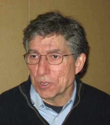
An expert on primary Raynaud’s phenomenon describes treatment approach
SNOWMASS, COLO. – Plenty of medication options exist for treatment of primary Raynaud’s phenomenon caused by reversible cutaneous arteriolar vasospasm. But actually, for most affected patients, no drug therapy is required, according to Dr. Fredrick M. Wigley.
“The environment – physical and emotional – has a huge impact on the severity of Raynaud’s phenomenon,” he emphasized at the Winter Rheumatology Symposium sponsored by the American College of Rheumatology.

“Whatever I tell you in terms of drugs, there’s nothing more potent than getting the patient out of a stressful, cold environment. So I tell patients who have digital ischemia to stop work, go home, get in bed, put the blanket up to your neck, keep warm, and often that’s more effective than the drug therapy that we try,” said Dr. Wigley, professor of medicine, associate head of rheumatology, and director of the scleroderma center at Johns Hopkins University, Baltimore.
Primary Raynaud’s phenomenon is typically reversible within 15-20 minutes after rewarming, he added.
Aggravators: Stress, trauma, smoking, certain drugs
Besides cold temperatures, another important aggravating factor in primary Raynaud’s phenomenon is emotional stress. Anxiety, like cold, increases sympathetic tone, triggering cutaneous vasoconstriction and rapid shunting of blood into the body core in vulnerable patients. Other major triggers of Raynaud’s are physical trauma – especially vibration – and smoking.
“I can tell you that in patients who have complicated Raynaud’s with digital lesions, if they continue to smoke, it doesn’t matter what drug you give them; you’re not going to get good control or fast healing of those digital lesions,” the rheumatologist continued.
Drugs known to aggravate Raynaud’s phenomenon include the serotonin agonists prescribed for migraine headaches, interferons, opiates, some cancer chemotherapy drugs, and medications used in treating attention-deficit/hyperactivity disorder.
“Thirty to forty percent of ADHD kids develop Raynaud’s phenomenon. Methylphenidate, dextroamphetamine, atomoxetine – those are definitely potent sympathomimetic drugs. And the vascular shunts in the skin are very sensitive to sympathomimetic agonists,” according to Dr. Wigley.
Poor evidence for behavioral therapies
The notion of biofeedback, acupuncture, herbals, autogenic training, and other nondrug treatments appeals to many, but in his view, there is no good supporting evidence. What clinical trials have been done have been negative.
“Those therapies have all sort of gone belly up. I wouldn’t be recommending behavioral therapies,” he continued.
When to use vasodilators
In the minority of patients with severe primary Raynaud’s phenomenon, which affects their quality of life and doesn’t sufficiently respond to strategies aimed at avoiding environmental triggers, calcium channel blocker monotherapy is king.
“Amlodipine is my favorite because it has less cardiac effects. I use the long-acting form rather than short-acting agents. I titrate the calcium channel blocker to the maximum tolerated dose before considering combining it with a second agent. It’s a rare patient that I don’t have good control with just a calcium channel blocker,” he said.
“Usually it’s at 15-20 mg/day of amlodipine when patients start getting edema and intolerance to the drug, so you’ve got a lot of room. You usually see about a 40% reduction in the severity of Raynaud’s with 5-10 mg/day. And understand that you’re not going to get rid of Raynaud’s phenomenon. But bringing it down to where you get improved quality of life, along with all the nondrug things that you do, can be all you need to do,” Dr. Wigley said.
When he needs to resort to a second vasodilator, it’s typically a phosphodiesterase-5 inhibitor.
“I use them,” the rheumatologist said. “The best benefit I’ve gotten is when I add it to a calcium channel blocker. If someone tolerates, say, 5 mg/day of amlodipine but not 10 mg, then I might add 10 mg of sildenafil t.i.d. [three times daily] to the calcium channel blocker.”
He was quick to add, however, that the supporting evidence base for the phosphodiesterase-5 inhibitors is weak. All of the studies to date have been small – too small, in fact, to persuade insurance companies to routinely authorize this use of the medications without a fight. For example, a recent double-blind, randomized, crossover trial showed udenafil and amlodipine had equal efficacy in reducing the frequency of Raynaud’s attacks and the phosphodiesterase-5 inhibitor achieved greater blood flow in the digital arteries (Rheumatology 2014;53:658-64); however, there were only 29 subjects, and the drug is not approved in the United States.
“I’m just talking about my own experience. The studies aren’t there,” Dr. Wigley observed. “The fact of the matter is we need a large, well-controlled, double-blind study to exactly define the role of the phosphodiesterase inhibitors.”
Botox in need of supporting evidence
OnabotulinumtoxinA (Botox) has become very popular. On the plus side, it has no adverse impact upon blood pressure, and in case series, 75%-100% of treated patients report significant improvement. The negatives? It’s costly, the benefit doesn’t kick in until after about 48 hours, and the supporting evidence to date is weak, although Dr. Wigley and coinvestigators have an ongoing placebo-controlled trial.
“I have to say, in my own experience in patients with garden-variety primary Raynaud’s, [onabotulinumtoxinA] has not been very exciting, but in patients with critical ischemia, where they’re losing a finger, I’ve been very impressed. So I do think that it works. We need evidence to prove it,” he said.
Alternative vasodilator options with some demonstrated benefit in mild disease include the selective serotonin reuptake inhibitor fluoxetine, the angiotensin receptor blocker losartan, cilostazol, pentoxifylline, and topical nitrates, although the benefits of nitrates tend to wane over time and headaches are often a problem.
Dr. Wigley reported serving as a consultant to Novartis and United Therapeutics and receiving research grants from KineMed, MedImmune, and CSL Behring.
SNOWMASS, COLO. – Plenty of medication options exist for treatment of primary Raynaud’s phenomenon caused by reversible cutaneous arteriolar vasospasm. But actually, for most affected patients, no drug therapy is required, according to Dr. Fredrick M. Wigley.
“The environment – physical and emotional – has a huge impact on the severity of Raynaud’s phenomenon,” he emphasized at the Winter Rheumatology Symposium sponsored by the American College of Rheumatology.
“Whatever I tell you in terms of drugs, there’s nothing more potent than getting the patient out of a stressful, cold environment. So I tell patients who have digital ischemia to stop work, go home, get in bed, put the blanket up to your neck, keep warm, and often that’s more effective than the drug therapy that we try,” said Dr. Wigley, professor of medicine, associate head of rheumatology, and director of the scleroderma center at Johns Hopkins University, Baltimore.
Primary Raynaud’s phenomenon is typically reversible within 15-20 minutes after rewarming, he added.
Aggravators: Stress, trauma, smoking, certain drugs
Besides cold temperatures, another important aggravating factor in primary Raynaud’s phenomenon is emotional stress. Anxiety, like cold, increases sympathetic tone, triggering cutaneous vasoconstriction and rapid shunting of blood into the body core in vulnerable patients. Other major triggers of Raynaud’s are physical trauma – especially vibration – and smoking.
“I can tell you that in patients who have complicated Raynaud’s with digital lesions, if they continue to smoke, it doesn’t matter what drug you give them; you’re not going to get good control or fast healing of those digital lesions,” the rheumatologist continued.
Drugs known to aggravate Raynaud’s phenomenon include the serotonin agonists prescribed for migraine headaches, interferons, opiates, some cancer chemotherapy drugs, and medications used in treating attention-deficit/hyperactivity disorder.
“Thirty to forty percent of ADHD kids develop Raynaud’s phenomenon. Methylphenidate, dextroamphetamine, atomoxetine – those are definitely potent sympathomimetic drugs. And the vascular shunts in the skin are very sensitive to sympathomimetic agonists,” according to Dr. Wigley.
Poor evidence for behavioral therapies
The notion of biofeedback, acupuncture, herbals, autogenic training, and other nondrug treatments appeals to many, but in his view, there is no good supporting evidence. What clinical trials have been done have been negative.
“Those therapies have all sort of gone belly up. I wouldn’t be recommending behavioral therapies,” he continued.
When to use vasodilators
In the minority of patients with severe primary Raynaud’s phenomenon, which affects their quality of life and doesn’t sufficiently respond to strategies aimed at avoiding environmental triggers, calcium channel blocker monotherapy is king.
“Amlodipine is my favorite because it has less cardiac effects. I use the long-acting form rather than short-acting agents. I titrate the calcium channel blocker to the maximum tolerated dose before considering combining it with a second agent. It’s a rare patient that I don’t have good control with just a calcium channel blocker,” he said.
“Usually it’s at 15-20 mg/day of amlodipine when patients start getting edema and intolerance to the drug, so you’ve got a lot of room. You usually see about a 40% reduction in the severity of Raynaud’s with 5-10 mg/day. And understand that you’re not going to get rid of Raynaud’s phenomenon. But bringing it down to where you get improved quality of life, along with all the nondrug things that you do, can be all you need to do,” Dr. Wigley said.
When he needs to resort to a second vasodilator, it’s typically a phosphodiesterase-5 inhibitor.
“I use them,” the rheumatologist said. “The best benefit I’ve gotten is when I add it to a calcium channel blocker. If someone tolerates, say, 5 mg/day of amlodipine but not 10 mg, then I might add 10 mg of sildenafil t.i.d. [three times daily] to the calcium channel blocker.”
He was quick to add, however, that the supporting evidence base for the phosphodiesterase-5 inhibitors is weak. All of the studies to date have been small – too small, in fact, to persuade insurance companies to routinely authorize this use of the medications without a fight. For example, a recent double-blind, randomized, crossover trial showed udenafil and amlodipine had equal efficacy in reducing the frequency of Raynaud’s attacks and the phosphodiesterase-5 inhibitor achieved greater blood flow in the digital arteries (Rheumatology 2014;53:658-64); however, there were only 29 subjects, and the drug is not approved in the United States.
“I’m just talking about my own experience. The studies aren’t there,” Dr. Wigley observed. “The fact of the matter is we need a large, well-controlled, double-blind study to exactly define the role of the phosphodiesterase inhibitors.”
Botox in need of supporting evidence
OnabotulinumtoxinA (Botox) has become very popular. On the plus side, it has no adverse impact upon blood pressure, and in case series, 75%-100% of treated patients report significant improvement. The negatives? It’s costly, the benefit doesn’t kick in until after about 48 hours, and the supporting evidence to date is weak, although Dr. Wigley and coinvestigators have an ongoing placebo-controlled trial.
“I have to say, in my own experience in patients with garden-variety primary Raynaud’s, [onabotulinumtoxinA] has not been very exciting, but in patients with critical ischemia, where they’re losing a finger, I’ve been very impressed. So I do think that it works. We need evidence to prove it,” he said.
Alternative vasodilator options with some demonstrated benefit in mild disease include the selective serotonin reuptake inhibitor fluoxetine, the angiotensin receptor blocker losartan, cilostazol, pentoxifylline, and topical nitrates, although the benefits of nitrates tend to wane over time and headaches are often a problem.
Dr. Wigley reported serving as a consultant to Novartis and United Therapeutics and receiving research grants from KineMed, MedImmune, and CSL Behring.
EXPERT ANALYSIS FROM THE WINTER RHEUMATOLOGY SYMPOSIUM
