User login
A Ripple Effect of Groin Skin Issues
For several months, a 60-year-old woman has had a rash on the right side of her groin, which her primary care provider diagnosed as a “probable yeast infection.” The patient was given a prescription for nystatin cream; when this did not help, she was prescribed a combination clotrimazole/betamethasone cream. Initial signs of improvement prompted her to continue application daily.
However, although the original rash cleared, other skin changes occurred in the area. These alarmed the patient’s primary care provider, who observed them during a follow-up exam. In response, the patient requested a referral to dermatology.

EXAMINATION
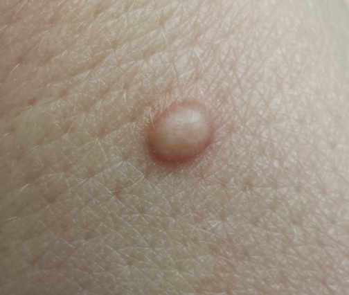
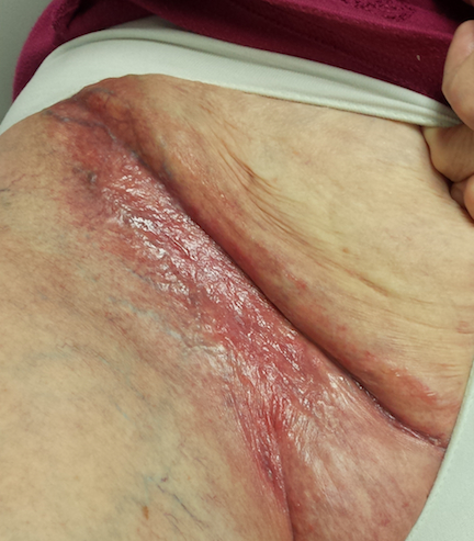
The entire right crural area is bright red and shiny. Multiple blood vessels are seen coursing over the surface of the area. There is no edema, increased warmth, or tenderness on palpation.
Punch biopsy shows marked epidermal atrophy and fails to show any evidence of cellular atypia.
What is the diagnosis?
DISCUSSION
When the patient was advised to stop using the combination cream, the “new” problem resolved quickly and her skin returned to normal. Although the source of the original rash remains unknown, what is clear is that the prescribed steroid induced marked atrophy. This then became “the problem.”
The groin is particularly susceptible to these types of changes. Why? Three reasons are occlusion, thin skin, and hydration. Read on for further explanation!
Occlusion, intentional or not, is known to potentiate the effects of topical steroids. The nature of intertriginous skin (skin folds) is that it effectively occludes the medicated skin, magnifying positive and negative effects. This means that the steroid works better and faster, but it also makes atrophy more likely (and equally quick to develop). When steroids are used for sufficient duration in susceptible areas—groin, axillae, under the breasts—focal areas of epidermis can wither totally, leaving an actual hole in the skin through which subcutaneous fat can be seen.
Furthermore, intertriginous skin is inherently thin (as are eyelids, neck skin, and the face). As a result, it takes less time for these changes to develop. The groin is especially prone to this problem because well-hydrated skin facilitates better penetration of the steroid.
This situation would likely have been avoided if the patient had used the prescribed product for only a week or two. A better option might have been a prescription for a weaker steroid (eg, hydocortisone 2.5%), although even that can be problematic if the medication is used for an extended period.
Though not quite as effective, topical NSAID preparations (calcineurin inhibitors such as pimecrolimus and tacrolimus) cause no such problems. They can help to reduce steroid use—for example, the patient can apply the steroid every third day and the calcineurin inhibitor on the other days.
The vehicle (the base in which the steroid is mixed) matters as well. Steroid ointments are inherently “self-occlusive,” especially in intertriginous areas—a fact that calls for increased caution in their use.
The atrophic look seen in this patient’s groin could have had other causes, such as T-cell or B-cell lymphoma. These were effectively ruled out by the biopsy.
TAKE-HOME LEARNING POINTS
• Epidermal atrophy is especially common on intertriginous skin (skin folds).
• The stronger the steroid (classes 1 through 7, with 1 being the strongest), the faster the atrophy occurs.
• Occlusion—caused by opposing skin folds or by other material (eg, socks, plastic wrap)—potentiates the positive and negative effects of topical steroids.
• Thin, moist skin allows better penetration by topical steroids, making them work better but also making atrophy more likely.
• Calcineurin inhibitors (eg, tacrolimus or pimecrolimus) have no such adverse effects and can help to reduce overuse of topical steroids in susceptible areas.
For several months, a 60-year-old woman has had a rash on the right side of her groin, which her primary care provider diagnosed as a “probable yeast infection.” The patient was given a prescription for nystatin cream; when this did not help, she was prescribed a combination clotrimazole/betamethasone cream. Initial signs of improvement prompted her to continue application daily.
However, although the original rash cleared, other skin changes occurred in the area. These alarmed the patient’s primary care provider, who observed them during a follow-up exam. In response, the patient requested a referral to dermatology.
EXAMINATION
The entire right crural area is bright red and shiny. Multiple blood vessels are seen coursing over the surface of the area. There is no edema, increased warmth, or tenderness on palpation.
Punch biopsy shows marked epidermal atrophy and fails to show any evidence of cellular atypia.
What is the diagnosis?
DISCUSSION
When the patient was advised to stop using the combination cream, the “new” problem resolved quickly and her skin returned to normal. Although the source of the original rash remains unknown, what is clear is that the prescribed steroid induced marked atrophy. This then became “the problem.”
The groin is particularly susceptible to these types of changes. Why? Three reasons are occlusion, thin skin, and hydration. Read on for further explanation!
Occlusion, intentional or not, is known to potentiate the effects of topical steroids. The nature of intertriginous skin (skin folds) is that it effectively occludes the medicated skin, magnifying positive and negative effects. This means that the steroid works better and faster, but it also makes atrophy more likely (and equally quick to develop). When steroids are used for sufficient duration in susceptible areas—groin, axillae, under the breasts—focal areas of epidermis can wither totally, leaving an actual hole in the skin through which subcutaneous fat can be seen.
Furthermore, intertriginous skin is inherently thin (as are eyelids, neck skin, and the face). As a result, it takes less time for these changes to develop. The groin is especially prone to this problem because well-hydrated skin facilitates better penetration of the steroid.
This situation would likely have been avoided if the patient had used the prescribed product for only a week or two. A better option might have been a prescription for a weaker steroid (eg, hydocortisone 2.5%), although even that can be problematic if the medication is used for an extended period.
Though not quite as effective, topical NSAID preparations (calcineurin inhibitors such as pimecrolimus and tacrolimus) cause no such problems. They can help to reduce steroid use—for example, the patient can apply the steroid every third day and the calcineurin inhibitor on the other days.
The vehicle (the base in which the steroid is mixed) matters as well. Steroid ointments are inherently “self-occlusive,” especially in intertriginous areas—a fact that calls for increased caution in their use.
The atrophic look seen in this patient’s groin could have had other causes, such as T-cell or B-cell lymphoma. These were effectively ruled out by the biopsy.
TAKE-HOME LEARNING POINTS
• Epidermal atrophy is especially common on intertriginous skin (skin folds).
• The stronger the steroid (classes 1 through 7, with 1 being the strongest), the faster the atrophy occurs.
• Occlusion—caused by opposing skin folds or by other material (eg, socks, plastic wrap)—potentiates the positive and negative effects of topical steroids.
• Thin, moist skin allows better penetration by topical steroids, making them work better but also making atrophy more likely.
• Calcineurin inhibitors (eg, tacrolimus or pimecrolimus) have no such adverse effects and can help to reduce overuse of topical steroids in susceptible areas.
Attempt at “Wart” Removal Backfires
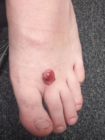
Two months ago, a tender lesion manifested on the dorsum of a 14-year-old boy’s right foot. Before it appeared, there was a tiny papule in the same spot; the boy’s mother says they thought it was a wart and attempted to remove it. As a result, the papule bled and became inflamed before quickly growing to its present form.
Besides being tender to touch, the lesion bleeds with minimal trauma. Attempts to remove it—using silver nitrate, liquid nitrogen, and triple-antibiotic cream—have all failed to have any positive impact.
The patient is reportedly otherwise healthy. He takes no medications of any kind.

EXAMINATION
The lesion is a 2-cm, domelike, red shiny nodule located on the mid-dorsum of the right foot. It is attached to the underlying skin by a thick sessile base. There is no surrounding erythema or other skin changes.
The site is anesthetized with local infiltrate of 1% lidocaine with epinephrine before deep shave biopsy is used to remove the lesion. The base is curetted, then cauterized for hemostasis.
PATHOLOGY
Microscopic examination shows a tangle of capillaries arranged in lobules separated by septae of connective tissue. A brisk inflammatory reaction and faint erosions are seen on the lesion’s surface.
What is the diagnosis?
DISCUSSION
First described by Ponce and Dor in 1897, pyogenic granuloma (PG) was thought to represent a kind of infection but turned out to be neither infectious nor granulomatous. It is now understood to represent the body’s attempt to heal a wound or other trauma (albeit this process goes awry for unknown reasons).
PG has a range of morphologic presentations; this case, with a berry-like friable papule, is fairly typical. PG is common on fingertips, lips, and faces (in children) and can even appear on gingival mucosae (particularly in first-trimester pregnancy). Other common areas of manifestation include ingrown toenails (lesion forms where the distal edge of the nail cuts into the lateral aspect of the nail bed) and the umbilical stump of newborns. An extremely common scenario is the child who cannot leave a wart or skin tag alone, picking and squeezing the lesion repeatedly until a PG forms.
PG is reactive in nature and not neoplastic. It is essentially vascular (other names for it include lobular capillary hemangioma and eruptive hemangioma), and the amount of blood that can flow from these lesions tends to frighten patients and parents.
While PG is completely benign, it is part of a differential of “look-alikes” that include nodular melanoma and metastatic cancer. For this reason, once removed, PGs are always sent for pathologic examination.
Removal should be done by deep shave, followed by electrodessication and curettage to prevent recurrence. Cryosurgery or silver nitrate application, though frequently done, is seldom effective. Significantly, neither yields a definitive diagnosis.
Certain drugs are associated with PG formation. These include isotretinoin, some chemotherapy drugs, and retroviral medications.
TAKE-HOME LEARNING POINTS
• Pyogenic granuloma (PG) is neither pyogenic nor granulomatous and is instead a reactive process.
• PG represents the body’s incomplete attempt to heal a wound.
• The trauma is often repetitive, particularly in PG associated with ingrown toenails or digitally manipulated by the patient.
• A berry-like appearance, sudden growth, and friability are all characteristic of PG.
• PG, though extremely common, needs to be removed to provide relief and to establish the lesion’s benign nature by pathologic examination.
Two months ago, a tender lesion manifested on the dorsum of a 14-year-old boy’s right foot. Before it appeared, there was a tiny papule in the same spot; the boy’s mother says they thought it was a wart and attempted to remove it. As a result, the papule bled and became inflamed before quickly growing to its present form.
Besides being tender to touch, the lesion bleeds with minimal trauma. Attempts to remove it—using silver nitrate, liquid nitrogen, and triple-antibiotic cream—have all failed to have any positive impact.
The patient is reportedly otherwise healthy. He takes no medications of any kind.
EXAMINATION
The lesion is a 2-cm, domelike, red shiny nodule located on the mid-dorsum of the right foot. It is attached to the underlying skin by a thick sessile base. There is no surrounding erythema or other skin changes.
The site is anesthetized with local infiltrate of 1% lidocaine with epinephrine before deep shave biopsy is used to remove the lesion. The base is curetted, then cauterized for hemostasis.
PATHOLOGY
Microscopic examination shows a tangle of capillaries arranged in lobules separated by septae of connective tissue. A brisk inflammatory reaction and faint erosions are seen on the lesion’s surface.
What is the diagnosis?
DISCUSSION
First described by Ponce and Dor in 1897, pyogenic granuloma (PG) was thought to represent a kind of infection but turned out to be neither infectious nor granulomatous. It is now understood to represent the body’s attempt to heal a wound or other trauma (albeit this process goes awry for unknown reasons).
PG has a range of morphologic presentations; this case, with a berry-like friable papule, is fairly typical. PG is common on fingertips, lips, and faces (in children) and can even appear on gingival mucosae (particularly in first-trimester pregnancy). Other common areas of manifestation include ingrown toenails (lesion forms where the distal edge of the nail cuts into the lateral aspect of the nail bed) and the umbilical stump of newborns. An extremely common scenario is the child who cannot leave a wart or skin tag alone, picking and squeezing the lesion repeatedly until a PG forms.
PG is reactive in nature and not neoplastic. It is essentially vascular (other names for it include lobular capillary hemangioma and eruptive hemangioma), and the amount of blood that can flow from these lesions tends to frighten patients and parents.
While PG is completely benign, it is part of a differential of “look-alikes” that include nodular melanoma and metastatic cancer. For this reason, once removed, PGs are always sent for pathologic examination.
Removal should be done by deep shave, followed by electrodessication and curettage to prevent recurrence. Cryosurgery or silver nitrate application, though frequently done, is seldom effective. Significantly, neither yields a definitive diagnosis.
Certain drugs are associated with PG formation. These include isotretinoin, some chemotherapy drugs, and retroviral medications.
TAKE-HOME LEARNING POINTS
• Pyogenic granuloma (PG) is neither pyogenic nor granulomatous and is instead a reactive process.
• PG represents the body’s incomplete attempt to heal a wound.
• The trauma is often repetitive, particularly in PG associated with ingrown toenails or digitally manipulated by the patient.
• A berry-like appearance, sudden growth, and friability are all characteristic of PG.
• PG, though extremely common, needs to be removed to provide relief and to establish the lesion’s benign nature by pathologic examination.
Common Presentation for Complex Condition
A month ago, a 33-year-old woman noticed skin changes on her arms and face. The affected areas have recently begun to itch and burn—particularly, the patient notes, since she spent an extended period in the sun over the weekend. She has used an antifungal cream (nystatin) on the rash, to no avail.
The patient denies joint pain, fever, and malaise. She has a sister with multiple sclerosis, but her family history is otherwise unremarkable. The patient’s only medication is oral contraceptives, which she has taken since the birth of her first and only child last year.

EXAMINATION
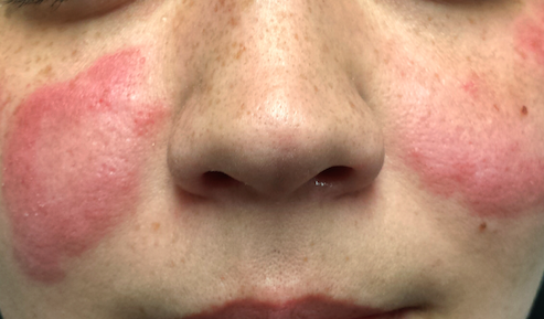
The patient is afebrile and in no particular distress. A florid red rash covers both cheeks, sparing the nose entirely. The margins are somewhat indurated and redder than the clearing centers. The follicular orifices are somewhat patulous, and a fine scale covers the affected areas of the face. The lateral brachial and triceps (sun-exposed) areas of both arms are similarly affected.
Punch biopsy reveals classic signs of lupus: vacuolar alteration of the basal cell layer, perivascular infiltrate around appendicial structures, and modest epidermal atrophy. Bloodwork yields no evidence of systemic lupus erythematosus (SLE).
What is the diagnosis?
DISCUSSION
Lupus, as a general topic, can be utterly confusing. Here are some facts that might help you make sense of the subject:
Purely cutaneous forms of lupus are commonly seen in dermatology; they manifest in sun-exposed skin as scaly annular lesions with clearing centers. Somewhat confusingly, however, cases of systemic lupus erythematosus (SLE) can present with similar cutaneous signs—and furthermore, patients with purely cutaneous lupus (subacute cutaneous lupus) may exhibit some systemic symptoms (just not enough to meet the strict criteria for SLE). Lupus in general is far more common in women than in men.
The “butterfly rash” seen in this case is uncommon but can occur in either cutaneous or systemic lupus. In most cases, though, this particular rash is a manifestation of seborrhea, psoriasis, rosacea or eczema—not lupus at all.
There are, of course, many other types of lupus. Another common form is discoid lupus (DLE), which manifests with round, scaly lesions on the head, neck, or ears; these are often misidentified as actinic keratosis, eczema, or psoriasis. DLE can be localized or generalized, purely cutaneous or a manifestation of SLE.
The key to diagnosis lies in first considering lupus in the differential and then biopsying the lesion (or sending the patient to someone who will). Once the clinical diagnosis is histologically confirmed (typical results include vacuolar interface dermatitis with sparse lymphocytic perivascular infiltrate), an immunologic workup is warranted. Also, depending on the predominant organ systems involved, the patient should be thoroughly evaluated by a dermatology or rheumatology specialist (or both).
Lupus is an autoimmune process, but in some ways it’s more useful to think of it as a form of vasculitis—which is why it can affect almost any organ system. The very first lupus patient I ever saw was in the psych ward having a psychotic break, which turned out to be secondary to a lupus-induced cerebritis. Since then, I’ve seen it affect the pericardium, kidneys, lungs, and joints. Lupus is even a major item in the differential of alopecia! SLE in particular is associated with an increase in thrombotic events and accounts for most early deaths from lupus (ie, within the first five years of diagnosis, when the cause of death is usually renal or pulmonary).
The patient in this case proved to have only cutaneous disease. She’ll respond nicely to a combination of sun protection and oral antimalarials (hydroxychloroquine) but will probably have recurrences every spring. Although unlikely to ever develop SLE, she is statistically more likely to develop other autoimmune diseases.
TAKE-HOME LEARNING POINTS
• Cutaneous lupus is more common than you might imagine. Lesions and eruptions in sun-exposed skin should prompt consideration of that item in the differential.
• Many forms of lupus have been identified, including neonatal lupus and overlapping syndromes involving lupus and lichen planus or even pemphigus.
• Though UV exposure is not always the cause, almost every type of lupus is worsened by UV light exposure.
• The differential for lupus is vast but includes psoriasis, sarcoidosis, dermatomyositis, and drug eruptions.
A month ago, a 33-year-old woman noticed skin changes on her arms and face. The affected areas have recently begun to itch and burn—particularly, the patient notes, since she spent an extended period in the sun over the weekend. She has used an antifungal cream (nystatin) on the rash, to no avail.
The patient denies joint pain, fever, and malaise. She has a sister with multiple sclerosis, but her family history is otherwise unremarkable. The patient’s only medication is oral contraceptives, which she has taken since the birth of her first and only child last year.
EXAMINATION
The patient is afebrile and in no particular distress. A florid red rash covers both cheeks, sparing the nose entirely. The margins are somewhat indurated and redder than the clearing centers. The follicular orifices are somewhat patulous, and a fine scale covers the affected areas of the face. The lateral brachial and triceps (sun-exposed) areas of both arms are similarly affected.
Punch biopsy reveals classic signs of lupus: vacuolar alteration of the basal cell layer, perivascular infiltrate around appendicial structures, and modest epidermal atrophy. Bloodwork yields no evidence of systemic lupus erythematosus (SLE).
What is the diagnosis?
DISCUSSION
Lupus, as a general topic, can be utterly confusing. Here are some facts that might help you make sense of the subject:
Purely cutaneous forms of lupus are commonly seen in dermatology; they manifest in sun-exposed skin as scaly annular lesions with clearing centers. Somewhat confusingly, however, cases of systemic lupus erythematosus (SLE) can present with similar cutaneous signs—and furthermore, patients with purely cutaneous lupus (subacute cutaneous lupus) may exhibit some systemic symptoms (just not enough to meet the strict criteria for SLE). Lupus in general is far more common in women than in men.
The “butterfly rash” seen in this case is uncommon but can occur in either cutaneous or systemic lupus. In most cases, though, this particular rash is a manifestation of seborrhea, psoriasis, rosacea or eczema—not lupus at all.
There are, of course, many other types of lupus. Another common form is discoid lupus (DLE), which manifests with round, scaly lesions on the head, neck, or ears; these are often misidentified as actinic keratosis, eczema, or psoriasis. DLE can be localized or generalized, purely cutaneous or a manifestation of SLE.
The key to diagnosis lies in first considering lupus in the differential and then biopsying the lesion (or sending the patient to someone who will). Once the clinical diagnosis is histologically confirmed (typical results include vacuolar interface dermatitis with sparse lymphocytic perivascular infiltrate), an immunologic workup is warranted. Also, depending on the predominant organ systems involved, the patient should be thoroughly evaluated by a dermatology or rheumatology specialist (or both).
Lupus is an autoimmune process, but in some ways it’s more useful to think of it as a form of vasculitis—which is why it can affect almost any organ system. The very first lupus patient I ever saw was in the psych ward having a psychotic break, which turned out to be secondary to a lupus-induced cerebritis. Since then, I’ve seen it affect the pericardium, kidneys, lungs, and joints. Lupus is even a major item in the differential of alopecia! SLE in particular is associated with an increase in thrombotic events and accounts for most early deaths from lupus (ie, within the first five years of diagnosis, when the cause of death is usually renal or pulmonary).
The patient in this case proved to have only cutaneous disease. She’ll respond nicely to a combination of sun protection and oral antimalarials (hydroxychloroquine) but will probably have recurrences every spring. Although unlikely to ever develop SLE, she is statistically more likely to develop other autoimmune diseases.
TAKE-HOME LEARNING POINTS
• Cutaneous lupus is more common than you might imagine. Lesions and eruptions in sun-exposed skin should prompt consideration of that item in the differential.
• Many forms of lupus have been identified, including neonatal lupus and overlapping syndromes involving lupus and lichen planus or even pemphigus.
• Though UV exposure is not always the cause, almost every type of lupus is worsened by UV light exposure.
• The differential for lupus is vast but includes psoriasis, sarcoidosis, dermatomyositis, and drug eruptions.
A Second Patient and a Double Diagnosis
A 45-year-old woman brings her daughter for evaluation of the daughter’s acne. However, during the appointment, an odd lesion is noted on the mother’s shoulder.
Once the daughter’s evaluation is completed, attention turns to the mother’s lesion, which she reports “has been there for years.” Until last year, it hadn’t changed—but since then, it has grown considerably and also darkened.
The patient has an extensive history of poorly tolerated sun exposure in her childhood and young adulthood. She says she is able to tan but “it only holds for a short time.”
EXAMINATION
The lesion, a 2.8-cm black plaque with irregular margins, is located on the crown of the right shoulder. The patient has somewhat sun-damaged, freckled type II skin.
Dermatoscopic examination reveals regularly spaced white pinpoint areas scattered over the lesion’s surface. Focally, there is definite black streaking and pigment clumping on the borders of the lesion.
The white spots are consistent with pseudocysts seen in seborrheic keratosis. But the clumping and streaming of pigment are features we might expect to see with melanoma.
What is the diagnosis?
DISCUSSION
This case illustrates at least two useful principles:
1. The “patient” is not always the one listed on the charge sheet. I’ve found at least four melanomas and innumerable basal cell carcinomas on friends and relations who happen to be in the room with “the patient.” I can’t pretend I didn’t see the lesion, whoever it’s on. Of course, we must prioritize the patient of record—but then turn our attention to the “new” lesion/patient.
2. There is no law that says a seborrheic keratosis (SK) cannot occur in the same location as a melanoma. It may be rare, but it’s not unheard of. In this case, there were signs of both; the only way to sort it out, safely, was to excise the entire lesion and submit it to pathology. This provides the pathologist with adequate tissue to judge the whole lesion.
As it happens, this case entailed both diagnoses: the SK on the surface and a melanoma in situ on the underside. The latter was confined to the upper epidermis (ie, did not penetrate into the dermis). Re-excision with 5-mm margins was done, just to be on the safe side. Had the melanoma been left in place, it could have become invasive with time (though it might have taken years).
SKs are the most common benign lesions seen in dermatology practices—this patient had several others on her trunk—but can coincide with other lesions/diagnoses (eg, cancer). The oddity of the shoulder lesion’s appearance (a shape known as the black sheep sign), along with the patient’s fair, sun-damaged skin, prompted dermatoscopic examination.
With the power to visualize lesions under polarized light at 10x magnification, we have developed an entire body of knowledge about the features of benign vs malignant lesions—making the dermatoscope a common and valuable tool in dermatology practices across the world.
TAKE-HOME LEARNING POINTS
• Although a rare occurrence, seborrheic keratosis and melanoma (or another lesion) can co-exist in the same location.
• The odd appearance of the lesion, combined with the patient’s fair, sun-damaged skin, was enough to trigger a closer look.
• Dermatoscopic examination (10x magnification with polarized light) can identify features of benign and malignant lesions. In this case, both were found.
• Complete excision is the gold standard for biopsy of lesions suspicious for melanoma.

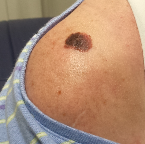
What Is This Large, Oddly Pigmented “Freckle”?
A 79-year-old man presents for a routine skin check, in the context of his 40-year history of nonmelanoma skin cancer. He grew up on a farm and then became a farmer himself, spending almost every day in the sun (usually without a hat). His skin burned easily but would take on a “tan” by the start of summer.
In the succeeding years, he developed so many skin cancers (and had them removed) that he lost count. All were basal cell or squamous cell carcinomas, predominately manifesting on his face, arms, and ears. Several required Mohs surgery for removal.

EXAMINATION
Abundant evidence of excessive sun exposure is seen on the patient’s skin: actinic keratoses on the forehead, ears, and neck, and multiple solar lentigines on the face, neck, and arms. On his left neck, below the ear, is a large, oddly pigmented, dark macular patch. Dermatoscopic examination reveals focal pigmentary clumping and streaming, which prompts the decision to perform an incisional biopsy. The darkest and most irregular part of the lesion is taken as a sample. The pathology report shows lentigo maligna.
What is lentigo maligna?
DISCUSSION
Lentigo maligna (LM), also known as Hutchinson freckle, is a type of melanoma in situ that is typically seen on sun-exposed skin. It has indistinct margins with predominantly brown and black coloration. LM is usually seen on older, mostly fair-skinned patients who have a history of extensive sun exposure. Its preferred sites include the face, ears, neck, and upper extremities.
LM is, by definition, entirely superficial and therefore safe. Its main significance is that it can become focally invasive to the dermis, a phenomenon termed lentigo maligna melanoma (LMM). When that occurs, the clusters of spindle-shaped atypical cells can then progress to a vertical growth phase, resulting in intravascular invasion that eventuates in metastasis.
LMs grow so slowly that they often escape detection; they are sometimes mistaken for solar lentigines (SL). They can “collide” with SLs or other benign lesions (eg, seborrheic keratoses), which can effectively camouflage them. Suspicion is usually triggered by change (color, size) in a lesion.
Biopsy is the only method of detection. In this case, the central portion of this large, oddly pigmented and bordered, multicolored patch was excised and the darkest, most irregular part of the lesion collected. This is the gold standard for biopsy of a potential melanoma. A large deep shave biopsy (“saucerization”) would have accomplished the same thing; studies show that neither process will cause metastasis. The main mistake to avoid is performing a single punch biopsy, which risks a false-negative result.
The definitive surgical approach is to remove the lesion with a 1-cm margin around it and into the underlying adipose layer. The wound can either be left to heal by secondary intention or closed primarily. Either way, this patient’s prognosis is excellent, unless the final pathology report indicates focal invasion. With a patient of this age, the LM could have been left alone—although, once discovered, LM is irresistibly compelling to treat.
TAKE-HOME LEARNING POINTS
• Lentigo maligna (LM) is a type of melanoma in situ with the potential to progress to lentigo maligna melanoma, an invasive form of early melanoma.
• LM is common on sun-exposed skin of older patients with a history of excessive sun exposure.
• Faces, ears, arms, and necks are common areas for LM to manifest.
• Biopsy of suspected melanoma should incorporate a significant portion of the darkest, most irregular part of the lesion; it can be done by incisional technique or deep saucerization.
• LM is also known as Hutchinson freckle, since it often resembles a large, irregularly bordered and pigmented freckle.
A 79-year-old man presents for a routine skin check, in the context of his 40-year history of nonmelanoma skin cancer. He grew up on a farm and then became a farmer himself, spending almost every day in the sun (usually without a hat). His skin burned easily but would take on a “tan” by the start of summer.
In the succeeding years, he developed so many skin cancers (and had them removed) that he lost count. All were basal cell or squamous cell carcinomas, predominately manifesting on his face, arms, and ears. Several required Mohs surgery for removal.
EXAMINATION
Abundant evidence of excessive sun exposure is seen on the patient’s skin: actinic keratoses on the forehead, ears, and neck, and multiple solar lentigines on the face, neck, and arms. On his left neck, below the ear, is a large, oddly pigmented, dark macular patch. Dermatoscopic examination reveals focal pigmentary clumping and streaming, which prompts the decision to perform an incisional biopsy. The darkest and most irregular part of the lesion is taken as a sample. The pathology report shows lentigo maligna.
What is lentigo maligna?
DISCUSSION
Lentigo maligna (LM), also known as Hutchinson freckle, is a type of melanoma in situ that is typically seen on sun-exposed skin. It has indistinct margins with predominantly brown and black coloration. LM is usually seen on older, mostly fair-skinned patients who have a history of extensive sun exposure. Its preferred sites include the face, ears, neck, and upper extremities.
LM is, by definition, entirely superficial and therefore safe. Its main significance is that it can become focally invasive to the dermis, a phenomenon termed lentigo maligna melanoma (LMM). When that occurs, the clusters of spindle-shaped atypical cells can then progress to a vertical growth phase, resulting in intravascular invasion that eventuates in metastasis.
LMs grow so slowly that they often escape detection; they are sometimes mistaken for solar lentigines (SL). They can “collide” with SLs or other benign lesions (eg, seborrheic keratoses), which can effectively camouflage them. Suspicion is usually triggered by change (color, size) in a lesion.
Biopsy is the only method of detection. In this case, the central portion of this large, oddly pigmented and bordered, multicolored patch was excised and the darkest, most irregular part of the lesion collected. This is the gold standard for biopsy of a potential melanoma. A large deep shave biopsy (“saucerization”) would have accomplished the same thing; studies show that neither process will cause metastasis. The main mistake to avoid is performing a single punch biopsy, which risks a false-negative result.
The definitive surgical approach is to remove the lesion with a 1-cm margin around it and into the underlying adipose layer. The wound can either be left to heal by secondary intention or closed primarily. Either way, this patient’s prognosis is excellent, unless the final pathology report indicates focal invasion. With a patient of this age, the LM could have been left alone—although, once discovered, LM is irresistibly compelling to treat.
TAKE-HOME LEARNING POINTS
• Lentigo maligna (LM) is a type of melanoma in situ with the potential to progress to lentigo maligna melanoma, an invasive form of early melanoma.
• LM is common on sun-exposed skin of older patients with a history of excessive sun exposure.
• Faces, ears, arms, and necks are common areas for LM to manifest.
• Biopsy of suspected melanoma should incorporate a significant portion of the darkest, most irregular part of the lesion; it can be done by incisional technique or deep saucerization.
• LM is also known as Hutchinson freckle, since it often resembles a large, irregularly bordered and pigmented freckle.
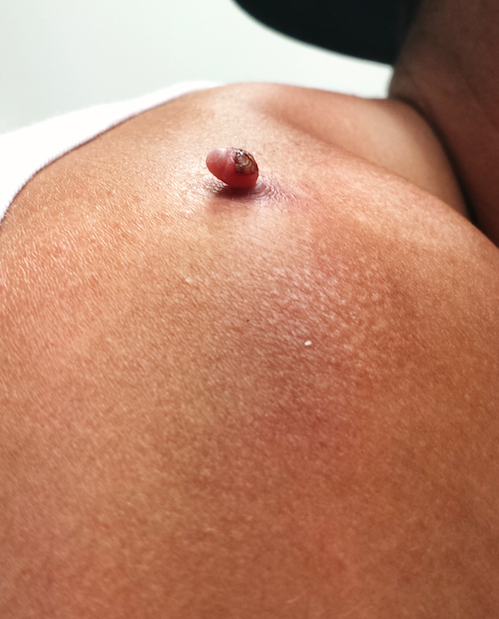
Tiny Lesion Turns Troublesome After Trauma
Four months ago, this 23-year-old man developed a lesion on his right shoulder. It appeared, as he recalls, over the course of a week and has subsequently grown. The lesion, which is now rather large, bleeds copiously with minor trauma. It causes only modest discomfort to the patient but considerable worry to his family.
The lesion was originally tiny and tag-like; the patient initially mistook it for a tick and tried to pull it off. Not only did that fail to work, it also seemed to irritate the lesion. At that point, it started to swell, eventually transforming into the lesion he presents with today.
The patient’s history is otherwise uneventful, and he reports taking no medications. He has had minimal sun exposure but says he tans easily when he does get some sun.

EXAMINATION
The lesion, measuring 5 mm x 2.5 mm, is a dark red and pedunculated papule on the crown of the right shoulder. It looks edematous and feels firm. The patient has otherwise unremarkable type IV skin.
Shave biopsy is performed, using a double-edged razor to make a shallow concave defect under the lesion. The wound is cauterized.
What is the diagnosis?
DISCUSSION
The pathology report confirmed the clinical suspicion of pyogenic granuloma, which, ironically, is neither pyogenic nor granulomatous. The condition acquired this name more than 100 years ago, based on assumptions about its origin. Microscopic examination revealed the highly vascular nature of these lesions, showing a field full of circles that represented the truncated ends of bundles of capillaries and venules. Although they are also called lobular capillary hemangiomas, the term pyogenic granuloma (PG) is still more commonly used.
PGs commonly manifest in the patient’s second or third decade of life, typically on extremities, chest, and nipples. This patient’s story is typical: His lesion began as a tag or wart that he then traumatized, creating a situation in which the body attempts (in vain) to heal the wound. Undisturbed, nearly all PGs would eventually wither and resolve with minimal scarring; however, that process is prolonged when the patient fails to “leave it alone.” (This is especially true in the case of young children.)
While this presentation is characteristic, there are other circumstances in which PGs develop. One is as a consequence of taking certain medications (eg, retinoids, antiretrovirals, and certain chemotherapy drugs). PGs are also commonly seen in the oral cavity of pregnant women in the third trimester and on the end of the umbilical stump in many newborns. Ingrown toenails are another common site; PGs will appear as glistening red, friable buttons of vascular tissue in the perionychial skin adjacent to the affected portion of the nail.
Shave biopsy is standard in such cases, not only to produce a cure but also to establish, via pathologic examination of the tissue, the correct diagnosis. (Nodular melanoma is the most prominent item in the differential; to miss that diagnosis would have dire consequences.) In terms of treatment, mere cautery or cryotherapy will not work and excision is seldom necessary. A deep shave will capture the entire lesion; if any remains, electrodessication and curettage will take care of it. Prior to such procedures, the patient (and family) needs to understand that scarring and pigment loss will occur.
In cases associated with medications or with ingrown toenails, the “cure” would be to withdraw the offending medications—although this is not always advisable for other, obvious reasons.
TAKE-HOME LEARNING POINTS
• Pyogenic granulomas (PG) have nothing to do with infection, nor are they truly granulomatous.
• PGs bleed readily with minor trauma and are often swollen and occasionally painful.
• PGs appear to represent, in most cases, the body’s frustrated attempt to heal a wound, most often a prick or pinch of a pre-existing lesion (eg, tag or mole).
• PGs are also seen in other contexts, such as with use of certain medications or in association with pregnancy.
• If treatment is attempted, the resulting specimen must be submitted for pathologic examination, since other lesions can mimic PGs (of most concern, nodular melanoma).
Four months ago, this 23-year-old man developed a lesion on his right shoulder. It appeared, as he recalls, over the course of a week and has subsequently grown. The lesion, which is now rather large, bleeds copiously with minor trauma. It causes only modest discomfort to the patient but considerable worry to his family.
The lesion was originally tiny and tag-like; the patient initially mistook it for a tick and tried to pull it off. Not only did that fail to work, it also seemed to irritate the lesion. At that point, it started to swell, eventually transforming into the lesion he presents with today.
The patient’s history is otherwise uneventful, and he reports taking no medications. He has had minimal sun exposure but says he tans easily when he does get some sun.
EXAMINATION
The lesion, measuring 5 mm x 2.5 mm, is a dark red and pedunculated papule on the crown of the right shoulder. It looks edematous and feels firm. The patient has otherwise unremarkable type IV skin.
Shave biopsy is performed, using a double-edged razor to make a shallow concave defect under the lesion. The wound is cauterized.
What is the diagnosis?
DISCUSSION
The pathology report confirmed the clinical suspicion of pyogenic granuloma, which, ironically, is neither pyogenic nor granulomatous. The condition acquired this name more than 100 years ago, based on assumptions about its origin. Microscopic examination revealed the highly vascular nature of these lesions, showing a field full of circles that represented the truncated ends of bundles of capillaries and venules. Although they are also called lobular capillary hemangiomas, the term pyogenic granuloma (PG) is still more commonly used.
PGs commonly manifest in the patient’s second or third decade of life, typically on extremities, chest, and nipples. This patient’s story is typical: His lesion began as a tag or wart that he then traumatized, creating a situation in which the body attempts (in vain) to heal the wound. Undisturbed, nearly all PGs would eventually wither and resolve with minimal scarring; however, that process is prolonged when the patient fails to “leave it alone.” (This is especially true in the case of young children.)
While this presentation is characteristic, there are other circumstances in which PGs develop. One is as a consequence of taking certain medications (eg, retinoids, antiretrovirals, and certain chemotherapy drugs). PGs are also commonly seen in the oral cavity of pregnant women in the third trimester and on the end of the umbilical stump in many newborns. Ingrown toenails are another common site; PGs will appear as glistening red, friable buttons of vascular tissue in the perionychial skin adjacent to the affected portion of the nail.
Shave biopsy is standard in such cases, not only to produce a cure but also to establish, via pathologic examination of the tissue, the correct diagnosis. (Nodular melanoma is the most prominent item in the differential; to miss that diagnosis would have dire consequences.) In terms of treatment, mere cautery or cryotherapy will not work and excision is seldom necessary. A deep shave will capture the entire lesion; if any remains, electrodessication and curettage will take care of it. Prior to such procedures, the patient (and family) needs to understand that scarring and pigment loss will occur.
In cases associated with medications or with ingrown toenails, the “cure” would be to withdraw the offending medications—although this is not always advisable for other, obvious reasons.
TAKE-HOME LEARNING POINTS
• Pyogenic granulomas (PG) have nothing to do with infection, nor are they truly granulomatous.
• PGs bleed readily with minor trauma and are often swollen and occasionally painful.
• PGs appear to represent, in most cases, the body’s frustrated attempt to heal a wound, most often a prick or pinch of a pre-existing lesion (eg, tag or mole).
• PGs are also seen in other contexts, such as with use of certain medications or in association with pregnancy.
• If treatment is attempted, the resulting specimen must be submitted for pathologic examination, since other lesions can mimic PGs (of most concern, nodular melanoma).
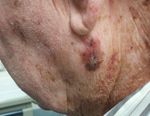
Excessive Sun Damage and a “Fungal Infection”?
A scaly lesion manifested on this 55-year-old man’s back several years ago. Over time, the lesion has grown, despite the application of several different OTC creams, including hydrocortisone and various antifungal creams. Previously asymptomatic, the lesion is now beginning to itch and occasionally bleed.
Additional history-taking reveals that for the first 10 years of his working life, the patient was a concrete finisher, working 12-hour shifts in the sun without a shirt, six days a week for months at a time. Aside from a 40-pack-year history of smoking, his health is “decent.”

EXAMINATION
The lesion is an 8 x 10–cm pink, scaly plaque with thready, well-defined scaly margins. Tiny focal erosions are seen in the peripheral leading edge. Closer inspection of the lesion’s center reveals multiple telangiectasias and a complete lack of hairs or hair follicles. Elsewhere on sun-exposed areas of the patient’s truncal skin, there are thousands of solar lentigines. Multiple small actinic keratoses are noted on his face as well.
What is the diagnosis?
DISCUSSION
Prior to consulting dermatology, this patient had talked extensively with his primary care provider (PCP) about the large lesion on his back. The PCP was quite sure the cause was a fungal organism and assured the patient that the prescribed antifungal cream would almost certainly cure it. This is a scenario in which almost every PCP will participate at some point early in his/her career. When treatment fails, the patient is then referred to dermatology—where a totally unexpected diagnosis is made.
This case represents the natural history of superficial basal cell carcinoma (SBCC), the appearance of which is at odds with any other kind of skin cancer, including the other types of basal cell carcinoma. SBCC is especially common on the shoulders and trunks of patients with a history of excessive sun exposure. The mid-back location on this patient is quite typical, as is the annular, scaly morphology.
The cancer—which originates in a row of cells at the base of the epidermis—effectively destroys surface structures such as skin lines and hair follicles as it spreads laterally. Left in place long enough, SBCCs can eventually become focally invasive and can even spread into regional lymph nodes. Metastasis from them is extremely rare but has been reported.
Treatment is therefore necessary and can range from surgical excision of smaller lesions to electrodessication and curettage to nonsurgical options, such as the application of imiquimod creams or 5-fluorouracil cream, or even radiation therapy. This man was treated with thrice-weekly application of imiquimod 3.75% cream for at least two months, which will cause irritation and scant drainage but will likely result in a cure.
One of the main benefits of his presentation to dermatology was the opportunity to encourage and facilitate yearly skin check-ups—important, given his risk for other sun-caused skin cancers (especially melanoma).
A major learning point to be gained from this case: Just because a lesion is annular and scaly doesn’t mean it’s fungal. The fungi have to have a source (animal, child, soil), and it helps a lot if the affected area is damp and warm (eg, groin or under a bulletproof vest). Immunosuppression (from corticosteroid use, especially) helps as well.
Besides fungal infection, the differential for this type of lesion includes Bowen’s disease (intraepidermal squamous cell carcinoma), psoriasis, and eczema. The fact that the lesion was fixed (did not come and go), grew steadily, and was on the directly sun-exposed skin of a person with a history of heavy sun exposure all pointed to the diagnosis.
TAKE-HOME LEARNING POINTS
• Superficial BCC (SBCC) has also been called “field fire” skin cancer because of the prominent, well-defined scaly border that expands peripherally and leaves a “scorched earth” look behind.
• SBCCs are common on skin directly exposed to the sun, especially backs and arms.
• SBCCs do not resemble other types of BCC at all and are therefore often mistaken for “fungal infection.”
• Pay as much attention to the patient as to the lesion. There was abundant evidence of excessive sun damage on this patient’s trunk, making him an ideal patient for skin cancer.
A scaly lesion manifested on this 55-year-old man’s back several years ago. Over time, the lesion has grown, despite the application of several different OTC creams, including hydrocortisone and various antifungal creams. Previously asymptomatic, the lesion is now beginning to itch and occasionally bleed.
Additional history-taking reveals that for the first 10 years of his working life, the patient was a concrete finisher, working 12-hour shifts in the sun without a shirt, six days a week for months at a time. Aside from a 40-pack-year history of smoking, his health is “decent.”
EXAMINATION
The lesion is an 8 x 10–cm pink, scaly plaque with thready, well-defined scaly margins. Tiny focal erosions are seen in the peripheral leading edge. Closer inspection of the lesion’s center reveals multiple telangiectasias and a complete lack of hairs or hair follicles. Elsewhere on sun-exposed areas of the patient’s truncal skin, there are thousands of solar lentigines. Multiple small actinic keratoses are noted on his face as well.
What is the diagnosis?
DISCUSSION
Prior to consulting dermatology, this patient had talked extensively with his primary care provider (PCP) about the large lesion on his back. The PCP was quite sure the cause was a fungal organism and assured the patient that the prescribed antifungal cream would almost certainly cure it. This is a scenario in which almost every PCP will participate at some point early in his/her career. When treatment fails, the patient is then referred to dermatology—where a totally unexpected diagnosis is made.
This case represents the natural history of superficial basal cell carcinoma (SBCC), the appearance of which is at odds with any other kind of skin cancer, including the other types of basal cell carcinoma. SBCC is especially common on the shoulders and trunks of patients with a history of excessive sun exposure. The mid-back location on this patient is quite typical, as is the annular, scaly morphology.
The cancer—which originates in a row of cells at the base of the epidermis—effectively destroys surface structures such as skin lines and hair follicles as it spreads laterally. Left in place long enough, SBCCs can eventually become focally invasive and can even spread into regional lymph nodes. Metastasis from them is extremely rare but has been reported.
Treatment is therefore necessary and can range from surgical excision of smaller lesions to electrodessication and curettage to nonsurgical options, such as the application of imiquimod creams or 5-fluorouracil cream, or even radiation therapy. This man was treated with thrice-weekly application of imiquimod 3.75% cream for at least two months, which will cause irritation and scant drainage but will likely result in a cure.
One of the main benefits of his presentation to dermatology was the opportunity to encourage and facilitate yearly skin check-ups—important, given his risk for other sun-caused skin cancers (especially melanoma).
A major learning point to be gained from this case: Just because a lesion is annular and scaly doesn’t mean it’s fungal. The fungi have to have a source (animal, child, soil), and it helps a lot if the affected area is damp and warm (eg, groin or under a bulletproof vest). Immunosuppression (from corticosteroid use, especially) helps as well.
Besides fungal infection, the differential for this type of lesion includes Bowen’s disease (intraepidermal squamous cell carcinoma), psoriasis, and eczema. The fact that the lesion was fixed (did not come and go), grew steadily, and was on the directly sun-exposed skin of a person with a history of heavy sun exposure all pointed to the diagnosis.
TAKE-HOME LEARNING POINTS
• Superficial BCC (SBCC) has also been called “field fire” skin cancer because of the prominent, well-defined scaly border that expands peripherally and leaves a “scorched earth” look behind.
• SBCCs are common on skin directly exposed to the sun, especially backs and arms.
• SBCCs do not resemble other types of BCC at all and are therefore often mistaken for “fungal infection.”
• Pay as much attention to the patient as to the lesion. There was abundant evidence of excessive sun damage on this patient’s trunk, making him an ideal patient for skin cancer.
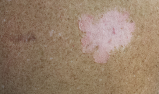
Are These Leg Lesions and a Family History of Diabetes Related?
Several months ago, a 51-year-old woman first noticed brownish red lesions on her leg. She initially dismissed them as insect bites and regarded them as mostly a cosmetic concern. When the lesions failed to disappear after six months, her primary care provider referred the patient to dermatology. (In the interim, a friend did suggest ringworm as a diagnosis, but the OTC tolnaftate cream the patient tried had no effect.)
The patient claims to be quite healthy, which she attributes to walking several miles a day and working out at the gym every other day. She denies any shortness of breath, unexplained fever, or night sweats. There is no personal history of diabetes, but she has had glucose tolerance tests every six months for years because of a significant family history of the disease.
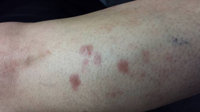
EXAMINATION
There are four intradermal shiny round nodules, averaging about 1.8 cm, on the upper anterior tibial area. The margins of the lesions are sharply drawn, and there is no surrounding erythema or increased warmth on palpation. The surfaces are quite uniform in texture.
Examination of her skin elsewhere reveals no noteworthy lesions. There is no palpable adenopathy in the groin on the affected side.
Results of a punch biopsy show interstitial and palisaded granulomas of the subcutaneous tissue, especially the dermis. Thickening of blood vessel walls and endothelial cell swelling are also noted.
What is the diagnosis?
DISCUSSION
Based on the appearance of the lesions, the patient’s family history, and the histopathologic findings, a diagnosis of necrobiosis lipoidica (NL) was made. When NL was first described in 1929, it was only seen in diabetic persons and thus was named necrobiosis lipoidica diabeticorum. By 1932, however, cases of NL were being reported in persons without diabetes—so the “diabeticorum” was dropped.
Only 0.3% of diabetic persons develop NL. In 15% of cases, NL precedes diabetes; in 60%, diabetes manifests first. Simultaneous diagnosis of both occurs about 25% of the time.
The cause of NL is still unknown. Most theories suggest it is related in some way to diabetes, since most persons with NL have a personal or family history of diabetes or register abnormal results to glucose tolerance testing. Furthermore, the microvascular changes seen in NL lesions are reminiscent of those seen in microangiopathic lesions in diabetic patients. Oddly enough, though, the presence or progression of NL does not correlate with how well controlled the patient’s diabetes is.
Other theories center on an autoimmune mechanism, based on demonstrable deposition of immunoglobulins, components of complement, and fibrinogen in vessel walls. High levels of TNF-a are found in many cases of NL, a fact that researchers are pursuing vigorously.
NL can manifest anywhere but is most common on the legs, where 75% of patients report no symptoms beyond a slight loss of sensation. The remaining 25% do report moderate to severe pain.
In terms of diagnosis, the appearance and history are sufficient in most cases. In this particular case, the lesions were fairly new and small, and the differential thus included such worrisome items as sarcoidosis or even Hansen’s disease. Less concerning differential items included granuloma annulare, other xanthomatous processes, and rheumatoid nodules.
In this case, a punch biopsy was necessary (the defect from which was closed carefully with interrupted sutures). Such procedures are discouraged in patients with larger and more advanced lesions, for fear of inducing a nonhealing wound. In fact, ulceration, usually from trauma, is the most feared complication of NL—and effective treatment is hard to come by.
This patient was treated with perilesional injection of triamcinolone suspension 10 mg per cc and given a prescription for 0.1% tacrolimus cream (to be applied twice daily). A follow-up visit was arranged for two months down the road. Her long-term prognosis is guarded, at best. Since she was already acutely aware of her risk for diabetes, there was no need for additional action in that arena.
Many other treatments have been tried for NL, with varying success, including dihydrochloroquine (Plaquenil and others) pentoxifylline, and TNF-a inhibitors.
TAKE-HOME LEARNING POINTS
• Early manifestations of necrobiosis lipoidica (NL) can be puzzling, and biopsy may be required for diagnosis.
• However, with advanced, obvious lesions, it is preferable to make the diagnosis of NL without biopsy, since punch biopsy can result in a nonhealing wound.
• Mature NL lesions become atrophic and yellowed, with marked telangiectasia formation on their surfaces.
• Just 0.3% of diabetic patients ever develop NL, but most affected persons have a family history of diabetes or an abnormal glucose tolerance test results, if not a personal history of diabetes.
Several months ago, a 51-year-old woman first noticed brownish red lesions on her leg. She initially dismissed them as insect bites and regarded them as mostly a cosmetic concern. When the lesions failed to disappear after six months, her primary care provider referred the patient to dermatology. (In the interim, a friend did suggest ringworm as a diagnosis, but the OTC tolnaftate cream the patient tried had no effect.)
The patient claims to be quite healthy, which she attributes to walking several miles a day and working out at the gym every other day. She denies any shortness of breath, unexplained fever, or night sweats. There is no personal history of diabetes, but she has had glucose tolerance tests every six months for years because of a significant family history of the disease.
EXAMINATION
There are four intradermal shiny round nodules, averaging about 1.8 cm, on the upper anterior tibial area. The margins of the lesions are sharply drawn, and there is no surrounding erythema or increased warmth on palpation. The surfaces are quite uniform in texture.
Examination of her skin elsewhere reveals no noteworthy lesions. There is no palpable adenopathy in the groin on the affected side.
Results of a punch biopsy show interstitial and palisaded granulomas of the subcutaneous tissue, especially the dermis. Thickening of blood vessel walls and endothelial cell swelling are also noted.
What is the diagnosis?
DISCUSSION
Based on the appearance of the lesions, the patient’s family history, and the histopathologic findings, a diagnosis of necrobiosis lipoidica (NL) was made. When NL was first described in 1929, it was only seen in diabetic persons and thus was named necrobiosis lipoidica diabeticorum. By 1932, however, cases of NL were being reported in persons without diabetes—so the “diabeticorum” was dropped.
Only 0.3% of diabetic persons develop NL. In 15% of cases, NL precedes diabetes; in 60%, diabetes manifests first. Simultaneous diagnosis of both occurs about 25% of the time.
The cause of NL is still unknown. Most theories suggest it is related in some way to diabetes, since most persons with NL have a personal or family history of diabetes or register abnormal results to glucose tolerance testing. Furthermore, the microvascular changes seen in NL lesions are reminiscent of those seen in microangiopathic lesions in diabetic patients. Oddly enough, though, the presence or progression of NL does not correlate with how well controlled the patient’s diabetes is.
Other theories center on an autoimmune mechanism, based on demonstrable deposition of immunoglobulins, components of complement, and fibrinogen in vessel walls. High levels of TNF-a are found in many cases of NL, a fact that researchers are pursuing vigorously.
NL can manifest anywhere but is most common on the legs, where 75% of patients report no symptoms beyond a slight loss of sensation. The remaining 25% do report moderate to severe pain.
In terms of diagnosis, the appearance and history are sufficient in most cases. In this particular case, the lesions were fairly new and small, and the differential thus included such worrisome items as sarcoidosis or even Hansen’s disease. Less concerning differential items included granuloma annulare, other xanthomatous processes, and rheumatoid nodules.
In this case, a punch biopsy was necessary (the defect from which was closed carefully with interrupted sutures). Such procedures are discouraged in patients with larger and more advanced lesions, for fear of inducing a nonhealing wound. In fact, ulceration, usually from trauma, is the most feared complication of NL—and effective treatment is hard to come by.
This patient was treated with perilesional injection of triamcinolone suspension 10 mg per cc and given a prescription for 0.1% tacrolimus cream (to be applied twice daily). A follow-up visit was arranged for two months down the road. Her long-term prognosis is guarded, at best. Since she was already acutely aware of her risk for diabetes, there was no need for additional action in that arena.
Many other treatments have been tried for NL, with varying success, including dihydrochloroquine (Plaquenil and others) pentoxifylline, and TNF-a inhibitors.
TAKE-HOME LEARNING POINTS
• Early manifestations of necrobiosis lipoidica (NL) can be puzzling, and biopsy may be required for diagnosis.
• However, with advanced, obvious lesions, it is preferable to make the diagnosis of NL without biopsy, since punch biopsy can result in a nonhealing wound.
• Mature NL lesions become atrophic and yellowed, with marked telangiectasia formation on their surfaces.
• Just 0.3% of diabetic patients ever develop NL, but most affected persons have a family history of diabetes or an abnormal glucose tolerance test results, if not a personal history of diabetes.
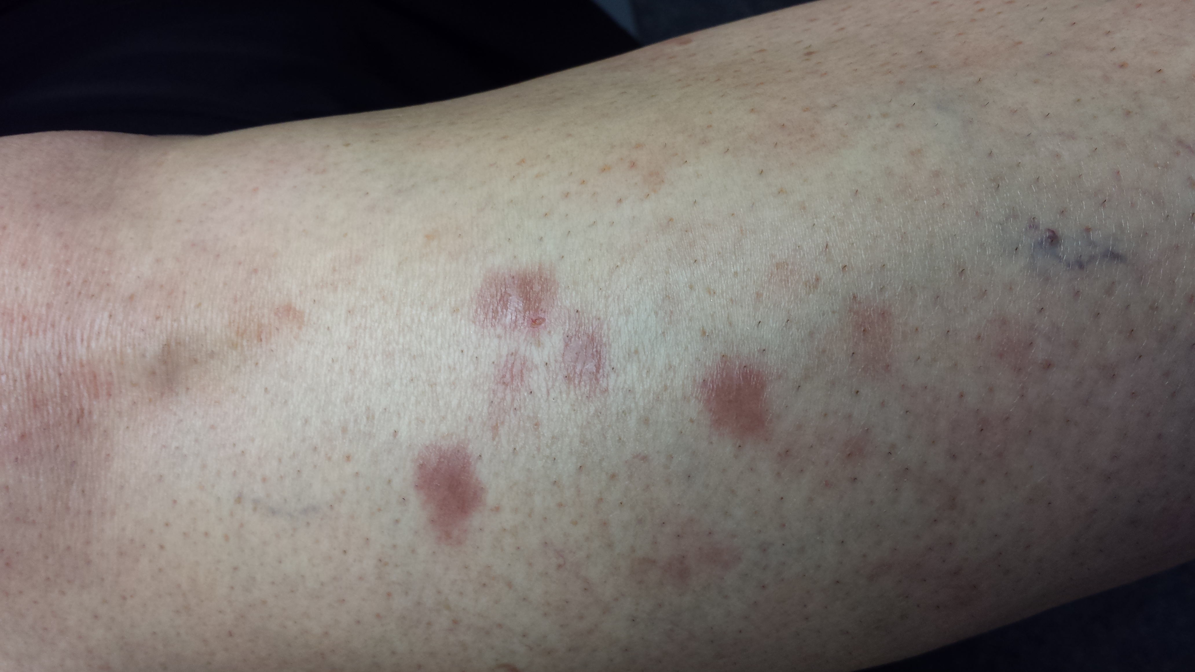
Finger Lesion Is Painful When Bumped
A 67-year-old man has had a small lesion on his index finger for several years. Recently, it has grown large enough to start interfering with his normal activities. The lesion is not painful unless the patient bumps it—but as it grows, avoiding such incidents is becoming more difficult.
The diagnosis has thus far been a mystery: What is it? The patient’s primary care provider didn’t know and, per the patient’s report, had no interest in trying to remove the lesion. Thus, the patient finds himself referred to dermatology.
The patient can recall no specific injury. He does, however, report that he worked for years as an upholsterer, shaping, sewing, and stapling heavy fabric onto chairs and sofas—often puncturing his fingers in the process.
His health is otherwise “decent.” He is taking medication for hypertension and an a-blocker due to “problems” with his prostate.
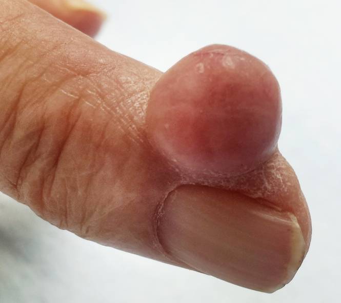
EXAMINATION
The finger lesion is a 2-cm, round, firm nodule with a smooth, telangiectatic surface. It is located on the perionychial area of patient’s index finger (lateral aspect). The lesion is attached to the finger by a broad base that rises seamlessly from the skin. It is not translucent or tender to touch. No similar lesions are seen on either hand, and no nodes are felt in the epitrochlear or axillary locations.
Within a few weeks, the patient returns to have the lesion excised. This is done with a combination of a digital block and local anesthesia, with a tourniquet to control bleeding. After primary closure, the lesion is submitted to pathology. The report indicates the lesion is a cyst, full of cheesy material.
What is the diagnosis?
DISCUSSION
This type of cyst is sometimes referred to as an implantation cyst and occurs when epidermal tissue, including sebaceous glands, is implanted into deeper tissue by a penetrating injury and continues to grow. Although this is an utterly common and benign diagnosis, the differential for a “fingeroma” includes digital mucous cyst, pyogenic granuloma, acquired digital fibrokeratoma, calcinosis cutis, tophaceous gout, wart, and giant cell tumor. Had the lesion been of bony origin, it could have represented connective tissue cancer (sarcoma) or even metastatic cancer.
Implantation cysts must be removed completely to prevent recurrence. The size and location of this particular cyst made it conducive to simple excision and primary closure. Larger lesions in more difficult locations often require the expertise of a hand surgeon for disposition.
TAKE-HOME LEARNING POINTS
• “Fingeromas” are quite common but have an extensive differential diagnosis—mostly benign, but occasionally malignant.
• Regarding malignancy, it is helpful to remember that any tissue in the body can undergo malignant transformation and that many types of primary cancers can metastasize to distant areas (including the hands).
• Removal, or at least biopsy, is the best way to differentiate between types of fingeroma lesions.
• Implantation cysts, as seen in this case, result from traumatic implantation of epidermal structures (including sebaceous glands) into deeper tissue, where they continue to grow.
• When in doubt, consider referring affected patients to dermatology or to a hand surgeon.
A 67-year-old man has had a small lesion on his index finger for several years. Recently, it has grown large enough to start interfering with his normal activities. The lesion is not painful unless the patient bumps it—but as it grows, avoiding such incidents is becoming more difficult.
The diagnosis has thus far been a mystery: What is it? The patient’s primary care provider didn’t know and, per the patient’s report, had no interest in trying to remove the lesion. Thus, the patient finds himself referred to dermatology.
The patient can recall no specific injury. He does, however, report that he worked for years as an upholsterer, shaping, sewing, and stapling heavy fabric onto chairs and sofas—often puncturing his fingers in the process.
His health is otherwise “decent.” He is taking medication for hypertension and an a-blocker due to “problems” with his prostate.
EXAMINATION
The finger lesion is a 2-cm, round, firm nodule with a smooth, telangiectatic surface. It is located on the perionychial area of patient’s index finger (lateral aspect). The lesion is attached to the finger by a broad base that rises seamlessly from the skin. It is not translucent or tender to touch. No similar lesions are seen on either hand, and no nodes are felt in the epitrochlear or axillary locations.
Within a few weeks, the patient returns to have the lesion excised. This is done with a combination of a digital block and local anesthesia, with a tourniquet to control bleeding. After primary closure, the lesion is submitted to pathology. The report indicates the lesion is a cyst, full of cheesy material.
What is the diagnosis?
DISCUSSION
This type of cyst is sometimes referred to as an implantation cyst and occurs when epidermal tissue, including sebaceous glands, is implanted into deeper tissue by a penetrating injury and continues to grow. Although this is an utterly common and benign diagnosis, the differential for a “fingeroma” includes digital mucous cyst, pyogenic granuloma, acquired digital fibrokeratoma, calcinosis cutis, tophaceous gout, wart, and giant cell tumor. Had the lesion been of bony origin, it could have represented connective tissue cancer (sarcoma) or even metastatic cancer.
Implantation cysts must be removed completely to prevent recurrence. The size and location of this particular cyst made it conducive to simple excision and primary closure. Larger lesions in more difficult locations often require the expertise of a hand surgeon for disposition.
TAKE-HOME LEARNING POINTS
• “Fingeromas” are quite common but have an extensive differential diagnosis—mostly benign, but occasionally malignant.
• Regarding malignancy, it is helpful to remember that any tissue in the body can undergo malignant transformation and that many types of primary cancers can metastasize to distant areas (including the hands).
• Removal, or at least biopsy, is the best way to differentiate between types of fingeroma lesions.
• Implantation cysts, as seen in this case, result from traumatic implantation of epidermal structures (including sebaceous glands) into deeper tissue, where they continue to grow.
• When in doubt, consider referring affected patients to dermatology or to a hand surgeon.
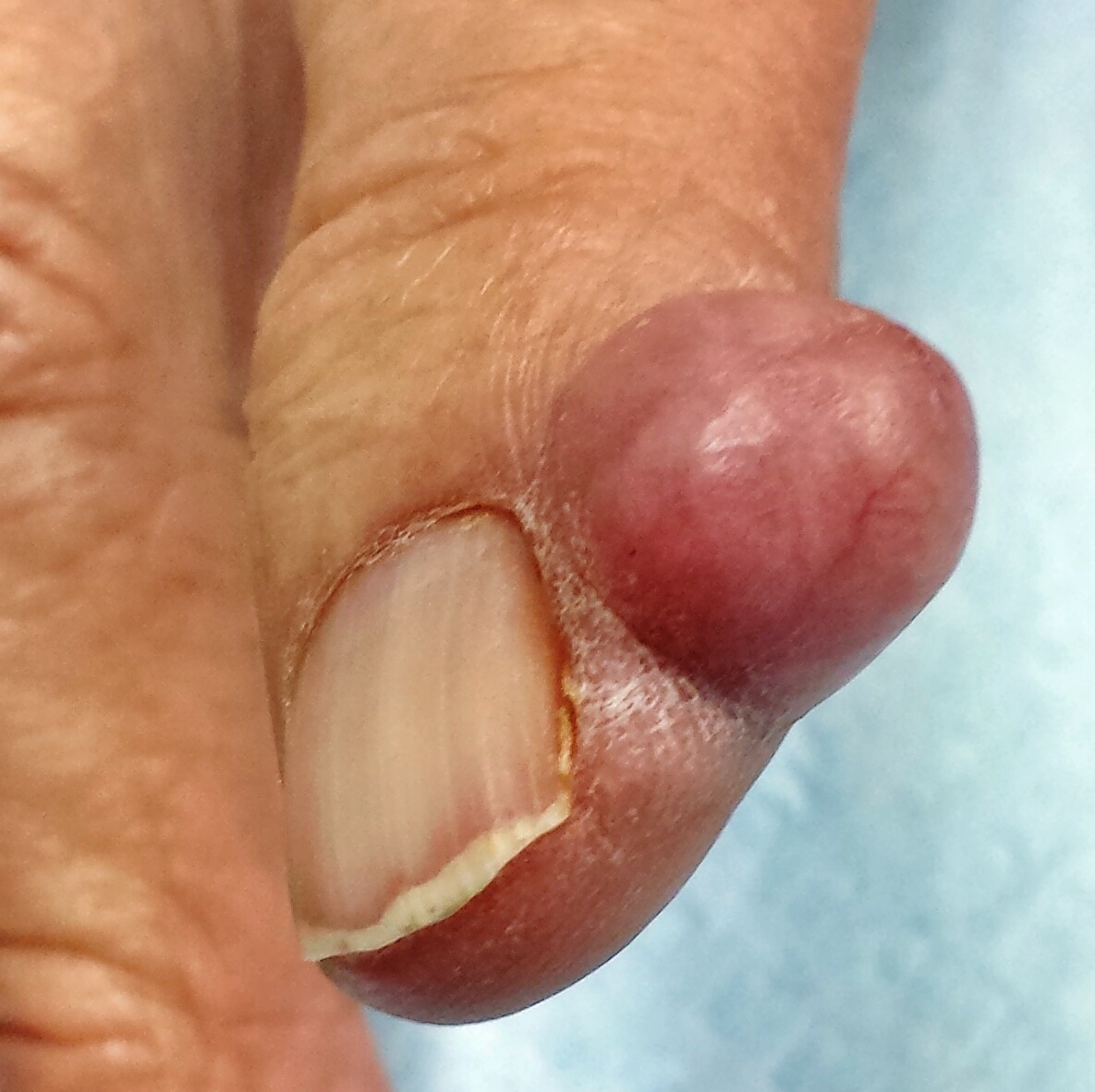
Irritated and Downright Painful
A 33-year-old woman presents with a lesion on the dorsum of her hand. Though it manifested several years ago, it was not problematic until recently, when it became irritated and downright painful. It has not changed in size and has never been red or swollen.
The patient denies ever having similar lesions elsewhere. She also denies any other serious health problems, specifically ophthalmologic problems.

EXAMINATION
The lesion, a 5-mm ovoid tan-orange papule, is located on the dorsum of her left hand. The surface is smooth, with a very firm feel. No skin changes are noted on the surrounding skin, and there are no palpable nodes in the arm or adjacent axilla.
In light of the patient’s concern, the lesion is excised, using an elliptical incision and minimal margins. The incision is carried down into superficial adipose tissue to ensure complete removal. Bleeding is controlled and the defect closed with simple interrupted sutures.
The pathology report shows numerous densely distributed polyhedral histiocytes. Many contain a large amount of cytoplasm.
What is the diagnosis?
DISCUSSION
The clinical presentation and pathology report were both consistent with an entity called juvenile xanthogranuloma (JXG). It usually presents, as in this case, as a solitary, firm, tan to orange papule or nodule. It is more commonly seen on the head and neck of young children, although it is by no means limited to that population (despite its name, which represents anachronistic derm terminology).
Based on the pathologic picture (typified by the biopsy report in this case), JXG may represent a disordered macrophage response to an unknown tissue injury that eventuates in a granulomatous reaction.
JXG is one of the more common members of a spectrum of histiocytic disorders that includes Langerhans cell histiocytosis. In addition to its more common cutaneous distribution, JXG has been seen in almost every internal organ as well. It is rarely associated with systemic manifestations.
The most significant extracutaneous manifestation of JXG is in the eye, where it is the most common cause of spontaneous hyphema in children (usually those younger than 2). This, in turn, can lead to a secondary glaucoma and eventual blindness. This is the main caveat with solitary cutaneous JXGs: They can be associated with ocular involvement, which would manifest as changes in the color of the iris or in the size of the globe itself. Fortunately, this is a rare complication.
Beyond the potential for ocular involvement, since the vast majority of cutaneous JXG lesions are easy to diagnose and self-limiting, there is no reason to remove or otherwise treat them.
The differential for cutaneous JXG includes Spitz tumor, molluscum, and intradermal nevus.
TAKE-HOME LEARNING POINTS
• Juvenile xanthogranuloma (JXG) lesions usually resolve on their own, without treatment.
• In rare instances, cutaneous JXG can be associated with ocular involvement, which manifests with either changes in iris color or in the size of the globe itself.
• JXG lesions are commonly seen on the head and neck of young children, usually as a solitary tan to orange firm papule.
• The differential for JXG includes molluscum, Spitz tumor, and nevus. Removal of JXG lesions is usually not necessary, but pathologic examination shows pathognomic changes that effectively distinguish it from its lookalikes.
A 33-year-old woman presents with a lesion on the dorsum of her hand. Though it manifested several years ago, it was not problematic until recently, when it became irritated and downright painful. It has not changed in size and has never been red or swollen.
The patient denies ever having similar lesions elsewhere. She also denies any other serious health problems, specifically ophthalmologic problems.
EXAMINATION
The lesion, a 5-mm ovoid tan-orange papule, is located on the dorsum of her left hand. The surface is smooth, with a very firm feel. No skin changes are noted on the surrounding skin, and there are no palpable nodes in the arm or adjacent axilla.
In light of the patient’s concern, the lesion is excised, using an elliptical incision and minimal margins. The incision is carried down into superficial adipose tissue to ensure complete removal. Bleeding is controlled and the defect closed with simple interrupted sutures.
The pathology report shows numerous densely distributed polyhedral histiocytes. Many contain a large amount of cytoplasm.
What is the diagnosis?
DISCUSSION
The clinical presentation and pathology report were both consistent with an entity called juvenile xanthogranuloma (JXG). It usually presents, as in this case, as a solitary, firm, tan to orange papule or nodule. It is more commonly seen on the head and neck of young children, although it is by no means limited to that population (despite its name, which represents anachronistic derm terminology).
Based on the pathologic picture (typified by the biopsy report in this case), JXG may represent a disordered macrophage response to an unknown tissue injury that eventuates in a granulomatous reaction.
JXG is one of the more common members of a spectrum of histiocytic disorders that includes Langerhans cell histiocytosis. In addition to its more common cutaneous distribution, JXG has been seen in almost every internal organ as well. It is rarely associated with systemic manifestations.
The most significant extracutaneous manifestation of JXG is in the eye, where it is the most common cause of spontaneous hyphema in children (usually those younger than 2). This, in turn, can lead to a secondary glaucoma and eventual blindness. This is the main caveat with solitary cutaneous JXGs: They can be associated with ocular involvement, which would manifest as changes in the color of the iris or in the size of the globe itself. Fortunately, this is a rare complication.
Beyond the potential for ocular involvement, since the vast majority of cutaneous JXG lesions are easy to diagnose and self-limiting, there is no reason to remove or otherwise treat them.
The differential for cutaneous JXG includes Spitz tumor, molluscum, and intradermal nevus.
TAKE-HOME LEARNING POINTS
• Juvenile xanthogranuloma (JXG) lesions usually resolve on their own, without treatment.
• In rare instances, cutaneous JXG can be associated with ocular involvement, which manifests with either changes in iris color or in the size of the globe itself.
• JXG lesions are commonly seen on the head and neck of young children, usually as a solitary tan to orange firm papule.
• The differential for JXG includes molluscum, Spitz tumor, and nevus. Removal of JXG lesions is usually not necessary, but pathologic examination shows pathognomic changes that effectively distinguish it from its lookalikes.