User login
Hair Loss in a 12-Year-Old
A mother brings her 12-year-old son to dermatology following a referral from the boy’s pediatrician. Several months ago, she noticed her son’s hair loss. The change had been preceded by a stressful period in which she and her husband divorced and one of the boy’s grandparents died unexpectedly.
Both the mother and other relatives and friends had observed the boy reaching for his scalp frequently and twirling his hair “absentmindedly.” When asked if the area in question bothers him, the boy always answers in the negative. Although he knows he should leave his scalp and hair alone, he says he finds it difficult to do so—even though he acknowledges the social liability of his hair loss. According to the mother, the more his family discourages his behavior, the more it persists.
EXAMINATION
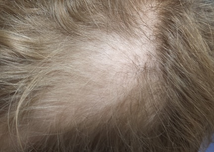
Distinct but incomplete hair loss is noted in an 8 x 10–cm area of his scalp crown. There is neither redness nor any disturbance to the skin there. On palpation, there is no tenderness or increased warmth. No nodes are felt in the adjacent head or neck. Hair pull test is negative.
Closer examination shows hairs of varying lengths in the affected area: many quite short, others of normal length, and many of intermediate length.
Blood work done by the referring pediatrician—including complete blood count, chemistry panel, antinuclear antibody test, and thyroid testing—yielded no abnormal results.
What is the diagnosis?
DISCUSSION
Hair loss, collectively termed alopecia, is a disturbing development, especially in a child. In this case, we had localized hair loss most likely caused by behavior that was not only witnessed by the boy’s parents but also admitted to by the patient. (We’re not always so fortunate.) Thus, it was fairly straightforward to diagnosis trichotillomania, also known as trichotillosis or hair-pulling disorder. This condition can mimic alopecia areata and tinea capitis.
In this case, the lack of epidermal change (scaling, redness, edema) and palpable adenopathy spoke loudly against fungal infection. The hair loss in alopecia areata (AA) is usually sharply defined and complete, which our patient’s hair loss was not. And the blood work that was done effectively ruled out systemic disease (an unlikely cause of localized hair loss in any case).
The jury is still out as to how exactly to classify trichotillomania (TTM). The new DSM-V lists it as an anxiety disorder, in part because it often appears in that context. What we do know is that girls are twice as likely as boys to be affected. And children ages 4 to 13 are seven times more likely than adults to develop TTM.
TTM can involve hair anywhere on the body, though children almost always confine their behavior to their scalp. Actual hair-pulling is not necessarily seen. Manipulation, such as the twirling in this case, is enough to weaken hair follicles, causing hair to fall out. In cases involving hair-pulling, a small percentage of patients actually ingest the hairs they’ve plucked out (trichophagia). Being indigestible, the hairs can accumulate in hairballs (trichobezoars).
Even though TTM is most likely a psychiatric disorder lying somewhere in the obsessive-compulsive spectrum, it is seen more often in primary care and dermatology offices. Scalp biopsy would certainly settle the matter, but a better alternative is simply shaving a dime-sized area of scalp and watching it for normal hair growth.
Most cases eventually resolve with time and persistent but gentle reminders, but a few will require psychiatric intervention. This typically includes habit reversal therapy or cognitive behavioral therapy, plus or minus combinations of psychoactive medications. (The latter decision depends on whether there psychiatric comorbidities.) Despite all these efforts, severe cases of TTM can persist for years or even a lifetime.
It remains to be seen how this particular patient responds to his parents’ efforts. It was an immense relief for them to know the cause of their son’s hair loss and that the condition is likely self-limiting.
TAKE-HOME LEARNING POINTS
• Trichotillomania (TTM) is an unusual form of localized hair loss, usually involving children’s scalps.
• TTM affects children ages 4 to 13 and at least twice as many girls as boys.
• TTM does not always involve actual plucking of hairs. Repetitive manipulation, such as twirling, can weaken the hairs enough to cause hair loss.
• Unlike alopecia areata (the main item in the alopecia differential for children), TTM is more likely to cause incomplete, poorly defined hair loss in an area where hairs of varying length can be seen.
• Usually self-limiting, TTM can require psychiatric attention, for which a variety of habit training techniques can be used.
A mother brings her 12-year-old son to dermatology following a referral from the boy’s pediatrician. Several months ago, she noticed her son’s hair loss. The change had been preceded by a stressful period in which she and her husband divorced and one of the boy’s grandparents died unexpectedly.
Both the mother and other relatives and friends had observed the boy reaching for his scalp frequently and twirling his hair “absentmindedly.” When asked if the area in question bothers him, the boy always answers in the negative. Although he knows he should leave his scalp and hair alone, he says he finds it difficult to do so—even though he acknowledges the social liability of his hair loss. According to the mother, the more his family discourages his behavior, the more it persists.
EXAMINATION
Distinct but incomplete hair loss is noted in an 8 x 10–cm area of his scalp crown. There is neither redness nor any disturbance to the skin there. On palpation, there is no tenderness or increased warmth. No nodes are felt in the adjacent head or neck. Hair pull test is negative.
Closer examination shows hairs of varying lengths in the affected area: many quite short, others of normal length, and many of intermediate length.
Blood work done by the referring pediatrician—including complete blood count, chemistry panel, antinuclear antibody test, and thyroid testing—yielded no abnormal results.
What is the diagnosis?
DISCUSSION
Hair loss, collectively termed alopecia, is a disturbing development, especially in a child. In this case, we had localized hair loss most likely caused by behavior that was not only witnessed by the boy’s parents but also admitted to by the patient. (We’re not always so fortunate.) Thus, it was fairly straightforward to diagnosis trichotillomania, also known as trichotillosis or hair-pulling disorder. This condition can mimic alopecia areata and tinea capitis.
In this case, the lack of epidermal change (scaling, redness, edema) and palpable adenopathy spoke loudly against fungal infection. The hair loss in alopecia areata (AA) is usually sharply defined and complete, which our patient’s hair loss was not. And the blood work that was done effectively ruled out systemic disease (an unlikely cause of localized hair loss in any case).
The jury is still out as to how exactly to classify trichotillomania (TTM). The new DSM-V lists it as an anxiety disorder, in part because it often appears in that context. What we do know is that girls are twice as likely as boys to be affected. And children ages 4 to 13 are seven times more likely than adults to develop TTM.
TTM can involve hair anywhere on the body, though children almost always confine their behavior to their scalp. Actual hair-pulling is not necessarily seen. Manipulation, such as the twirling in this case, is enough to weaken hair follicles, causing hair to fall out. In cases involving hair-pulling, a small percentage of patients actually ingest the hairs they’ve plucked out (trichophagia). Being indigestible, the hairs can accumulate in hairballs (trichobezoars).
Even though TTM is most likely a psychiatric disorder lying somewhere in the obsessive-compulsive spectrum, it is seen more often in primary care and dermatology offices. Scalp biopsy would certainly settle the matter, but a better alternative is simply shaving a dime-sized area of scalp and watching it for normal hair growth.
Most cases eventually resolve with time and persistent but gentle reminders, but a few will require psychiatric intervention. This typically includes habit reversal therapy or cognitive behavioral therapy, plus or minus combinations of psychoactive medications. (The latter decision depends on whether there psychiatric comorbidities.) Despite all these efforts, severe cases of TTM can persist for years or even a lifetime.
It remains to be seen how this particular patient responds to his parents’ efforts. It was an immense relief for them to know the cause of their son’s hair loss and that the condition is likely self-limiting.
TAKE-HOME LEARNING POINTS
• Trichotillomania (TTM) is an unusual form of localized hair loss, usually involving children’s scalps.
• TTM affects children ages 4 to 13 and at least twice as many girls as boys.
• TTM does not always involve actual plucking of hairs. Repetitive manipulation, such as twirling, can weaken the hairs enough to cause hair loss.
• Unlike alopecia areata (the main item in the alopecia differential for children), TTM is more likely to cause incomplete, poorly defined hair loss in an area where hairs of varying length can be seen.
• Usually self-limiting, TTM can require psychiatric attention, for which a variety of habit training techniques can be used.

Slowly Spreading Hand Lesions
Several months ago, lesions appeared on both of this 48-year-old woman’s hands. They have been completely unresponsive to any of the treatments prescribed by her primary care provider, including oral (terbinafine) and topical antifungal medications.
The lesions, which are asymptomatic, manifested slowly. They first appeared on the dorsa of her hands and then gradually spread until they covered the central portion of both hands. The lesions wax and wane in terms of thickness and extent but never disappear.
The patient denies similar lesions elsewhere. She also denies contact with animals or children. She has never been immunosuppressed and is quite healthy aside from her skin problems. She has a family history but no personal history of diabetes.
EXAMINATION
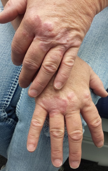
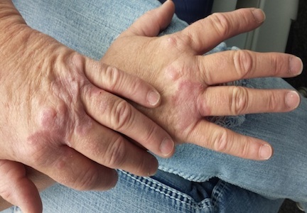
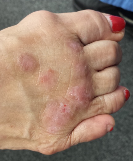
The condition affects both hands equally. It is composed of intradermal reddish brown papules and plaques that cover the metacarpal areas and extend onto the proximal interphalangeal skin and the distal dorsa. No epidermal component (scale or other broken skin surface) is seen.
The margins of the lesions are arciform, slightly raised, and smooth. The centers of several are concave.
What is the diagnosis?
DISCUSSION
This is the classic presentation of granuloma annulare (GA), in terms of morphology, distribution, and patient gender. Though this common condition can manifest in a number of locations and in men, it tends to favor the extensor surfaces of the extremities of women. The dorsum of the foot, anterior tibial skin, and elbows are typical locations that make it a relatively easy condition to recognize.
GA can be more difficult to identify, however, when it is generalized or deeper in the dermis. The latter, termed subcutaneous GA, is most common in children, who present with large dark annular patches of discoloration too deep in the dermis to produce any palpable surface changes. The more rare generalized GA can be difficult to diagnose and treat. Biopsy of suspected generalized or subcutaneous GA is necessary to differentiate it from its lookalikes, such as secondary syphilis, lichen planus, sarcoidosis, and tinea.
The annular borders of GA lesions are notorious for deceiving the unwary practitioner into diagnosing fungal infection. But the latter is, by definition, an infection of the outermost layers of the skin on which fungal organisms feed. That process almost invariably produces significant scaling, especially on the periphery of the lesions. Such scaling is completely absent on GA lesions, which also have a papularity and induration rarely seen with superficial fungal infections. In this particular case, the patient denied any potential source for a fungal infection (animals, children) and denied the itching we would expect with one.
The cause of GA is unknown, but it appears to represent a reaction to an unknown antigen. One previous hypothesis held that it was connected to diabetes, but this was proved false.
Ordinary GA resolves on its own (though this can take months)—which is fortunate, since there is no uniformly successful treatment. Many things have been tried, including topical or intralesional steroids and cryotherapy, with varying levels of success. Generalized GA is even more difficult to treat, can linger for years, and can be highly pruritic. Treatments have included hydroxychloroquine, dapsone, potassium iodide, isotretinoin, and PUVA. More recently, biologics are being used with some success.
The bigger problem with GA is simply to consider it in the differential for odd, annular, or generalized lesions.
TAKE-HOME LEARNING POINTS
• Granuloma annulare (GA) is often mistaken for fungal infection, but it is an intradermal rather than epidermal process.
• The extensor surfaces of the extremities of women are favored sites for GA lesions.
• Biopsy of suspected GA lesions confirms the diagnosis while ruling out other items in the differential (eg, sarcoidosis and lichen planus).
• GA has no connection to diabetes.
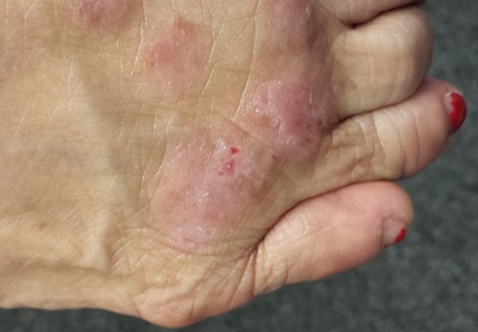
Several months ago, lesions appeared on both of this 48-year-old woman’s hands. They have been completely unresponsive to any of the treatments prescribed by her primary care provider, including oral (terbinafine) and topical antifungal medications.
The lesions, which are asymptomatic, manifested slowly. They first appeared on the dorsa of her hands and then gradually spread until they covered the central portion of both hands. The lesions wax and wane in terms of thickness and extent but never disappear.
The patient denies similar lesions elsewhere. She also denies contact with animals or children. She has never been immunosuppressed and is quite healthy aside from her skin problems. She has a family history but no personal history of diabetes.
EXAMINATION
The condition affects both hands equally. It is composed of intradermal reddish brown papules and plaques that cover the metacarpal areas and extend onto the proximal interphalangeal skin and the distal dorsa. No epidermal component (scale or other broken skin surface) is seen.
The margins of the lesions are arciform, slightly raised, and smooth. The centers of several are concave.
What is the diagnosis?
DISCUSSION
This is the classic presentation of granuloma annulare (GA), in terms of morphology, distribution, and patient gender. Though this common condition can manifest in a number of locations and in men, it tends to favor the extensor surfaces of the extremities of women. The dorsum of the foot, anterior tibial skin, and elbows are typical locations that make it a relatively easy condition to recognize.
GA can be more difficult to identify, however, when it is generalized or deeper in the dermis. The latter, termed subcutaneous GA, is most common in children, who present with large dark annular patches of discoloration too deep in the dermis to produce any palpable surface changes. The more rare generalized GA can be difficult to diagnose and treat. Biopsy of suspected generalized or subcutaneous GA is necessary to differentiate it from its lookalikes, such as secondary syphilis, lichen planus, sarcoidosis, and tinea.
The annular borders of GA lesions are notorious for deceiving the unwary practitioner into diagnosing fungal infection. But the latter is, by definition, an infection of the outermost layers of the skin on which fungal organisms feed. That process almost invariably produces significant scaling, especially on the periphery of the lesions. Such scaling is completely absent on GA lesions, which also have a papularity and induration rarely seen with superficial fungal infections. In this particular case, the patient denied any potential source for a fungal infection (animals, children) and denied the itching we would expect with one.
The cause of GA is unknown, but it appears to represent a reaction to an unknown antigen. One previous hypothesis held that it was connected to diabetes, but this was proved false.
Ordinary GA resolves on its own (though this can take months)—which is fortunate, since there is no uniformly successful treatment. Many things have been tried, including topical or intralesional steroids and cryotherapy, with varying levels of success. Generalized GA is even more difficult to treat, can linger for years, and can be highly pruritic. Treatments have included hydroxychloroquine, dapsone, potassium iodide, isotretinoin, and PUVA. More recently, biologics are being used with some success.
The bigger problem with GA is simply to consider it in the differential for odd, annular, or generalized lesions.
TAKE-HOME LEARNING POINTS
• Granuloma annulare (GA) is often mistaken for fungal infection, but it is an intradermal rather than epidermal process.
• The extensor surfaces of the extremities of women are favored sites for GA lesions.
• Biopsy of suspected GA lesions confirms the diagnosis while ruling out other items in the differential (eg, sarcoidosis and lichen planus).
• GA has no connection to diabetes.
Penile Rash Worries Man (And Wife)
A 61-year-old man presents with an asymptomatic but worrisome (to him) penile rash that manifested over a two-day period several weeks ago. OTC miconazole cream, triple-antibiotic creams, and most recently, a two-day course of fluconazole (200 mg bid) have not helped.
The patient denies any sexual exposure outside his marriage, as does his wife. Both are in good health in other respects, although the wife has been treated for lymphoma (long since declared cured).
At first, the patient denies any other skin problems. However, with specific questioning, he admits to having a chronic scaly and slightly itchy rash that periodically appears in the same places: bilateral brows, nasolabial folds, various spots in his beard, and both external ear canals.
Additional history taking reveals that the patient has been under a great deal of stress lately. His father recently died, leaving him to deal with a number of issues, and his work shift changed, requiring him to sleep during the day and work at night.
EXAMINATION
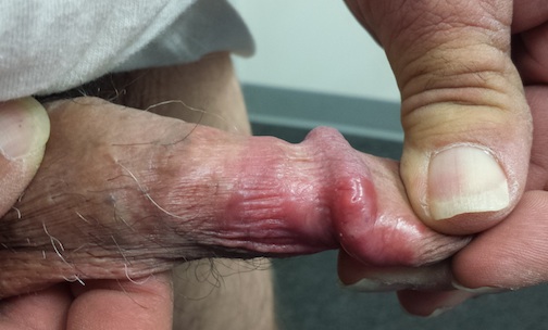
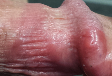
The penile rash is macular, smooth, strikingly red, and shiny. It covers the dorsal, distal penile shaft, spilling onto the glans. It looks wet but is quite dry. His groin, upper intergluteal area, and axillae are free of any changes.
A fine pink rash is seen in the glabella, extending into both brows. Similar areas of focal scaling on pink bases are also noted in the beard area and in both external auditory meati.
What is the diagnosis?
DISCUSSION
This is the way seborrheic dermatitis (SD) acts: It sits around for years, causing minor problems in common areas (eg, face, scalp, ears, and beard). Then the stress of life intrudes, the candle is being burnt from both ends, and SD blossoms, appearing in places such as the chest, groin, suprapubic area, axillae, and genitals.
On the genitals, SD is often bright red, with little if any scale. Why? The moisture and friction particular to the genital and intertriginous (skin on skin) areas prevent scale from accumulating.
Penile rashes generally get everyone’s attention, including that of the patient’s sexual partner(s). For the average nonclinician, the list of possible explanations is infinite—and worrisome. The situation is, in some ways, worse for the average primary care provider, who will almost always decree such a rash a “yeast infection” (though it seldom responds to anti-yeast medications—for good reason: Otherwise healthy circumcised men almost never develop yeast infections anywhere, let alone on the penis).
If you remove “yeast” from the differential, the average primary care provider will be lost. An abbreviated differential list for penile rashes and conditions includes:
• Psoriasis
• Seborrhea
• Contact vs irritant dermatitis
• Eczema/lichen simplex chronicus
• Lichen sclerosis et atrophicus (traditionally termed balanitis xerotica obliterans or BXO when it occurs on the penis)
• Lichen planus
• Reiter syndrome
• Scabies
• Erythroplasia of Queyrat (superficial squamous cell carcinoma)
• Melanoma
• Herpes simplex
• Molluscum
• Pearly penile papules
• Tyson’s glands (prominent pilosebaceous glands on the penile shaft)
• Angiokeratoma of Fordyce
• Condyloma accuminata
• Primary syphilis chancre
• Fixed-drug eruption
(Note that while yeast is not on this list, it certainly could be considered in an uncircumcised hyperglycemic patient.)
The top five items on the list would cover 90% of patients with penile rashes. Seborrheic dermatitis is utterly common but only rarely recognized when it occurs in unusual areas (as in this case).
For this patient, I prescribed topical hydrocortisone 2.5% cream (to be applied bid to all affected areas) and advised him to shampoo the areas daily with ketoconazole-containing shampoo. But the main thing I did for him, and his wife, was provide peace of mind about all the terrible conditions he didn’t have. With treatment, his penile lesions resolved within two weeks.
TAKE-HOME LEARNING POINTS
• Seborrheic dermatitis (SD) is common in the scalp, on the face, and in the ears, but it can also appear on the chest, axillae, or genitals.
• Yeast infections are quite uncommon in otherwise healthy men.
• Stress is often a triggering factor with worsening SD.
• Asking about and finding corroborative areas of involvement (face, ears, brows, scalp) can help in diagnosing atypical cases of SD.
A 61-year-old man presents with an asymptomatic but worrisome (to him) penile rash that manifested over a two-day period several weeks ago. OTC miconazole cream, triple-antibiotic creams, and most recently, a two-day course of fluconazole (200 mg bid) have not helped.
The patient denies any sexual exposure outside his marriage, as does his wife. Both are in good health in other respects, although the wife has been treated for lymphoma (long since declared cured).
At first, the patient denies any other skin problems. However, with specific questioning, he admits to having a chronic scaly and slightly itchy rash that periodically appears in the same places: bilateral brows, nasolabial folds, various spots in his beard, and both external ear canals.
Additional history taking reveals that the patient has been under a great deal of stress lately. His father recently died, leaving him to deal with a number of issues, and his work shift changed, requiring him to sleep during the day and work at night.
EXAMINATION
The penile rash is macular, smooth, strikingly red, and shiny. It covers the dorsal, distal penile shaft, spilling onto the glans. It looks wet but is quite dry. His groin, upper intergluteal area, and axillae are free of any changes.
A fine pink rash is seen in the glabella, extending into both brows. Similar areas of focal scaling on pink bases are also noted in the beard area and in both external auditory meati.
What is the diagnosis?
DISCUSSION
This is the way seborrheic dermatitis (SD) acts: It sits around for years, causing minor problems in common areas (eg, face, scalp, ears, and beard). Then the stress of life intrudes, the candle is being burnt from both ends, and SD blossoms, appearing in places such as the chest, groin, suprapubic area, axillae, and genitals.
On the genitals, SD is often bright red, with little if any scale. Why? The moisture and friction particular to the genital and intertriginous (skin on skin) areas prevent scale from accumulating.
Penile rashes generally get everyone’s attention, including that of the patient’s sexual partner(s). For the average nonclinician, the list of possible explanations is infinite—and worrisome. The situation is, in some ways, worse for the average primary care provider, who will almost always decree such a rash a “yeast infection” (though it seldom responds to anti-yeast medications—for good reason: Otherwise healthy circumcised men almost never develop yeast infections anywhere, let alone on the penis).
If you remove “yeast” from the differential, the average primary care provider will be lost. An abbreviated differential list for penile rashes and conditions includes:
• Psoriasis
• Seborrhea
• Contact vs irritant dermatitis
• Eczema/lichen simplex chronicus
• Lichen sclerosis et atrophicus (traditionally termed balanitis xerotica obliterans or BXO when it occurs on the penis)
• Lichen planus
• Reiter syndrome
• Scabies
• Erythroplasia of Queyrat (superficial squamous cell carcinoma)
• Melanoma
• Herpes simplex
• Molluscum
• Pearly penile papules
• Tyson’s glands (prominent pilosebaceous glands on the penile shaft)
• Angiokeratoma of Fordyce
• Condyloma accuminata
• Primary syphilis chancre
• Fixed-drug eruption
(Note that while yeast is not on this list, it certainly could be considered in an uncircumcised hyperglycemic patient.)
The top five items on the list would cover 90% of patients with penile rashes. Seborrheic dermatitis is utterly common but only rarely recognized when it occurs in unusual areas (as in this case).
For this patient, I prescribed topical hydrocortisone 2.5% cream (to be applied bid to all affected areas) and advised him to shampoo the areas daily with ketoconazole-containing shampoo. But the main thing I did for him, and his wife, was provide peace of mind about all the terrible conditions he didn’t have. With treatment, his penile lesions resolved within two weeks.
TAKE-HOME LEARNING POINTS
• Seborrheic dermatitis (SD) is common in the scalp, on the face, and in the ears, but it can also appear on the chest, axillae, or genitals.
• Yeast infections are quite uncommon in otherwise healthy men.
• Stress is often a triggering factor with worsening SD.
• Asking about and finding corroborative areas of involvement (face, ears, brows, scalp) can help in diagnosing atypical cases of SD.
Baby Has Rash; Parents Feel Itchy
A 5-month-old baby is brought in by his parents for evaluation of a rash that manifested on his hands several weeks ago. It then spread to his arms and trunk and is now essentially everywhere except his face. Despite a number of treatment attempts, including use of oral antibiotics (cephalexin suspension 125/5 cc) and OTC topical steroid creams, the problem has persisted.
Prior to dermatology, they had consulted a pediatrician. He suggested the child might have scabies, for which he prescribed permethrin cream. The parents tried it, but it made little if any difference.
Neither the child nor his parents are atopic. However, both parents have recently started to feel itchy.
EXAMINATION
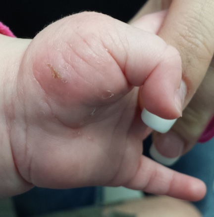
The child is afebrile and in no acute distress. Hundreds of tiny papules are scattered on his trunk, arms, and legs, with a particular concentration on his palms. Several of the papules, on closer examination, prove to be vesicles (ie, filled with clear fluid).
One of these lesions, on the child’s volar wrist, is scraped and the sample examined under the microscope. Magnification at 10x power reveals an adult scabies organism and a number of rugby ball–shaped eggs.
Both parents are also examined and found to have probable scabies as well. The mother’s lesions are concentrated around the anterior axillary areas and waistline. The father’s are on his volar wrists and penis.
What is the diagnosis?
DISCUSSION
This case nicely illustrates several issues revolving around the diagnosis of scabies. One might think this would be a simple matter: Diagnose, then treat. Alas, it is seldom so.
For one thing, the diagnosis of scabies needs to be confirmed, whenever possible, with microscopic findings of scabetic elements. Without this, patient and provider confidence are lacking—a situation that often leads to shotgun treatment.
In addition, had the diagnosis been confirmed prior to presentation to dermatology, the previously consulted providers might have considered treating the whole family and trying to identify the source of the infestation. Both of these are absolutely crucial to successful treatment.
Several factors make the diagnosis of scabies difficult in infants. Any part of an infant’s thin, soft, relatively hairless skin is fair game (whereas, in adults, scabies rarely affects skin above the neck). Furthermore, although infants with scabies undoubtedly itch—probably just as much as adults—they are totally inept excoriators and even worse historians. In contrast, adults with scabies will scratch continuously while in the exam room and complain bitterly 24/7.
Once the diagnosis is established, a crucial element of dealing effectively with scabies is education—in this case, of the parents. They must understand the nature of the problem in specific terms. For example, scabies cannot be caught from or given to nonhuman hosts (eg, animals). And while I advise affected families to clean areas such as beds, sofas, and bathrooms, I also emphasize that the organism does not reside in or multiply on inanimate objects. Despite my best efforts, though, some families become almost hysterical: steam-cleaning every surface, calling pest control, washing bedding and towels multiple times, and calling me three times a day.
Families must also understand that treatment of all household members must be coordinated and done twice, seven to 10 days apart, in order to kill freshly hatched organisms. This child was treated with permethrin 5% cream, applied to the entire body and left on overnight, then washed off the next morning (twice per the schedule outlined above). In addition to permethrin, the adults were treated with ivermectin (200 mcg/kg) on the same schedule. Even with these extensive measures, recurrence would not be surprising.
Most often, when treatment “fails,” it is because the diagnosis was not scabies in the first place. In confirmed cases, treatment will be unsuccessful if all family members are not adequately (and concurrently) treated. Another problem occurs when the actual source is outside the home (daycare, sleepovers, sexual partner) and remains unidentified—dooming the family to recurrence. (Institutional scabies—from nursing homes, group living, etc—can be far more difficult to deal with and is beyond the scope of this article.)
The differential for scabies includes—most significantly—atopic dermatitis, which it can closely resemble.
TAKE-HOME LEARNING POINTS
• Scabies can show up almost anywhere on an infant’s body, because the skin is so thin, hairless, and soft.
• If the baby has scabies, chances are the parents and siblings have it too.
• Someone brings scabies into the family, and unless the source is identified and treated, the problem will recur.
• Microscopic examination (KOH) for scabetic elements is a crucial component of diagnosis and treatment.
• Scabies sarcoptes var humani is species-specific and cannot be given to or caught from an animal.
• Permethrin cream 5% is considered safe for infants ages 2 months and older.
A 5-month-old baby is brought in by his parents for evaluation of a rash that manifested on his hands several weeks ago. It then spread to his arms and trunk and is now essentially everywhere except his face. Despite a number of treatment attempts, including use of oral antibiotics (cephalexin suspension 125/5 cc) and OTC topical steroid creams, the problem has persisted.
Prior to dermatology, they had consulted a pediatrician. He suggested the child might have scabies, for which he prescribed permethrin cream. The parents tried it, but it made little if any difference.
Neither the child nor his parents are atopic. However, both parents have recently started to feel itchy.
EXAMINATION
The child is afebrile and in no acute distress. Hundreds of tiny papules are scattered on his trunk, arms, and legs, with a particular concentration on his palms. Several of the papules, on closer examination, prove to be vesicles (ie, filled with clear fluid).
One of these lesions, on the child’s volar wrist, is scraped and the sample examined under the microscope. Magnification at 10x power reveals an adult scabies organism and a number of rugby ball–shaped eggs.
Both parents are also examined and found to have probable scabies as well. The mother’s lesions are concentrated around the anterior axillary areas and waistline. The father’s are on his volar wrists and penis.
What is the diagnosis?
DISCUSSION
This case nicely illustrates several issues revolving around the diagnosis of scabies. One might think this would be a simple matter: Diagnose, then treat. Alas, it is seldom so.
For one thing, the diagnosis of scabies needs to be confirmed, whenever possible, with microscopic findings of scabetic elements. Without this, patient and provider confidence are lacking—a situation that often leads to shotgun treatment.
In addition, had the diagnosis been confirmed prior to presentation to dermatology, the previously consulted providers might have considered treating the whole family and trying to identify the source of the infestation. Both of these are absolutely crucial to successful treatment.
Several factors make the diagnosis of scabies difficult in infants. Any part of an infant’s thin, soft, relatively hairless skin is fair game (whereas, in adults, scabies rarely affects skin above the neck). Furthermore, although infants with scabies undoubtedly itch—probably just as much as adults—they are totally inept excoriators and even worse historians. In contrast, adults with scabies will scratch continuously while in the exam room and complain bitterly 24/7.
Once the diagnosis is established, a crucial element of dealing effectively with scabies is education—in this case, of the parents. They must understand the nature of the problem in specific terms. For example, scabies cannot be caught from or given to nonhuman hosts (eg, animals). And while I advise affected families to clean areas such as beds, sofas, and bathrooms, I also emphasize that the organism does not reside in or multiply on inanimate objects. Despite my best efforts, though, some families become almost hysterical: steam-cleaning every surface, calling pest control, washing bedding and towels multiple times, and calling me three times a day.
Families must also understand that treatment of all household members must be coordinated and done twice, seven to 10 days apart, in order to kill freshly hatched organisms. This child was treated with permethrin 5% cream, applied to the entire body and left on overnight, then washed off the next morning (twice per the schedule outlined above). In addition to permethrin, the adults were treated with ivermectin (200 mcg/kg) on the same schedule. Even with these extensive measures, recurrence would not be surprising.
Most often, when treatment “fails,” it is because the diagnosis was not scabies in the first place. In confirmed cases, treatment will be unsuccessful if all family members are not adequately (and concurrently) treated. Another problem occurs when the actual source is outside the home (daycare, sleepovers, sexual partner) and remains unidentified—dooming the family to recurrence. (Institutional scabies—from nursing homes, group living, etc—can be far more difficult to deal with and is beyond the scope of this article.)
The differential for scabies includes—most significantly—atopic dermatitis, which it can closely resemble.
TAKE-HOME LEARNING POINTS
• Scabies can show up almost anywhere on an infant’s body, because the skin is so thin, hairless, and soft.
• If the baby has scabies, chances are the parents and siblings have it too.
• Someone brings scabies into the family, and unless the source is identified and treated, the problem will recur.
• Microscopic examination (KOH) for scabetic elements is a crucial component of diagnosis and treatment.
• Scabies sarcoptes var humani is species-specific and cannot be given to or caught from an animal.
• Permethrin cream 5% is considered safe for infants ages 2 months and older.

Itch–Scratch–Itch: Can the Cycle Be Broken?
A 67-year-old woman has had a very itchy rash on the dorsa of both feet for almost a year. In addition to consulting her primary care provider, she has presented to a number of medical venues, including urgent care clinics. Different products have been prescribed—including clotrimazole cream, nystatin powder, and OTC hydrocortisone 1% cream—none of which produced any beneficial effect. So the patient finally self-refers to dermatology.
She reports that at one point, she was convinced her shoes were the source of the problem. But trying new shoes and even going entirely barefoot during a two-week vacation at the beach made no difference.
The patient admits that it is “impossible” to leave the lesions alone, because they are so itchy. She knows that “scratching can’t be good,” so she tries to just rub them, often with a wet washcloth.
Aside from the foot rash, her health is excellent. Her only medications are NSAIDs for mild arthritis.
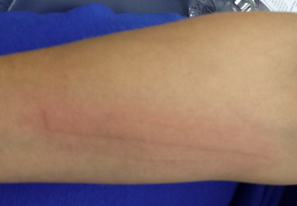
EXAMINATION
The lesions are confined to the forefeet. There are about five on the right foot and three on the left. The lesions are dark purplish round plaques with planar surfaces that are shiny but have a white frosting-like finish. On average, they measure 1.8 cm in diameter. No surrounding inflammation is appreciated. The patient has otherwise unremarkable type IV skin.
What is the diagnosis?
DIAGNOSIS
Punch biopsy confirms the expected diagnosis of lichen planus.
DISCUSSION
Lichen planus (LP) is a very common problem seen in dermatology offices worldwide. LP represents an immune response of unknown origin and may be found in association with other diseases of altered immunity (eg, dermatomyositis, alopecia areata, vitiligo, morphea, and myasthenia gravis). Some studies support the theory that LP is caused by hepatitis C.
The most common forms of LP lend themselves to a useful mnemonic device that uses the letter “P” to describe common features of the disease: purple, papular or plaquish, planar (flat) surfaces, polygonal shapes, pruritic, penile (frequent location), and finally, “puzzling” to the clinician.
In contrast to this particular case, LP commonly affects flexural surfaces, such as the volar wrist. LP can also affect nails (with dystrophy or onycholysis), the scalp, and, most notoriously, the oral mucosae, where it can cause ulcerations and intense burning. Oral lesions often present with a lacy white look on the buccal mucosal surfaces.
LP is only one of a number of skin diseases that “koebnerize” (ie, form along lines of trauma, such as a scratch). The resulting linear collection of planar purple papules—known as the Koebner phenomenon—is useful for diagnosis.
Biopsy is often needed to confirm the diagnosis. It will show hyperkeratotic epidermis with irregular acanthosis. In the upper dermis, there is often an infiltrating band of lymphocytic and histiocytic cells, along with many Langerhans cells, that effectively obliterates the dermo-epidermal junction (a pathognomic finding of LP).
Most cases of LP eventually resolve, usually within months, though some can persist for years. Treatment can be problematic, especially when the disease is widespread or manifests in difficult areas, such as the mouth or scalp. This particular patient’s problem was relatively simple to treat with topical clobetasol cream under occlusion (bid for three weeks). Had that not worked, we could have tried intralesional injection of triamcinolone (10 mg per cc).
This case was typical of LP seen on the legs of older patients with darker skin. In these patients, the lesions tend to become hypertrophic and darker than the usual light pink to purple seen in those with fairer skin.
The differential for LP includes psoriasis, fixed-drug eruption, granuloma annulare, and nummular eczema.
TAKE-HOME LEARNING POINTS
• Lichen planus (LP) is a commonly encountered inflammatory condition that classically affects flexural skin, such as the volar wrist.
• LP lesions can often be seen in a linear configuration, following the line of a scratch or other trauma, a phenomenon known as the Koebner phenomenon (the isomorphic linear response), which can be helpful diagnostically.
• The “Ps” of LP include papular, purple, planar, plaquish, pruritic, penile, polygonal, and puzzling.
• LP can also affect hair follicles, nails, and oral mucosa.
• LP can present with hypertrophic plaques, especially on legs and feet.
A 67-year-old woman has had a very itchy rash on the dorsa of both feet for almost a year. In addition to consulting her primary care provider, she has presented to a number of medical venues, including urgent care clinics. Different products have been prescribed—including clotrimazole cream, nystatin powder, and OTC hydrocortisone 1% cream—none of which produced any beneficial effect. So the patient finally self-refers to dermatology.
She reports that at one point, she was convinced her shoes were the source of the problem. But trying new shoes and even going entirely barefoot during a two-week vacation at the beach made no difference.
The patient admits that it is “impossible” to leave the lesions alone, because they are so itchy. She knows that “scratching can’t be good,” so she tries to just rub them, often with a wet washcloth.
Aside from the foot rash, her health is excellent. Her only medications are NSAIDs for mild arthritis.
EXAMINATION
The lesions are confined to the forefeet. There are about five on the right foot and three on the left. The lesions are dark purplish round plaques with planar surfaces that are shiny but have a white frosting-like finish. On average, they measure 1.8 cm in diameter. No surrounding inflammation is appreciated. The patient has otherwise unremarkable type IV skin.
What is the diagnosis?
DIAGNOSIS
Punch biopsy confirms the expected diagnosis of lichen planus.
DISCUSSION
Lichen planus (LP) is a very common problem seen in dermatology offices worldwide. LP represents an immune response of unknown origin and may be found in association with other diseases of altered immunity (eg, dermatomyositis, alopecia areata, vitiligo, morphea, and myasthenia gravis). Some studies support the theory that LP is caused by hepatitis C.
The most common forms of LP lend themselves to a useful mnemonic device that uses the letter “P” to describe common features of the disease: purple, papular or plaquish, planar (flat) surfaces, polygonal shapes, pruritic, penile (frequent location), and finally, “puzzling” to the clinician.
In contrast to this particular case, LP commonly affects flexural surfaces, such as the volar wrist. LP can also affect nails (with dystrophy or onycholysis), the scalp, and, most notoriously, the oral mucosae, where it can cause ulcerations and intense burning. Oral lesions often present with a lacy white look on the buccal mucosal surfaces.
LP is only one of a number of skin diseases that “koebnerize” (ie, form along lines of trauma, such as a scratch). The resulting linear collection of planar purple papules—known as the Koebner phenomenon—is useful for diagnosis.
Biopsy is often needed to confirm the diagnosis. It will show hyperkeratotic epidermis with irregular acanthosis. In the upper dermis, there is often an infiltrating band of lymphocytic and histiocytic cells, along with many Langerhans cells, that effectively obliterates the dermo-epidermal junction (a pathognomic finding of LP).
Most cases of LP eventually resolve, usually within months, though some can persist for years. Treatment can be problematic, especially when the disease is widespread or manifests in difficult areas, such as the mouth or scalp. This particular patient’s problem was relatively simple to treat with topical clobetasol cream under occlusion (bid for three weeks). Had that not worked, we could have tried intralesional injection of triamcinolone (10 mg per cc).
This case was typical of LP seen on the legs of older patients with darker skin. In these patients, the lesions tend to become hypertrophic and darker than the usual light pink to purple seen in those with fairer skin.
The differential for LP includes psoriasis, fixed-drug eruption, granuloma annulare, and nummular eczema.
TAKE-HOME LEARNING POINTS
• Lichen planus (LP) is a commonly encountered inflammatory condition that classically affects flexural skin, such as the volar wrist.
• LP lesions can often be seen in a linear configuration, following the line of a scratch or other trauma, a phenomenon known as the Koebner phenomenon (the isomorphic linear response), which can be helpful diagnostically.
• The “Ps” of LP include papular, purple, planar, plaquish, pruritic, penile, polygonal, and puzzling.
• LP can also affect hair follicles, nails, and oral mucosa.
• LP can present with hypertrophic plaques, especially on legs and feet.
Years of Gardening, and Now a Facial Lesion
The lesion on the face of this 72-year-old woman has slowly grown over a period of years but has never caused pain. On several occasions, she has consulted primary care providers (PCPs), most of whom prescribed oral antibiotics for possible infection. However, these medications never helped.
She recently developed oral health problems and established care with a new PCP, who insisted she see a dermatologist about the facial lesion. The patient, who is “terrified” of needles, resisted this advice at first but finally agreed once her family got involved.
The patient reports a history of extensive sun exposure in childhood, when she worked in the fields, that continued into adulthood; she maintained a garden until recently. Her response to the sun is invariably a deep tan, which lasts all summer.
EXAMINATION
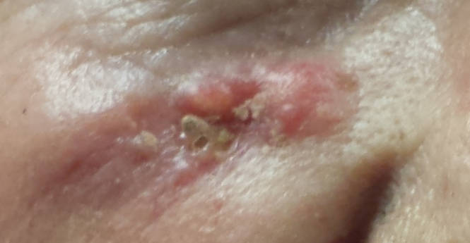
The lesion, almost 6 cm wide and more than 4 cm long, is located in the right infraorbital facial cheek area, well below the eyelid margin. It is focally eroded, peripherally erythematous, and quite firm to the touch. Fortunately, it is still mobile and not fixed to the underlying tissue. There are no palpable lymph nodes in the area and no change in sensation from one side of the face to the other.
A punch biopsy is performed under local infiltrate with 1% lidocaine with epinephrine. The 4-mm defect is closed with 2 x 5-0 nylon sutures and the specimen submitted to pathology. The report confirms the clinical suspicion of basal cell carcinoma (BCC).
Continue for Joe Monroe's discussion >>
DISCUSSION
When they are allowed to reach this size, BCCs can become a real problem. In extreme instances, they can erode into the face and even into bone. Patients can lose ears, noses, or even their entire face to this relentless but “safe” cancer. Given enough time and bad luck, BCC can metastasize to local lymph nodes and even to the brain or lung; it is even occasionally a cause of death.
Patients often ask why we have to remove BCCs if they’re usually “safe.” Without trying to scare them, we describe cases such as this one as the inevitable outcome of prolonged neglect and/or an aggressive tumor. BCCs almost never heal without treatment and almost always grow wider and deeper over time. Some are extremely indolent, taking 20 years to become noticeable, while others are more aggressive. In addition to the clinical behavior of any given BCC, there are also histologic clues to how aggressive a particular BCC might be.
This patient was referred to a Mohs surgeon, who will be able to do two things that require specialized training:
1. He’ll remove the biopsy-proven cancer with scalpel surgery, and while the patient is still present, check the margins for residual cancer. If the margins are positive, he’ll go back to the area and remove more, repeating this step until clear wide and deep microscopic margins are visualized by frozen section technique. (It’s important to note that the exact way the frozen specimen is processed and examined permits evaluation of the entire margin—top, bottom, sides—making it considerably different from ordinary frozen sections.)
2. Then, the Mohs surgeon will have the skill to close the defect in an acceptable way: usually by primary closure, sometimes with flaps or with grafts. All of this is typically done on an outpatient basis, on the same visit, although it may take most of a morning or afternoon.
Mohs surgery was pioneered in the 1930s by Frederic Mohs, MD, a general surgeon, as a way to address large and/or aggressive cancers located in difficult areas (eg, face or genitals). It is not indicated for ordinary, relatively small skin cancers on the arms, legs, or trunk.
Some BCCs and squamous cell carcinomas develop in especially difficult areas, such as the eyelids, or involve large areas of the ear. These lesions may require the attention of relevant surgical specialists, such as oculoplastic or head and neck surgeons.
This patient will undergo Mohs surgery in the near future, and her defect will probably be closed primarily. The depth and histologic markers indicating exceptional aggression may dictate a further step: Radiation therapy may be required to minimize the likelihood of recurrence. Her chances of a complete cure are 95% to 97% with Mohs surgery alone.
TAKE-HOME LEARNING POINTS
• Basal cell carcinomas (BCCs) are often mistaken for infection.
• BCCs almost never heal on their own, grow slowly but steadily, and can reach prodigious size and depth.
• BCCs can metastasize to local nodes or even to the brain and lung.
• Ordinary excision is adequate for most BCCs, but Mohs surgery is indicated for larger lesions located in difficult areas (eg, face, scalp or groin).
• Simple shave biopsy is adequate to diagnose most BCCs.
The lesion on the face of this 72-year-old woman has slowly grown over a period of years but has never caused pain. On several occasions, she has consulted primary care providers (PCPs), most of whom prescribed oral antibiotics for possible infection. However, these medications never helped.
She recently developed oral health problems and established care with a new PCP, who insisted she see a dermatologist about the facial lesion. The patient, who is “terrified” of needles, resisted this advice at first but finally agreed once her family got involved.
The patient reports a history of extensive sun exposure in childhood, when she worked in the fields, that continued into adulthood; she maintained a garden until recently. Her response to the sun is invariably a deep tan, which lasts all summer.
EXAMINATION
The lesion, almost 6 cm wide and more than 4 cm long, is located in the right infraorbital facial cheek area, well below the eyelid margin. It is focally eroded, peripherally erythematous, and quite firm to the touch. Fortunately, it is still mobile and not fixed to the underlying tissue. There are no palpable lymph nodes in the area and no change in sensation from one side of the face to the other.
A punch biopsy is performed under local infiltrate with 1% lidocaine with epinephrine. The 4-mm defect is closed with 2 x 5-0 nylon sutures and the specimen submitted to pathology. The report confirms the clinical suspicion of basal cell carcinoma (BCC).
Continue for Joe Monroe's discussion >>
DISCUSSION
When they are allowed to reach this size, BCCs can become a real problem. In extreme instances, they can erode into the face and even into bone. Patients can lose ears, noses, or even their entire face to this relentless but “safe” cancer. Given enough time and bad luck, BCC can metastasize to local lymph nodes and even to the brain or lung; it is even occasionally a cause of death.
Patients often ask why we have to remove BCCs if they’re usually “safe.” Without trying to scare them, we describe cases such as this one as the inevitable outcome of prolonged neglect and/or an aggressive tumor. BCCs almost never heal without treatment and almost always grow wider and deeper over time. Some are extremely indolent, taking 20 years to become noticeable, while others are more aggressive. In addition to the clinical behavior of any given BCC, there are also histologic clues to how aggressive a particular BCC might be.
This patient was referred to a Mohs surgeon, who will be able to do two things that require specialized training:
1. He’ll remove the biopsy-proven cancer with scalpel surgery, and while the patient is still present, check the margins for residual cancer. If the margins are positive, he’ll go back to the area and remove more, repeating this step until clear wide and deep microscopic margins are visualized by frozen section technique. (It’s important to note that the exact way the frozen specimen is processed and examined permits evaluation of the entire margin—top, bottom, sides—making it considerably different from ordinary frozen sections.)
2. Then, the Mohs surgeon will have the skill to close the defect in an acceptable way: usually by primary closure, sometimes with flaps or with grafts. All of this is typically done on an outpatient basis, on the same visit, although it may take most of a morning or afternoon.
Mohs surgery was pioneered in the 1930s by Frederic Mohs, MD, a general surgeon, as a way to address large and/or aggressive cancers located in difficult areas (eg, face or genitals). It is not indicated for ordinary, relatively small skin cancers on the arms, legs, or trunk.
Some BCCs and squamous cell carcinomas develop in especially difficult areas, such as the eyelids, or involve large areas of the ear. These lesions may require the attention of relevant surgical specialists, such as oculoplastic or head and neck surgeons.
This patient will undergo Mohs surgery in the near future, and her defect will probably be closed primarily. The depth and histologic markers indicating exceptional aggression may dictate a further step: Radiation therapy may be required to minimize the likelihood of recurrence. Her chances of a complete cure are 95% to 97% with Mohs surgery alone.
TAKE-HOME LEARNING POINTS
• Basal cell carcinomas (BCCs) are often mistaken for infection.
• BCCs almost never heal on their own, grow slowly but steadily, and can reach prodigious size and depth.
• BCCs can metastasize to local nodes or even to the brain and lung.
• Ordinary excision is adequate for most BCCs, but Mohs surgery is indicated for larger lesions located in difficult areas (eg, face, scalp or groin).
• Simple shave biopsy is adequate to diagnose most BCCs.
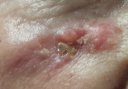
Young Patient With “Bumps” Near Her Mouth
A 12-year-old girl is referred by her pediatrician for evaluation and treatment of facial lesions. She’s had them for almost five years, and several treatment attempts have been unsuccessful. Most recently, she tried topical adapalene gel for presumed acne, but this only irritated the area and had no beneficial effect on the “bumps.” Oral antibiotics did not help, either.
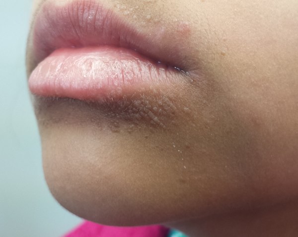
The presenting complaint is complicated somewhat by the fact that the same area of her skin is often dark—a problem thought to be related. The patient admits to licking her lips frequently (something her parents have also observed).
The patient’s history is significant for Hodgkin’s lymphoma, diagnosed and successfully treated five years ago. It was around that time that the bumps first appeared, while she was undergoing chemotherapy.
EXAMINATION
There is a ring of sharply demarcated, modest hyperpigmentation around the entire perioral area, extending outward about 2 cm. Closer questioning reveals that this aspect of the problem “comes and goes.”
The main concern is the collection of planar, pinkish brown papules around the mouth. These are mostly located inferior to the lips, although a few tiny papules are on the maxilla. All papules are uniform in size (≤ 1 mm) and shape. None are inflamed or umbilicated.
Continue for Joe Monroe's diagnosis and discussion >>
DISCUSSION
The HPV family has more than 100 distinct DNA types, several of which have particular morphologic characteristics and favored locations. Depending on the affected site, warts can assume many appearances, including planar (flat-topped) papules that generally favor the legs and face of a child or adolescent. Chemotherapy made this patient susceptible, and the lack of a diagnosis in the intervening years compounded the problem.
The perioral hyperpigmentation is, of course, secondary to her lip licking, which dries and irritates the thin facial skin. On a blond or red-haired, fair-skinned patient, it would appear pink or red. But with our patient’s type IV skin, inflammation begets darkening. The two problems are probably unrelated.
Treatment of flat warts on the face of a young woman is problematic, especially when dark skin is a factor. Treatments such as liquid nitrogen would not only be painful if applied to the face but could also cause permanent pigment changes. Irritants and acids (eg, cantharidin or salicylic acid) would have that same potential, so we tend to treat this area with the least invasive, least irritating modalities, knowing that the perfect treatment has yet to be invented.
A sinecatechins ointment —a botanical product made from the extract of green tea leaves—was prescribed. The patient was instructed to apply it three times per day for a month, then return to the clinic for re-evaluation. Treatment alternatives at that point will include imiquimod, which stimulates the skin to produce interferon (known to discourage viral growth) and 5-fluorouracil (5FU) cream, which has also been used with some success for warts. (For the record, I have no financial interest in any of these products.)
Chances are good that with time and treatment, the patient’s warts will disappear. It should be noted that the differential included acne, molluscum contagiosum, and perioral dermatitis. A simple shallow shave biopsy could have been performed to confirm the diagnosis, if necessary.
TAKE-HOME LEARNING POINTS
• The HPV family of warts contains more than 100 distinct genomic types, which can present in a wide variety of morphologies.
• Flat warts (verucca planae) are commonly seen on faces and legs but can appear in other locations. Shaving spreads them.
• No perfect treatment for warts exists. All have drawbacks, including potential for irritation, pain, and dyschromia—the latter a particular problem for patients with darker skin.
• For these patients, sinecatechins ointment, imiquimod, and 5-fluorouracil cream are decent choices.
A 12-year-old girl is referred by her pediatrician for evaluation and treatment of facial lesions. She’s had them for almost five years, and several treatment attempts have been unsuccessful. Most recently, she tried topical adapalene gel for presumed acne, but this only irritated the area and had no beneficial effect on the “bumps.” Oral antibiotics did not help, either.
The presenting complaint is complicated somewhat by the fact that the same area of her skin is often dark—a problem thought to be related. The patient admits to licking her lips frequently (something her parents have also observed).
The patient’s history is significant for Hodgkin’s lymphoma, diagnosed and successfully treated five years ago. It was around that time that the bumps first appeared, while she was undergoing chemotherapy.
EXAMINATION
There is a ring of sharply demarcated, modest hyperpigmentation around the entire perioral area, extending outward about 2 cm. Closer questioning reveals that this aspect of the problem “comes and goes.”
The main concern is the collection of planar, pinkish brown papules around the mouth. These are mostly located inferior to the lips, although a few tiny papules are on the maxilla. All papules are uniform in size (≤ 1 mm) and shape. None are inflamed or umbilicated.
Continue for Joe Monroe's diagnosis and discussion >>
DISCUSSION
The HPV family has more than 100 distinct DNA types, several of which have particular morphologic characteristics and favored locations. Depending on the affected site, warts can assume many appearances, including planar (flat-topped) papules that generally favor the legs and face of a child or adolescent. Chemotherapy made this patient susceptible, and the lack of a diagnosis in the intervening years compounded the problem.
The perioral hyperpigmentation is, of course, secondary to her lip licking, which dries and irritates the thin facial skin. On a blond or red-haired, fair-skinned patient, it would appear pink or red. But with our patient’s type IV skin, inflammation begets darkening. The two problems are probably unrelated.
Treatment of flat warts on the face of a young woman is problematic, especially when dark skin is a factor. Treatments such as liquid nitrogen would not only be painful if applied to the face but could also cause permanent pigment changes. Irritants and acids (eg, cantharidin or salicylic acid) would have that same potential, so we tend to treat this area with the least invasive, least irritating modalities, knowing that the perfect treatment has yet to be invented.
A sinecatechins ointment —a botanical product made from the extract of green tea leaves—was prescribed. The patient was instructed to apply it three times per day for a month, then return to the clinic for re-evaluation. Treatment alternatives at that point will include imiquimod, which stimulates the skin to produce interferon (known to discourage viral growth) and 5-fluorouracil (5FU) cream, which has also been used with some success for warts. (For the record, I have no financial interest in any of these products.)
Chances are good that with time and treatment, the patient’s warts will disappear. It should be noted that the differential included acne, molluscum contagiosum, and perioral dermatitis. A simple shallow shave biopsy could have been performed to confirm the diagnosis, if necessary.
TAKE-HOME LEARNING POINTS
• The HPV family of warts contains more than 100 distinct genomic types, which can present in a wide variety of morphologies.
• Flat warts (verucca planae) are commonly seen on faces and legs but can appear in other locations. Shaving spreads them.
• No perfect treatment for warts exists. All have drawbacks, including potential for irritation, pain, and dyschromia—the latter a particular problem for patients with darker skin.
• For these patients, sinecatechins ointment, imiquimod, and 5-fluorouracil cream are decent choices.
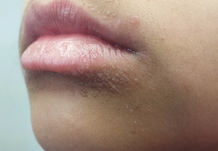
Red and Swollen Doesn’t Mean “Infection”
Six month ago, this 36-year-old man’s left fourth finger began to bother him. He’s tried topical antibiotics (triple-antibiotic cream and mupirocin), hot soaks in solutions of Epsom salts, application of colloidal silver solution, and courses of two different oral antibiotics (cephalexin and ciprofloxacin). None have provided any relief from the pain.
The patient admits removing a hangnail from the nail in question, causing a little bleeding. Then followed the slow onset of chronic pain and swelling, which has never been severe but is painful enough to interfere with normal activities—particularly his job, which requires extensive computer time.
EXAMINATION
The medial perionychial area of his left fourth finger is modestly swollen and red and markedly tender to touch. No pus can be expressed from the area, and there are no palpable lymph nodes in either the epitrochlear or axillary locations of this arm. No lymphangiitic streaking is seen on the hand or arm. The affected area is a small section of the nail fold, where glistening, friable tissue is seen in the proximal invaginated area.
After a brief discussion of treatment alternatives, the decision is made to anesthetize the digit by means of a digital block, using 2 cc of 2% plain lidocaine. Once anesthesia is achieved, the hand is elevated above the chest and a tourniquet applied to minimize bleeding. The back of the patient’s hand is placed on his chest, then the swollen nail fold is pulled back. A 3-mm remnant of hangnail, still attached proximally, is revealed. It is removed and the area curetted. The site is cleaned and dressed, then the tourniquet is released.
Within 2 weeks, the finger is no longer swollen or painful.
Continue for Joe Monroe's discussion and take-home learning points >>
DISCUSSION
“Infection” is only one potential cause of redness, swelling, increased warmth, and localized pain. Classically termed rubor, tumor, calor, and dolor, these are indicators of inflammation, which can occur in many conditions besides “infection.” One tragic example is inflammatory breast cancer, which is all too often mistreated as mastitis until the lack of response to treatment finally forces—often belatedly—consideration of other items in the differential.
A far less dramatic example is the case described, in which a simple hangnail is incompletely removed, leaving a shard of nail that then digs into the perionychial skin as it grows out. This sets into motion a healing process that cannot proceed to resolution, because the tissue is re-injured every time the finger strikes the computer keyboard. This not only causes the wound to get stuck in a certain phase of healing (angioneogenesis) but also prevents completion of the process. The tissue’s response is what we see with this patient, invariably (and erroneously) called “infection.”
This is basically the identical process we see with ingrown toenails, except for the unfortunate fact that we stand upright, compress the toe with a shoe, and walk on it—all of which greatly magnify the pain, redness, and swelling. With toes more than fingers, we also tend to see the production of a button of granulated tissue at the site. This results from ongoing, inappropriate angioneogenesis. Sometimes termed pyogenic granuloma (or sclerosing hemangioma), this tissue is quite friable and bleeds copiously with any amount of trauma.
Ironically, acute bacterial paronychia of the fingers, usually caused by ordinary staph, can start in much the same way (without, of course, the retained nail shard). However, it presents with more focal concentration of redness and swelling, a collection of thick, green pus, and exquisite tenderness, all of which is relieved by simple incision and drainage.
With this patient, and those with ingrown toenails, it’s quite compelling to prescribe oral antibiotics. But these never help, and for good reason: The problem is intolerance of the “foreign body,” not infection. The solution is to “reboot” the healing process by removing the offending nail shard.
TAKE-HOME LEARNING POINTS
• While “ingrown fingernails” are far less common than ingrown toenails, both are caused by nail fragments slicing into live tissue.
• The cure is to remove the offending fragment, which allows the wound to heal.
• For ingrown toenails, an extra step is often necessary: destroying the offending segment of nail matrix with curettement and/or application of phenol.
• Anesthesia for digits should never be accomplished by local infiltration of the affected area for these kinds of procedures. Instead, employ a digital block technique, which is far less painful and provides complete anesthesia when done properly.
• Resist the urge to reflexively diagnose “infection” when confronted with redness, swelling, etc. Instead, consider other potential causes for inflammation first.
A Journey Into the Patient's Mouth
An 18-year-old boy is brought by his parents for evaluation of several relatively minor skin problems, including warts, moles, and skin tags. The patient’s history is notable for multiple as-yet-unexplained bony fractures that have occurred with minimal trauma since he was born. He has a brother who was diagnosed with osteogenesis imperfecta, yet, despite an extensive workup, the patient’s constellation of findings has thus far failed to corroborate that or any other definitive diagnosis.
As an afterthought, the parents also ask about changes they have noted on the patient's gums.
There is no history of seizures, and the patient takes no medication regularly.
EXAMINATION
The potential connection to a heritable syndrome draws attention to the patient’s teeth and gums. The latter appear to be overgrown and, as such, far more prominent than normal. They also appear a bit inflamed, especially the portion of the gingiva bordering the teeth. Advanced gingival recession is noted in some of these locations. Closer examination reveals extensive plaque formation on the adjacent teeth.
During the exam, the patient is observed to be a mouth-breather. His parents corroborate this fact.
Continue for Joe Monroe's discussion and learning points >>
DISCUSSION
The most common cause of gingival overgrowth (GO) is thought to be gingivitis, itself a result of inflammation caused by dental plaque and plaque-associated bacteria. The obvious gingivitis, gingival recession, and extensive dental plaque seen in this patient are more common in younger, mouth-breathing persons. Higher levels of growth hormone might account for the gingival growth, while the dehydration of teeth and gums promoted by mouth-breathing appears to exacerbate gingivitis.
Formerly called gingival hyperplasia, the term gingival overgrowth has been posited as more inclusive of all forms of such findings. Advocates of this change in terminology point out that confirmation of the diagnosis of gingival hypertrophy (an increase in the size of individual cells) or gingival hyperplasia (an increase in the number of cells) would require biopsy and microscopic examination.
Three major categories of potential causes of GO are commonly described in the literature. They include (in descending order of frequency):
1. Inflammation: such as that described, with apparent connection with gingivitis
2. Drugs: A number of medications have been associated with GO, including anticonvulsants such as phenytoin (approximately 50% of cases), calcium channel blockers such as nifedipine (20%), and immunosuppressants such as cyclosporine (30%).
3. Systemic causes: These include pregnancy, puberty, vitamin C deficiency, leukemia, and granulomatous diseases such as Wegener granulomatosis. It can also manifest as a perineoplastic sign (associated with known or occult cancers).
Treatment, of course, depends on the cause. Even when drugs are to blame, however, co-existing gingivitis can be a contributing factor; it can be addressed with plaque scaling, better dental care, or even gingivectomy. Drugs can be changed or eliminated or doses can be reduced. Other, less common systemic conditions associated with GO can be sought through additional history, laboratory work, and more extensive examination.
TAKE-HOME LEARNING POINTS
• The term gingival hypertrophy is being replaced by the more inclusive gingival overgrowth (GO).
• The most common cause of GO is chronic gingivitis.
• Drug classes associated with GO include anticonvulsants, calcium channel blockers, and immunosuppressants.
• Systemic diseases can trigger GO; these include pregnancy, puberty, and cancer (eg, leukemia).
• Addressing oral hygiene is a good place to start treatment of GO, regardless of the trigger, since it often co-exists with other causation.

Appearing and Disappearing Scratch Marks
A 41-year-old man complains of “red lines” that appear on his skin soon after minor scratching trauma. They manifest rapidly but disappear within minutes (occasionally as long as an hour), leaving no trace. They cause no symptoms but are nonetheless disturbing to the patient.
In terms of his overall health, the patient has few problems beyond mild seasonal allergies. However, he reports experiencing a great deal of stress recently. He denies taking any prescription medications regularly, though in the spring, he often takes OTC antihistamines to ward off sneezing and itchy eyes.
EXAMINATION
A linear wheal appears after the skin is scratched with the dull edge of a fingernail. The initial reaction to the scratch is a macular red flare, followed within seconds by a broadening band of erythema. Seconds later, the frank wheal appears. The lesion is warmer than the surrounding skin but not at all tender. As expected, it clears completely within a few minutes, leaving no sign of its ever having been there.
On the next page: Diagnosis and Joe Monroe's Discussion >>
DISCUSSION
This form of urticaria is called dermatographism (literally, “skin writing”) and is of unknown origin. It affects 2% to 5% of the population. Most affected patients are asymptomatic, although perhaps 10% of them will report itching and burning. This condition exhibits neither gender nor race bias.
Dermatographism is the most common form of the physical urticarias, which can also be caused by pressure, cold, and vibration. The speed with which the wheal forms is remarkable and unique to histamine-mediated conditions, though the exact mechanism is debatable. (Its three-stage formation is collectively referred to as the triple response of Lewis.) One theory holds that the trauma of scratching provokes an antigen to interact with mast cell bound IgE, triggering the release of inflammatory mediators (eg, histamine, bradykinins, or leukotrienes). Bolstering this theory is the observation that 75% of patients with hypereosinophilic syndrome have dermatographism.
Dermatographism effectively confirms the presence of traditional urticaria, itself more common in atopic patients. The rapidity of the appearance and disappearance of urticaria, a phenomenon termed evanescence, often presents a challenge to diagnosis until it is confirmed.
Most patients with dermatographism first note it in their second to third decade of life. When it’s congenital, symptomatic dermatographism suggests the possibility of a mast cell disorder, such as urticaria pigmentosa or systemic mastocytosis. These can be confirmed by biopsy and/or measurement of histamine in a 24-hour urine sample.
TAKE-HOME LEARNING POINTS
• Dermatographism is an extremely common form of physical urticaria.
• It is asymptomatic in the majority of cases and disappears on its own in minutes to hours.
• It can herald the presence of more serious chronic symptomatic urticaria.
• The rapid appearance of the linear wheal and its subsequent rapid disappearance, termed evanescence, are unique to urticarial disorders.
• The phenomena by which the initial red linear flare widens quickly before forming the evanescent linear wheal are collectively known as the “triple response of Lewis.”