User login
ESC: Obstructive sleep apnea often complicates heart failure
LONDON – The majority of patients with severe heart failure had sleep-disordered breathing and, in most affected patients, this manifested as obstructive sleep apnea, in an analysis of more than 1,000 German heart failure patients enrolled in a multicenter registry.
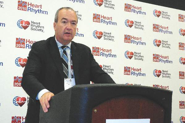
“The vast majority of heart failure patients with sleep-disordered breathing [SDB] have obstructive sleep apnea, which differs from previous results,” said Dr. Olaf Oldenburg at the annual congress of the European Society of Cardiology. Possible reasons why this German registry had different findings, compared with prior reports, were its inclusion of heart failure patients with milder symptoms, inclusion of patients with preserved ejection fraction, and inclusion of more women, suggested Dr. Oldenburg, director of the sleep laboratory at the Heart and Diabetes Center of Ruhr University of Bochum in Bad Oeynhausen, Germany.

His finding that nearly two-thirds of the heart failure patients with SDB in this registry had obstructive sleep apnea and that one-third had moderate or severe obstructive sleep apnea was notable because this remains a form of sleep-disordered breathing that can be treated, he said. “There is still enough evidence to treat obstructive sleep apnea” in heart failure patients when it has a moderate or severe presentation, which is defined as causing 15 or more apnea-hypopnea events/hour during sleep. “Obstructive sleep apnea is definitely not a compensatory mechanism in heart failure,” Dr. Oldenburg said.
He highlighted the ongoing need to treat more severe obstructive sleep apnea in heart failure patients because this form of SDB sharply contrasts with the results of the SERVE-HF trial, also reported at the congress, which showed that in patients with advanced heart failure and central sleep apnea nocturnal treatment with adaptive servo ventilation failed to provide benefit and also appeared to boost patient mortality (N Engl J Med. 2015 Sep 17;373[12]:1095-105).
Following that report “we need to think about which heart failure patients to treat” with nocturnal ventilation, and differentiate between heart failure patients with obstructive sleep apnea and those with central sleep apnea, Dr. Oldenburg said.
The data he reported came from the SchlaHF-XT (Sleep Disordered Breathing in Heart Failure) registry, which enrolled patients with heart failure and reduced or preserved ejection fraction and any New York Heart Association functional class treated either at German hospitals or in physician offices. He reported data for 1,186 fully assessed and classified patients, who averaged 68 years old and two-thirds of whom were men. Slightly more than half had heart failure with reduced ejection fraction, and about half had New York Heart Association class II heart failure, a quarter had class III heart failure, with the remaining patients divided roughly equally between class I and IV.
Screening for SDB showed that 24% had no SDB, 37% had mild SBD, 21% had moderate SDB, and 19% had severe SDB (percentages total 101% because of rounding). Among those with SDB, 64% had obstructive sleep apnea, 22% had central sleep apnea, and the remaining 14% had either a mixed form of sleep apnea or were not classifiable.
The analysis also showed that moderate and severe SDB, the forms that require treatment, occurred more often among patients with heart failure and reduced ejection fraction, 43%, compared with patients with heart failure and preserved ejection fraction, who had a 36% prevalence of SDB requiring treatment. Moderate or severe central sleep apnea occurred in 15% of patients with reduced ejection fraction and in 9% of patients with preserved ejection fraction.
A second report at the congress by Dr. Oldenburg showed that the duration of time when a patient’s oxygen saturation fell below 90% was a better gauge of the severity of SDB than was the traditional measure of the apnea-hypopnea index (AHI), the average number of apnea-hypopnea episodes a patient has during an hour of sleep. For this analysis, he used data collected on 963 patients with chronic, stable heart failure with reduced ejection fraction who underwent a comprehensive sleep study with pulse oximetry measurements during 2002-2013.
The results showed that while the measured AHI significantly linked with the 5-year mortality rate of these patients, the relationship became statistically insignificant after researchers adjusted for age, sex, body mass index, heart failure severity, ejection fraction, medications, and other clinical variables.
In contrast, the average time a patient spent with an oxygen saturation level below 90% overnight strong linked with 5-year mortality even after adjusting for all these covariables. The analysis showed that each hour of sleep a heart failure patient spent with an oxygen saturation level below 90% linked with a relative 16% reduction in 5-year survival. Patients in the quartile with the greatest amount of time spent with a oxygen saturation level below 90% had a 50% 5-year mortality rate, while those in the quartile with the least amount of time spent with severely depressed oxygen saturation had a 30% 5-year mortality rate.
Based on this finding “you need to look at the effect of SDB and not just the apnea-hypopnea index” when assessing SDB in patients with heart failure, Dr. Oldenburg said.
On Twitter @mitchelzoler
LONDON – The majority of patients with severe heart failure had sleep-disordered breathing and, in most affected patients, this manifested as obstructive sleep apnea, in an analysis of more than 1,000 German heart failure patients enrolled in a multicenter registry.
“The vast majority of heart failure patients with sleep-disordered breathing [SDB] have obstructive sleep apnea, which differs from previous results,” said Dr. Olaf Oldenburg at the annual congress of the European Society of Cardiology. Possible reasons why this German registry had different findings, compared with prior reports, were its inclusion of heart failure patients with milder symptoms, inclusion of patients with preserved ejection fraction, and inclusion of more women, suggested Dr. Oldenburg, director of the sleep laboratory at the Heart and Diabetes Center of Ruhr University of Bochum in Bad Oeynhausen, Germany.
His finding that nearly two-thirds of the heart failure patients with SDB in this registry had obstructive sleep apnea and that one-third had moderate or severe obstructive sleep apnea was notable because this remains a form of sleep-disordered breathing that can be treated, he said. “There is still enough evidence to treat obstructive sleep apnea” in heart failure patients when it has a moderate or severe presentation, which is defined as causing 15 or more apnea-hypopnea events/hour during sleep. “Obstructive sleep apnea is definitely not a compensatory mechanism in heart failure,” Dr. Oldenburg said.
He highlighted the ongoing need to treat more severe obstructive sleep apnea in heart failure patients because this form of SDB sharply contrasts with the results of the SERVE-HF trial, also reported at the congress, which showed that in patients with advanced heart failure and central sleep apnea nocturnal treatment with adaptive servo ventilation failed to provide benefit and also appeared to boost patient mortality (N Engl J Med. 2015 Sep 17;373[12]:1095-105).
Following that report “we need to think about which heart failure patients to treat” with nocturnal ventilation, and differentiate between heart failure patients with obstructive sleep apnea and those with central sleep apnea, Dr. Oldenburg said.
The data he reported came from the SchlaHF-XT (Sleep Disordered Breathing in Heart Failure) registry, which enrolled patients with heart failure and reduced or preserved ejection fraction and any New York Heart Association functional class treated either at German hospitals or in physician offices. He reported data for 1,186 fully assessed and classified patients, who averaged 68 years old and two-thirds of whom were men. Slightly more than half had heart failure with reduced ejection fraction, and about half had New York Heart Association class II heart failure, a quarter had class III heart failure, with the remaining patients divided roughly equally between class I and IV.
Screening for SDB showed that 24% had no SDB, 37% had mild SBD, 21% had moderate SDB, and 19% had severe SDB (percentages total 101% because of rounding). Among those with SDB, 64% had obstructive sleep apnea, 22% had central sleep apnea, and the remaining 14% had either a mixed form of sleep apnea or were not classifiable.
The analysis also showed that moderate and severe SDB, the forms that require treatment, occurred more often among patients with heart failure and reduced ejection fraction, 43%, compared with patients with heart failure and preserved ejection fraction, who had a 36% prevalence of SDB requiring treatment. Moderate or severe central sleep apnea occurred in 15% of patients with reduced ejection fraction and in 9% of patients with preserved ejection fraction.
A second report at the congress by Dr. Oldenburg showed that the duration of time when a patient’s oxygen saturation fell below 90% was a better gauge of the severity of SDB than was the traditional measure of the apnea-hypopnea index (AHI), the average number of apnea-hypopnea episodes a patient has during an hour of sleep. For this analysis, he used data collected on 963 patients with chronic, stable heart failure with reduced ejection fraction who underwent a comprehensive sleep study with pulse oximetry measurements during 2002-2013.
The results showed that while the measured AHI significantly linked with the 5-year mortality rate of these patients, the relationship became statistically insignificant after researchers adjusted for age, sex, body mass index, heart failure severity, ejection fraction, medications, and other clinical variables.
In contrast, the average time a patient spent with an oxygen saturation level below 90% overnight strong linked with 5-year mortality even after adjusting for all these covariables. The analysis showed that each hour of sleep a heart failure patient spent with an oxygen saturation level below 90% linked with a relative 16% reduction in 5-year survival. Patients in the quartile with the greatest amount of time spent with a oxygen saturation level below 90% had a 50% 5-year mortality rate, while those in the quartile with the least amount of time spent with severely depressed oxygen saturation had a 30% 5-year mortality rate.
Based on this finding “you need to look at the effect of SDB and not just the apnea-hypopnea index” when assessing SDB in patients with heart failure, Dr. Oldenburg said.
On Twitter @mitchelzoler
AT THE ESC CONGRESS 2015
Key clinical point: Significant sleep disordered breathing is common among heart failure patients, most often manifesting as obstructive sleep apnea.
Major finding: In a real-world population of heart failure patients 64% of those with sleep disordered breathing had obstructive sleep apnea.
Data source: The SchlaHF-XT registry with 1,1816 heart failure patients enrolled at several German hospitals and private practices.
Disclosures: The SchlaHF-XT registry was sponsored by ResMed. Dr. Oldenburg said that he has received research support from several companies.

ESC: Registry confirms liver dysfunction’s heart-failure importance
LONDON – Hepatic dysfunction, which is beginning to be appreciated as an important complication of patients with acute heart failure, boosted the relative rate of death in patients with acute heart failure by 57% in an analysis of more than 5,000 patients followed for a year in a registry maintained by the European Society of Cardiology (ESC).
The registry data also showed that at entry into the registry the acute heart failure patients had an 8% prevalence of hepatic dysfunction.
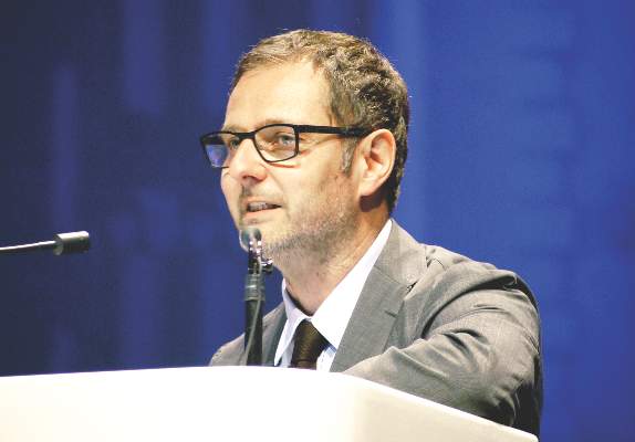
These observations on the prevalence and impact of liver dysfunction in acute heart failure patients highlight a new recognition that acute heart failure can trigger morbidity and mortality through its hepatic effects in a manner similar to the better-appreciated effect that acute heart failure has on renal and pulmonary function, Dr. Alexandre Mebazaa said at the annual congress of the European Society of Cardiology.

“Everybody knows about the kidney, but not many know about the liver,” said Dr. Mebazaa in an interview. Recent studies on mechanisms of organ damage during acute heart failure episodes indicates that the liver, kidneys, and lungs are all damaged by fluid congestion in these organs, he said. “Fluid overload leads to organ dysfunction,” and this complication seems relatively resistant to the diuretic treatment that acute heart failure patients typically receive when they are hospitalized for decompensation episodes. “New approaches are needed to remove fluid from the organs. Diuretics remove fluid from the blood, but not from the organs,” said Dr. Mebazaa, a professor of anesthesiology and critical care medicine at Lariboisière Hospital in Paris.
The data Dr. Mebazaa reported at the congress were the first 1-year follow-up data from the ESC’s Heart Failure Long-Term Registry, begun in 2012 and involving centers from more than 30 countries in Europe as well as in North Africa and the Middle East. The panel of ESC members who oversee this registry “selected centers dedicated to the database,” he said.
The registry followed 5,039 patients with acute heart failure for at least 1 year, as well as 7,401 patients diagnosed with chronic heart failure. Another striking, though not surprising finding of the 1-year follow-up data was the disparity in mortality and hospitalization rates between these two subgroups.
After 1 year, the mortality rate was 24% among the acute heart failure patients, compared with 6% among the chronic heart failure patients. Dr. Mebazaa called the acute heart failure mortality rate “terrible.”
Hospitalization rates in the two subgroups over the 1-year follow-up ran to 19% among the acute heart failure patients ands 10% for chronic heart failure patients. The combined rate of death or hospitalization over 1 year was 36% in the acute heart failure patients and 15% in those with chronic heart failure.
These data “show that we really need new treatments for acute heart failure to reduce rehospitalizations and deaths,” Dr. Mebazaa said.
A multivariate adjusted analysis identified several baseline factors that significantly linked with mortality among the acute heart-failure patients during the 1-year follow-up. In addition to hepatic dysfunction linking with a 57% increased rate of death other factors included age, which linked with a 24% increased rate for every 5 years of older age; New York Heart Association class III or IV severity, which linked with 50% higher mortality; renal dysfunction, which linked with a 52% higher mortality rate; and aortic stenosis, linked with a 54% increased mortality rate.
The finding that hepatic dysfunction was an independent mortality risk in acute heart failure confirmed a finding that Dr. Mebazaa and his associates reported in 2013 in a post hoc analysis of 1,134 patients with acute heart failure who had been enrolled in a drug-intervention trial (Eur Heart J. 2013 Sep 15;34:742-9). The registry results confirm for the first time that finding in an independent and much larger group of patients, Dr. Mebazaa said.
On Twitter @mitchelzoler
LONDON – Hepatic dysfunction, which is beginning to be appreciated as an important complication of patients with acute heart failure, boosted the relative rate of death in patients with acute heart failure by 57% in an analysis of more than 5,000 patients followed for a year in a registry maintained by the European Society of Cardiology (ESC).
The registry data also showed that at entry into the registry the acute heart failure patients had an 8% prevalence of hepatic dysfunction.
These observations on the prevalence and impact of liver dysfunction in acute heart failure patients highlight a new recognition that acute heart failure can trigger morbidity and mortality through its hepatic effects in a manner similar to the better-appreciated effect that acute heart failure has on renal and pulmonary function, Dr. Alexandre Mebazaa said at the annual congress of the European Society of Cardiology.
“Everybody knows about the kidney, but not many know about the liver,” said Dr. Mebazaa in an interview. Recent studies on mechanisms of organ damage during acute heart failure episodes indicates that the liver, kidneys, and lungs are all damaged by fluid congestion in these organs, he said. “Fluid overload leads to organ dysfunction,” and this complication seems relatively resistant to the diuretic treatment that acute heart failure patients typically receive when they are hospitalized for decompensation episodes. “New approaches are needed to remove fluid from the organs. Diuretics remove fluid from the blood, but not from the organs,” said Dr. Mebazaa, a professor of anesthesiology and critical care medicine at Lariboisière Hospital in Paris.
The data Dr. Mebazaa reported at the congress were the first 1-year follow-up data from the ESC’s Heart Failure Long-Term Registry, begun in 2012 and involving centers from more than 30 countries in Europe as well as in North Africa and the Middle East. The panel of ESC members who oversee this registry “selected centers dedicated to the database,” he said.
The registry followed 5,039 patients with acute heart failure for at least 1 year, as well as 7,401 patients diagnosed with chronic heart failure. Another striking, though not surprising finding of the 1-year follow-up data was the disparity in mortality and hospitalization rates between these two subgroups.
After 1 year, the mortality rate was 24% among the acute heart failure patients, compared with 6% among the chronic heart failure patients. Dr. Mebazaa called the acute heart failure mortality rate “terrible.”
Hospitalization rates in the two subgroups over the 1-year follow-up ran to 19% among the acute heart failure patients ands 10% for chronic heart failure patients. The combined rate of death or hospitalization over 1 year was 36% in the acute heart failure patients and 15% in those with chronic heart failure.
These data “show that we really need new treatments for acute heart failure to reduce rehospitalizations and deaths,” Dr. Mebazaa said.
A multivariate adjusted analysis identified several baseline factors that significantly linked with mortality among the acute heart-failure patients during the 1-year follow-up. In addition to hepatic dysfunction linking with a 57% increased rate of death other factors included age, which linked with a 24% increased rate for every 5 years of older age; New York Heart Association class III or IV severity, which linked with 50% higher mortality; renal dysfunction, which linked with a 52% higher mortality rate; and aortic stenosis, linked with a 54% increased mortality rate.
The finding that hepatic dysfunction was an independent mortality risk in acute heart failure confirmed a finding that Dr. Mebazaa and his associates reported in 2013 in a post hoc analysis of 1,134 patients with acute heart failure who had been enrolled in a drug-intervention trial (Eur Heart J. 2013 Sep 15;34:742-9). The registry results confirm for the first time that finding in an independent and much larger group of patients, Dr. Mebazaa said.
On Twitter @mitchelzoler
AT THE ESC CONGRESS 2015
Key clinical point: Liver dysfunction was a statistically significant, independent predictor of 1-year mortality in acute heart-failure patients.
Major finding: Acute heart-failure patients had a 57% relative increased rate of 1-year death, compared with patients without liver dysfunction.
Data source: One-year follow-up of 5,039 patients with acute heart failure enrolled in a multinational registry maintained by the European Society of Cardiology.
Disclosures: Dr. Mebazaa has received speaking honoraria and consulting fees from 11 drug or diagnostic companies.

First MRI-compatible implantable defibrillator approved
The Food and Drug Administration approved the first MRI-compatible implantable cardioverter defibrillator on Sept. 14, starting a new era of convenience and flexibility when performing MRI scans on patients who carry this type of cardiac implant.
Eventually most, if not all, implantable cardioverter defibrillators (ICDs) will have MRI compatibility, electrophysiologists predicted, a change that’s already been occurring for pacemakers following FDA approval of the first MRI-compatible pacemaker in 2011.

“This is a major step forward and sets a new standard” for ICDs, commented Dr. Rod S. Passman, professor of medicine and electrophysiologist at Northwestern University, Chicago, who has not been involved in developing MRI-compatible ICDs. “If a patient has an ICD, they should be able to go to any community emergency room and get what could be a life-saving MRI. MRI scans of patients with ICDs should not be limited to experienced academic centers. Ultimately all [cardiac] devices will be MRI compatible,” Dr. Passman said in an interview.
“There is no downside” to the newly approved, MRI-compatible ICD, said Dr. Michael R. Gold, an electrophysiologist at the Medical University of South Carolina, Charleston, who led the pivotal study that showed the device’s safety and efficacy during and after MRI scanning. Dr. Gold first reported results from the Evera MRI Study at the Heart Rhythm Society annual meeting in May and in a concurrently published report (J Am Soc Cardiol. 2015;65[24]:2581-8). The study enrolled 275 patients at 42 centers. Medtronic, the company that makes the newly approved ICD, plans to begin U.S. sales the week of Sept. 20, a company spokesperson said.
“I’m not sure I’d use it in all patients” who need an ICD once it’s on the market, admitted Dr. Gold. For example, some patients already have ICD leads in place that are not MRI compatible, so placing an ICD capable of MRI exposure in such patients would be moot, he noted. In other cases, the patient might best receive an ICD model made by a different manufacturer because of other device features.
“Every physician will need to choose the ICD that is best for each patient, so I don’t think we’ll see immediate, wholesale adoption [of the MRI-compatible ICD], but I expect the field will move in this direction,” Dr. Passman said.
Dr. Gold agreed that, as time goes by, the ICD models sold for U.S. patients increasingly will be MRI compatible, although that might take several years to happen.
For example, the transition to pacemakers that are MRI compatible has been gradual and incomplete, even though the first of these came onto the U.S. market in 2011.
“Only two pacemaker manufacturers sell MRI-compatible devices in the United States,” noted Dr. Gold, professor of medicine and director of the division of adult cardiology at the university. Several other manufacturers produce MRI-compatible pacemakers, but so far they have not sought FDA approval for these and they only sell them outside the United States, he noted.
“Many physicians don’t think about a patient’s long-term needs [for MRI] and may instead focus on the device they are most comfortable with” or a device with other attractive features, Dr. Passman said.
The possibility of performing MRI on a patient with an ICD is not totally new. A relatively small number of sophisticated U.S. centers have been performing MRIs on patients with conventional ICDs or pacemakers for several years, especially in circumstances when the MRI was considered vitally needed. Some of these centers have participated in the MagnaSafe registry, which reported results documenting the safety and efficacy of the procedure at a cardiology meeting in 2013.
“If you take certain precautions, the risk from MRI appears to be quite low in the most experienced hands, although even in experienced hands adverse events have been reported,” said Dr. Passman. Performing an MRI on a patient with a conventional ICD or pacemaker is also a relatively labor-intensive process that requires temporarily reprogramming the device, closely monitoring the patient during the MRI scan, and then checking out the device thoroughly after the scan to make sure it is functioning correctly.
In addition to the extra labor and uncertainty about outcome, running an MRI scan on a conventional cardiac device is generally not reimbursed by insurers and creates medicolegal exposure, Dr. Gold noted. “Even though it can be done, it often is not done, and it clearly compromises patient care,” he said.
Although the Medtronic unit was the first to get FDA approval, a competitor model seems on track to also hit the U.S. market soon. Two different ICD models made by Biotronik showed safety and efficacy in a study with 153 patients (Heart Rhythm. 2015 doi. org/10.1016/j.hrthm.2015.06.002). Biotronik has submitted an application to the FDA to market these ICDs and associated leads as MRI compatible, and an agency decision is pending, a company spokeswoman said.
The Evera MRI Study was sponsored by Medtronic, which produces and will market the Evera ICD. Dr. Gold has been a consultant to and has received research and speaker funding from Medtronic as well as from Boston Scientific and St. Jude. Dr. Passman has received research support from Medtronic but not for work on ICDs.
On Twitter @mitchelzoler
The Food and Drug Administration approved the first MRI-compatible implantable cardioverter defibrillator on Sept. 14, starting a new era of convenience and flexibility when performing MRI scans on patients who carry this type of cardiac implant.
Eventually most, if not all, implantable cardioverter defibrillators (ICDs) will have MRI compatibility, electrophysiologists predicted, a change that’s already been occurring for pacemakers following FDA approval of the first MRI-compatible pacemaker in 2011.
“This is a major step forward and sets a new standard” for ICDs, commented Dr. Rod S. Passman, professor of medicine and electrophysiologist at Northwestern University, Chicago, who has not been involved in developing MRI-compatible ICDs. “If a patient has an ICD, they should be able to go to any community emergency room and get what could be a life-saving MRI. MRI scans of patients with ICDs should not be limited to experienced academic centers. Ultimately all [cardiac] devices will be MRI compatible,” Dr. Passman said in an interview.
“There is no downside” to the newly approved, MRI-compatible ICD, said Dr. Michael R. Gold, an electrophysiologist at the Medical University of South Carolina, Charleston, who led the pivotal study that showed the device’s safety and efficacy during and after MRI scanning. Dr. Gold first reported results from the Evera MRI Study at the Heart Rhythm Society annual meeting in May and in a concurrently published report (J Am Soc Cardiol. 2015;65[24]:2581-8). The study enrolled 275 patients at 42 centers. Medtronic, the company that makes the newly approved ICD, plans to begin U.S. sales the week of Sept. 20, a company spokesperson said.
“I’m not sure I’d use it in all patients” who need an ICD once it’s on the market, admitted Dr. Gold. For example, some patients already have ICD leads in place that are not MRI compatible, so placing an ICD capable of MRI exposure in such patients would be moot, he noted. In other cases, the patient might best receive an ICD model made by a different manufacturer because of other device features.
“Every physician will need to choose the ICD that is best for each patient, so I don’t think we’ll see immediate, wholesale adoption [of the MRI-compatible ICD], but I expect the field will move in this direction,” Dr. Passman said.
Dr. Gold agreed that, as time goes by, the ICD models sold for U.S. patients increasingly will be MRI compatible, although that might take several years to happen.
For example, the transition to pacemakers that are MRI compatible has been gradual and incomplete, even though the first of these came onto the U.S. market in 2011.
“Only two pacemaker manufacturers sell MRI-compatible devices in the United States,” noted Dr. Gold, professor of medicine and director of the division of adult cardiology at the university. Several other manufacturers produce MRI-compatible pacemakers, but so far they have not sought FDA approval for these and they only sell them outside the United States, he noted.
“Many physicians don’t think about a patient’s long-term needs [for MRI] and may instead focus on the device they are most comfortable with” or a device with other attractive features, Dr. Passman said.
The possibility of performing MRI on a patient with an ICD is not totally new. A relatively small number of sophisticated U.S. centers have been performing MRIs on patients with conventional ICDs or pacemakers for several years, especially in circumstances when the MRI was considered vitally needed. Some of these centers have participated in the MagnaSafe registry, which reported results documenting the safety and efficacy of the procedure at a cardiology meeting in 2013.
“If you take certain precautions, the risk from MRI appears to be quite low in the most experienced hands, although even in experienced hands adverse events have been reported,” said Dr. Passman. Performing an MRI on a patient with a conventional ICD or pacemaker is also a relatively labor-intensive process that requires temporarily reprogramming the device, closely monitoring the patient during the MRI scan, and then checking out the device thoroughly after the scan to make sure it is functioning correctly.
In addition to the extra labor and uncertainty about outcome, running an MRI scan on a conventional cardiac device is generally not reimbursed by insurers and creates medicolegal exposure, Dr. Gold noted. “Even though it can be done, it often is not done, and it clearly compromises patient care,” he said.
Although the Medtronic unit was the first to get FDA approval, a competitor model seems on track to also hit the U.S. market soon. Two different ICD models made by Biotronik showed safety and efficacy in a study with 153 patients (Heart Rhythm. 2015 doi. org/10.1016/j.hrthm.2015.06.002). Biotronik has submitted an application to the FDA to market these ICDs and associated leads as MRI compatible, and an agency decision is pending, a company spokeswoman said.
The Evera MRI Study was sponsored by Medtronic, which produces and will market the Evera ICD. Dr. Gold has been a consultant to and has received research and speaker funding from Medtronic as well as from Boston Scientific and St. Jude. Dr. Passman has received research support from Medtronic but not for work on ICDs.
On Twitter @mitchelzoler
ESC: Noncardiac surgery in HCM patients warrants special attention
LONDON – Hypertrophic cardiomyopathy patients undergoing noncardiac surgery posted significantly worse 30-day composite outcomes than did closely matched controls undergoing the same sorts of surgical procedures.
“Our recommendation is that when hypertrophic cardiomyopathy patients need noncardiac surgery they should be evaluated and treated at an experienced center,” Dr. Milind Y. Desai concluded at the annual congress of the European Society of Cardiology.

There is a dearth of data on outcomes of noncardiac surgery in patients with hypertrophic cardiomyopathy (HCM). This was the impetus for Dr. Desai and his coinvestigators at the Cleveland Clinic to conduct a case-control study involving 92 consecutive adults with HCM undergoing intermediate- or high-cardiovascular-risk noncardiac surgery and 184 controls matched for age, gender, and type of surgery. Enrollment was restricted to HCM patients who hadn’t previously undergone septal myectomy or alcohol ablation.
The primary outcome was the 30-day composite of postoperative death, MI, stroke, or heart failure. The incidence was 10% in the HCM group, significantly greater than the 3% rate in controls. Moreover, 4% of HCM patients developed postoperative atrial fibrillation, compared with none of the controls. Three deaths occurred among the 92 HCM patients, an incidence twice that in the control group.
The special challenge of noncardiac surgery in HCM patients is that their heart condition is characterized by systolic anterior motion of the mitral valve, dynamic left ventricular outflow tract obstruction, diastolic dysfunction, and mitral regurgitation. The rapid blood pressure and fluid shifts that occur during noncardiac surgery require special attention in such patients, Dr. Desai observed.
The HCM patients in this series received such attention, he added. They were more likely than controls to be on beta-blocker therapy at surgery, they received lower doses of ephedrine intraoperatively so as to avoid aggravating outflow tract obstruction, and they spent half as much time as controls with a systolic blood pressure below 90 mm Hg or a heart rate greater than 100 bpm.
“Care was taken to make sure these patients did not decompensate,” he noted.
Dr. Desai reported no financial conflicts regarding this study, conducted free of commercial support.
LONDON – Hypertrophic cardiomyopathy patients undergoing noncardiac surgery posted significantly worse 30-day composite outcomes than did closely matched controls undergoing the same sorts of surgical procedures.
“Our recommendation is that when hypertrophic cardiomyopathy patients need noncardiac surgery they should be evaluated and treated at an experienced center,” Dr. Milind Y. Desai concluded at the annual congress of the European Society of Cardiology.
There is a dearth of data on outcomes of noncardiac surgery in patients with hypertrophic cardiomyopathy (HCM). This was the impetus for Dr. Desai and his coinvestigators at the Cleveland Clinic to conduct a case-control study involving 92 consecutive adults with HCM undergoing intermediate- or high-cardiovascular-risk noncardiac surgery and 184 controls matched for age, gender, and type of surgery. Enrollment was restricted to HCM patients who hadn’t previously undergone septal myectomy or alcohol ablation.
The primary outcome was the 30-day composite of postoperative death, MI, stroke, or heart failure. The incidence was 10% in the HCM group, significantly greater than the 3% rate in controls. Moreover, 4% of HCM patients developed postoperative atrial fibrillation, compared with none of the controls. Three deaths occurred among the 92 HCM patients, an incidence twice that in the control group.
The special challenge of noncardiac surgery in HCM patients is that their heart condition is characterized by systolic anterior motion of the mitral valve, dynamic left ventricular outflow tract obstruction, diastolic dysfunction, and mitral regurgitation. The rapid blood pressure and fluid shifts that occur during noncardiac surgery require special attention in such patients, Dr. Desai observed.
The HCM patients in this series received such attention, he added. They were more likely than controls to be on beta-blocker therapy at surgery, they received lower doses of ephedrine intraoperatively so as to avoid aggravating outflow tract obstruction, and they spent half as much time as controls with a systolic blood pressure below 90 mm Hg or a heart rate greater than 100 bpm.
“Care was taken to make sure these patients did not decompensate,” he noted.
Dr. Desai reported no financial conflicts regarding this study, conducted free of commercial support.
AT THE ESC CONGRESS 2015
Key clinical point: Hypertrophic cardiomyopathy patients undergoing noncardiac surgery have significantly worse outcomes than do matched controls undergoing similar operations.
Major finding: The 30-day composite endpoint of death, MI, stroke, or heart failure occurred in 10% of hypertrophic cardiomyopathy patients who underwent noncardiac surgery, compared with 3% of matched controls.
Data source: A case-control study comparing 30-day outcomes in 92 consecutive hypertrophic cardiomyopathy patients undergoing intermediate- or high-cardiovascular-risk noncardiac surgery and 184 matched controls.
Disclosures: This study was conducted free of commercial support, and the presenter reported having no financial conflicts.

ESC: Further data indicate no excess heart failure risk with lixisenatide or sitagliptin
LONDON – Further data from two large-scale diabetes trials continue to indicate that there is no increased risk for heart failure with either lixisenatide or sitagliptin compared to placebo.
The findings from ELIXA (the evaluation of lixisenatide in acute coronary syndromes) and TECOS (the Trial Evaluating Cardiovascular Outcomes With Sitagliptin), presented at the annual congress of the European Society of Cardiology, provide reassuring evidence of the cardiovascular safety of both drugs, and add to the trials’ results previously presented at the 2015 annual scientific sessions of the American Diabetes Association, according to the key investigators for the separately-run trials.

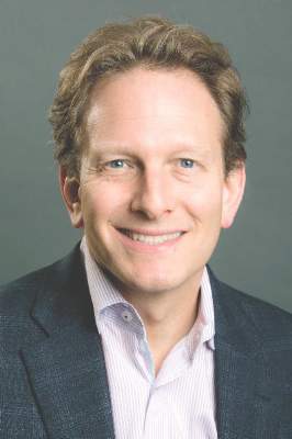
“ELIXA demonstrates the cardiovascular safety of lixisenatide,” Dr. Eldrin Lewis, associate professor of medicine at Harvard Medical School and of Brigham and Women’s Hospital in Boston. “Additional analyses indicate safety with respect to heart failure events as well as all-cause mortality, and the hazard ratio after heart failure hospitalizations demonstrate that this endpoint is very meaningful in diabetes as it is in other populations” he added. Furthermore, the neutral effects of lixisenatide were seen across a wide spectrum of heart failure risk.
TECOS investigator Dr. Paul Armstrong of the University of Alberta in Canada noted similar safety findings with sitagliptin. “We found no increase in the risk of heart failure or related adverse outcomes after sitagliptin therapy,” he said at a press briefing. “We can safely conclude that sitagliptin can be used in patients with type 2 diabetes without concern for worsening heart failure.”
ELIXA and TECOS were two phase 3, randomized, double blind placebo-controlled trials specifically designed to look at cardiovascular outcomes after treatment with the glucagon-like peptide 1 (GLP-1) receptor agonist lixisenatide (intended trade name Lyxumia) or the dipeptyl peptidase 4 (DPP-4) inhibitor sitagliptin (Januvia). The trials enrolled 6,068 and 14,671 patients, respectively, and were performed in the wake of prior trials that had suggested an increased risk for hospitalization for heart failure in patients treated with other DPP-4 inhibitors including saxagliptin (Onglyza) in the SAVOR-TIMI 53 trial and alogliptin (Nesina) in the EXAMINE (Examination of Cardiovascular Outcomes with Alogliptin versus Standard of Care) trial.
Although powered to show superiority of lixisenatide versus placebo in terms of cardiovascular safety, ELIXA was only able to establish noninferiority (HR, 1.02), but results were below limits for noninferiority set by the United States Food and Drug Administration, Dr. Lewis observed. In the updated analysis of ELIXA there was no difference between treatment with the lixisenatide and placebo for the primary composite cardiovascular endpoint plus heart failure hospitalization (hazard ratio, 0.97, 95% confidence interval 0.85-1.10) after 3 years of follow-up. There were also no differences in the rates of heart failure hospitalization (HR, 0.96, 95% CI 0.75-1.23), the primary endpoint plus heart failure hospitalization and coronary revascularization (HR=1.00, 95% CI 0.90-1.11), or death from any cause (HR, 0.94, 95% CI 0.78-1.13).Although the risk for heart failure hospitalization was found to be nine times higher in patients who had a prior history of heart failure in ELIXA than in those who did not there was there was no difference between treatment with the GLP-1 agonist or placebo in patients with (9.7% vs. 10.2%, HR, 0.93, 95% CI 0.66-1.30) or without (2.4% vs. 2.5%, HR, 0.97, 95% CI 0.67-1.40) a history of the disease.
The primary findings of TECOS have been published (doi:10.1056/NEJMoa1501352) and showed sitagliptin to be noninferior to placebo for the primary composite cardiovascular endpoint (HR,0.98; 95% CI, 0.88-1.09; P less than .001). Heart failure hospitalization rates were similar (HR, 1.00; 95% CI, 0.83-1.20; P = .98).
In the updated analysis of TECOS, the prespecified secondary endpoint of time to first hospitalization for heart failure did not differ between the placebo and sitagliptin arms (HR, 1.0; 95% CI, 0.84-1.20, P = .95), nor were there any differences between the treatments in terms of other prespecified secondary endpoints of hospitalization for heart failure or cardiovascular death (HR, 1.02, 95% CI 0.90-1.14, P =.74), or hospitalization for heart failure or all-cause death (HR, 1.00, 95% CI 0.90-1.11, P = .93).
Dr. Armstrong acknowledged that the findings of TECOS were in contrast to the SAVOR-TIMI 53 and EXAMINE trials but that this could be due to several factors: differences in the patients enrolled, the background care provided, and variation in the acquisition or definition of heart failure events between the trials. It could also be due to intrinsic differences among the DPP-4 inhibitors themselves or “chance.”
Dr. Gabriel Steg of Hôpital Bichat in Paris, France who provided independent comment after the presentation of the ELIXA data questioned the value of continuing to perform large-scale noninferiority studies assessing the cardiovascular safety of novel diabetes medicines. More than 150,000 patients have been studied in such trials to date which have shown noninferiority for cardiovascular outcomes.
“Non-inferiority trials are often mistakenly being interpreted as ‘lack of efficacy’ trials and can among nonspecialists generate skepticism regarding treatment of diabetes or the need to control glycemia,” he said. “My conclusion would be to suggest that it is time to stop wasting resources on noninferiority trials and to work on truly improving diabetes care.”
ELXIA was sponsored by Sanofi. Dr. Lewis disclosed receiving research funding from Sanofi, and other drug companies. TECOS was sponsored by Merck. Dr. Armstrong disclosed receiving research funding, educational grants and consulting fees from Merck as well as other companies. Dr. Steg had received research grants from and has acted as a consultant for Sanofi, Merck, and other companies.
LONDON – Further data from two large-scale diabetes trials continue to indicate that there is no increased risk for heart failure with either lixisenatide or sitagliptin compared to placebo.
The findings from ELIXA (the evaluation of lixisenatide in acute coronary syndromes) and TECOS (the Trial Evaluating Cardiovascular Outcomes With Sitagliptin), presented at the annual congress of the European Society of Cardiology, provide reassuring evidence of the cardiovascular safety of both drugs, and add to the trials’ results previously presented at the 2015 annual scientific sessions of the American Diabetes Association, according to the key investigators for the separately-run trials.
“ELIXA demonstrates the cardiovascular safety of lixisenatide,” Dr. Eldrin Lewis, associate professor of medicine at Harvard Medical School and of Brigham and Women’s Hospital in Boston. “Additional analyses indicate safety with respect to heart failure events as well as all-cause mortality, and the hazard ratio after heart failure hospitalizations demonstrate that this endpoint is very meaningful in diabetes as it is in other populations” he added. Furthermore, the neutral effects of lixisenatide were seen across a wide spectrum of heart failure risk.
TECOS investigator Dr. Paul Armstrong of the University of Alberta in Canada noted similar safety findings with sitagliptin. “We found no increase in the risk of heart failure or related adverse outcomes after sitagliptin therapy,” he said at a press briefing. “We can safely conclude that sitagliptin can be used in patients with type 2 diabetes without concern for worsening heart failure.”
ELIXA and TECOS were two phase 3, randomized, double blind placebo-controlled trials specifically designed to look at cardiovascular outcomes after treatment with the glucagon-like peptide 1 (GLP-1) receptor agonist lixisenatide (intended trade name Lyxumia) or the dipeptyl peptidase 4 (DPP-4) inhibitor sitagliptin (Januvia). The trials enrolled 6,068 and 14,671 patients, respectively, and were performed in the wake of prior trials that had suggested an increased risk for hospitalization for heart failure in patients treated with other DPP-4 inhibitors including saxagliptin (Onglyza) in the SAVOR-TIMI 53 trial and alogliptin (Nesina) in the EXAMINE (Examination of Cardiovascular Outcomes with Alogliptin versus Standard of Care) trial.
Although powered to show superiority of lixisenatide versus placebo in terms of cardiovascular safety, ELIXA was only able to establish noninferiority (HR, 1.02), but results were below limits for noninferiority set by the United States Food and Drug Administration, Dr. Lewis observed. In the updated analysis of ELIXA there was no difference between treatment with the lixisenatide and placebo for the primary composite cardiovascular endpoint plus heart failure hospitalization (hazard ratio, 0.97, 95% confidence interval 0.85-1.10) after 3 years of follow-up. There were also no differences in the rates of heart failure hospitalization (HR, 0.96, 95% CI 0.75-1.23), the primary endpoint plus heart failure hospitalization and coronary revascularization (HR=1.00, 95% CI 0.90-1.11), or death from any cause (HR, 0.94, 95% CI 0.78-1.13).Although the risk for heart failure hospitalization was found to be nine times higher in patients who had a prior history of heart failure in ELIXA than in those who did not there was there was no difference between treatment with the GLP-1 agonist or placebo in patients with (9.7% vs. 10.2%, HR, 0.93, 95% CI 0.66-1.30) or without (2.4% vs. 2.5%, HR, 0.97, 95% CI 0.67-1.40) a history of the disease.
The primary findings of TECOS have been published (doi:10.1056/NEJMoa1501352) and showed sitagliptin to be noninferior to placebo for the primary composite cardiovascular endpoint (HR,0.98; 95% CI, 0.88-1.09; P less than .001). Heart failure hospitalization rates were similar (HR, 1.00; 95% CI, 0.83-1.20; P = .98).
In the updated analysis of TECOS, the prespecified secondary endpoint of time to first hospitalization for heart failure did not differ between the placebo and sitagliptin arms (HR, 1.0; 95% CI, 0.84-1.20, P = .95), nor were there any differences between the treatments in terms of other prespecified secondary endpoints of hospitalization for heart failure or cardiovascular death (HR, 1.02, 95% CI 0.90-1.14, P =.74), or hospitalization for heart failure or all-cause death (HR, 1.00, 95% CI 0.90-1.11, P = .93).
Dr. Armstrong acknowledged that the findings of TECOS were in contrast to the SAVOR-TIMI 53 and EXAMINE trials but that this could be due to several factors: differences in the patients enrolled, the background care provided, and variation in the acquisition or definition of heart failure events between the trials. It could also be due to intrinsic differences among the DPP-4 inhibitors themselves or “chance.”
Dr. Gabriel Steg of Hôpital Bichat in Paris, France who provided independent comment after the presentation of the ELIXA data questioned the value of continuing to perform large-scale noninferiority studies assessing the cardiovascular safety of novel diabetes medicines. More than 150,000 patients have been studied in such trials to date which have shown noninferiority for cardiovascular outcomes.
“Non-inferiority trials are often mistakenly being interpreted as ‘lack of efficacy’ trials and can among nonspecialists generate skepticism regarding treatment of diabetes or the need to control glycemia,” he said. “My conclusion would be to suggest that it is time to stop wasting resources on noninferiority trials and to work on truly improving diabetes care.”
ELXIA was sponsored by Sanofi. Dr. Lewis disclosed receiving research funding from Sanofi, and other drug companies. TECOS was sponsored by Merck. Dr. Armstrong disclosed receiving research funding, educational grants and consulting fees from Merck as well as other companies. Dr. Steg had received research grants from and has acted as a consultant for Sanofi, Merck, and other companies.
AT THE ESC CONGRESS 2015
Key clinical point:Patients with type 2 diabetes who are treated with lixisenatide or sitagliptin are at no greater risk for heart failure than if they are given placebo.
Major finding: The rates of heart failure hospitalization versus placebo were comparable for either lixisenatide (HR, 0.96, 95% CI 0.75-1.23) or sitagliptin (HR, 1.00; 95% CI, 0.83-1.20; P = .98).
Data source: ELIXA and TECOS: Two separately run, randomized, double blind, placebo-controlled cardiovascular safety trials of over 20,000 patients with type 2 diabetes mellitus.
Disclosures: ELIXA was sponsored by Sanofi, the maker of lixisenatide. Dr. Lewis disclosed receiving research funding from Sanofi. TECOS was sponsored by Merck, the maker of sitagliptin. Dr. Armstrong disclosed receiving research funding, educational grants and consulting fees from Merck as well as other companies. Dr. Steg had received research grants from Sanofi, and has acted as a consultant for Sanofi, Merck, and other companies.

ESC: Heart failure risk climbs early in childhood cancer survivors
LONDON – Childhood cancer survivors face an increased risk of developing heart failure starting at a much younger age than the cardiac disease typically appears, according to a Dutch national cohort study.
“Health care professionals need to be aware of these findings and recognize the risk of developing heart failure in young people, even years after their childhood cancer treatment,” Dr. Elizabeth A.M. Feijen advised at the annual congress of the European Society of Cardiology.

She presented an analysis from the Dutch Childhood Oncology Group LATER (Late Effects of Childhood Cancer) study which entailed collecting followup data on more than 5,000 Dutch 5-year survivors of childhood cancer. After a median followup of 20 years, and at an average age of 27.4 years, 2% of the national cohort had been diagnosed with heart failure.
Of 113 childhood cancer survivors with heart failure, 9 had undergone heart transplantation, 11 required a pacemaker and/or implantable cardioverter-defibrillator, and 22 had died due to their heart failure, reported Dr. Feijen of the Academic Medical Center of Amsterdam.
The cumulative incidence of heart failure was 1.1% by age 20, 2.3% by age 30, 3.9% by age 40, and 5.4% by 50 years of age.
In a multivariate regression analysis, the significant risk factors for heart failure after childhood cancer were treatment with anthracyclines, with an associated 1.8-fold increased risk per 100 mg/m2 of medication; mitoxantrone, with a 2.3-fold risk per 25 mg/m2; chest radiation to the heart, with a 1.9-fold risk; and the use of cyclophosphamide, with an associated 1.8-fold increased risk of subsequent heart failure.
The DCOG LATER study is supported by the Dutch Cancer Society and the Children Cancer Free Foundation. Dr. Feijen reported having no financial conflicts.
LONDON – Childhood cancer survivors face an increased risk of developing heart failure starting at a much younger age than the cardiac disease typically appears, according to a Dutch national cohort study.
“Health care professionals need to be aware of these findings and recognize the risk of developing heart failure in young people, even years after their childhood cancer treatment,” Dr. Elizabeth A.M. Feijen advised at the annual congress of the European Society of Cardiology.
She presented an analysis from the Dutch Childhood Oncology Group LATER (Late Effects of Childhood Cancer) study which entailed collecting followup data on more than 5,000 Dutch 5-year survivors of childhood cancer. After a median followup of 20 years, and at an average age of 27.4 years, 2% of the national cohort had been diagnosed with heart failure.
Of 113 childhood cancer survivors with heart failure, 9 had undergone heart transplantation, 11 required a pacemaker and/or implantable cardioverter-defibrillator, and 22 had died due to their heart failure, reported Dr. Feijen of the Academic Medical Center of Amsterdam.
The cumulative incidence of heart failure was 1.1% by age 20, 2.3% by age 30, 3.9% by age 40, and 5.4% by 50 years of age.
In a multivariate regression analysis, the significant risk factors for heart failure after childhood cancer were treatment with anthracyclines, with an associated 1.8-fold increased risk per 100 mg/m2 of medication; mitoxantrone, with a 2.3-fold risk per 25 mg/m2; chest radiation to the heart, with a 1.9-fold risk; and the use of cyclophosphamide, with an associated 1.8-fold increased risk of subsequent heart failure.
The DCOG LATER study is supported by the Dutch Cancer Society and the Children Cancer Free Foundation. Dr. Feijen reported having no financial conflicts.
AT THE ESC CONGRESS 2015
Key clinical point: Survivors of childhood cancer face a risk of developing heart failure that grows with time but is already elevated by age 20.
Major finding: The cumulative incidence of heart failure in survivors of childhood cancer is 1.1% by age 20, 2.3% by age 30, and 3.9% by age 40 years.
Data source: This was a national study of more than 5,000 Dutch 5-year survivors of childhood cancer who were followed for a median of 20.2 years.
Disclosures: The DCOG LATER study is supported by the Dutch Cancer Society and the Children Cancer Free Foundation. he presenter reported having no financial conflicts.
ESC: Zero risk of death in pregnant women with severe aortic stenosis
LONDON – The mortality risk for pregnant women with moderate or severe aortic stenosis is close to zero in contemporary practice, according to data from the large, international ROPAC registry.
That being said, more than one-third of women with severe aortic stenosis will require hospitalization for cardiac reasons during their pregnancy, with heart failure the number-one cause for admission, Dr. Stefan Orwat reported at the annual congress of the European Society of Cardiology.

Severe fetal complications are rare. However, preterm birth, small for gestational age, and neonatal Apgar scores below 7 are quite common in pregnancies involving severe aortic stenosis, as defined by a prepregnancy transaortic peak gradient of 64 mm Hg or more, according to Dr. Orwat of University Hospital in Muenster, Germany.
ROPAC (the Registry on Pregnancy and Cardiac disease) is a unique ongoing, global, prospective, observational registry created in order to advance understanding of the risks of pregnancy in the setting of structural heart disease. Prior reports from other sources have often provided conflicting guidance because of relatively small patient sample sizes. That’s been especially true with regard to aortic stenosis (AS), the cardiologist said.
Out of 2,966 pregnancies in women with various forms of structural heart disease in ROPAC, Dr. Orwat focused on the 99 pregnancies in 96 women with moderate or severe AS, 21 of whom had a prosthetic heart valve in place prior to pregnancy; 65 of the women had moderate AS as evidenced by an echocardiographic prepregnancy peak gradient of 36-63 mm Hg.
No maternal deaths occurred during pregnancy or within 1 week afterward. However, 13% of women with moderate AS and 35% with severe AS were hospitalized for cardiac reasons during their pregnancy. Most of the admissions were for new or worsening heart failure, which occurred at a median of 28 weeks’ gestation.
The risk of heart failure hospitalization during pregnancy was quite low – in the 6%-8% range – among women with baseline prepregnancy asymptomatic or symptomatic moderate AS or asymptomatic severe AS. In contrast, the rate shot up to 26% in women with symptomatic and severe AS prior to pregnancy.
There were a couple of hospitalizations for arrhythmias and one for endocarditis. Unlike in some prior reports by other research groups, there were no cases of pulmonary embolism, valve thrombosis, cerebrovascular events, or deep vein thrombosis. The presence of a mechanical valve with its required rigorous anticoagulation wasn’t related to worse outcomes in this series, unlike in some others.
The heart failure was generally manageable medically. Only two patients required intervention. One developed endocarditis, then underwent balloon aortic valvuloplasty and aortic valve replacement with a mechanical valve 4 months into pregnancy, with subsequent vaginal delivery at 38 weeks. The other patient, who was unaware she had AS prior to pregnancy, presented with severe AS and a depressed left ventricular ejection fraction at 20 weeks’ gestation. She responded well to valvuloplasty, had an uneventful vaginal delivery at 38 weeks, then underwent aortic valve replacement.
In a multivariate analysis, only two independent predictors of maternal hospitalization during pregnancy were identified: the presence of symptoms prior to pregnancy, and the prepregnancy hemodynamic severity of AS.
Preterm birth prior to 37 weeks occurred in 16% of pregnancies involving moderate and 36% of those featuring severe AS. An Apgar score below 7 occurred in 5% of cases of moderate and 16% of severe AS. The small for gestational age rate was 3% with moderate AS and ballooned to 21% with severe AS. The only independent predictor of fetal complications was the degree of elevation in peak aortic gradient prior to pregnancy.
Audience questions focused on challenging scenarios. Would you allow a pregnancy to continue, Dr. Orwat was asked, if a woman with AS developed an aortic gradient of 90 mm Hg during the first trimester?
“It depends on whether she was asymptomatic prior to pregnancy. If so, I think it’s a low-risk situation, and you can carry on with the pregnancy,” he replied. Remember, he added, it’s the baseline gradient that’s important. Cardiac output goes up during the first trimester as a matter of course, and so will the gradient.
Dr. Orwat emphasized that symptomatic severe AS is an indication for aortic valve replacement, and he would advise an affected patient to undergo the surgery prior to pregnancy.
Audience member Dr. Fiona Walker rose to advocate ordering a prepregnancy treadmill exercise test in all patients with asymptomatic severe AS in order to learn whether they are likely to remain asymptomatic during the stresses of pregnancy. In a patient with severe AS who remains asymptomatic with exercise, it’s probably safer to go through pregnancy without prior valve replacement than to put in a mechanical valve.
“Patients with mechanical valves in pregnancy are one of the highest-risk groups we look after,” commented Dr. Walker, head of the maternal cardiology program at University College London Hospitals.
But another audience member advocated aortic valve replacement prior to pregnancy in all women with severe AS, asymptomatic or not.
“Nowadays, if you are afraid of a mechanical valve, you can put in a bioprosthetic one, although I know that’s considered controversial. I see that asymptomatic severe aortic stenosis seems quite safe in ROPAC, but I still consider severe aortic stenosis one of the truly dangerous problems in pregnancy. Maybe I’ll change my mind after ROPAC is published, but right now I don’t think we’re going to leave a severe aortic stenosis prior to pregnancy without intervention,” declared Dr. Avraham Shotan, head of the Heart Institute at Hillel Yaffe Medical Center in Hadera, Israel.
Session cochair Dr. Christa Gohlke-Baerwolf of Bad Krozingen (Germany) Hospital mediated the dispute and closed discussion by observing, “Severe aortic stenosis is not just one thing, it’s a continuum, and such patients should be evaluated by a multidisciplinary team very carefully before starting pregnancy.“
ROPAC is sponsored by the ESC. Dr. Orwat reported having no financial conflicts regarding his study.
LONDON – The mortality risk for pregnant women with moderate or severe aortic stenosis is close to zero in contemporary practice, according to data from the large, international ROPAC registry.
That being said, more than one-third of women with severe aortic stenosis will require hospitalization for cardiac reasons during their pregnancy, with heart failure the number-one cause for admission, Dr. Stefan Orwat reported at the annual congress of the European Society of Cardiology.
Severe fetal complications are rare. However, preterm birth, small for gestational age, and neonatal Apgar scores below 7 are quite common in pregnancies involving severe aortic stenosis, as defined by a prepregnancy transaortic peak gradient of 64 mm Hg or more, according to Dr. Orwat of University Hospital in Muenster, Germany.
ROPAC (the Registry on Pregnancy and Cardiac disease) is a unique ongoing, global, prospective, observational registry created in order to advance understanding of the risks of pregnancy in the setting of structural heart disease. Prior reports from other sources have often provided conflicting guidance because of relatively small patient sample sizes. That’s been especially true with regard to aortic stenosis (AS), the cardiologist said.
Out of 2,966 pregnancies in women with various forms of structural heart disease in ROPAC, Dr. Orwat focused on the 99 pregnancies in 96 women with moderate or severe AS, 21 of whom had a prosthetic heart valve in place prior to pregnancy; 65 of the women had moderate AS as evidenced by an echocardiographic prepregnancy peak gradient of 36-63 mm Hg.
No maternal deaths occurred during pregnancy or within 1 week afterward. However, 13% of women with moderate AS and 35% with severe AS were hospitalized for cardiac reasons during their pregnancy. Most of the admissions were for new or worsening heart failure, which occurred at a median of 28 weeks’ gestation.
The risk of heart failure hospitalization during pregnancy was quite low – in the 6%-8% range – among women with baseline prepregnancy asymptomatic or symptomatic moderate AS or asymptomatic severe AS. In contrast, the rate shot up to 26% in women with symptomatic and severe AS prior to pregnancy.
There were a couple of hospitalizations for arrhythmias and one for endocarditis. Unlike in some prior reports by other research groups, there were no cases of pulmonary embolism, valve thrombosis, cerebrovascular events, or deep vein thrombosis. The presence of a mechanical valve with its required rigorous anticoagulation wasn’t related to worse outcomes in this series, unlike in some others.
The heart failure was generally manageable medically. Only two patients required intervention. One developed endocarditis, then underwent balloon aortic valvuloplasty and aortic valve replacement with a mechanical valve 4 months into pregnancy, with subsequent vaginal delivery at 38 weeks. The other patient, who was unaware she had AS prior to pregnancy, presented with severe AS and a depressed left ventricular ejection fraction at 20 weeks’ gestation. She responded well to valvuloplasty, had an uneventful vaginal delivery at 38 weeks, then underwent aortic valve replacement.
In a multivariate analysis, only two independent predictors of maternal hospitalization during pregnancy were identified: the presence of symptoms prior to pregnancy, and the prepregnancy hemodynamic severity of AS.
Preterm birth prior to 37 weeks occurred in 16% of pregnancies involving moderate and 36% of those featuring severe AS. An Apgar score below 7 occurred in 5% of cases of moderate and 16% of severe AS. The small for gestational age rate was 3% with moderate AS and ballooned to 21% with severe AS. The only independent predictor of fetal complications was the degree of elevation in peak aortic gradient prior to pregnancy.
Audience questions focused on challenging scenarios. Would you allow a pregnancy to continue, Dr. Orwat was asked, if a woman with AS developed an aortic gradient of 90 mm Hg during the first trimester?
“It depends on whether she was asymptomatic prior to pregnancy. If so, I think it’s a low-risk situation, and you can carry on with the pregnancy,” he replied. Remember, he added, it’s the baseline gradient that’s important. Cardiac output goes up during the first trimester as a matter of course, and so will the gradient.
Dr. Orwat emphasized that symptomatic severe AS is an indication for aortic valve replacement, and he would advise an affected patient to undergo the surgery prior to pregnancy.
Audience member Dr. Fiona Walker rose to advocate ordering a prepregnancy treadmill exercise test in all patients with asymptomatic severe AS in order to learn whether they are likely to remain asymptomatic during the stresses of pregnancy. In a patient with severe AS who remains asymptomatic with exercise, it’s probably safer to go through pregnancy without prior valve replacement than to put in a mechanical valve.
“Patients with mechanical valves in pregnancy are one of the highest-risk groups we look after,” commented Dr. Walker, head of the maternal cardiology program at University College London Hospitals.
But another audience member advocated aortic valve replacement prior to pregnancy in all women with severe AS, asymptomatic or not.
“Nowadays, if you are afraid of a mechanical valve, you can put in a bioprosthetic one, although I know that’s considered controversial. I see that asymptomatic severe aortic stenosis seems quite safe in ROPAC, but I still consider severe aortic stenosis one of the truly dangerous problems in pregnancy. Maybe I’ll change my mind after ROPAC is published, but right now I don’t think we’re going to leave a severe aortic stenosis prior to pregnancy without intervention,” declared Dr. Avraham Shotan, head of the Heart Institute at Hillel Yaffe Medical Center in Hadera, Israel.
Session cochair Dr. Christa Gohlke-Baerwolf of Bad Krozingen (Germany) Hospital mediated the dispute and closed discussion by observing, “Severe aortic stenosis is not just one thing, it’s a continuum, and such patients should be evaluated by a multidisciplinary team very carefully before starting pregnancy.“
ROPAC is sponsored by the ESC. Dr. Orwat reported having no financial conflicts regarding his study.
AT THE ESC CONGRESS 20015
Key clinical point: Pregnancy in women with aortic stenosis now carries a near-zero risk of maternal mortality.
Major finding: Maternal mortality was zero in 99 pregnancies in 96 women with moderate or severe aortic stenosis.
Data source: The ROPAC registry is an ongoing, prospective, global, observational registry devoted to women with structural heart disease.
Disclosures: The registry is sponsored by the European Society of Cardiology. The presenter reported having no financial conflicts of interest.
VIDEO: Patients use CardioMEMS feedback to improve self-management
Some of the highly selected heart failure patients who have received the CardioMEMS device at Brigham and Women’s Hospital in Boston have shown clinically meaningful behavior changes in response to the feedback they receive on fluctuations in their pulmonary artery pressures. This feedback has prompted patients to change things like their salt and fluid intake so that they have fewer spikes in their pulmonary-artery diastolic pressure, an important step toward reducing their need for hospitalization because of acute decompensation episodes and possibly improving their long-term outcomes, Dr. Eldrin F. Lewis said in an interview.
“In our clinical practice we notice that when patients get feedback on their pulmonary-artery pressure they often change their behavior. In some patients their pulmonary-artery pressures normalized and stayed in the normal range more consistently,” said Dr. Lewis, a heart failure cardiologist at Brigham and Women’s. This apparent effect of daily monitoring of pulmonary artery pressure using CardioMEMS on patient behavior had not previously been assessed in the device’s clinical trials.
Those trials documented that diligent monitoring of pulmonary artery pressures and tweaking therapy to optimize those pressures led to significant reduction in heart failure hospitalizations, but Dr. Lewis and other heart failure specialists speculate that patients may reap other benefits.
“If we can dramatically reduce heart failure hospitalization rates in these patients, that should eventually translate into improved survival, and I think that by keeping fluid levels down it should also improve exercise capacity and quality of life,” he said.
The CardioMEMS monitoring system is marketed by St. Jude Medical. Dr. Lewis has no disclosures that involve St. Jude. He has received research grants from Amgen, Novartis, and Sanofi.
The video associated with this article is no longer available on this site. Please view all of our videos on the MDedge YouTube channel
On Twitter @mitchelzoler

ESC: Most heart failure patients’ rehospitalizations are for other reasons
LONDON – More than half of all patients hospitalized for heart failure were readmitted within 1 year of discharge in a large Italian population-based study.
Results from the ARNO-CORE CardioVascular Observatory database showed that 56.6% of 41,417 patients with heart failure who were discharged alive and prescribed at least one drug for heart failure were readmitted to the hospital at least once in the following year, Dr. Aldo Maggioni said at the annual congress of the European Society of Cardiology.
This averaged out to 2.1 readmissions per person, and importantly, those readmissions were frequently for noncardiovascular reasons, he said. Indeed, 23,826 (49.1%) of rehospitalizations were due to respiratory disease, gastrointestinal disease, cancer, trauma, or other reasons.
“This clearly implies that if we want to impact the total burden of [heart failure], we are not to just consider the reduction in hospitalization for heart failure, but we have to take into account the multiplicity of causes that these patients have,” commented Dr. Maggioni of the National Association of Hospital Cardiologists Research Center in Florence, Italy.
The ARNO-CORE CardioVascular Observatory is a large, retrospective observational study looking at the clinical epidemiology of patients with heart failure. Unlike clinical trial populations and even specialty-run heart failure registries, the ARNO database aims to provide a more representative picture of heart failure and its treatment in a routine practice setting, Dr. Maggioni noted.
Of the 41,413 patients with acute heart failure during 2008-2012 included in the present analysis, just over one-quarter (27%) were admitted to a cardiology unit, with the majority (50%) admitted to internal medicine or general medicine practices, and 14% seen in geriatric departments, 2% in functional recovery or rehabilitation units, 2% in pulmonary units, and the remainder in other practices.
The mean age of patients was 78 years, which was older than that seen in heart failure trials such as the RELAX-AHF study (72 years) and the ESC-HF Long-Term Registry(71 years). There was also a higher percentage of women, compared with the percentage for the trial and the other registry population (51% vs. 38% and 27%, respectively), and there were higher incidences of chronic obstructive pulmonary disease (31% vs. 16% and 20%) and depression (21% in the ARNO population vs. 8% in the ESC-HF Long-Term Registry). A substantial percentage (31%) had diabetes (48% in RELAX-AHF and 39% in the ESC registry), but a lower percentage was found to have coexistent chronic kidney disease (4% vs. 26% in the ESC registry). The reason for the latter difference was perhaps that it was possible to more accurately include only those patients with chronic kidney disease using the administrative coding, Dr. Maggioni said.
In terms of pharmacologic treatments, fewer patients treated in routine practice than in the clinical trial or ESC registry settings received guideline-recommended therapies. Two-thirds (66%) of patients in the ARNO database were taking ACE inhibitors or angiotensin II receptor blockers (ARBs), compared with 72% and 77% of the RELAX-AHF and ESC registry populations. Furthermore only about half (52%) were taking beta-blockers, compared with 89% and 72% of the trial and other ESC registry populations.
Importantly, patients were being treated with suboptimal doses, he said, and adherence rates at 1 year were 58% for ACE inhibitors and ARBs and 62% for beta-blockers.
All-cause mortality at 1 year was 28.7%, which was higher than that seen in clinical studies such as EVEREST (26.1%) and at 6 months in the RELAX-AHF study (9.2%). It was also slightly higher than that seen in the ESC-HF Long-Term Registry (23.6%).
“The yearly cost for a patient with heart failure is high and mainly driven by hospitalizations,” Dr. Maggioni said. The estimated costs per patient per year were $13,295.04, which is higher than that for cardiovascular disease in general ($10,689.59) and lower than for acute coronary syndrome ($16,664.74). Of this cost, roughly 83% is linked to hospitalization and less than 10% to medications and 7% to specialist diagnostic procedures.
“Given the poor prognosis and the high health care costs associated with hospital readmissions, even small improvements in the global heart failure patient care can have a substantial impact on patient quality of life and health care costs,” Dr. Maggioni suggested. He added that better adherence to guideline-recommended medications could also play a vital role.The study was partially supported by Novartis Pharma Italy. Dr. Maggioni disclosed serving on committees of heart failure trials sponsored by Bayer Healthcare, Cardiorentis, and Novartis Pharma Italy.
*This story was updated 9/10/2015.
AT THE ESC CONGRESS 2015
Key clinical point: Hospital readmission in patients with heart failure is often a result of noncardiovascular causes, warranting a multidisciplinary approach.
Major finding: More than half (56.6%) of 41,417 patients with heart failure studied were rehospitalized within 1 year, mostly for noncardiovascular reasons (49.1%).
Data source: The ARNO-CORE CardioVascular Observatory, a large retrospective observational study looking at the clinical epidemiology of patients with heart failure.
Disclosures: The study was partially supported by Novartis Pharma Italy. Dr. Maggioni disclosed serving on committees of heart failure trials sponsored by Bayer Healthcare, Cardiorentis, and Novartis Pharma Italy.
Collaboration helped reduce heart failure readmissions
Hospitals that participated in the Southeast Michigan “See You in 7 Collaborative” experienced substantial reductions in 30-day readmission rates and Medicare payments related to heart failure (HF) care.
Increases in postdischarge 7-day follow-up were modest “but associated processes aimed at this goal may have improved the overall transition from inpatient to outpatient care,” Dr. Scott L. Hummel reported online Sept. 9 in JACC: Heart Failure. “Our study suggests that regional hospital collaboration to share best practices can be an effective strategy to reduce HF readmissions and associated costs.”

Established in 2011, the Southeast Michigan See You in 7 Collaborative is an effort by the Greater Detroit Area Council, the American College of Cardiology’s Michigan Chapter, the Michigan Peer Review Organization, and 11 previously nonaffiliated hospitals to increase 7-day postdischarge follow-up and reduce all-cause 30-day readmission rates in HF patients. Over a 1-year period, Dr. Hummel, a cardiologist at the University of Michigan, Ann Arbor, and his associates examined the rates of 7-day follow-up and 30-day readmissions in Medicare fee-for-service HF patients among the 10 collaborating hospitals, and compared the findings to trends in the state’s 82 nonparticipating hospitals. The preintervention period studied was May 2011-April 2012; the intervention occurred from May 2012-April 2013.
During the intervention period, the rates of 7-day postdischarge follow-up increased significantly but remained low in both groups (from 31.1% to 34.4% in collaborating hospitals, and from 30.2% to 32.6% in nonparticipating hospitals). At the same time, the rates of unadjusted readmissions significantly decreased in both groups (from 29% to 27.3% in collaborating hospitals and from 26.4% to 25.8% in nonparticipating hospitals). The researchers also found that the mean risk-standardized 30-day, all-cause readmission rates improved significantly among collaborating hospitals (from 31.1% to 28.5%), but significantly less so among nonparticipating hospitals (from 26.7% to 26.1%).
Finally, combined Medicare payments for inpatient and 30 days of postdischarge care decreased by an average of $182 per beneficiary in collaborating hospitals and by $63 in nonparticipating hospitals (JACC Heart Fail. 2015 Sept 9; [doi:10.1016/j.jchf.2015.06.007]).
The researchers acknowledged certain limitations of the study, including its observational design and the fact that administrative data sets were used to determine the outcomes of interest. “Given the observational nature of the study, we cannot confirm that improvements in 7-day follow-up or 30-day readmission rates directly resulted from See You in 7 Collaborative participation,” they noted.
The study was funded in part by the Robert Wood Johnson Foundation. Dr. Hummel disclosed that he is supported by a grant from the National Heart, Lung, and Blood Institute. The remaining study authors reported having no financial disclosures.
The investigators should be congratulated for a well-designed intervention that was multifaceted, allowing hospitals to experiment locally with the innovations that proved most useful to the individual health care environment. Unfortunately, the hospitals in the Collaborative were unable to significantly increase 7-day follow-up over the course of 1 year; 7-day follow-up rose from 31% to 34% only, compared with 32% to 34% in matched hospitals in Michigan. Even 14-day follow-up was less than 50% for various hospital groups in the study. These rates are consistent with other national data and are an important reflection of the current state of health care in the United States, yet it remains unclear why it is so difficult to move the needle.
Despite the lack of improvement in early follow-up, all-cause 30-day, risk-standardized readmission rates decreased more in Collaborative hospitals (31.1% to 28.5%), compared with other hospitals in Michigan (26.7% to 26.1%) during the 1-year study, including a cohort of hospitals matched for size, region, and demographics. Nevertheless, it is unclear which tools in the toolkit led to this improvement. Because of other changes noted in the environment, these results were possibly brought about by overall cultural or environmental attention toward readmission among more motivated hospitals. To address this important source of bias, future studies should consider randomization schemes of different interventions, such as cluster-randomized trials or embedded randomization.
There will undoubtedly be more opportunities to establish a better evidence base leveraging national, reusable research infrastructure such as the National Patient-Centered Clinical Research Network (PCORnet), which can serve as a platform for testing new health system delivery models.
Dr. Adrian F. Hernandez and Dr. Adam D. Devore are with the department of medicine at Duke University, as well as the Duke Clinical Research Institute, both in Durham, N.C. They made these comments in an accompanying editorial (JACC Heart Fail. 2015 Sept 9; [doi:10.1016/j.jchf.2015.07.004]). Dr. Hernandez reported receiving honoraria from Amgen, AstraZeneca, Janssen, Merck and Novartis; and research support from the Agency for Healthcare Research and Quality, American Heart Association, Amgen, AstraZeneca, Novartis, Merck, the National Heart, Lung, and Blood Institute, and the Patient Centered Outcomes Research Institute. Dr. Devore reported research funding from Amgen, AHA, Maquet, Novartis, and Thoratec; and consultant fees from Maquet.
Hospitals that participated in the Southeast Michigan “See You in 7 Collaborative” experienced substantial reductions in 30-day readmission rates and Medicare payments related to heart failure (HF) care.
Increases in postdischarge 7-day follow-up were modest “but associated processes aimed at this goal may have improved the overall transition from inpatient to outpatient care,” Dr. Scott L. Hummel reported online Sept. 9 in JACC: Heart Failure. “Our study suggests that regional hospital collaboration to share best practices can be an effective strategy to reduce HF readmissions and associated costs.”
Established in 2011, the Southeast Michigan See You in 7 Collaborative is an effort by the Greater Detroit Area Council, the American College of Cardiology’s Michigan Chapter, the Michigan Peer Review Organization, and 11 previously nonaffiliated hospitals to increase 7-day postdischarge follow-up and reduce all-cause 30-day readmission rates in HF patients. Over a 1-year period, Dr. Hummel, a cardiologist at the University of Michigan, Ann Arbor, and his associates examined the rates of 7-day follow-up and 30-day readmissions in Medicare fee-for-service HF patients among the 10 collaborating hospitals, and compared the findings to trends in the state’s 82 nonparticipating hospitals. The preintervention period studied was May 2011-April 2012; the intervention occurred from May 2012-April 2013.
During the intervention period, the rates of 7-day postdischarge follow-up increased significantly but remained low in both groups (from 31.1% to 34.4% in collaborating hospitals, and from 30.2% to 32.6% in nonparticipating hospitals). At the same time, the rates of unadjusted readmissions significantly decreased in both groups (from 29% to 27.3% in collaborating hospitals and from 26.4% to 25.8% in nonparticipating hospitals). The researchers also found that the mean risk-standardized 30-day, all-cause readmission rates improved significantly among collaborating hospitals (from 31.1% to 28.5%), but significantly less so among nonparticipating hospitals (from 26.7% to 26.1%).
Finally, combined Medicare payments for inpatient and 30 days of postdischarge care decreased by an average of $182 per beneficiary in collaborating hospitals and by $63 in nonparticipating hospitals (JACC Heart Fail. 2015 Sept 9; [doi:10.1016/j.jchf.2015.06.007]).
The researchers acknowledged certain limitations of the study, including its observational design and the fact that administrative data sets were used to determine the outcomes of interest. “Given the observational nature of the study, we cannot confirm that improvements in 7-day follow-up or 30-day readmission rates directly resulted from See You in 7 Collaborative participation,” they noted.
The study was funded in part by the Robert Wood Johnson Foundation. Dr. Hummel disclosed that he is supported by a grant from the National Heart, Lung, and Blood Institute. The remaining study authors reported having no financial disclosures.
FROM JACC: HEART FAILURE
Key clinical point: An interhospital collaborative approach was associated with substantial reductions in 30-day readmission rates and Medicare payments among heart failure patients.
Major finding: Over the course of 1 year, all-cause 30-day, risk-standardized readmission rates decreased more in collaborating hospitals (31.1% to 28.5%; P less than .001), compared with other hospitals in Michigan (26.7% to 26.1%; P = .02).
Data source: An observational study of Medicare heart failure patients discharged from 10 collaborating hospitals participating in the Southeast Michigan See You in 7 Collaborative.
Disclosures: The study was funded in part by the Robert Wood Johnson Foundation. Dr. Hummel disclosed that he is supported by a grant from the National Heart, Lung, and Blood Institute. The remaining study authors reported having no financial disclosures.