User login
ISCHEMIA-EXTEND: Conservative stable CAD management holds up
CHICAGO – The case for survival equipoise between an invasive or conservative strategy for managing patients with stable coronary disease and moderate or severe cardiac ischemia grew stronger with an additional 2.5 years of median follow-up of the landmark ISCHEMIA trial.
During a median follow-up of 5.7 years in ISCHEMIA-EXTEND – and as long as 7 years – patients randomized to an upfront invasive strategy regardless of their symptoms had an all-cause mortality rate of 12.7%, compared with a 13.4% rate in the patients randomized to the conservative, medication-based management strategy that employed revascularization only when the medical approach failed to resolve their angina. This survival difference fell far short of significance (adjusted hazard ratio, 1.00; 95% confidence interval, 0.85-1.18), solidifying a finding first seen in the main ISCHEMIA results when they came out 3 years before, in late 2019, Judith S. Hochman, MD, said at the American Heart Association scientific sessions.
The new results “provide evidence for patients with chronic coronary disease and their physicians as they decide whether to add invasive management to guideline-directed medical therapy,” concluded Dr. Hochman, professor and senior associate dean for clinical sciences at New York University Langone Health. Simultaneous with her report, the extended follow-up results also appeared in an article published online in Circulation.
Nil probability of a survival benefit
“The probability over 5.7 years that a patient’s risk of dying is lower with the invasive strategy is nil, which means: Go with the patient’s preference. Not undergoing revascularization is a reasonable strategy because there is no excess mortality,” Dr. Hochman said in an interview. The trial’s extended follow-up provides “much more robust evidence” for the neutral effect on survival. The investigators plan to further follow-up out to a maximum of 10 years to continue to monitor for a signal of a mortality difference.
“These findings might help physicians in shared decision-making as to whether to add invasive management to guideline-directed medical management in selected patients with chronic coronary artery disease and moderate or severe ischemia,” commented M. Cecilia Bahit, MD, designated discussant for the report and chief of cardiology for INECO Neurosciences in Rosario, Argentina.
The original ISCHEMIA results had also shown that invasive intervention can improve the quality of life in patients who have angina as a result of their coronary disease, but also showed “minimal benefits” from an invasive approach in asymptomatic patients, who comprised 35% of the study cohort of 5,179 patients.
While ISCHEMIA enrolled patients with moderate to severe coronary ischemia identified with noninvasive testing, it excluded certain patients for whom an invasive strategy is recommended, including those with unprotected left main coronary stenoses of at least 50%, a recent acute coronary syndrome event, a left ventricular ejection fraction of less than 35%, more advanced functional limitations from heart failure, or advanced chronic kidney disease.
Follow-up without adjudication
The extended follow-up included 4,825 patients from the initial cohort, with data collected from 4,540 patients. One limitation of the follow-up was that the cause of death was not adjudicated as it had been during the initial follow-up phase. It instead relied on unconfirmed information collected either from patients’ families or national databases. The demographics and clinical profiles of the study participants available for extended follow-up closely matched the entire original study cohort.
The additional follow-up also revealed a significant survival benefit from the invasive approach for cardiovascular deaths, with an incidence of 8.6% in the conservative arm and 6.4% in the invasive group, an adjusted 22% relative reduction in this outcome favoring the invasive strategy (95% CI, 0.63-0.96). This difference had appeared as a nonsignificant signal in the initial 3.2-year median follow-up.
However, this significant benefit from the invasive strategy was counterbalanced by a surprising and inexplicable increase in deaths from noncardiovascular causes in those managed with the invasive strategy. Noncardiovascular deaths occurred in 5.5% of those in the invasive arm and in 4.4% of those in the conservative arm, a significant adjusted 44% relative increase in this outcome associated with invasive management. Again, this difference was not as clearly apparent after the initial follow-up phase.
“The increase in noncardiovascular deaths with the invasive strategy surprisingly persisted over time and offset” the cardiovascular survival benefit from upfront invasive treatment, explained Dr. Hochman. A prior report from the investigators looked in depth at the noncardiovascular deaths during the initial follow-up phase and found that most of the excess was caused by malignancies, although why this happened in the invasively treated patients remains a mystery.
Staying alive is what patients care about
“I think that interventional cardiologists who favor an invasive strategy will be excited to see this significant reduction in cardiovascular deaths, but patients don’t care what they die from. What patients care about is whether they are dead or alive,” Dr. Hochman noted.
But B. Hadley Wilson, MD, an interventional cardiologist and vice president of the American College of Cardiology, had a somewhat different take on these findings.
“We need to consider the significant decrease in cardiovascular mortality, as we sort out the conundrum” of the increase in noncardiovascular deaths,” he said in an interview. “Hopefully, the 10-year outcomes will help answer this.”
But until more information is available, the ISCHEMIA and ISCHEMIA-EXTEND results have already helped advance the conversation that patients with stable coronary disease and their families have with clinicians about management decisions.
“I love that ISCHEMIA highlighted the importance of shared decision making and a heart team approach,” said Dr. Wilson, executive vice chair of the Sanger Heart & Vascular Institute of Atrium Health in Charlotte, N.C.
Anecdotally, ISCHEMIA reduced invasive management
After the initial ISCHEMIA results were published nearly 3 years ago, “I think use of invasive treatment for these patients has decreased, although I have seen no numbers” that document this, said Dr. Wilson. “I think most interventional cardiologists would say that ISCHEMIA has had an impact,” with fewer patients who match the trial’s enrollment criteria undergoing invasive management.
“Anecdotally, cardiologists are reviewing the ISCHEMIA data with their patients,” agreed Dr. Hochman, who added that no actual data have yet appeared to document this, nor do data yet document a change in the use of invasive management. “It takes time to measure the impact.”
To expedite the shared decision-making process for these patients, the ISCHEMIA researchers are planning to make available an app that will allow patients and physicians to enter clinical and demographic data and see a calculated estimate of their future cardiovascular disease risk and how amenable it may be to modification by invasive management, Dr. Hochman said. The app would be available on the ISCHEMIA study website in 2023.
ISCHEMIA and ISCHEMIA EXTEND received no commercial funding. Dr. Hochman and Dr. Wilson had no disclosures. Dr. Bahit has received honoraria from Behring, Boehringer Ingelheim, Bristol-Myers Squibb, Janssen, MSD, and Pfizer.
AT AHA 2022
In CABG, radial artery works best for second key graft: RAPCO at 15 years
Lower risk of MACE shown
CHICAGO – With more than 15 years of follow-up from two related trials, the best conduit for the second most important target vessel in coronary artery bypass grafting (CABG) appears to be resolved.
The radial artery (RA) graft is linked with a lower risk of major adverse cardiac events (MACE) relative to a saphenous vein (SV) or the free right internal thoracic artery (FRITA).
On the basis of these findings, “a radial artery graft should be considered in all isolated CABG operations unless there are contraindications,” reported David L. Hare, MBBS, director of research in the department of cardiology, University of Melbourne.
For the primary graft, there is general agreement that the left internal thoracic artery (LITA) is the first choice for the left anterior descending vessel, but the optimal graft for the second most important target has never been established, according to Dr. Hare.
Almost 25 years ago, two randomized controlled trials called RAPCO-RITA and RAPCO-SV were initiated to address the question. There is now 15 years of follow-up for both of the RAPCO (Radial Artery Patency and Clinical Outcomes) trials, which were presented together at the American Heart Association scientific sessions.
Two trials conducted simultaneously
The RAPCO-RITA trial randomized CABG patients less than 70 years of age (less than 60 years in those with diabetes) to grafting of the second target vessel with an RA or FRITA graft. The RAPCO-SV trial randomized those 70 years or older (60 years or older with diabetes) to an RA or SV graft.
The two primary endpoints were graft patency at 10 years and a composite MACE at 10 years. The assessment of the MACE endpoint, which consisted of cardiovascular mortality, acute myocardial infarction, and coronary revascularization, was later amended to include a comparison at 15 years.
Ten-year patency results, favoring the RA in both studies, were previously published in Circulation. In the new data presented at the meeting, the RA was associated with a significant reduction in MACE relative to the comparator graft in both studies.
“The main driver was a reduction in all-cause mortality,” Dr. Hare reported.
In RAPCO-RITA, 394 patients were randomized with follow-up data available for all but 1 patient at 15 years. Similarly, only 1 patient was lost to follow-up among the 225 randomized in RAPCO-SV. In both studies, baseline characteristics were well balanced.
MACE curves separate at 5 years
In RAPCO-RITA, the MACE survival curves began to separate at about 5 years and then gradually widened. By 15 years, the lower rate of MACE in the RA group (38% vs. 48%) translated into a 26% relative reduction (hazard ratio, 0.74; P = .04).
In RAPCO-SV, the pattern was similar, by 15 years, the rates of MACE were 60% and 73% for the RA and SV groups, respectively, translating into a 29% relative reduction (HR, 0.71; P = .04).
There was no heterogeneity in benefit across prespecified subgroups such as presence or absence of diabetes, gender, or age. In RAPCO-RITA, there was 8% absolute and 31% relative reduction in all-cause mortality. In RAPCO-SV, the absolute and relative reductions were 11% and 26%.
When the trial was initiated, Dr. Hare hypothesized that RITA would prove more durable than RA, so the outcome was not anticipated.
“This is the first randomized controlled trial program to address the question,” said Dr. Hare, who noted that there have been numerous retrospective and case control analyses that have produced mixed results in the past.
Discussant praises trial quality
The AHA-invited discussant, Marc Ruel, MD, chair of cardiac surgery, University of Ottawa (Ont.) Heart Institute, called these data “important,” and he congratulated Dr. Hare for conducting the first randomized trial to address the question about second graft durability.
However, he noted that, although the study was randomized, it was not blinded, and he questioned whether postoperative care, in particular, was similar. He also pointed out that the MACE rate seemed high, particularly among the older patients randomized in RAPCO-SV.
“All of the patients were referred to an independently run CABG rehab program that was quite separate from the trial but that provided identical mandated care,” Dr. Hare responded, indicating that there was no opportunity for differences in postprocedural management.
In the United States, the SV graft is often preferred on the basis of easy harvesting and handling characteristics, according to Dr. Hare, who estimated that fewer than 10% of the 200,000 CABG procedures performed in the United States employ the RA conduit for second target vessels. He believes the RAPCO trials data support a change.
“My personal view is [that, on the basis of] this data, given that it is from a controlled trial rather than from patient-level meta-analyses, all isolated CABG operations should be using a radial graft if it is suitable,” Dr. Hare said.
Dr. Hare reports financial relationships with Abbott, Amgen, AstraZeneca, Bayer, Boehringer-Ingelheim, CSL-Biotherapies, Lundbeck, Menarini, Merck, Novartis, Pfizer, Regeneron, Sanofi, Servier, and Vifor. Dr. Ruel reports financial relationships with Cryolife, Edwards, and Medtronic.
Lower risk of MACE shown
Lower risk of MACE shown
CHICAGO – With more than 15 years of follow-up from two related trials, the best conduit for the second most important target vessel in coronary artery bypass grafting (CABG) appears to be resolved.
The radial artery (RA) graft is linked with a lower risk of major adverse cardiac events (MACE) relative to a saphenous vein (SV) or the free right internal thoracic artery (FRITA).
On the basis of these findings, “a radial artery graft should be considered in all isolated CABG operations unless there are contraindications,” reported David L. Hare, MBBS, director of research in the department of cardiology, University of Melbourne.
For the primary graft, there is general agreement that the left internal thoracic artery (LITA) is the first choice for the left anterior descending vessel, but the optimal graft for the second most important target has never been established, according to Dr. Hare.
Almost 25 years ago, two randomized controlled trials called RAPCO-RITA and RAPCO-SV were initiated to address the question. There is now 15 years of follow-up for both of the RAPCO (Radial Artery Patency and Clinical Outcomes) trials, which were presented together at the American Heart Association scientific sessions.
Two trials conducted simultaneously
The RAPCO-RITA trial randomized CABG patients less than 70 years of age (less than 60 years in those with diabetes) to grafting of the second target vessel with an RA or FRITA graft. The RAPCO-SV trial randomized those 70 years or older (60 years or older with diabetes) to an RA or SV graft.
The two primary endpoints were graft patency at 10 years and a composite MACE at 10 years. The assessment of the MACE endpoint, which consisted of cardiovascular mortality, acute myocardial infarction, and coronary revascularization, was later amended to include a comparison at 15 years.
Ten-year patency results, favoring the RA in both studies, were previously published in Circulation. In the new data presented at the meeting, the RA was associated with a significant reduction in MACE relative to the comparator graft in both studies.
“The main driver was a reduction in all-cause mortality,” Dr. Hare reported.
In RAPCO-RITA, 394 patients were randomized with follow-up data available for all but 1 patient at 15 years. Similarly, only 1 patient was lost to follow-up among the 225 randomized in RAPCO-SV. In both studies, baseline characteristics were well balanced.
MACE curves separate at 5 years
In RAPCO-RITA, the MACE survival curves began to separate at about 5 years and then gradually widened. By 15 years, the lower rate of MACE in the RA group (38% vs. 48%) translated into a 26% relative reduction (hazard ratio, 0.74; P = .04).
In RAPCO-SV, the pattern was similar, by 15 years, the rates of MACE were 60% and 73% for the RA and SV groups, respectively, translating into a 29% relative reduction (HR, 0.71; P = .04).
There was no heterogeneity in benefit across prespecified subgroups such as presence or absence of diabetes, gender, or age. In RAPCO-RITA, there was 8% absolute and 31% relative reduction in all-cause mortality. In RAPCO-SV, the absolute and relative reductions were 11% and 26%.
When the trial was initiated, Dr. Hare hypothesized that RITA would prove more durable than RA, so the outcome was not anticipated.
“This is the first randomized controlled trial program to address the question,” said Dr. Hare, who noted that there have been numerous retrospective and case control analyses that have produced mixed results in the past.
Discussant praises trial quality
The AHA-invited discussant, Marc Ruel, MD, chair of cardiac surgery, University of Ottawa (Ont.) Heart Institute, called these data “important,” and he congratulated Dr. Hare for conducting the first randomized trial to address the question about second graft durability.
However, he noted that, although the study was randomized, it was not blinded, and he questioned whether postoperative care, in particular, was similar. He also pointed out that the MACE rate seemed high, particularly among the older patients randomized in RAPCO-SV.
“All of the patients were referred to an independently run CABG rehab program that was quite separate from the trial but that provided identical mandated care,” Dr. Hare responded, indicating that there was no opportunity for differences in postprocedural management.
In the United States, the SV graft is often preferred on the basis of easy harvesting and handling characteristics, according to Dr. Hare, who estimated that fewer than 10% of the 200,000 CABG procedures performed in the United States employ the RA conduit for second target vessels. He believes the RAPCO trials data support a change.
“My personal view is [that, on the basis of] this data, given that it is from a controlled trial rather than from patient-level meta-analyses, all isolated CABG operations should be using a radial graft if it is suitable,” Dr. Hare said.
Dr. Hare reports financial relationships with Abbott, Amgen, AstraZeneca, Bayer, Boehringer-Ingelheim, CSL-Biotherapies, Lundbeck, Menarini, Merck, Novartis, Pfizer, Regeneron, Sanofi, Servier, and Vifor. Dr. Ruel reports financial relationships with Cryolife, Edwards, and Medtronic.
AT AHA 2022
Puzzling, unique ECG from pig-to-human transplanted heart
In the first transplant of a genetically altered pig heart into a human in January, initial unexpected, prolonged ECG readings apparently did not affect the heart’s function, although the organ suddenly began to fail at day 50.
A study of these ECG changes, scheduled for presentation by Calvin Kagan, MD, and colleagues at the American Heart Association scientific sessions, offers insight into this novel operation.
As widely reported, the patient, 57-year-old David Bennett of Maryland, had end-stage heart disease and was a poor candidate for a ventricular assist device and was ineligible for a human heart, when he consented to be the first human to be transplanted with a pig heart that had a number of genes added or subtracted with the goal, in part, to prevent rejection.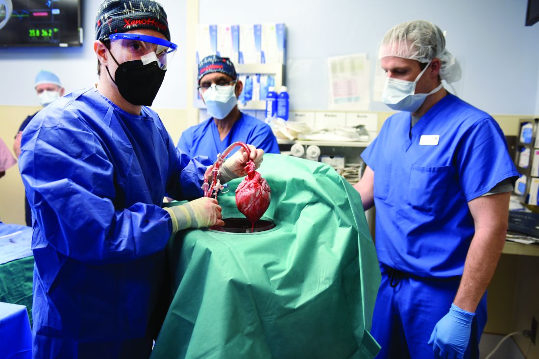
The heart initially performed well after it was transplanted in an operation at the University of Maryland School of Medicine (UMSOM) in Baltimore on Jan. 7, but failed in the second month, and Mr. Bennett died on March 9.
The Food and Drug Administration had granted emergency authorization for the surgery through its expanded access (compassionate use) program, coauthor Muhammad Mohiuddin, MD, said in an interview.
“We have learned a lot and hope we can do more,” said Dr. Mohiuddin, scientific and program director of the cardiac xenotransplantation program at UMSOM.
“Suddenly on day 50, the heart started to get thicker and was not relaxing enough,” explained senior author Timm-Michael Dickfeld, MD, PhD, director of electrophysiology research at UMSOM. A biopsy revealed substantial buildup of interstitial fluid that restricted movement. The fluid was replaced by fibrous tissue, leading to irreversible damage.
Persistent, prolonged ECG parameters
In the heart from a genetically modified pig, three genes associated with antibody-mediated rejection and a gene associated with pig heart tissue growth had been inactivated and six human genes associated with immune acceptance had been added. The donor pig was supplied by Revivicor (Blacksburg, Va.).
The patient’s immunosuppressant therapy included an experimental antirejection medication (Kiniksa Pharmaceuticals; Lexington, Mass.).
The patient had daily 12-lead ECGs after the transplant.
In prior research using a pig heart transplanted into a pig body, ECG readings showed a short PR interval (50-120 ms), short QRS duration (70-90 ms) and short QT intervals (260-380 ms).
However, in the transplanted xenograft heart, the initial ECG readings showed a longer PR interval of 190 ms, QRS duration of 138 ms, and QT of 538 ms.
Prolonged intrinsic PR intervals remained stable during the postoperative course (210 ms, range 142-246 ms).
QRS duration also remained prolonged (145 ms, range 116-192 ms), but shortened during the postoperative course (days 21-40 vs. 41-60: 148 ms vs. 132 ms; P < .001).
Increased QT persisted (509 ms, range 384-650 ms) with dynamic fluctuations. The shortest QT duration was observed on day 14 (P < .001).
“In a human heart, when those parameters get longer, this can indicate signs of electrical or myocardial disease,” Dr. Dickfeld explained in a press release from the AHA.
“The QRS duration may prolong when, for example, the muscle and the electrical system itself is diseased, and that is why it takes a long time for electricity to travel from cell to cell and travel from one side of the heart to the other,” he said.
“In the human heart, the QT duration is correlated with an increased risk of abnormal heart rhythms,” he noted. “In our patient, it was concerning that the QT measure was prolonged. While we saw some fluctuations, the QT measure remained prolonged during the whole 61 days.”
‘Interesting study’
Two experts who were not involved with this research weighed in on the findings for this news organization.
“This very interesting study reinforces the difficulties in xenotransplantation, and the need for more research to be able to safely monitor recipients, as baseline values are unknown,” said Edward Vigmond, PhD.
Dr. Vigmond, from the Electrophysiology and Heart Modeling Institute at the University of Bordeaux in France, published a related study about a model of translation of pig to human electrophysiology.
The ECG is sensitive to the electrical activation pattern of the heart, along with the cellular and tissue electrical properties, he noted.
“Although pigs and humans may be similar in size, there are many differences between them,” Dr. Vigmond observed, including “the extent of the rapid conduction system of the heart, the number of nuclei in the muscle cells, the proteins in the cell membrane which control electrical activity, the orientation of the heart and thorax, and the handling of calcium inside the cell.”
“On top of this,” he continued, “donor hearts are denervated, so they no longer respond to nervous modulation, and circulating compounds in the blood which affect heart function vary between species.
“With all these differences, it is not surprising that the ECG of a pig heart transplanted into a human resembles neither that of a human nor that of a pig,” Dr. Vigmond said.
“It is interesting to note that the humanized-gene-edited porcine heart exhibited abnormal electrical conduction parameters from the outset,” said Mandeep R. Mehra, MD.
“Whether these changes were due to the gene modifications (i.e., already inherent in the pig ECG prior to transplant) or a result of the transplant operation challenges (such as the ischemia reperfusion injury and early immunological interactions) is uncertain and should be clarified,” said Dr. Mehra, of Harvard Medical School and Brigham and Women’s Medicine in Boston.
“Knowledge of these changes is important to determine whether a simple ECG parameter may be useful to identify changes that could indicate developing pathology,” Dr. Mehra added.
“In the older days of human transplantation, we often used ECG parameters such as a change in voltage amplitude to identify signals for rejection,” he continued. “Whether such changes occurred in this case could be another interesting aspect to explore as changes occurred in cardiac performance in response to the physiological and pathological challenges that were encountered in this sentinel case.”
The study authors reported having no outside sources of funding.
A version of this article first appeared on Medscape.com.
FROM AHA 2022
ACC/AHA issues updated guidance on aortic disease
focusing on surgical intervention considerations, consistent imaging practices, genetic and familial screenings, and the importance of multidisciplinary care.
“There has been a host of new evidence-based research available for clinicians in the past decade when it comes to aortic disease. It was time to reevaluate and update the previous, existing guidelines,” Eric M. Isselbacher, MD, MSc, chair of the writing committee, said in a statement.
“We hope this new guideline can inform clinical practices with up-to-date and synthesized recommendations, targeted toward a full multidisciplinary aortic team working to provide the best possible care for this vulnerable patient population,” added Dr. Isselbacher, codirector of the Thoracic Aortic Center at Massachusetts General Hospital, Boston.
The 2022 ACC/AHA Guideline for the Diagnosis and Management of Aortic Disease was simultaneously published online in the Journal of the American College of Cardiology and Circulation.
The new guideline replaces the 2010 ACCF/AHA Guidelines for the Diagnosis and Management of Patients With Thoracic Aortic Disease and the 2015 Surgery for Aortic Dilation in Patients With Bicuspid Aortic Valves: A Statement of Clarification From the ACC/AHA Task Force on Clinical Practice Guidelines.
The new guideline is intended to be used with the 2020 ACC/AHA Guideline for the Management of Patients With Valvular Heart Disease.
It brings together guidelines for both the thoracic and abdominal aorta and is targeted to cardiovascular clinicians involved in the care of people with aortic disease, including general cardiovascular care clinicians and emergency medicine clinicians, the writing group says.
Among the key recommendations in the new guideline are the following:
- Screen first-degree relatives of individuals diagnosed with aneurysms of the aortic root or ascending thoracic aorta, or those with aortic dissection to identify individuals most at risk for aortic disease. Screening would include genetic testing and imaging.
- Be consistent in the way CT or MRI are obtained and reported; in the measurement of aortic size and features; and in how often images are used for monitoring before and after repair surgery or other intervention. Ideally, all surveillance imaging for an individual should be done using the same modality and in the same lab, the guideline notes.
- For individuals who require aortic intervention, know that outcomes are optimized when surgery is performed by an experienced surgeon working in a multidisciplinary aortic team. The new guideline recommends “a specialized hospital team with expertise in the evaluation and management of aortic disease, in which care is delivered in a comprehensive, multidisciplinary manner.”
- At centers with multidisciplinary aortic teams and experienced surgeons, the threshold for surgical intervention for sporadic aortic root and ascending aortic aneurysms has been lowered from 5.5 cm to 5.0 cm in select individuals, and even lower in specific scenarios among patients with heritable thoracic aortic aneurysms.
- In patients who are significantly smaller or taller than average, surgical thresholds may incorporate indexing of the aortic root or ascending aortic diameter to either patient body surface area or height, or aortic cross-sectional area to patient height.
- Rapid aortic growth is a risk factor for rupture and the definition for rapid aneurysm growth rate has been updated. Surgery is now recommended for patients with aneurysms of aortic root and ascending thoracic aorta with a confirmed growth rate of ≥ 0.3 cm per year across 2 consecutive years or ≥ 0.5 cm in 1 year.
- In patients undergoing aortic root replacement surgery, valve-sparing aortic root replacement is reasonable if the valve is suitable for repair and when performed by experienced surgeons in a multidisciplinary aortic team.
- Patients with acute type A aortic dissection, if clinically stable, should be considered for transfer to a high-volume aortic center to improve survival. The operative repair of type A aortic dissection should entail at least an open distal anastomosis rather than just a simple supracoronary interposition graft.
- For management of uncomplicated type B aortic dissection, there is an increasing role for . Clinical trials of repair of thoracoabdominal aortic aneurysms with endografts are reporting results that suggest endovascular repair is an option for patients with suitable anatomy.
- Shared decision-making between the patient and multidisciplinary aortic team is highly encouraged, especially when the patient is on the borderline of thresholds for repair or eligible for different types of surgical repair.
- Shared decision-making should also be used with individuals who are pregnant or may become pregnant to consider the risks of pregnancy in individuals with aortic disease.
The guideline was developed in collaboration with and endorsed by the American Association for Thoracic Surgery, the American College of Radiology, the Society of Cardiovascular Anesthesiologists, the Society for Cardiovascular Angiography and Interventions, the Society of Thoracic Surgeons, and the Society for Vascular Medicine.
It has been endorsed by the Society of Interventional Radiology and the Society for Vascular Surgery.
A version of this article first appeared on Medscape.com.
FROM CIRCULATION
Combo thrombolytic approach fails to reduce ICH in stroke
A study evaluating a new approach using a combination of two thrombolytics designed to reduce bleeding risk in patients with acute ischemic stroke has not shown any benefit on the primary outcome of all intracranial hemorrhage (ICH).
However, there were some encouraging findings including a trend towards a reduction in symptomatic ICH, researchers report, and the combination approach did not show any depletion of fibrinogen levels, which suggests a potential lower bleeding risk.
“Although the main results of this study are neutral, we are encouraged that the combination approach with a low dose of alteplase followed by the new mutant pro-urokinase product looked as effective as full-dose alteplase alone, and there were some promising signs signaling a potential lower bleeding risk,” senior investigator, Diederik Dippel, MD, Erasmus University Medical Center, Rotterdam, the Netherlands, told this news organization.
The DUMAS study (Dual Thrombolytic Therapy With Mutant Pro-Urokinase and Low Dose Alteplase for Ischemic Stroke) was presented at the World Stroke Congress in Singapore by study coauthor Nadinda van der Ende, MD, also from Erasmus University Medical Center.
She pointed out that thrombolysis with intravenous alteplase increases the likelihood of a good outcome in acute ischemic stroke but can cause symptomatic intracranial hemorrhage, which can be associated with death and major disability.
Mutant pro-urokinase is a new thrombolytic agent, in development by Thrombolytic Science, Cambridge, Mass., formed by changing one amino acid in pro-urokinase to make it more stable. It is more fibrin specific than alteplase and therefore believed to have a lower risk of intracranial hemorrhage.
Fibrin is formed as the last step in the clotting process, and the precursor of fibrin in the blood is fibrinogen, Dr. van der Ende noted. Alteplase depletes fibrinogen, contributing to its increased bleeding risk, but mutant pro-urokinase is not believed to affect fibrinogen.
“Mutant pro-urokinase does not bind to intact fibrin. It only binds to fibrin that has already been primed by alteplase,” she explained.
The hypothesis behind the current study is that giving a small dose of alteplase will break down fibrin in the clot enough to expose the binding sites for mutant pro-urokinase, which can then be given to continue to lyse the clot.
As alteplase has a short half-life, it disappears quickly, and new fibrin is not affected. As mutant pro-urokinase can only lyse fibrin that is primed with alteplase, new hemostatic clots should stay intact. Animal studies have shown less bleeding from distant sites with this approach, Dr. van der Ende said.
The primary analysis of the phase 2 DUMAS study included 238 patients with mild ischemic stroke (median National Institutes of Health Stroke Scale [NIHSS] score 3) who met the standard criteria for IV alteplase.
They were randomized to alteplase alone at the regular dose of 0.9 mg/kg (max 90 mg) with a 10% bolus and the remaining given over 60 minutes; or to a combination of a 5-mg bolus of IV alteplase followed by mutant pro-urokinase at a dose of 40 mg given over 60 minutes.
The primary outcome was the rate of all intracranial hemorrhage (symptomatic and asymptomatic) detected by neuroimaging.
This occurred in 14% of patients in the full-dose alteplase group vs. 13% of patients in the combined alteplase/mutant pro-urokinase group, a nonsignificant difference: adjusted odds ratio, 0.99 (95% confidence interval, 0.46-2.14).
Secondary outcomes showed no significant differences in NIHSS scores at 24 hours or 5-7 days; functional outcome as measured by a shift analysis of the Modified Rankin Scale (mRS); final infarct volume; or perfusion deficit.
However, blood fibrinogen levels were not depleted and significantly higher in the alteplase/mutant pro-urokinase group than in the full-dose alteplase alone group.
In terms of safety, symptomatic ICH occurred in three patients in the alteplase group (3%) and in none (0%) in the combined alteplase/mutant pro-urokinase group; death occurred in 4% vs. 2% patients respectively; and major extracranial hemorrhage occurred in 1% in both groups.
Dr. Van der Ende concluded that the study showed an overall low rate of ICH; a combination of alteplase and mutant pro-urokinase was not superior to alteplase alone in reducing ICH rates in this population of patients with minor stroke; and mutant pro-urokinase appeared to be safe and, unlike alteplase, did not show any reduction in fibrinogen levels.
“We think the lack of an effect on fibrinogen with this new combination of a small alteplase bolus followed by mutant pro-urokinase infusion is promising,” Dr. Dippel commented. “The fact that there was no symptomatic ICH with the combination treatment is also encouraging. Although the primary endpoint of this trial was neutral, we still believe this is a very interesting approach, with the potential for reduced bleeding, compared with alteplase alone, but we need larger numbers to see an effect on outcomes.”
Dr. Dippel also pointed out that the study included only patients with minor stroke who were not eligible for endovascular therapy, and these patients have a low risk of a poor outcome and a low bleeding risk.
They are hoping to do another study in patients with more severe stroke, who have a higher bleeding risk and would have more to gain from this combination approach.
Because many patients with severe stroke now have immediate thrombectomy if they present to a comprehensive stroke center, a trial in severe stroke patients would have to be done in primary stroke centers, so if the patents are referred to thrombectomy, the thrombolytic would have a chance to work, Dr. Dippel added.
Commenting on the study for this news organization, Stefan Kiechl, MD, Medical University of Innsbruck (Austria), who is cochair of the World Stroke Congress scientific committee, said, “Alteplase is not fibrin specific, and also causes a degeneration of fibrinogen, which results in ‘fibrinogen depletion coagulopathy.’ It is assumed that 20%-40% of intracerebral bleeding after thrombolysis with alteplase is caused by this problem. DUMAS tests the combination of a substantially reduced alteplase [5 mg] dose plus mutant pro-urokinase to avoid this problem.”
The new thrombolysis protocol, however, did not result in a lower bleeding risk, compared to the comparator alteplase,” he added. “The main limitation of this study is that mainly patients with minor strokes were included. Patients with moderate and severe strokes, who have a substantial risk of bleeding, were not adequately addressed.”
The DUMAS trial was funded by an unrestricted grant from Thrombolytic Science, paid to the institution. Dr. Van der Ende and Dr. Dippel report no relevant disclosures.
A version of this article first appeared on Medscape.com.
FROM WSC 2022
Marital stress tied to worse outcome in young MI patients
Severe marital stress was associated with worse recovery after myocardial infarction in a large U.S. cohort of married/partnered patients aged 55 years or younger.
Compared with patients who reported no or mild marital stress a month after their MI, patients who reported severe marital stress had worse physical and mental health, worse generic and cardiovascular quality of life, more frequent angina symptoms, and a greater likelihood of having a hospital readmission a year later.
These findings held true after adjusting for gender, age, race/ethnicity, and baseline health status (model 1) and after further adjusting for education and income levels and employment and insurance status (model 2).
A greater percentage of women than men reported having severe marital stress (39% vs. 30%; P = .001).
Cenjing Zhu, MPhil, a PhD candidate at Yale University, New Haven, Conn., and colleagues will present this study at the American Heart Association scientific sessions.
The results show that “both patients and care providers should be aware that stress experienced in one’s everyday life, such as marital stress, can affect AMI [acute MI] recovery,” Ms. Zhu said in an email.
Health care providers should consider incorporating screening for everyday stress during follow-up patient visits to better spot people at high risk of a poor recovery and further hospitalizations, she added. When possible, they could guide patients to resources to help them manage and reduce their stress levels.
According to Ms. Zhu, the findings suggest that “managing personal stress may be as important as managing other clinical risk factors during the recovery process.”
This study in younger patients with MI “shows that high levels of marital stress impair heart attack recovery, and women have greater impairment in their heart attack recovery compared to men,” AHA spokesperson Nieca Goldberg, MD, who was not involved with this research, told this news organization.
The study shows that “clinicians have to incorporate mental health as part of their assessment of all patients,” said Dr. Goldberg, a clinical associate professor of medicine at New York University and medical director of Atria New York City.
“Our mental health impacts our physical health,” she noted. “Questions about marital stress should be included as part of an overall assessment of mental health. This means assessing all patients for stress, anxiety, and depression.”
Patients who are experiencing marital stress should share the information with their doctor and discuss ways to be referred to therapists and cardiac rehabilitation providers, she said. “My final thought is, women have often been told that their cardiac symptoms are due to stress by doctors. Now we know stress impacts physical health and [is] no longer an excuse but a contributing factor to our physical health.”
Does marital stress affect young MI recovery?
Previous literature has linked psychological stress with worse cardiovascular outcomes, Ms. Zhu noted.
However, little is known about the prognostic impact of marital stress on 1-year health outcomes for younger people who survive an MI.
To investigate this, the researchers analyzed data from participants in the Variation in Recovery: Role of Gender on Outcomes of Young AMI Patients (VIRGO) study.
The current study comprised 1,593 adults, including 1,020 female participants (64%), who were treated for MI at 103 hospitals in 30 U.S. states.
VIRGO enrolled participants in a 2:1 female-to-male ratio so as to enrich the inclusion of women, Ms. Zhu explained.
In the study, “partnered” participants were individuals who self-reported as “living as married/living with a partner.” There were 126 such patients (8%) in the current study.
The mean age of the patients was 47, and about 90% were 40-55 years old. Three quarters were White, 13% were Black, and 7% were Hispanic.
Marital stress was assessed on the basis of patients’ replies to 17 questions in the Stockholm Marital Stress Scale regarding the quality of their emotional and sexual relationships with their spouses/partners.
The researchers divided patients into three groups on the basis of their marital stress: mild or absent (lowest quartile), moderate (second quartile), and severe (upper two quartiles).
At 1 year after their MI, patients replied to questionnaires that assessed their health, quality of life, and depressive and angina symptoms. Hospital readmissions were determined on the basis of self-reports and medical records.
Compared to participants who reported no or mild marital stress, those who reported severe mental stress had significantly worse scores for physical and mental health and generic and cardiovascular quality of life, after adjusting for baseline health and demographics. They had worse scores for mental health and quality of life, after further adjusting for socioeconomic status.
In the fully adjusted model, patients who reported severe marital stress were significantly more likely to report more frequent chest pain/angina (odds ratio, 1.49; 95% confidence interval, 1.06-2.10; P = .023) and to have been readmitted to hospital for any cause (OR, 1.45; 95% CI, 1.04-2.00; P = .006), compared with the patients who reported no or mild marital stress.
Study limitations include the fact that the findings are based on self-reported questionnaire replies; they may not be generalizable to patients in other countries; and they do not extend beyond a period of 1 year.
The researchers call for further research “to understand this complex relationship and potential causal pathway associated with these findings.”
“Additional stressors beyond marital stress, such as financial strain or work stress, may also play a role in young adults’ recovery, and the interaction between these factors require further research,” Ms. Zhu noted in a press release from the AHA.
The study was funded by Canadian Institutes of Health Research. The VIRGO study was funded by the National Heart, Lung, and Blood Institute. Ms. Zhu and Dr. Goldberg have disclosed no relevant financial relationships.
A version of this article first appeared on Medscape.com.
FROM AHA 2022
AHA 2022 to recapture in-person vibe but preserve global reach
That a bustling medical conference can have global reach as it unfolds is one of the COVID pandemic’s many lessons for science. Hybrid meetings such as the American Heart Association scientific sessions, getting underway Nov. 5 in Chicago and cyberspace, are one of its legacies.
The conference is set to recapture the magic of the in-person Scientific Sessions last experienced in Philadelphia in 2019. But planners are mindful of a special responsibility to younger clinicians and scientists who entered the field knowing only the virtual format and who may not know “what it’s like in a room when major science is presented or to present posters and have people come by for conversations,” Manesh R. Patel, MD, chair of the AHA 2022 Scientific Sessions program committee, told this news organization.
Still, the pandemic has underlined the value of live streaming for the great many who can’t attend in person, Dr. Patel said. At AHA 2022, virtual access doesn’t mean only late breaking and featured presentations; more than 70 full sessions will be streamed from Friday through Monday.
Overall, the conference has more than 800 sessions on the schedule, about a third are panels or invited lectures and two-thirds are original reports on the latest research. At the core of the research offerings, 78 studies and analyses are slated across 18 Late-Breaking Science (LBS) and Featured Science (FS) sessions from Saturday through Monday. At least 30 presentations and abstracts will enter the peer-reviewed literature right away with their simultaneous online publication, Dr. Patel said.
More a meet-and-greet than a presentation, the Puppy Snuggles Booth will make a return appearance in Chicago after earning rave reviews at the 2019 Sessions in Philadelphia. All are invited to take a breather from their schedules to pet, cuddle, and play with a passel of pups, all in need of homes and available for adoption. The experience’s favorable effect on blood pressure is almost guaranteed.
LBS and FS highlights
“It’s an amazing year for Late Breaking Science and Featured Science at the Scientific Sessions,” Dr. Patel said of the presentations selected for special attention after a rigorous review process. “We have science that is as broad and as deep as we’ve seen in years.”
Saturday’s two LBS sessions kick off the series with studies looking at agents long available in heart failure and hypertension but lacking solid supporting evidence, “pretty large randomized trials that are, we think, going to affect clinical practice as soon as they are presented,” Dr. Patel said.
They include TRANSFORM-HF, a comparison of the loop diuretics furosemide and torsemide in patients hospitalized with heart failure. And the Diuretic Comparison Project (DCP), with more than 13,000 patients with hypertension assigned to the diuretics chlorthalidone or hydrochlorothiazide, “is going to immediately impact how people think about blood pressure management,” Dr. Patel said.
Other highlights in the hypertension arena include the CRHCP trial, the MB-BP study, the Rich Life Project, and the polypill efficacy and safety trial QUARTET-USA, all in Sunday’s LBS-4; and the FRESH, PRECISION, and BrigHTN trials, all in LBS-9 on Monday.
Other heart failure trials joining TRANSFORM-HF in the line-up include IRONMAN, which revisited IV iron therapy in iron-deficient patients, in LBS-2 on Saturday and, in FS-4 on Monday, BETA3LVH and STRONG-HF, the latter a timely randomized test of pre- and post-discharge biomarker-driven uptitration of guideline-directed heart failure meds.
STRONG-HF was halted early, the trial’s nonprofit sponsor announced only weeks ago, after patients following the intensive uptitration strategy versus usual care showed a reduced risk of death or heart failure readmission; few other details were given.
Several sessions will be devoted to a rare breed of randomized trial, one that tests the efficacy of traditional herbal meds or nonprescription supplements against proven medications. “These are going to get a lot of people’s interest, one can imagine, because they are on common questions that patients bring to the clinic every day,” Dr. Patel said.
Such studies include CTS-AMI, which explored the traditional Chinese herbal medicine tongxinluo in ST-segment elevation myocardial infarction, in LBS-3 on Sunday, and SPORT in Sunday’s LBS-5, a small randomized comparison of low-dose rosuvastatin, cinnamon, garlic, turmeric, an omega-3 fish-oil supplement, a plant sterol, red yeast rice, and placebo for any effects on LDL-C levels.
Other novel approaches to dyslipidemia management are to be covered in RESPECT-EPA and OCEAN(a)-DOSE, both in LBS-5 on Sunday, and all five presentations in Monday’s FS-9, including ARCHES-2, SHASTA-2, FOURIER-OLE, and ORION-3.
The interplay of antiplatelets and coronary interventions will be explored in presentations called OPTION, in LBS-6 on Sunday, and HOST-EXAM and TWILIGHT, in FS-6 on Monday.
Coronary and peripheral-vascular interventions are center stage in reports on RAPCO in LBS-3 and BRIGHT-4 in LBS-6, both on Sunday, and BEST-CLI in LBS-7 and the After-80 Study in FS-6, both on Monday.
Several Monday reports will cover comorbidities and complications associated with COVID-19, including PREVENT-HD in LBS-7, and PANAMO, FERMIN, COVID-NET, and a secondary analysis of the DELIVER trial in FS-5.
Rebroadcasts for the Pacific Rim
The sessions will also feature several evening rebroadcasts of earlier LBS sessions that meeting planners scored highly for scientific merit and potential clinical impact but also for their “regional pull,” primarily for our colleagues in Asia, Dr. Patel said.
The first two LBS sessions presented live during the day in Chicago will be rebroadcast that evening as, for example, Sunday morning and afternoon fare in Tokyo and Singapore. And LBS-5 live Sunday afternoon will rebroadcast that night as a Monday mid-morning session in, say, Hong Kong or Seoul.
This year’s AHA meeting spans the range of cardiovascular care, from precision therapies, such as gene editing or specific drugs, to broad strategies that consider, for example, social determinants of health, Dr. Patel said. “I think people, when they leave the Scientific Sessions, will feel very engaged in the larger conversation about how you impact very common conditions globally.”
A version of this article first appeared on Medscape.com.
Collateral flow flags stroke patients for late thrombectomy
Patients with acute ischemic stroke presenting late at the hospital can be selected for endovascular thrombectomy by the presence of collateral flow on CT angiography (CTA), a new study shows.
The MR CLEAN-LATE trial found that patients selected for thrombectomy in this way had a greater chance of a better functional outcome than patients who did not receive endovascular therapy.
The study was presented at the 14th World Stroke Congress in Singapore by study investigator Susanne Olthuis, MD, of Maastricht (the Netherlands) University Medical Center.
Patients in the intervention group were more likely to show a benefit on the primary endpoint of modified Rankin Scale (mRS) score at 90 days with a significant common odds ratio of 1.68, a finding that received applause from attendees of the plenary WSC session at which the study was presented.
“This means that patients treated with endovascular therapy in this trial had about a 1.7 times higher chance of achieving a better functional outcome at 90 days,” Dr. Olthuis said.
“Selection based on collateral flow identifies an additional group of patients eligible for late-window endovascular therapy in addition to those eligible based on perfusion and clinical criteria,” Dr. Olthuis concluded.
“We recommend implementation of collateral selection in routine clinical practice as it is time efficient. The CTA is already available, and it involves a low-complexity assessment. The only distinction that needs to be made is whether or not there are any collaterals visible on CTA. If collaterals are absent or there is any doubt, then CT perfusion [CTP] imaging can still be used,” she added.
Co–principal investigator Wim H. van Zwam, MD, interventional radiologist at Maastricht, said in a comment:“My take-home message is that now in the late window we can select patients based on the presence of collaterals on CT angiography, which makes selection easier and faster and more widely available.
“If any collaterals are seen – and that is easily done just by looking at the CTA scan – then the patient can be selected for endovascular treatment,” Dr. van Zwam added. “We don’t need to wait for calculations of core and penumbra volumes from the CTP scan. There will also be additional patients who can benefit from endovascular therapy who do not fulfill the CTP criteria but do have visible collaterals.”
Explaining the background to the study, Dr. Olthuis noted that endovascular thrombectomy for large vessel occlusion stroke is safe and effective if performed within 6 hours and the effect then diminishes over time. In the original trial of endovascular treatment, MR CLEAN, patients with higher collateral grades had more treatment benefit, leading to the hypothesis that the assessment of collateral blood flow could help identify patients who would still benefit in the late time window.
The current MR CLEAN-LATE trial therefore set out to compare safety and efficacy of endovascular therapy in patients with acute ischemic stroke in the anterior circulation presenting within 6-24 hours from symptom onset with patients selected based on the presence of collateral flow on CTA.
At the time the trial was starting, the DAWN and DEFUSE 3 trials reported showing benefit of endovascular therapy in patients presenting in the late window who had been selected for endovascular treatment based on a combination of perfusion imaging and clinical criteria, so patients who fitted these criteria were also excluded from MR CLEAN-LATE as they would now be eligible for endovascular therapy under the latest clinical guidelines.
But the study continued, as “we believed collateral selection may still be able to identify an additional group of patients that may benefit from endovascular therapy in the late window,” Dr. Olthuis said.
The trial randomly assigned 502 such patients with a National Institutes of Health Stroke Scale (NIHSS) score of at least 2 and with collateral flow grades of 1-3 to receive endovascular therapy (intervention) or control.
Safety data showed a slightly but nonsignificantly higher mortality rate at 90 days in the control group (30%) versus 24% in the intervention group.
The rate of symptomatic intracranial hemorrhage was higher in the intervention group (6.7%) versus 1.6% in the control group, but Dr. Olthuis pointed out that the rate of sICH in the intervention group was similar to that in the endovascular groups of the DAWN and DEFUSE 3 trials.
The primary endpoint – mRS score at 90 days – showed a shift toward better outcome in the intervention group, with an adjusted common OR of 1.68 (95% confidence interval, 1.21-2.33).
The median mRS score in the intervention group was 3 (95% CI, 2-5) versus 4 (95% CI, 2-6) in the control group.
Secondary outcomes also showed benefits for the intervention group for the endpoints of mRS score 0-1 versus 2-6 (OR, 1.63); mRS 0-2 versus 3-6 (OR 1.54); and mRS 0-3 versus 4-6 (OR, 1.74).
In addition, NIHSS score was reduced by 17% at 24 hours and by 27% by 5-7 days or discharge in the intervention group. Recanalization at 24 hours was also improved in the intervention group (81% vs. 52%) and infarct size was reduced by 32%.
Dr. Olthuis explained that collateral grade was defined as the amount of collateral flow in the affected hemisphere as a percentage of the contralateral site, with grade 0 correlating to an absence of collaterals (and these were the only patients excluded).
Grade 1 included patients with 50% or less collaterals, grade 2 more than 50%, and grade 3 excellent collaterals – around 100%. “We included grade 1, 2 and 3, and subgroup analysis suggested no treatment interactions between different collateral grades in the patients included,” she said.
Dr. van Zwam noted that there has been evidence from other studies suggesting that the presence of collateral flow could be used to select patients for late thrombectomy, but MR CLEAN-LATE is the first randomized trial to show this and provides confirmation that this strategy is valid.
“Our results show that patients can be selected with just standard CT angiography imaging and that CT perfusion is not necessary. This will make it easier and faster to select patients especially for centers in low-resource areas who do not yet have CT perfusion imaging,” he commented.
“But even in centers where CT perfusion imaging is performed, these results should mean that we do not have to wait to analyze these results before going ahead with thrombectomy. It will also give us an additional tool, as some patients do not meet the criteria on perfusion imaging but still have identifiable collaterals and thus would now qualify for endovascular thrombectomy,” he added.
Could collateral assessment replace CT perfusion?
Commenting on the MR CLEAN-LATE trial, Stefan Kiechl, MD, Medical University of Innsbruck (Austria), who is cochair of the WSC scientific committee, said it was an “excellent study.”
“This study does not rely on advanced imaging (e.g., mismatch) and criteria can easily be interpreted on CT/CTA. If the study is published and all details are available this study may substantially ease endovascular therapy in the late time window,” Dr. Kiechl told this news organization.
Also commenting, Urs Fischer, MD, chairman of the department of neurology at the University Hospital Basel (Switzerland), who was not involved with MR CLEAN-LATE, said: “This is another study that has nicely shown that endovascular therapy in patients in the later time window is highly effective.”
Dr. Fischer said he was not surprised by the results.
“I was expecting the trial to be positive,” he said. “What we can say is that endovascular therapy in patients with proximal vessel occlusion is a very effective intervention – probably one of the most important interventions in the history of medicine – and now we have another subgroup to whom we can offer this therapy. So, this is an important study that will improve the outcome of many further patients.”
Yvo Roos, MD, professor of acute neurology at University Medical Center, Amsterdam, who was a MR CLEAN-LATE investigator, agreed that the trial has the potential to increase number of patients who can be treated with endovascular therapy.
But both Dr. Roos and Dr. Fischer were not convinced that collateral assessment would replace CT perfusion as the first-line choice in selecting patients for endovascular treatment.
“We need to see what kind of patients were included in the trial and what kind of perfusion imaging characteristics they had, to see how they compare with patients selected by perfusion imaging,” Dr. Roos noted. “I think CT perfusion is here. But if the data shows that collateral score is better able to identify patients for endovascular treatment than CT perfusion, then this has the potential to change practice. But that needs to be shown.”
All patients screened for the MR CLEAN-LATE trial also received CT perfusion imaging as part of the standard imaging protocol, and many were selected for endovascular therapy directly on this basis, so would not have entered the trial. The researchers plan to analyze these results and to compare how the two approaches differ.
MR CLEAN-LATE is an investigator-driven study, funded by the Dutch Heart Foundation, the Brain Foundation Netherlands, and Medtronic. The study was designed and conducted, analyzed, and interpreted by the investigators independently of all sponsors. Dr. Olthuis reported no relevant financial relationships.
A version of this article first appeared on Medscape.com.
IM residents rate cardiology low on work-life balance
Both male and female internal medicine (IM) residents prioritized work-life balance, such as stable working hours and family friendliness, when considering career choices, and cardiology was perceived to fall short in this area, an updated survey revealed.
Originally conducted in 2010, the survey aimed to understand IM residents’ professional development preferences and perceptions of cardiology as a specialty. That survey demonstrated a discordance between what residents valued in making a career choice and their perceptions of a career in cardiology.
The discordance remained in 2020, with residents even more likely than their predecessors to report negative perceptions of cardiology.
Compared with residents surveyed in 2010, respondents in 2020 placed higher value on all aspects of work-life balance and of having role models who demonstrated a successful balance. The value change was particularly notable for men.
“While our survey does not elucidate why this is, speculation could be made that this value on work-life balance is generational and prominent in the youngest generations entering all professional fields, not just medicine,” lead author Meghan York, MD, of Beth Israel Deaconess Medical Center, Harvard Medical School, Boston, told this news organization.
“There is also an interesting trend that dual-career couples are on the rise in the U.S.,” she said. “This may reflect that trend, [with] men in medical fields possibly taking on more domestic responsibility and requiring more work flexibility to do that.”
Regarding perceptions, she added, cardiology tends to show resident cardiologists who are working in inpatient services with “ballooning and unpredictable hours,” rather than those who are working in more time-controlled clinics. Therefore, “their prime exposure to physicians is not truly representative of the career.” The study was published online in JAMA Cardiology.
‘Lack of diversity’
The updated surveys were sent by various means to close to 30,000 residents, and were completed by 840 (mean age, 29; 50% male; 55% White). Cardiology was a favored subspecialty choice among men, with 46.5% reporting they were considering it vs. 29.7% of women. Women were more likely to report never having considered cardiology as a career choice (37.6%) compared with men (22.3%).
The survey incorporated a 5-point Likert scale of 1 (not important) to 5 (extremely important) for some of the questions.
The most important professional development preferences for respondents were positive role models (4.56), stimulating career (3.81), family friendly (3.78), patient focus (3.70), stable work hours (3.66), female or race friendly (3.33), professional challenges (3.21), and financial benefits (3.20).
The cardiology perception statements with the highest agreement were:
- Interferes with family life during training (3.93).
- Having met positive role models or having positive views of cardiovascular disease as a topic (3.85).
- Reasonable compensation (3.69).
- Adverse job conditions (3.16).
- Field lacks diversity (2.90).
Compared with the 2010 survey, the 2020 findings indicated increased importance on work-life balance components for both male and female residents, with a greater change among males.
In addition, 2020 respondents were more likely than their predecessors to report negative perceptions of cardiology, such as too much overnight or weekend call, challenging to have children during fellowship, and lack of diversity.
“The culture of the subspecialty of cardiology has not improved to become significantly more diverse or inclusive, whereas other specialties and subspecialties have, and residents interact with cardiologists frequently and can see that,” Dr. York noted.
“As women now make up greater than 50% of medical students,” she said, “it is reasonable to focus on women in medical school and residency to bring them into the field of cardiology. But as racial and ethnic minority groups are also massively underrepresented in medical school, recruitment into medicine needs to start much earlier, in high school and college.
“Creating and supporting rotations that embed residents in the outpatient cardiology setting and exposure to more longitudinal experiences will provide a more realistic picture of the career,” she concluded.
ACC ‘at the forefront’
“Work-life balance looks different for each and every individual, but there are some themes that we need to think about,” Lisa Rose-Jones, MD, chair of the American College of Cardiology’s Program Directors and Graduate Medical Educators Section, said in her comments on the study. “The ACC is really at the forefront of this. They are putting together different work groups to focus on ‘how can we have some innovations?’ ”
The ACC is seeking mentors as part of its workforce diversity efforts among African American/Black, Hispanic/LatinX and Women’s IM cardiology programs, she noted. Furthermore, on Oct. 13, the organization released its 2022 health policy statement on career flexibility in cardiology, which calls for more leeway for cardiologists to deal with common life events without jeopardizing their careers.
Dr. Rose-Jones, director of the training program in cardiovascular disease at the University of North Carolina at Chapel Hill, said that because both male and female residents placed a high value on work-life balance, “we’ve got to think about how we can have flexibility in our work hours. That is critically important. Health systems need to be able to accommodate working families that may need to alter traditional 9 to 5 work hours to meet the demands of being a successful cardiologist and also being a parent.”
In addition, she said, “We need to have very clear policies at every institution on gender-related and parent-related discrimination. Data show that many female trainees are still being questioned on their family planning. That is absolutely not appropriate. It is none of our business. While we continue to do that, we continue to create stigma in our field.”
Like Dr. York, she noted generational differences in the doctors who are coming up now. “They’ve seen burnout firsthand and want to have a well-balanced life that includes medicine, but also life outside of the hospital,” Dr. Rose-Jones said. “So, those of us in cardiology really need to look deep inside and make changes. We need to be thoughtful about how we can be innovative.”
No commercial funding or conflicts of interest were declared.
A version of this article first appeared on Medscape.com.
FROM JAMA CARDIOLOGY
FDA OKs Medtronic lead for left bundle branch pacing
Labeling for a Medtronic pacing lead, already indicated for stimulation of the His bundle, has been expanded to include the left bundle branch (LBB), the company announced on Oct. 17.
The U.S. Food and Drug Administration previously expanded the Medtronic SelectSecure MRI SureScan Model 3830 lead’s approval in 2018 to include His-bundle pacing. “Now this cardiac lead is approved for pacing and sensing at the bundle of His or in the left bundle branch area as an alternative to apical pacing in the right ventricle in a single- or dual-chamber pacing system,” Medtronic states in a press release.

The Model 3830 lead was initially approved for atrial or right ventricular pacing and sensing, the announcement says, and now “has more than 20 years of proven performance and reliability.”
The newly expanded conduction system pacing indication is “based on evidence from multiple sources spanning more than 20,000 treated patients,” for which the company cited “Medtronic data on file.”
A version of this article first appeared on Medscape.com.
Labeling for a Medtronic pacing lead, already indicated for stimulation of the His bundle, has been expanded to include the left bundle branch (LBB), the company announced on Oct. 17.
The U.S. Food and Drug Administration previously expanded the Medtronic SelectSecure MRI SureScan Model 3830 lead’s approval in 2018 to include His-bundle pacing. “Now this cardiac lead is approved for pacing and sensing at the bundle of His or in the left bundle branch area as an alternative to apical pacing in the right ventricle in a single- or dual-chamber pacing system,” Medtronic states in a press release.
The Model 3830 lead was initially approved for atrial or right ventricular pacing and sensing, the announcement says, and now “has more than 20 years of proven performance and reliability.”
The newly expanded conduction system pacing indication is “based on evidence from multiple sources spanning more than 20,000 treated patients,” for which the company cited “Medtronic data on file.”
A version of this article first appeared on Medscape.com.