User login
Prediction, Management of Sjögren-Related Lymphomas Gain Ground With New Studies
, particularly mucosa-associated lymphoid tissue (MALT) lymphoma, based on recent findings that confirmed a key early biomarker and found that a systemic treatment strategy reduced Sjögren disease activity and the risk for lymphoma relapse.
Two European studies published in The Lancet Rheumatology — one a case-control study reporting that rheumatoid factor (RF) was an early and strong predictor of Sjögren disease–related MALT lymphoma and the other a retrospective study that found a combination of chemotherapy and anti-CD20 therapy with rituximab as a first-line treatment for lymphoma was more effective than localized treatment or watch-and-wait approach in minimizing autoimmune activity and treating the lymphoma — potentially shed new light on strategies to manage Sjögren disease–related lymphoma.
A commentary accompanying the studies noted that 5%-10% of patients with Sjögren disease will develop non-Hodgkin B-cell lymphoma, with marginal lymphoma the most common type of low-grade lymphoma. The commentary, led by Suzanne Arends, MD, a rheumatologist at the University of Groningen in Groningen, the Netherlands, found the studies “clinically relevant” but stated that the lack of consistent definitions between the two studies along with their retrospective nature prevent any “definitive conclusions.”
High Lymphoma Risk in Sjögren Disease
“It is the autoimmune disease in which the risk of lymphoma is the highest, a 10- to 20-fold increase of the risk of lymphoma in this disease,” Xavier Mariette, MD, PhD, co-senior author of the retrospective treatment study, said of Sjögren disease.
These lymphomas are predominantly the marginal zone type, specifically MALT occurring in the salivary glands, the same site of the autoimmune disease, said Dr. Mariette, who is the head of Rheumatology and professor at Université Paris-Saclay and Hôpital Bicêtre. Autoimmune B cells become lymphomatous. “So there is a continuity between autoimmunity and lymphoma genesis,” Dr. Mariette told this news organization. Typically, hematologists do not treat the lymphoma if it doesn’t migrate beyond the salivary glands, he said.

Dr. Mariette said his group’s findings make the case for a more aggressive treatment.
“When patients got the systemic treatment, there was a decreased risk of flare of the autoimmune disease of Sjögren’s, but there was no effect on the lymphoma formation,” Dr. Mariette said. “And when these patients have combined therapy, immunotherapy plus chemotherapy, compared to single immunotherapy, they did have improvement of the lymphoma progression-free survival.”
Their multicenter study enrolled 106 patients with Sjögren disease who developed lymphoma, 64% (n = 68) of whom had MALT, 13% (n = 14) of whom had other marginal zone subtypes, and the same percentage with diffuse large B-cell lymphoma. With a median follow-up of 7 years, 32 patients with marginal zone subtypes who had combination chemotherapy and anti-CD20 therapy had a 64% greater chance of lymphoma progression-free survival than 18 of their counterparts who received anti-CD20 monotherapy. Overall, outcomes for Sjögren disease systemic activity or survival were no different between the combination therapy and monotherapy arms.
Patients who had a systemic approach had a 57% reduced risk for new Sjögren disease activity compared with those who had first-line surgery or radiation (16%, n = 13) or underwent watch and wait (23%, n = 19).
The study strengthens the argument for a systemic treatment approach over localized therapy “because patients with Sjögren’s have a higher degree of development of MALT lymphoma of the salivary glands,” Juan Pablo Alderuccio, MD, a hematologist and lymphoma clinical site disease group leader at the Sylvester Comprehensive Cancer Center at the University of Miami Health Systems, Miami, Florida, told this news organization.
“We already knew that the combination of chemotherapy with rituximab usually achieves a better outcome,” Dr. Alderuccio added, citing a 2017 clinical trial that found combined chemotherapy with chlorambucil plus rituximab improved progression-free survival compared with either therapy alone. The latest retrospective study from France reinforces that, he said.
“The study also shows it’s very important to consider treatment-related specificities — to select the most appropriate treatment for these patients,” Dr. Alderuccio added.
RF Biomarker
The case-control study by researchers in Italy and Greece included 80 patients with Sjögren-related MALT lymphoma matched to controls with Sjögren disease who did not have lymphoma.
“We showed that rheumatoid factor positivity at the time of Sjögren’s disease diagnosis serves as the most reliable and temporally distant independent predictor of MALT lymphoma development,” lead author Andreas Goules, MD, a pathophysiologist at the National and Kapodistrian University of Athens, Athens, Greece, told this news organization.
He added that the study found that specific biomarkers in addition to RF positivity were signs of a high risk for MALT lymphoma and a more advanced stage of Sjögren disease–related lymphomagenesis. They included high systemic disease activity, measured as a European Alliance of Associations for Rheumatology Sjögren’s Syndrome Disease Activity Index ≥ 5, and specific B-cell manifestations, such as cryoglobulinemia, salivary gland enlargement, hypocomplementemia, and palpable purpura.
“Ideally, all patients should be evaluated at the time of diagnosis for the presence of RF and undergo a minor salivary gland biopsy to exclude an underlying ongoing lymphoproliferative process,” Dr. Goules said.
RF-positive patients with Sjögren disease require a closer follow-up to identify an advanced stage of lymphoma development, he added.
“It is well known that Sjögren’s disease is characterized by an increased mortality rate, compared to the general population, mainly due to the related lymphomas,” Dr. Goules added. “Thus, the early diagnosis of MALT lymphoma, which is associated with a better prognosis, is expected to improve the overall clinical outcome of Sjögren’s disease patients.”
Rheumatologists and hematologists should employ a similar strategy for Sjögren disease–related large B-cell lymphomas, he said.
“The pathogenetic mechanisms of these two lymphoma types are vastly different, so it wouldn’t be surprising if an entirely different risk factor emerges,” Dr. Goules said. “However, given the rarity of diffuse large B-cell lymphomas, much larger multinational cohorts will be necessary to obtain clinically and pathogenetically meaningful results.”
Alan Baer, MD, a rheumatologist and founder of the Sjögren’s Disease Clinic at Johns Hopkins University in Baltimore, noted Dr. Goules and colleagues are not the first to identify RF, along with a host of other clinical and laboratory findings, as a risk factor for lymphoma in patients with Sjögren disease. “The current study validates rheumatoid factor as an independent risk factor present at a time that is temporally distant from the time of lymphoma diagnosis,” he said.
However, he cautioned that RF alone isn’t highly predictive of Sjögren-related lymphoma. Up to 60% of patients with Sjögren disease are positive for RF at the time of the diagnosis, Dr. Baer said.
“Thus, the finding of rheumatoid factor alone does not necessarily mandate closer surveillance of this group of patients, with the potential for more frequent clinical exams, imaging, and laboratory testing,” he said. “Such an approach has the risk of subjecting patients to unnecessary testing, including invasive procedures.”
More detailed findings, such as if a certain RF level was more predictive of lymphoma or whether other features in combination with RF heightened the risk, would be helpful, he said.
What Future Studies Should Look At
The studies call for further research into biomarkers for Sjögren disease–related lymphoma and treatment of the disease, both Dr. Mariette and Dr. Goules said.
Dr. Goules said a multicenter prospective study is needed to measure RF positivity and RF titers over time and determine whether higher levels mean an increased risk for lymphoma development or a shorter time interval until lymphoma onset. “Such a study requires a large number of RF-positive Sjögren’s disease patients who would be followed up for a long period of time,” Dr. Goules said.
To further evaluate treatment approaches for Sjögren disease–related lymphoma, Dr. Mariette said, a prospective study should compare the watch-and-wait approach with combination chemotherapy and anti-CD20 therapy. “It would be difficult to run because the primary endpoint would be lymphoma progression–free survival, and the secondary would be Sjögren’s relapse and mortality, but it would take a lot of time,” he said.
He added, “It’s a reason why this retrospective study is important. Maybe if we had another retrospective study reaching the same conclusion, I think it would be very, very strong evidence.”
Funding for the case-control study came from the European Commission–Horizon 2020 program. The retrospective treatment study had no outside funding. Dr. Mariette disclosed financial relationships with AstraZeneca, Bristol-Myers Squibb, Galapagos, GlaxoSmithKline, Novartis, and Pfizer. Dr. Alderuccio, Dr. Goules, and Dr. Baer had no relevant relationships to disclose.
A version of this article first appeared on Medscape.com.
, particularly mucosa-associated lymphoid tissue (MALT) lymphoma, based on recent findings that confirmed a key early biomarker and found that a systemic treatment strategy reduced Sjögren disease activity and the risk for lymphoma relapse.
Two European studies published in The Lancet Rheumatology — one a case-control study reporting that rheumatoid factor (RF) was an early and strong predictor of Sjögren disease–related MALT lymphoma and the other a retrospective study that found a combination of chemotherapy and anti-CD20 therapy with rituximab as a first-line treatment for lymphoma was more effective than localized treatment or watch-and-wait approach in minimizing autoimmune activity and treating the lymphoma — potentially shed new light on strategies to manage Sjögren disease–related lymphoma.
A commentary accompanying the studies noted that 5%-10% of patients with Sjögren disease will develop non-Hodgkin B-cell lymphoma, with marginal lymphoma the most common type of low-grade lymphoma. The commentary, led by Suzanne Arends, MD, a rheumatologist at the University of Groningen in Groningen, the Netherlands, found the studies “clinically relevant” but stated that the lack of consistent definitions between the two studies along with their retrospective nature prevent any “definitive conclusions.”
High Lymphoma Risk in Sjögren Disease
“It is the autoimmune disease in which the risk of lymphoma is the highest, a 10- to 20-fold increase of the risk of lymphoma in this disease,” Xavier Mariette, MD, PhD, co-senior author of the retrospective treatment study, said of Sjögren disease.
These lymphomas are predominantly the marginal zone type, specifically MALT occurring in the salivary glands, the same site of the autoimmune disease, said Dr. Mariette, who is the head of Rheumatology and professor at Université Paris-Saclay and Hôpital Bicêtre. Autoimmune B cells become lymphomatous. “So there is a continuity between autoimmunity and lymphoma genesis,” Dr. Mariette told this news organization. Typically, hematologists do not treat the lymphoma if it doesn’t migrate beyond the salivary glands, he said.
Dr. Mariette said his group’s findings make the case for a more aggressive treatment.
“When patients got the systemic treatment, there was a decreased risk of flare of the autoimmune disease of Sjögren’s, but there was no effect on the lymphoma formation,” Dr. Mariette said. “And when these patients have combined therapy, immunotherapy plus chemotherapy, compared to single immunotherapy, they did have improvement of the lymphoma progression-free survival.”
Their multicenter study enrolled 106 patients with Sjögren disease who developed lymphoma, 64% (n = 68) of whom had MALT, 13% (n = 14) of whom had other marginal zone subtypes, and the same percentage with diffuse large B-cell lymphoma. With a median follow-up of 7 years, 32 patients with marginal zone subtypes who had combination chemotherapy and anti-CD20 therapy had a 64% greater chance of lymphoma progression-free survival than 18 of their counterparts who received anti-CD20 monotherapy. Overall, outcomes for Sjögren disease systemic activity or survival were no different between the combination therapy and monotherapy arms.
Patients who had a systemic approach had a 57% reduced risk for new Sjögren disease activity compared with those who had first-line surgery or radiation (16%, n = 13) or underwent watch and wait (23%, n = 19).
The study strengthens the argument for a systemic treatment approach over localized therapy “because patients with Sjögren’s have a higher degree of development of MALT lymphoma of the salivary glands,” Juan Pablo Alderuccio, MD, a hematologist and lymphoma clinical site disease group leader at the Sylvester Comprehensive Cancer Center at the University of Miami Health Systems, Miami, Florida, told this news organization.
“We already knew that the combination of chemotherapy with rituximab usually achieves a better outcome,” Dr. Alderuccio added, citing a 2017 clinical trial that found combined chemotherapy with chlorambucil plus rituximab improved progression-free survival compared with either therapy alone. The latest retrospective study from France reinforces that, he said.
“The study also shows it’s very important to consider treatment-related specificities — to select the most appropriate treatment for these patients,” Dr. Alderuccio added.
RF Biomarker
The case-control study by researchers in Italy and Greece included 80 patients with Sjögren-related MALT lymphoma matched to controls with Sjögren disease who did not have lymphoma.
“We showed that rheumatoid factor positivity at the time of Sjögren’s disease diagnosis serves as the most reliable and temporally distant independent predictor of MALT lymphoma development,” lead author Andreas Goules, MD, a pathophysiologist at the National and Kapodistrian University of Athens, Athens, Greece, told this news organization.
He added that the study found that specific biomarkers in addition to RF positivity were signs of a high risk for MALT lymphoma and a more advanced stage of Sjögren disease–related lymphomagenesis. They included high systemic disease activity, measured as a European Alliance of Associations for Rheumatology Sjögren’s Syndrome Disease Activity Index ≥ 5, and specific B-cell manifestations, such as cryoglobulinemia, salivary gland enlargement, hypocomplementemia, and palpable purpura.
“Ideally, all patients should be evaluated at the time of diagnosis for the presence of RF and undergo a minor salivary gland biopsy to exclude an underlying ongoing lymphoproliferative process,” Dr. Goules said.
RF-positive patients with Sjögren disease require a closer follow-up to identify an advanced stage of lymphoma development, he added.
“It is well known that Sjögren’s disease is characterized by an increased mortality rate, compared to the general population, mainly due to the related lymphomas,” Dr. Goules added. “Thus, the early diagnosis of MALT lymphoma, which is associated with a better prognosis, is expected to improve the overall clinical outcome of Sjögren’s disease patients.”
Rheumatologists and hematologists should employ a similar strategy for Sjögren disease–related large B-cell lymphomas, he said.
“The pathogenetic mechanisms of these two lymphoma types are vastly different, so it wouldn’t be surprising if an entirely different risk factor emerges,” Dr. Goules said. “However, given the rarity of diffuse large B-cell lymphomas, much larger multinational cohorts will be necessary to obtain clinically and pathogenetically meaningful results.”
Alan Baer, MD, a rheumatologist and founder of the Sjögren’s Disease Clinic at Johns Hopkins University in Baltimore, noted Dr. Goules and colleagues are not the first to identify RF, along with a host of other clinical and laboratory findings, as a risk factor for lymphoma in patients with Sjögren disease. “The current study validates rheumatoid factor as an independent risk factor present at a time that is temporally distant from the time of lymphoma diagnosis,” he said.
However, he cautioned that RF alone isn’t highly predictive of Sjögren-related lymphoma. Up to 60% of patients with Sjögren disease are positive for RF at the time of the diagnosis, Dr. Baer said.
“Thus, the finding of rheumatoid factor alone does not necessarily mandate closer surveillance of this group of patients, with the potential for more frequent clinical exams, imaging, and laboratory testing,” he said. “Such an approach has the risk of subjecting patients to unnecessary testing, including invasive procedures.”
More detailed findings, such as if a certain RF level was more predictive of lymphoma or whether other features in combination with RF heightened the risk, would be helpful, he said.
What Future Studies Should Look At
The studies call for further research into biomarkers for Sjögren disease–related lymphoma and treatment of the disease, both Dr. Mariette and Dr. Goules said.
Dr. Goules said a multicenter prospective study is needed to measure RF positivity and RF titers over time and determine whether higher levels mean an increased risk for lymphoma development or a shorter time interval until lymphoma onset. “Such a study requires a large number of RF-positive Sjögren’s disease patients who would be followed up for a long period of time,” Dr. Goules said.
To further evaluate treatment approaches for Sjögren disease–related lymphoma, Dr. Mariette said, a prospective study should compare the watch-and-wait approach with combination chemotherapy and anti-CD20 therapy. “It would be difficult to run because the primary endpoint would be lymphoma progression–free survival, and the secondary would be Sjögren’s relapse and mortality, but it would take a lot of time,” he said.
He added, “It’s a reason why this retrospective study is important. Maybe if we had another retrospective study reaching the same conclusion, I think it would be very, very strong evidence.”
Funding for the case-control study came from the European Commission–Horizon 2020 program. The retrospective treatment study had no outside funding. Dr. Mariette disclosed financial relationships with AstraZeneca, Bristol-Myers Squibb, Galapagos, GlaxoSmithKline, Novartis, and Pfizer. Dr. Alderuccio, Dr. Goules, and Dr. Baer had no relevant relationships to disclose.
A version of this article first appeared on Medscape.com.
FROM THE LANCET RHEUMATOLOGY
Experts Highlight Challenges That Remain for AI Devices in Triaging Skin Cancer
Emerging according to researchers and dermatologists investigating AI.
While some AI-integrated devices designed to triage skin lesions have emerged, including one that received Food and Drug Administration (FDA) clearance earlier in 2024, it may be some time before AI has a meaningful clinical impact in dermatology and, more specifically, the diagnosis of skin cancer, Ivy Lee, MD, a dermatologist in Pasadena, California, and chair of the American Academy of Dermatology’s augmented intelligence committee, told this news organization.
“It hasn’t really translated into clinical practice yet,” Dr. Lee said of AI in dermatology. “There have been significant advances in terms of the technical possibility and feasibility of these tools, but the translation and integration of AI into actual clinical work flows to benefit patients beyond academic research studies has been limited.” More studies and more “easily accessible and digestible information” are needed to evaluate AI tools in dermatologic practice.
“In dermatology, we’re on a cusp with AI,” said Rebecca Hartman, MD, MPH, chief of dermatology at the VA Boston Healthcare System and director of melanoma epidemiology at Brigham and Women’s Hospital, Boston, Massachusetts. “I think it’s going to come and change what we do,” which is especially true for any image-based specialty,” including radiology and pathology, in addition to dermatology.
Dr. Hartman led a study of one of these emerging technologies, the handheld elastic scattering spectroscopy device DermaSensor, which was cleared by the FDA in January for evaluating skin lesions suggestive of skin cancer.
Early AI Devices for Skin Cancer Detection
At the American Society for Laser Medicine and Surgery (ASLMS) meeting in April, a panel explored a number of algorithms with dermatologic applications that use AI to triage skin lesions, including DermaSensor.
Raman spectroscopy, which contains a handheld Raman probe, a diode laser, and a detecting spectrograph. A laser beam — which at 1.56 W/cm2 is below the maximum permissible exposure — focuses on the skin target with a 3.5-mm spot, gathers data on the target, and feeds it back into the unit that houses the algorithm that evaluates the spot analysis. It’s still in the investigative phase. A clinical trial, published almost 5 years ago, demonstrated a sensitivity of 90%-99% and a specificity of 24%-66% for skin cancer.
A dermatoscope called Sklip clips onto a smartphone and performs what company cofounder Alexander Witkowski, MD, PhD, described as an “optical painless virtual biopsy” for at-home use. The device uploads the captured image to an AI platform for analysis. It received FDA breakthrough device designation in 2022. At the ASLMS meeting, Dr. Witkowski said that clinical performance showed the device had a 97% sensitivity and 30% specificity for skin cancer.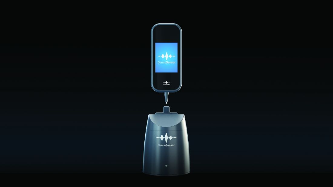
DermaSensor, described in the study conducted by Dr. Hartman and others as a noninvasive, point-and-click spectrometer, is a wireless handheld piece that weighs about 10 ounces. The unit captures five recordings to generate a spectral reading, which an algorithm in the software unit analyzes. The study found a sensitivity of 95.5% and specificity of 32.5% for melanoma detection with the device.
The target market for DermaSensor is primary care physicians, and, according to the FDA announcement in January, it is indicated for evaluating skin lesions “suggestive” of melanoma, basal cell carcinoma (BCC), and/or squamous cell carcinoma (SCC) in patients aged 40 and older to “assist healthcare providers in determining whether to refer a patient to a dermatologist.”
So Many Cases, So Few Dermatologists
In dermatology, AI devices have the potential to streamline the crushing burden of diagnosing skin cancer, said Yun Liu, PhD, a senior staff scientist at Google Research, Mountain View, California, who’s worked on developing machine-learning tools in dermatology among other medical fields. “Many people cannot access dermatology expertise when they most need it, ie, without waiting a long time. This causes substantial morbidity for patients,” Dr. Liu said in an interview.

His own research of an AI-based tool to help primary care physicians and nurse practitioners in teledermatology practices diagnose skin conditions documented the shortage of dermatologists to triage lesions, including a finding that only about one quarter of skin conditions are seen by a specialist and that nonspecialists play a pivotal role in the management of skin lesions.
The Centers for Disease Control and Prevention reports that about 6.1 million adults are treated for BCC and SCCs each year. The American Medical Association estimates that 13,200 active dermatologists practice in the United States.
Overcoming Barriers to AI in Dermatology
Before AI makes significant inroads in dermatology, clinicians need to see more verifiable data, said Roxana Daneshjou, MD, PhD, assistant professor of biomedical data science and dermatology at Stanford University, Stanford, California. “One of the challenges is having the availability of models that actually improve clinical care because we have some very early prospective trials on different devices, but we don’t have large-scale randomized clinical trials of AI devices showing definitive behaviors such as improved patient outcomes, that it helps curb skin cancer, or it catches it like dermatologists but helps reduce the biopsy load,” she said. “You need good data.”

Another challenge she noted was overcoming biases built into medicine. “A lot of the image-based models are built on datasets depicting skin disease on White skin, and those models don’t work so well on people with brown and black skin, who have historically had worse outcomes and also have been underrepresented in dermatology,” said Dr. Daneshjou, an associate editor of NEJM AI.
There’s also the challenge of getting verified AI models into the clinic. “Similar to many medical AI endeavors, developing a proof-of-concept or research prototype is far easier and faster than bringing the development to real users,” Dr. Liu said. “In particular, it is important to conduct thorough validation studies on various patient populations and settings and understand how these AI tools can best fit into the workflow or patient journey.”
A study published in 2023 documented progress Google made in deploying AI models in retina specialty clinics in India and Thailand, Dr. Liu noted.
Another challenge is to avoid overdiagnosis with these new technologies, Dr. Hartman said. Her group’s study showed the DermaSensor had a positive predictive value of 16% and a negative predictive value of 98.5%. “I think there’s some question about how this will factor into overdiagnosis. Could this actually bombard dermatologists more if the positive predictive value’s only 16%?”
One key to dermatologists accepting AI tools is having a transparent process for validating them, Dr. Lee said. “Even with FDA clearance, we don’t have the transparency we need as clinicians, researchers, and advocates of machine learning and AI in healthcare.”
But, Dr. Lee noted, the FDA in June took a step toward illuminating its validation process when it adopted guiding principles for transparency for machine learning–enabled devices. “Once we can get more access to this information and have more transparency, that’s where we can think about actually about making the decision to implement or not implement into local healthcare settings,” she said. The process was further enabled by a White House executive order in October 2023 on the safe, secure, and trustworthy development and use of AI.
The experience with telehealth during the COVID-19 pandemic, when patients and providers quickly embraced the technology to stay connected, serves as a potential template for AI, Dr. Lee noted. “As we’d seen with telehealth through the pandemic, you also need the cultural evolution and the development of the infrastructure around it to actually make sure this is a sustainable implementation and a scalable implementation in healthcare.”
Dr. Lee had no relevant relationships to disclose. Dr. Hartman received funding from DermaSensor for a study. Dr. Witkowski is a cofounder of Sklip. Dr. Liu is an employee of Google Research. Dr. Daneshjou reported financial relationships with MD Algorithms, Revea, and L’Oreal.
A version of this article first appeared on Medscape.com.
Emerging according to researchers and dermatologists investigating AI.
While some AI-integrated devices designed to triage skin lesions have emerged, including one that received Food and Drug Administration (FDA) clearance earlier in 2024, it may be some time before AI has a meaningful clinical impact in dermatology and, more specifically, the diagnosis of skin cancer, Ivy Lee, MD, a dermatologist in Pasadena, California, and chair of the American Academy of Dermatology’s augmented intelligence committee, told this news organization.
“It hasn’t really translated into clinical practice yet,” Dr. Lee said of AI in dermatology. “There have been significant advances in terms of the technical possibility and feasibility of these tools, but the translation and integration of AI into actual clinical work flows to benefit patients beyond academic research studies has been limited.” More studies and more “easily accessible and digestible information” are needed to evaluate AI tools in dermatologic practice.
“In dermatology, we’re on a cusp with AI,” said Rebecca Hartman, MD, MPH, chief of dermatology at the VA Boston Healthcare System and director of melanoma epidemiology at Brigham and Women’s Hospital, Boston, Massachusetts. “I think it’s going to come and change what we do,” which is especially true for any image-based specialty,” including radiology and pathology, in addition to dermatology.
Dr. Hartman led a study of one of these emerging technologies, the handheld elastic scattering spectroscopy device DermaSensor, which was cleared by the FDA in January for evaluating skin lesions suggestive of skin cancer.
Early AI Devices for Skin Cancer Detection
At the American Society for Laser Medicine and Surgery (ASLMS) meeting in April, a panel explored a number of algorithms with dermatologic applications that use AI to triage skin lesions, including DermaSensor.
Raman spectroscopy, which contains a handheld Raman probe, a diode laser, and a detecting spectrograph. A laser beam — which at 1.56 W/cm2 is below the maximum permissible exposure — focuses on the skin target with a 3.5-mm spot, gathers data on the target, and feeds it back into the unit that houses the algorithm that evaluates the spot analysis. It’s still in the investigative phase. A clinical trial, published almost 5 years ago, demonstrated a sensitivity of 90%-99% and a specificity of 24%-66% for skin cancer.
A dermatoscope called Sklip clips onto a smartphone and performs what company cofounder Alexander Witkowski, MD, PhD, described as an “optical painless virtual biopsy” for at-home use. The device uploads the captured image to an AI platform for analysis. It received FDA breakthrough device designation in 2022. At the ASLMS meeting, Dr. Witkowski said that clinical performance showed the device had a 97% sensitivity and 30% specificity for skin cancer.
DermaSensor, described in the study conducted by Dr. Hartman and others as a noninvasive, point-and-click spectrometer, is a wireless handheld piece that weighs about 10 ounces. The unit captures five recordings to generate a spectral reading, which an algorithm in the software unit analyzes. The study found a sensitivity of 95.5% and specificity of 32.5% for melanoma detection with the device.
The target market for DermaSensor is primary care physicians, and, according to the FDA announcement in January, it is indicated for evaluating skin lesions “suggestive” of melanoma, basal cell carcinoma (BCC), and/or squamous cell carcinoma (SCC) in patients aged 40 and older to “assist healthcare providers in determining whether to refer a patient to a dermatologist.”
So Many Cases, So Few Dermatologists
In dermatology, AI devices have the potential to streamline the crushing burden of diagnosing skin cancer, said Yun Liu, PhD, a senior staff scientist at Google Research, Mountain View, California, who’s worked on developing machine-learning tools in dermatology among other medical fields. “Many people cannot access dermatology expertise when they most need it, ie, without waiting a long time. This causes substantial morbidity for patients,” Dr. Liu said in an interview.
His own research of an AI-based tool to help primary care physicians and nurse practitioners in teledermatology practices diagnose skin conditions documented the shortage of dermatologists to triage lesions, including a finding that only about one quarter of skin conditions are seen by a specialist and that nonspecialists play a pivotal role in the management of skin lesions.
The Centers for Disease Control and Prevention reports that about 6.1 million adults are treated for BCC and SCCs each year. The American Medical Association estimates that 13,200 active dermatologists practice in the United States.
Overcoming Barriers to AI in Dermatology
Before AI makes significant inroads in dermatology, clinicians need to see more verifiable data, said Roxana Daneshjou, MD, PhD, assistant professor of biomedical data science and dermatology at Stanford University, Stanford, California. “One of the challenges is having the availability of models that actually improve clinical care because we have some very early prospective trials on different devices, but we don’t have large-scale randomized clinical trials of AI devices showing definitive behaviors such as improved patient outcomes, that it helps curb skin cancer, or it catches it like dermatologists but helps reduce the biopsy load,” she said. “You need good data.”
Another challenge she noted was overcoming biases built into medicine. “A lot of the image-based models are built on datasets depicting skin disease on White skin, and those models don’t work so well on people with brown and black skin, who have historically had worse outcomes and also have been underrepresented in dermatology,” said Dr. Daneshjou, an associate editor of NEJM AI.
There’s also the challenge of getting verified AI models into the clinic. “Similar to many medical AI endeavors, developing a proof-of-concept or research prototype is far easier and faster than bringing the development to real users,” Dr. Liu said. “In particular, it is important to conduct thorough validation studies on various patient populations and settings and understand how these AI tools can best fit into the workflow or patient journey.”
A study published in 2023 documented progress Google made in deploying AI models in retina specialty clinics in India and Thailand, Dr. Liu noted.
Another challenge is to avoid overdiagnosis with these new technologies, Dr. Hartman said. Her group’s study showed the DermaSensor had a positive predictive value of 16% and a negative predictive value of 98.5%. “I think there’s some question about how this will factor into overdiagnosis. Could this actually bombard dermatologists more if the positive predictive value’s only 16%?”
One key to dermatologists accepting AI tools is having a transparent process for validating them, Dr. Lee said. “Even with FDA clearance, we don’t have the transparency we need as clinicians, researchers, and advocates of machine learning and AI in healthcare.”
But, Dr. Lee noted, the FDA in June took a step toward illuminating its validation process when it adopted guiding principles for transparency for machine learning–enabled devices. “Once we can get more access to this information and have more transparency, that’s where we can think about actually about making the decision to implement or not implement into local healthcare settings,” she said. The process was further enabled by a White House executive order in October 2023 on the safe, secure, and trustworthy development and use of AI.
The experience with telehealth during the COVID-19 pandemic, when patients and providers quickly embraced the technology to stay connected, serves as a potential template for AI, Dr. Lee noted. “As we’d seen with telehealth through the pandemic, you also need the cultural evolution and the development of the infrastructure around it to actually make sure this is a sustainable implementation and a scalable implementation in healthcare.”
Dr. Lee had no relevant relationships to disclose. Dr. Hartman received funding from DermaSensor for a study. Dr. Witkowski is a cofounder of Sklip. Dr. Liu is an employee of Google Research. Dr. Daneshjou reported financial relationships with MD Algorithms, Revea, and L’Oreal.
A version of this article first appeared on Medscape.com.
Twice-Yearly PrEP Gives ‘Huge’ 100% Protection
Twice-yearly injections are 100% effective in preventing new infections, according to the final results from the PURPOSE 1 trial of lenacapavir.
For weeks, the HIV community has been talking about this highly anticipated clinical trial and whether the strong — and to many, surprising — interim results would hold at final presentation at the International AIDS Conference 2024 in Munich, Germany.
Presenting the results, Linda-Gail Bekker, MD, director of the Desmond Tutu HIV Center at the University of Cape Town, South Africa, reported zero new infections in those who got the shots in the study of about 5000 young women. In the group given daily oral preexposure prophylaxis (PrEP), roughly 2% contracted HIV from infected partners.
“A twice-yearly PrEP choice could overcome some of the adherence and persistence challenges and contribute critically to our quest to reduce HIV infection in women around the world,” Dr. Bekker said about the results, which were published simultaneously in The New England Journal of Medicine.
PURPOSE 1 confirmed that lenacapavir is a “breakthrough” for HIV prevention, said International AIDS Society president Sharon Lewin, PhD, MBBS. It has “huge public health potential,” said Dr. Lewin, the AIDS 2024 conference cochair and director of the Peter Doherty Institute for Infection and Immunity at the University of Melbourne in Australia.
Lenacapavir is a novel, first-in-class multistage HIV-1 capsid inhibitor with a long half-life, which enables the twice-yearly dosing.
PURPOSE 1 enrolled women aged 15-25 years who were at risk for HIV in South Africa and Uganda, with a primary endpoint of HIV infection. Because of the previously announced interim results, which showed the injection was preventing infections, study sponsor Gilead Sciences discontinued the randomized phase of the trial and shifted to an open-label design for lenacapavir.
“One hundred percent efficacy is more that we could ever have hoped for a potential prevention efficacy,” said Christoph Spinner, MD, MBA, an infectious disease specialist at the University Hospital of the Technical University of Munich and AIDS 2024 conference cochair.
Dr. Spinner added that while this is the first study of lenacapavir for PrEP, it’s also the first to explore outcomes of emtricitabine-tenofovir in cisgender women.
Strong Adherence Rates
The twice-yearly injection demonstrated adherence rates above 90% in the trial for both the 6- and 12-month injection intervals.
“Adherence was 91.5% at week 26 and 92.8% at week 52,” Dr. Bekker reported.
The trial compared three PrEP options including the lenacapavir injection to once-daily oral emtricitabine 200 mg and tenofovir-alafenamide 25 mg (F/TAF) and once-daily emtricitabine 200 mg and tenofovir–disoproxil fumarate 300 mg (F/TDF).
“Most participants in both the F/TAF and F/TDF groups had low adherence, and this declined over time,” Dr. Bekker reported. At 52 weeks, the vast majority of patients on both oral therapies had low adherence with dosing, defined at less than two doses a week.
Dr. Bekker called the adherence to the oral agents in this trial “disappointing.”
Findings from the trial underscore the challenges of adherence to a daily oral medication, Rochelle Walensky, MD, and Lindsey Baden, MD, from the Harvard Kennedy School of Government and Harvard Business School in Cambridge, Massachusetts, wrote in an editorial accompanying the published results.
With almost 92% attendance for the twice-yearly lenacapavir injections, the “well-done,” large, randomized, controlled trial “exemplifies not only that women can dependably adhere to this administration schedule, but also that levels of an HIV-1 capsid inhibitor can remain high enough over a period of 6 months to reliably prevent infection,” they added.
Another key focus of the presentation was adverse events. The rate of adverse events grade 3 or more in the lenacapavir arm was 4.1%, Bekker said, which is slightly lower than the rates in the oral arms. The rates of serious adverse events were 2.8% for lenacapavir, 4% for F/TAF and 3.3% for F/TDF.
Injection Site Reactions
Injection site reactions occurred in 68% of the lenacapavir group, including 63% with subcutaneous nodules.
The injection can form “a drug depot which may be palpable as a nodule,” Dr. Bekker said. In the placebo group, 34% of patients had injection-site reactions and 16% had nodules. Nearly all injection-site reactions were grade 1 or 2, she said. “Higher grade injection-site reactions were rare and not serious and occurred in a similar percentage in lenacapavir and placebo,” she said.
Overall, more than 25,000 injections of lenacapavir have been given, Dr. Bekker said, and four patients discontinued treatment because of injection-site reactions. “Reporting of injection-site reactions, including nodules, decreased with subsequent doses,” she said.
Contraception was not a requirement for enrollment in the study, Dr. Bekker pointed out, and pregnancy outcomes across the treatment arms were similar to the general population.
First in a Series of Trials
This is the first in a series of PURPOSE trials, Bekker reported. The phase 3 PURPOSE 2 trial, enrolling 3000 gay men, transgender women, transgender men and gender nonbinary people who have sex with male partners, is the second pivotal trial now underway.
Three other smaller trials are in the clinic in the United States and Europe.
PURPOSE 1 participants will continue to access lenacapavir until the product is available in South Africa and Uganda, Dr. Bekker said. Trial sponsor Gilead Sciences is also developing a direct licensing program to expedite generic access to the drug in high-incidence, resource-limited countries, she said.
Dr. Walensky and Dr. Baden report that lenacapavir currently costs about $43,000 annually in the United States. “But the results of the PURPOSE 1 trial have now created a moral imperative to make lenacapavir broadly accessible and affordable as PrEP” to people who were enrolled, as well as all those who are similarly eligible and could benefit.
So now we have a PrEP product with high efficacy, they added. “That is great news for science but not (yet) great for women.”
Given the high pregnancy rate among participants in the PURPOSE 1 trial, Dr. Walensky and Dr. Baden point out the assessment of lenacapavir safety is a priority. They are also interested in learning more about drug resistance with this new option.
“I f approved and delivered — rapidly, affordably, and equitably — to those who need or want it, this long-acting tool could help accelerate global progress in HIV prevention,” Dr. Lewin said.
Now, she added, “we eagerly await results from PURPOSE 2.”
A version of this article first appeared on Medscape.com.
FROM AIDS 2024
How Dermatologists Can Safeguard Against Malpractice Claims
for liability. Dermatologists can protect themselves by understanding malpractice trends and taking preventive steps, such as making sure NPOs have appropriate training and using a rigorous informed consent process, according to a dermatology resident and a dermatologist who have researched recent trends in dermatology lawsuits.
“It’s really important that physicians recognize their responsibility when delegating procedures to nonphysician operators and the physician’s role in supervision of these procedures,” Scott Stratman, MD, MPH, a dermatology resident at the Icahn School of Medicine at Mount Sinai, New York City, told this news organization. He led a study recently published in the Journal of the American Academy of Dermatology, which found that the majority (52%) of malpractice cases for cutaneous energy-based device procedures in the LexisNexis database from 1985 to September 2023 involved NPOs. The study did not break the data down between different types of NPOs.

Trends in Dermatology Malpractice
This follows a similar trend reported in a 2014 study led by Mathew M. Avram, MD, JD, director of the MGH Dermatology Laser and Cosmetic Center at Massachusetts General Hospital, Boston. The study analyzed liability claims related to cutaneous laser surgery performed by nonphysicians from January 1999 to December 2012.

“With nonphysician litigation data, we saw trend lines beginning in 2008 where the proportion of cases began to increase,” Dr. Avram said at the American Society for Laser Medicine and Surgery (ASLMS) meeting on April 12, 2024. “Over a period of 2008-2012, it went from 36% of cases to about 78%,” he said.
About a quarter (23.4%) of those were in medical offices; 76.6% were in nontraditional settings such as medical spas, he added. The proportion of NPOs was similar in a 2022 study that looked at causes of litigation in cutaneous laser surgery from 2012 to 2020, Dr. Avram said. Again, neither study broke down cases involving NPOs by specific type, but the 2014 study reported that 64% of cases by NPOs occurred outside of a traditional medical setting.
“So it seems that the location and potentially the supervision are issues that are important to patient safety,” Dr. Avram said at the meeting. While state laws regarding laser delegation vary widely, “depending on where you practice, it’s incumbent upon you to know that.”
Dr. Avram and colleagues were also the authors of a study published in June in Dermatologic Surgery that looked at the reasons behind ligations involving dermatologists in a retrospective analysis of 48 state and federal cases between 2011 and 2022. The majority of cases — 54.2% — were for unexpected harm, followed by wrong or delayed diagnoses, which accounted for a third of litigations.
Dr. Stratman’s study found that laser hair removal was the most common procedure for malpractice claims in dermatology among cutaneous energy-based device procedures. Complications from energy-based devices included burns, scarring, and pigmentation changes.
The growth of malpractice suits involving NPOs could be because NPOs are performing a greater proportion of dermatologic procedures, “particularly those practicing without direct supervision, such as in the context of a medical spa,” Dr. Stratman said in the interview. “Again, this highlights a physician’s responsibility in delegating these kinds of procedures to NPOs.”
Training Is a Must — But Not Standardized
Comprehensive training for physicians, staff, NPOs, and physicians “is all necessary and paramount in order to diminish adverse outcomes and legal risk, and then, of course, all these practitioners, be it staff or [NPOs], and, of course, physicians, are all held to the same standard of care,” Dr. Stratman said.
However, he added, “There is really no standardized training to operate these devices. That being said, it’s really important to know that both providers and facility owners have a significant obligation to their patients to make sure that their staff in their centers are appropriately trained.”
Training not only involves protocols and procedures but also how to handle patient interactions, Dr. Stratman said.
The legal concept of respondeat superior applies when nonphysicians participate in a patient’s care, Dr. Avram said at the ASLMS meeting. The physician is held liable for a nonphysician’s “negligence provided he or she is an employee receiving a salary [and] benefits and is performing within the scope of his or her duty,” regardless of whether the physician saw the patient or not at that visit, he said. Again, supervision of nonphysician laser procedures varies from state to state, he added.
“So the take-home point is to provide excellent training and appropriate supervision, and if you’re the owner of that practice, you are liable in the event of negligence even though you never were part of the treatment,” Dr. Avram said.
Ins and Outs of Informed Consent
When a patient outcome is less than desirable, or at least less than what the patient expected, a transparent and thorough informed consent process can protect the practice and physician, Dr. Avram said at the meeting.
“Malpractice and consent have nothing to do with each other,” he said. “Consent is getting permission to do a procedure. It’s needed actually for any medical intervention that you perform. What you need to do is to provide information to enable the patient or guardian or to choose knowledgeably among reasonable medical alternatives. This places the patient in control of the course of their medical treatment.”
The information conveyed to the patient should include the diagnosis, the medical causes, the nature and purpose of the treatment, and the risks and alternatives of procedure, “particularly if they’re high risk,” Dr. Avram said.
“Failure to obtain informed consent constitutes a civil battery, and the physician is liable for civil damages,” he said. “The patient need only show that he or she was not informed of the medical nature of the medical touching; physical injury is not necessary.”
A battery could occur if a procedure extends beyond the scope or area of treatment the patient agreed to — for example, extending a liposuction to an area that wasn’t originally targeted, or extending a laser procedure to an area of the body as a presumed favor to the patient. “It does not require a standard of care or an expert witness,” Dr. Avram said. “One only needs to show nonconsensual touching.”
Informed consents should include plain language, he said. “The whole idea is the patient understands what the risks and benefits are,” Dr. Avram said. “You don’t need to use medical jargon.” As an example, he suggested using the term “blisters” instead of “bullae.” If the treatment involves an off-label procedure, include that too, he said.
He also advised avoiding blanket authorizations. “Courts disfavor them,” he noted. “They need more specificity. So those are not valid.”
Dr. Stratman added that providers should think about the setting in which they obtain informed consent. “It’s really important that providers are consenting their patients in private and quiet places, free from distractions, that they accommodate patients who might have disabilities or limitations in English proficiency, using a teach-back method to help patients understand or demonstrate their understanding of the procedure in order to gauge comprehension,” he said.
Both Dr. Avram and Dr. Stratman pointed out that another strategy to prevent malpractice is to build trusting patient-provider relationships. “The patient-provider relationship is paramount not only to the success of the procedure but to the clinical visit as a whole,” Dr. Stratman said.
That’s a two-way street, he added. Patients should be able to trust that their provider provides them with the best treatment based on their own history, and providers should also be able to trust that patients are providing them with an accurate history, asking relevant questions, or expressing any level of apprehension about the procedure or visit. “The patient-provider relationship is everything,” Dr. Stratman said.
Dr. Stratman and Dr. Avram had no relevant disclosures.
A version of this article appeared on Medscape.com.
for liability. Dermatologists can protect themselves by understanding malpractice trends and taking preventive steps, such as making sure NPOs have appropriate training and using a rigorous informed consent process, according to a dermatology resident and a dermatologist who have researched recent trends in dermatology lawsuits.
“It’s really important that physicians recognize their responsibility when delegating procedures to nonphysician operators and the physician’s role in supervision of these procedures,” Scott Stratman, MD, MPH, a dermatology resident at the Icahn School of Medicine at Mount Sinai, New York City, told this news organization. He led a study recently published in the Journal of the American Academy of Dermatology, which found that the majority (52%) of malpractice cases for cutaneous energy-based device procedures in the LexisNexis database from 1985 to September 2023 involved NPOs. The study did not break the data down between different types of NPOs.
Trends in Dermatology Malpractice
This follows a similar trend reported in a 2014 study led by Mathew M. Avram, MD, JD, director of the MGH Dermatology Laser and Cosmetic Center at Massachusetts General Hospital, Boston. The study analyzed liability claims related to cutaneous laser surgery performed by nonphysicians from January 1999 to December 2012.
“With nonphysician litigation data, we saw trend lines beginning in 2008 where the proportion of cases began to increase,” Dr. Avram said at the American Society for Laser Medicine and Surgery (ASLMS) meeting on April 12, 2024. “Over a period of 2008-2012, it went from 36% of cases to about 78%,” he said.
About a quarter (23.4%) of those were in medical offices; 76.6% were in nontraditional settings such as medical spas, he added. The proportion of NPOs was similar in a 2022 study that looked at causes of litigation in cutaneous laser surgery from 2012 to 2020, Dr. Avram said. Again, neither study broke down cases involving NPOs by specific type, but the 2014 study reported that 64% of cases by NPOs occurred outside of a traditional medical setting.
“So it seems that the location and potentially the supervision are issues that are important to patient safety,” Dr. Avram said at the meeting. While state laws regarding laser delegation vary widely, “depending on where you practice, it’s incumbent upon you to know that.”
Dr. Avram and colleagues were also the authors of a study published in June in Dermatologic Surgery that looked at the reasons behind ligations involving dermatologists in a retrospective analysis of 48 state and federal cases between 2011 and 2022. The majority of cases — 54.2% — were for unexpected harm, followed by wrong or delayed diagnoses, which accounted for a third of litigations.
Dr. Stratman’s study found that laser hair removal was the most common procedure for malpractice claims in dermatology among cutaneous energy-based device procedures. Complications from energy-based devices included burns, scarring, and pigmentation changes.
The growth of malpractice suits involving NPOs could be because NPOs are performing a greater proportion of dermatologic procedures, “particularly those practicing without direct supervision, such as in the context of a medical spa,” Dr. Stratman said in the interview. “Again, this highlights a physician’s responsibility in delegating these kinds of procedures to NPOs.”
Training Is a Must — But Not Standardized
Comprehensive training for physicians, staff, NPOs, and physicians “is all necessary and paramount in order to diminish adverse outcomes and legal risk, and then, of course, all these practitioners, be it staff or [NPOs], and, of course, physicians, are all held to the same standard of care,” Dr. Stratman said.
However, he added, “There is really no standardized training to operate these devices. That being said, it’s really important to know that both providers and facility owners have a significant obligation to their patients to make sure that their staff in their centers are appropriately trained.”
Training not only involves protocols and procedures but also how to handle patient interactions, Dr. Stratman said.
The legal concept of respondeat superior applies when nonphysicians participate in a patient’s care, Dr. Avram said at the ASLMS meeting. The physician is held liable for a nonphysician’s “negligence provided he or she is an employee receiving a salary [and] benefits and is performing within the scope of his or her duty,” regardless of whether the physician saw the patient or not at that visit, he said. Again, supervision of nonphysician laser procedures varies from state to state, he added.
“So the take-home point is to provide excellent training and appropriate supervision, and if you’re the owner of that practice, you are liable in the event of negligence even though you never were part of the treatment,” Dr. Avram said.
Ins and Outs of Informed Consent
When a patient outcome is less than desirable, or at least less than what the patient expected, a transparent and thorough informed consent process can protect the practice and physician, Dr. Avram said at the meeting.
“Malpractice and consent have nothing to do with each other,” he said. “Consent is getting permission to do a procedure. It’s needed actually for any medical intervention that you perform. What you need to do is to provide information to enable the patient or guardian or to choose knowledgeably among reasonable medical alternatives. This places the patient in control of the course of their medical treatment.”
The information conveyed to the patient should include the diagnosis, the medical causes, the nature and purpose of the treatment, and the risks and alternatives of procedure, “particularly if they’re high risk,” Dr. Avram said.
“Failure to obtain informed consent constitutes a civil battery, and the physician is liable for civil damages,” he said. “The patient need only show that he or she was not informed of the medical nature of the medical touching; physical injury is not necessary.”
A battery could occur if a procedure extends beyond the scope or area of treatment the patient agreed to — for example, extending a liposuction to an area that wasn’t originally targeted, or extending a laser procedure to an area of the body as a presumed favor to the patient. “It does not require a standard of care or an expert witness,” Dr. Avram said. “One only needs to show nonconsensual touching.”
Informed consents should include plain language, he said. “The whole idea is the patient understands what the risks and benefits are,” Dr. Avram said. “You don’t need to use medical jargon.” As an example, he suggested using the term “blisters” instead of “bullae.” If the treatment involves an off-label procedure, include that too, he said.
He also advised avoiding blanket authorizations. “Courts disfavor them,” he noted. “They need more specificity. So those are not valid.”
Dr. Stratman added that providers should think about the setting in which they obtain informed consent. “It’s really important that providers are consenting their patients in private and quiet places, free from distractions, that they accommodate patients who might have disabilities or limitations in English proficiency, using a teach-back method to help patients understand or demonstrate their understanding of the procedure in order to gauge comprehension,” he said.
Both Dr. Avram and Dr. Stratman pointed out that another strategy to prevent malpractice is to build trusting patient-provider relationships. “The patient-provider relationship is paramount not only to the success of the procedure but to the clinical visit as a whole,” Dr. Stratman said.
That’s a two-way street, he added. Patients should be able to trust that their provider provides them with the best treatment based on their own history, and providers should also be able to trust that patients are providing them with an accurate history, asking relevant questions, or expressing any level of apprehension about the procedure or visit. “The patient-provider relationship is everything,” Dr. Stratman said.
Dr. Stratman and Dr. Avram had no relevant disclosures.
A version of this article appeared on Medscape.com.
Mediterranean Diet Lowers Tachyarrhythmia in Paroxysmal AF
BOSTON — A Mediterranean diet with extra virgin olive oil (EVOO) significantly reduced the risk for tachyarrhythmia recurrence after atrial fibrillation (AF) ablation in patients with paroxysmal disease, but the diet had less of an impact on patients with persistent AF, a new study showed.
“An intervention with the Mediterranean diet with EVOO produced a nonsignificant reduction in any atrial tachycardia in a selected population after undergoing atrial fibrillation ablation, but this intervention produced a significant reduction in any atrial tachyarrhythmias in patients with paroxysmal AF,” said Maria Teresa Barrio-Lopez, MD, PhD, an electrophysiologist at University Hospital HM Monteprincipe in Madrid, Spain, who presented results from the PREDIMAR trial at the Heart Rhythm Society (HRS) 2024 annual meeting.
The PREDIMAR study enrolled 720 patients from the larger PREDIMED study, which showed that patients without AF at enrollment and who followed a Mediterranean diet enriched with EVOO had a 38% lower rate of incidental AF than control individuals.
PREDIMAR evaluated the impact of the diet on arrhythmia recurrence in patients after ablation. The patients were randomized in a 1:1 ratio to either the dietary intervention group or the control group.
PREDIMAR Study Results
However, among the 431 patients with paroxysmal AF, 25.2% in the diet group and 34.7% in the control group had no tachyarrhythmia recurrence, which translates into a 31% lower risk in the diet group.
In this study, the diet was rich in fish, nuts, fruits, and vegetables and was complemented with EVOO. Participants were also permitted moderate wine consumption.
The intervention involved dietitians who remotely followed patients and made periodic telephone calls to encourage them to stay on the diet. Participants had weight and body measurements taken at baseline and at 3, 6, 12, and 18 months and underwent an ECG at 6, 12, and 18 months. Labs were obtained at baseline and at 12 months. Participants were also given educational materials throughout the intervention.
Average scores, based on a scale of 0-13, excluding an item for wine intake, were 7.8 in the diet group and 7.2 in the control group.
Daily average alcohol intake was higher in the diet group than in the control group (9.8 vs 8.2 g), but “the weight of the patient during the study didn’t change in any group,” Dr. Barrio-Lopez reported.
Baseline characteristics were similar in the two groups. About 60% were taking antiarrhythmic drugs, and about 84% were taking anticoagulants.
‘A Tour de Force’
PREDIMAR was “really a tour de force,” Christine Albert, MD, MPH, chair of cardiology at the Smidt Heart Institute at the Cedars-Sinai Medical Center in Los Angeles, California, said during a commentary presented at HRS. “We talk about how we’re going to do these dietary interventions and weight loss and all the risk-factor reduction, and they pulled it off with 700 individuals and also did it in a way that was very novel.”
This is the first large-scale dietary intervention trial of patients with AF. However, Dr. Albert noted later in an interview, the Mediterranean diet poses potential challenges for some people with AF.
“The Mediterranean diet recommends that people drink wine, but then there’s clear evidence that abstinence from alcohol actually reduces recurrences of atrial fibrillation, so even though there are a lot of things about the Mediterranean diet that are probably healthy and good for atrial fibrillation, that aspect of it might be working against the patient,” she explained.
The finding that patients in the Mediterranean diet group experienced no significant weight loss could be counterintuitive when it comes to preventing AF. But “you could adapt the diet for AF,” Dr. Albert said. You could “leave out the wine and focus more on weight loss if the patient is obese because those are also the pillars of what we’ve learned for patients with atrial fibrillation.”
Making weight loss a key component of the study could be significant for the American population. “At least in the United States, that’s a huge part of the risk factors for atrial fibrillation after ablation,” she said.
The remote follow-up component of the PREDIMAR study is also intriguing. “I think what’s most exciting about what they did is, they showed they can do all these things remotely,” Dr. Albert added.
Dr. Barrio-Lopez had no relevant financial relationships. Dr. Albert disclosed relationships with Abbott, Roche Diagnostics, St. Jude Medical, Boston Scientific, Medtronic, and Element Science.
A version of this article appeared on Medscape.com.
FROM HRS 2024
Anticoagulation Shows No Benefit in Preventing Second Stroke
BOSTON — Patients who have had a stroke are thought to be at a higher risk for another one, but oral anticoagulation with edoxaban led to no discernible reduction in the risk for a second stroke, and the risk for major bleeding was more than quadruple the risk with no anticoagulation, a subanalysis of a major European trial has shown.
“There is no interaction between prior stroke or TIA [transient ischemic attack] and the treatment effect, and this is true for the primary outcome and the safety outcome,” Paulus Kirchoff, MD, director of cardiology at the University Heart and Vascular Center in Hamburg, Germany, said during his presentation of a subanalysis of the NOAH-AFNET 6 trial at the annual meeting of the Heart Rhythm Society (HRS) 2024. However, “there is a signal for more safety events in patients randomized to anticoagulation with a prior stroke.”
The subanalysis involved 253 patients who had had a stroke or TIA and who had device-detected atrial fibrillation (AF) from the overall NOAH-AFNET 6 population of 2536 patients, which enrolled patients 65 years and older with at least one additional CHA2DS-VASc risk factor and patients 75 years and older with device-detected subclinical AF episodes of at least 6 minutes. Patients were randomized to either edoxaban or no anticoagulation, but 53.9% of the no-anticoagulation group was taking aspirin at trial enrollment. Anticoagulation with edoxaban was shown to have no significant impact on stroke rates or other cardiovascular outcomes.
Subanalysis Results
In the subanalysis, a composite of stroke, systemic embolism, and cardiovascular death — the primary outcome — was similar in the edoxaban and no-anticoagulation groups (14/122 patients [11.5%] vs 16/131 patients [12.2%]; 5.7% vs 6.3% per patient-year).
The rate of recurrent stroke was also similar in the edoxaban and no-anticoagulation groups (4 of 122 patients [3.3%] vs 6 of 131 patients [4.6%]; 1.6% vs 2.3% per patient-year). And there were eight cardiovascular deaths in each group.
However, edoxaban patients had significantly higher rates of major bleeding.
“This is a subanalysis, so what we see in terms of the number of patients with events is not powered for a definitive answer, but we do see that there were 10 major bleeds in the group of patients with a prior stroke or TIA in NOAH,” Dr. Kirchoff reported. “Eight of those 10 major bleeds occurred in patients randomized to edoxaban.”
Results from the NOAH-AFNET 6 trial have been compared with those from the ARTESiA trial, which compared apixaban anticoagulation with aspirin in patients with subclinical AF and was also presented at HRS 2024. ARTESiA showed that apixaban significantly lowered the risk for stroke and systemic embolism.
“In ARTESiA, everyone was on aspirin when they were randomized to no anticoagulation; in NOAH, only about half were on aspirin,” Dr. Kirchoff said.
Both studies had similar outcomes for cardiovascular death in the anticoagulation and no-anticoagulation groups. “It’s not significant; it may be chance, but it’s definitely not the reduction in death that we have seen in the anticoagulant trials,” Dr. Kirchoff said. “When you look at the meta-analyses of the early anticoagulation trials, there’s a one third reduction in death, and here we’re talking about a smaller reduction.”
This research points to a need for a better way to evaluate stroke risk. “We need new markers,” Dr. Kirchoff said. “Some of them may be in the blood or imaging, genetics maybe, and one thing that really emerges from my perspective is that we now have the first evidence to suggest that patients with a very low atrial fibrillation burden have a low stroke rate.”
More research is needed to better understand AF characteristics and stroke risk, he said.
AF Care Enters a ‘Gray Zone’
The NOAH-AFNET 6 results, coupled with those from ARTESiA, are changing the paradigm for anticoagulation in patients with stroke, said Taya Glotzer, MD, an electrophysiologist at the Hackensack University Medical Center in Hackensack, New Jersey, who compiled her own analysis of the studies’ outcomes.
“In ARTESiA, the stroke reduction was only 0.44% a year, with a number needed to treat of 250,” she said. “In the NOAH-AFNET 6 main trial, the stroke reduction was 0.2%, with the number needed to treat of 500, and in the NOAH prior stroke patients, there was a 0.7% reduction, with a number needed to treat of 143.”
None of these trials would meet the standard for a class 1 recommendation for anticoagulation with a reduction of even 1%-2% per year, she noted, but they do show that the stroke rate “is very, very low” in prior patients with stroke.
“Prior to 2024, we knew what was black and white; we knew who to anticoagulate and who not to anticoagulate. And now we are in a gray zone, trying to balance the risk of stroke and bleeding. We have to individualize or hope for substudies, perhaps using the CHA2DS-VASc score or other information about the left atrium, to help us make decisions in these patients. It’s not just going to be black and white,” she said.
Dr. Kirchoff had no relevant financial relationships to disclose. Dr. Glotzer disclosed financial relationships with Medtronic, Abbott, Boston Scientific, and MediaSphere Medical.
A version of this article first appeared on Medscape.com.
FROM HRS 2024
Why Are We Undertreating So Many Pulmonary Embolisms?
LONG BEACH, CALIFORNIA — A small fraction of patients with pulmonary embolism (PE) who are eligible for advanced therapies are actually getting them, reported investigators who conducted a big data analysis.
“Advanced PE therapy seems to be vulnerable to disparate use, and perhaps underuse,” Sahil Parikh, MD, a cardiovascular interventionalist at the Columbia University Medical Center in New York, said when he presented results from the REAL-PE study at the Society for Cardiovascular Angiography and Interventions (SCAI) 2024 Scientific Sessions.
The underuse of advanced PE therapies is “the controversy,” Dr. Parikh said after his presentation. “It remains unclear what the role of invasive therapy is in the management of so-called high-intermediate–risk people. There isn’t a Class 1 guideline recommendation, and there is a very rapidly evolving trend that we’re increasingly treating these patients invasively,” he said.
“However, if you come to these meetings [such as SCAI], you might think everyone is getting one of these devices, but these data show that’s not the case,” Dr. Parikh said.
The analysis mined deidentified data from Truveta, a collective of health systems that provides regulatory-grade electronic health record data for research.
The researchers accessed data on patients treated with ultrasound-assisted catheter-directed thrombolysis or mechanical thrombectomy, identified from claims codes. Patient characteristics — age, race, ethnicity, sex, comorbidities, and diagnoses — were also accessed for the analysis. Earlier results were published in the January issue of the Journal of the Society for Cardiovascular Angioplasty Interventions.
Less Intervention for Black Patients and Women
White patients were more likely to receive advanced therapy than were Black patients (0.5% vs 0.37%; P = .000), Dr. Parikh reported, and women were less likely to receive advanced therapy than were men (0.41% vs 0.55%; P = .000).
The only discernable differences in outcomes were in major bleeding events in the 7 days after the procedure, which affected more White patients than it did Black patients (13.9% vs 9.3%) and affected more women than it did men (16.6% vs 11.1%).
What’s noteworthy about this study is that it demonstrates the potential of advanced data analytics to identify disparities in care and outcomes, Dr. Parikh said during his presentation. “These analyses provide a means of evaluating disparities in real clinical practice, both in the area of PE and otherwise, and may also be used for real-time monitoring of clinical decision-making and decisional support,” he said. “We do think that both novel and established therapies can benefit equally from similar types of analyses.”
Big Data Signaling Disparities
“That’s where these data are helpful,” Dr. Parikh explained. They provide “a real snapshot of how many procedures are being performed and in what kinds of patients. The low number of patients getting the procedure would suggest that there are probably more patients who would be eligible for treatment based on some of the emerging consensus documents, and they’re not receiving them.”
The data are “hypotheses generating,” Dr. Parikh said in an interview. “These hypotheses have to be evaluated further in more granular databases.”
REAL-PE is also a “clarion call” for clinical trials of investigative devices going forward, he said. “In those trials, we need to endeavor to enroll enough women and men, minority and nonminority patients so that we can make meaningful assessments of differences in efficacy and safety.”
This study is “real proof that big data can be used to provide information on outcomes for patients in a very rapid manner; that’s really exciting,” said Ethan Korngold, MD, chair of structural and interventional cardiology at the Providence Health Institute in Portland, Oregon. “This is an area of great research with great innovation, and it’s proof that, with these type of techniques using artificial intelligence and big data, we can generate data quickly on how we’re doing and what kind of patients we’re reaching.”
Findings like these may also help identify sources of the disparities, Dr. Korngold added.
“This shows we need to be reaching every patient with advanced therapies,” he said. “Different hospitals have different capabilities and different expertise in this area and they reach different patient populations. A lot of the difference in utilization stems from this fact,” he said.
“It just underscores the fact that we need to standardize our treatment approaches, and then we need to reach every person who’s suffering from this disease,” Dr. Korngold said.
A version of this article appeared on Medscape.com.
Study of AI for Retina Disease Finds Many Unusable Images
Artificial intelligence (AI) has drawn interest in ophthalmology for its potential to track disease trends in huge populations, such as the 38.4 million people in the United States with diabetes who are at risk for diabetic eye disease. However, a recent study using AI to detect diabetic retinopathy from retinal photo screenings has found wide disparities in the quality of data being fed into the algorithm.
And screening photos captured in nine primary care settings were three times more likely to be unusable than those obtained in two ophthalmology clinics, a study at Temple University in Philadelphia found. The results of the new research were reported at the Association for Research in Vision and Ophthalmology (ARVO) 2024 annual meeting.
“AI-assisted diabetic retinopathy screenings were more successful when completed in the ophthalmology clinic setting compared to the primary care setting,” study leader Madelyn Class, a medical student at Temple, told this news organization. One key difference, Ms. Class said, was that the specialty clinics used a photographer training in capturing ophthalmic images, while the primary care sites had medical assistants taking the photos.
Challenges of Screening in Primary Care
The American Diabetes Association acknowledged in a 2017 position statement that retinal photography has the potential to bring screening into settings where optometrists or ophthalmologists are unavailable. This study showed the potential may not yet be realized.
In the primary care setting, 42.5% of retinal photos were ungradable compared with 14.5% in the specialty settings.
The number of patients diagnosed with more-than-mild diabetic retinopathy also varied significantly between the two settings — 13% in primary care and 24% in ophthalmology — as did the rates of follow-up appointments: 58% and 80%, respectively.
“It seems user error played a role in the quality of photographs that were taken,” Ms. Class said. “Some of the images we received from the primary care settings were actually of the eyelid, or even the curtains on the wall, rather than the fundus.
“All the camera operators in the study received training on the imaging device,” Ms. Class added. “This suggests that some of the photographers were rushed, out of practice, or simply no longer interested in taking photos,” she said. “Apparently, we will have to continuously monitor the performance of each photographer to ensure that quality photos are being taken.”
The findings may also point to the need for using different equipment for screening in primary care, Ms. Class added. “Robotic as opposed to manual cameras may help eliminate some of the user error that was experienced with primary care screenings,” she said.
Need for Training ‘Fixable’
These findings demonstrate the challenges of capturing usable retinal images outside of an eye care professional’s office, according to Jennifer Lim, MD, director of the retina service at the University of Illinois Chicago.
“This study illustrates that implementation is the rub of AI,” Dr. Lim told this news organization. “Getting primary care doctors and clinics to want to adopt and figure out how to implement AI screening [for diabetic retinopathy] in a healthcare system is difficult, so I applaud the Temple University system for trying to integrate retinal photography-based AI screening into the primary care outpatient centers and comparing outcomes to the ophthalmology clinics.”
The study showed that photographers need not only initial training but also monitoring to avoid ungradable images, Dr. Lim added, a problem that is “fixable.”
“It’s going to take a lot of work to get the message out to the primary care practices that these autonomous, cloud-based systems are available and effective for detecting retinopathy,” she said.
But the effort is worth it, she added: “It doesn’t take much time to take these photos for diabetic retinopathy screening, and the potential benefits are huge because the earlier you diagnose diabetic retinopathy that’s more than mild, the more likely the patient can be sent for eye care in a timely fashion and thus prevent visual loss from diabetic retinopathy.”
Ms. Class had no relevant disclosures. Dr. Lim disclosed a past relationship with Eyenuk, the maker of retinal screening cameras.
A version of this article appeared on Medscape.com .
Vigilance Needed in Gout Treatment to Reduce CVD Risks
NEW YORK — Urate, the culprit of gout, affects the vasculature in multiple ways that can raise cardiovascular risk (CV) in an individual with gout, and following guidelines for gout treatment, including the use of colchicine, can be the key to reducing those risks.
“Guideline-concordant gout treatment, which is essentially an anti-inflammatory urate-lowering strategy, at least improves arterial physiology and likely reduces cardiovascular risk,” Michael H. Pillinger, MD, told attendees at the 4th Annual Cardiometabolic Risk in Inflammatory Conditions conference. Dr. Pillinger is professor of medicine and biochemistry and molecular pharmacology at New York University Grossman School of Medicine, New York City, who has published multiple studies on gout.
He cited evidence that has shown soluble urate stimulates the production of C-reactive protein (CRP), which is a predictor of cardiovascular disease (CVD). Another study, in which Dr. Pillinger participated, demonstrated that gout patients have impaired vascular endothelial function associated with a chronic, low-level inflammatory state, he said.
“There’s good evidence that urate itself affects the vasculature in multiple ways, and I suspect this may be a model for other metabolic effects on vasculature,” Dr. Pillinger said. “Patients with gout have abnormal endothelium in ways that really convey vascular risk.”
Gout, Inflammation, and CVD
However, for rheumatologists to study the association between gout-related inflammation and CVD is “very, very hard,” Dr. Pillinger added. “But I do think that the mechanisms by which gout induces biological changes in the vasculature may provide insights into cardiovascular disease in general.”
One way to evaluate the effects of gout on the endothelium in the clinic is to measure flow-mediated dilation. This technique involves placing an ultrasound probe over the brachial artery and measuring the baseline artery diameter. Then, with the blood pressure cuff over the forearm, inflate it to reduce flow, then release the cuff and measure the brachial artery diameter after the endothelium releases vasodilators.
Dr. Pillinger and colleagues evaluated this technique in 34 patients with gout and 64 controls and found that patients with gout had an almost 50% decrease in flow-mediated dilation, he said. “Interestingly, the higher the urate, the worse the flow; the more the inflammation, the worse the flow, so seemingly corresponding with the severity of the gout,” he said. That raised an obvious question, Dr. Pillinger continued: “If you can treat the gout, can you improve the flow-mediated dilation?”
His group answered that question with a study in 38 previously untreated patients with gout, giving them colchicine 0.6 mg twice daily for a month plus a urate-lowering xanthine oxidase inhibitor (allopurinol or febuxostat) to treat them to a target urate level of < 6 mg/dL. “We saw an increase in endothelial function, and it normalized,” Dr. Pillinger said.
However, some study participants didn’t respond. “They were people with well-established other cardiovascular comorbidities — hypertension, hyperlipidemia,” he said. “I think some people just have vessels that are too damaged to get at them just by fixing their gout problem or their inflammation.”
That means patients with gout need to be treated with colchicine early on to avoid CV problems, Dr. Pillinger added. “We ought to get to them before they have the other problems,” he said.
Managing gout, and the concomitant CV problems, requires vigilance both during and in between flares, Dr. Pillinger said after his presentation.
“We have always taught that patients between flares basically look like people with no gout, but we do know now that patients with gout between flares tend to have what you might call ‘subclinical’ inflammation: CRPs and ESRs [erythrocyte sedimentation rates] that are higher than those of the general population, though not so excessive that they might grab attention,” he said. “We also know that many, if not all, patients between flares have urate deposited in or around their joints, but how these two relate is not fully established.”
Better treatment within 3 months of an acute gout flare may reduce the risk for CV events, he said, but that’s based on speculation more so than clinical data.
Potential Benefits of Targeting Inflammation
“More chronically, we know from the cardiologists’ studies that anti-inflammatory therapy should reduce risk in the high-risk general population,” Dr. Pillinger said. “There are no prospective studies confirming that this approach will work among gout patients, but there is no reason why it shouldn’t work — except perhaps that gout patients may have higher inflammation than the general population and also have more comorbidities, so they could perhaps be more resistant.”
Dr. Pillinger said that his group’s studies and another led by Daniel Solomon, MD, at Brigham and Women’s Hospital in Boston, have indicated that anti-inflammatory strategies in gout will lower CV risk.
“And interestingly,” he added, “our data suggest that colchicine use may lower risk not only in high-risk gout patients but also in gout patients who start with no CAD [coronary artery disease] but who seem to have less incident CAD on colchicine. I see this as identifying that gout patients are intrinsically at high risk for CAD, even if they don’t actually have any, so they represent a population for whom lowering chronic inflammation may help prevent incident disease.”
Dr. Pillinger provided more evidence that the understanding of the relationship between gout, gout flares, and CV risk is evolving, said Michael S. Garshick, MD, who attended the conference and is head of the Cardio-Rheumatology Program at NYU Langone, New York City.
“There’s epidemiologic evidence supporting the association,” Dr. Garshick told this news organization after the conference. “We think that most conditions with immune system activation do tend to have an increased risk of some form of cardiovascular disease, but I think the relationship with gout has been highly underpublicized.”
Many patients with gout tend to have a higher prevalence of traditional cardiometabolic issues, which may compound the relationship, Dr. Garshick added. “However, I would argue that with this patient subset that it doesn’t matter because gout patients have a higher risk of traditional risk factors, and you have to [treat-to-target] those traditional risk factors.”
While the clinical evidence of a link between gout and atherosclerosis may not be conclusive, enough circumstantial evidence exists to believe that treating gout will reduce CV risks, he said. “Some of the imaging techniques do suggest that gouty crystals [are] in the atherosclerotic plaque of gout patients,” Dr. Garshick added. Dr. Pillinger’s work, he said, “is showing us that there are different pathways to develop atherosclerosis.”
Dr. Pillinger disclosed relationships with Federation Bio, Fortress Biotech, Amgen, Scilex, Hikma Pharmaceuticals, LG Chem, and Olatec Therapeutics. Dr. Garshick disclosed relationships with Kiniksa Pharmaceuticals, Agepha Pharma, Bristol Myers Squibb, and Horizon Therapeutics.
A version of this article appeared on Medscape.com.
Don’t Leave CVD Risk in RA Undertreated Despite Unresolved Questions
NEW YORK — Patients with rheumatoid arthritis (RA) carry a high risk for cardiovascular events, but mounting clinical evidence suggests they’re being undertreated to manage that risk. Rheumatologists should consider a patient with RA’s cardiovascular disease (CVD) status before deciding on RA treatments, a researcher of cardiometabolic disorders advised.
“The ORAL Surveillance trial suggests that we need to consider cardiovascular risk factors and maybe do additional screening in these patients before we use RA therapies,” Jon T. Giles, MD, PhD, director of the Cedars-Sinai Inflammatory Arthritis Clinical Center at Cedars-Sinai in Los Angeles, told attendees at the 4th Annual Cardiometabolic Risk in Inflammatory Conditions conference.
Underuse of Statins
ORAL Surveillance enrolled 4362 patients with RA aged 50 years and older with at least one cardiovascular risk factor. About 23% of all patients were taking statins, as were about half of patients with a history of atherosclerotic CVD (ASCVD).

“A lot of those people should have been on statins,” Dr. Giles said in an interview. “Not because of their RA but because of their risk factors, and then RA brings it up another notch.” In the population with ASCVD, Dr. Giles added, “It should have been more like 70% and 80%. If we’re talking about a disease that has enhanced cardiovascular risk, then the adoption of standard care that you would do for anybody in the general population should be at that standard and maybe above.”
Multiple studies have documented the underlying risk for CVD events, CV mortality, and subclinical atherosclerosis in people with RA, Dr. Giles noted in his presentation. Physiologically, the RA-specific risk factors most linked to CVD risk are systemic inflammation/cytokine excess and specific circulating T-cell and intermediate monocyte subsets, or both, Dr. Giles said.
Disease-Modifying Antirheumatic Drugs (DMARDs) and CVD Risk
Likewise, research in the past decade has linked methotrexate and tumor necrosis factor (TNF) inhibitors to reduced ASCVD events in RA. Another study showed that abatacept had an effect similar to that of etanercept in patients with RA, and the ENTRACTE trial, for which Dr. Giles was the lead author, demonstrated that tocilizumab matched etanercept in reducing CV events.
The ORAL Surveillance investigators also reported that patients with RA who were receiving the Janus kinase (JAK) inhibitor tofacitinib had a higher risk for major adverse cardiovascular events and cancers than those on TNF therapy, Dr. Giles noted. While statins in combination with JAK inhibitors may have the potential to provide a balance for controlling CV risk in patients with RA, he said later that the potential of JAK inhibitors in reducing CVD risk in RA “is still unsettled.”
The ongoing TARGET trial is further evaluating the impact of DMARDs on vascular inflammation in RA, said Dr. Giles, who’s also a trial principal investigator. TARGET is randomizing 115 patients with RA who didn’t respond to methotrexate to a TNF inhibitor or the addition of sulfasalazine and hydroxychloroquine to their methotrexate. Patients can be on low-intensity but not high-intensity statin therapy, Dr. Giles said.
TARGET results reported last year demonstrated an 8% decrease in arterial fluorodeoxyglucose (FDG) uptake on PET-CT in both treatment arms. Previous studies, Dr. Giles noted, have shown a potential link between FDG and histologic markers of inflammation. “An 8% decrease in vascular FDG is in line with what you would expect from statin treatment,” he said.
TARGET results published in April showed that a measure of a cluster of 12 cytokines and other inflammatory mediators, known as the multibiomarker disease activity (MBDA) score and marketed under the brand name Vectra DA, may help determine arterial FDG uptake. “Those who had a low MBDA score at week 24 actually had the greatest reduction in the arterial FDG,” he said.
Those results were driven entirely by low serum amyloid A (SAA) levels, Dr. Giles said. Those same results didn’t hold for patients in whom SAA and C-reactive protein were correlated.
“So, there’s more to come here,” Dr. Giles said. “We’re looking at other, much larger biomarker panels.”
Nonetheless, he said, sufficient evidence exists to conclude that treating RA to target reduces CV events. “The idea is that at every visit that you see an RA patient, you measure their disease activity, and if they’re not at the target of low disease activity or remission, then you change their therapy to improve that,” he said in an interview.
But an evidence-based guideline is needed to improve coverage of CVD risks in patients with RA, Dr. Giles said. “There is a movement afoot” for a guideline, he said. “If you just did what is supposed to happen for a general population, you would make some improvements. The risk-benefit [ratio] for statins for people with RA has been looked at, and it’s very favorable.”
Unanswered Questions
Dr. Giles noted that the ORAL Surveillance trial has left a number of questions unanswered about the role of JAK inhibitors in managing CVD risk in patients with RA. “The issue that we’re trying to ask is, is it just the TNF inhibitors may be better? Is this a subpopulation issue, or was it just bad luck from the purposes of this one trial? Granted, it was a very large trial, but you can still have luck in terms of getting an effect that’s not accurate.”
Dr. Giles’ “gut feeling” on JAK inhibitors is that they’re not causing harm, but that they’re not as effective as TNF inhibitors in ameliorating CV risks in patients with RA.
Michael S. Garshick, MD, who attended the conference and is head of the cardio-rheumatology program at NYU Langone Health, concurred that a number of unanswered questions persist over the treatment of CVD risk in RA — and autoimmune disease in general.
“I think we’re still trying to prove that DMARDs reduce cardiovascular risk in autoimmune conditions,” he said. “The epidemiologic data would suggest, yes, that inflammation prevention is beneficial for cardiovascular disease, but the TARGET trial suggested that vascular inflammation improved by treating RA, but that biologic therapy wasn’t better than traditional triple therapy.”
Other questions remain unanswered, Dr. Garshick said.
“Is there a specific immunotherapy that is most beneficial to reduce heart disease in patients with an autoimmune condition, whether it’s rheumatoid arthritis, psoriasis, or lupus?”
Dr. Garshick said he’s specifically interested in the residual risk that exists after treating the autoimmunity. “Do you still have a higher risk for heart disease, and if so, why? Is there something else going on that we can’t see?”
The biggest unanswered question, he said, is “How can we do a better job of recognizing heart disease risk in these patients? That’s the low-hanging fruit that people are studying, but across many of those studies, patients have higher rates of blood pressure, cholesterol issues, obesity, diabetes, and many times, we’re not adequately treating these comorbidities.”
That, Dr. Garshick said, may be a result of physician fatigue. “And so [treatment of these comorbidities is] kicked down the road for a year or years,” he added.
Dr. Giles disclosed financial relationships with Pfizer, AbbVie, Eli Lilly, and Novartis. Dr. Garshick disclosed relationships with Kiniksa Pharmaceuticals, Agepha Pharma, Bristol Myers Squibb, and Horizon Therapeutics.
A version of this article appeared on Medscape.com.
NEW YORK — Patients with rheumatoid arthritis (RA) carry a high risk for cardiovascular events, but mounting clinical evidence suggests they’re being undertreated to manage that risk. Rheumatologists should consider a patient with RA’s cardiovascular disease (CVD) status before deciding on RA treatments, a researcher of cardiometabolic disorders advised.
“The ORAL Surveillance trial suggests that we need to consider cardiovascular risk factors and maybe do additional screening in these patients before we use RA therapies,” Jon T. Giles, MD, PhD, director of the Cedars-Sinai Inflammatory Arthritis Clinical Center at Cedars-Sinai in Los Angeles, told attendees at the 4th Annual Cardiometabolic Risk in Inflammatory Conditions conference.
Underuse of Statins
ORAL Surveillance enrolled 4362 patients with RA aged 50 years and older with at least one cardiovascular risk factor. About 23% of all patients were taking statins, as were about half of patients with a history of atherosclerotic CVD (ASCVD).
“A lot of those people should have been on statins,” Dr. Giles said in an interview. “Not because of their RA but because of their risk factors, and then RA brings it up another notch.” In the population with ASCVD, Dr. Giles added, “It should have been more like 70% and 80%. If we’re talking about a disease that has enhanced cardiovascular risk, then the adoption of standard care that you would do for anybody in the general population should be at that standard and maybe above.”
Multiple studies have documented the underlying risk for CVD events, CV mortality, and subclinical atherosclerosis in people with RA, Dr. Giles noted in his presentation. Physiologically, the RA-specific risk factors most linked to CVD risk are systemic inflammation/cytokine excess and specific circulating T-cell and intermediate monocyte subsets, or both, Dr. Giles said.
Disease-Modifying Antirheumatic Drugs (DMARDs) and CVD Risk
Likewise, research in the past decade has linked methotrexate and tumor necrosis factor (TNF) inhibitors to reduced ASCVD events in RA. Another study showed that abatacept had an effect similar to that of etanercept in patients with RA, and the ENTRACTE trial, for which Dr. Giles was the lead author, demonstrated that tocilizumab matched etanercept in reducing CV events.
The ORAL Surveillance investigators also reported that patients with RA who were receiving the Janus kinase (JAK) inhibitor tofacitinib had a higher risk for major adverse cardiovascular events and cancers than those on TNF therapy, Dr. Giles noted. While statins in combination with JAK inhibitors may have the potential to provide a balance for controlling CV risk in patients with RA, he said later that the potential of JAK inhibitors in reducing CVD risk in RA “is still unsettled.”
The ongoing TARGET trial is further evaluating the impact of DMARDs on vascular inflammation in RA, said Dr. Giles, who’s also a trial principal investigator. TARGET is randomizing 115 patients with RA who didn’t respond to methotrexate to a TNF inhibitor or the addition of sulfasalazine and hydroxychloroquine to their methotrexate. Patients can be on low-intensity but not high-intensity statin therapy, Dr. Giles said.
TARGET results reported last year demonstrated an 8% decrease in arterial fluorodeoxyglucose (FDG) uptake on PET-CT in both treatment arms. Previous studies, Dr. Giles noted, have shown a potential link between FDG and histologic markers of inflammation. “An 8% decrease in vascular FDG is in line with what you would expect from statin treatment,” he said.
TARGET results published in April showed that a measure of a cluster of 12 cytokines and other inflammatory mediators, known as the multibiomarker disease activity (MBDA) score and marketed under the brand name Vectra DA, may help determine arterial FDG uptake. “Those who had a low MBDA score at week 24 actually had the greatest reduction in the arterial FDG,” he said.
Those results were driven entirely by low serum amyloid A (SAA) levels, Dr. Giles said. Those same results didn’t hold for patients in whom SAA and C-reactive protein were correlated.
“So, there’s more to come here,” Dr. Giles said. “We’re looking at other, much larger biomarker panels.”
Nonetheless, he said, sufficient evidence exists to conclude that treating RA to target reduces CV events. “The idea is that at every visit that you see an RA patient, you measure their disease activity, and if they’re not at the target of low disease activity or remission, then you change their therapy to improve that,” he said in an interview.
But an evidence-based guideline is needed to improve coverage of CVD risks in patients with RA, Dr. Giles said. “There is a movement afoot” for a guideline, he said. “If you just did what is supposed to happen for a general population, you would make some improvements. The risk-benefit [ratio] for statins for people with RA has been looked at, and it’s very favorable.”
Unanswered Questions
Dr. Giles noted that the ORAL Surveillance trial has left a number of questions unanswered about the role of JAK inhibitors in managing CVD risk in patients with RA. “The issue that we’re trying to ask is, is it just the TNF inhibitors may be better? Is this a subpopulation issue, or was it just bad luck from the purposes of this one trial? Granted, it was a very large trial, but you can still have luck in terms of getting an effect that’s not accurate.”
Dr. Giles’ “gut feeling” on JAK inhibitors is that they’re not causing harm, but that they’re not as effective as TNF inhibitors in ameliorating CV risks in patients with RA.
Michael S. Garshick, MD, who attended the conference and is head of the cardio-rheumatology program at NYU Langone Health, concurred that a number of unanswered questions persist over the treatment of CVD risk in RA — and autoimmune disease in general.
“I think we’re still trying to prove that DMARDs reduce cardiovascular risk in autoimmune conditions,” he said. “The epidemiologic data would suggest, yes, that inflammation prevention is beneficial for cardiovascular disease, but the TARGET trial suggested that vascular inflammation improved by treating RA, but that biologic therapy wasn’t better than traditional triple therapy.”
Other questions remain unanswered, Dr. Garshick said.
“Is there a specific immunotherapy that is most beneficial to reduce heart disease in patients with an autoimmune condition, whether it’s rheumatoid arthritis, psoriasis, or lupus?”
Dr. Garshick said he’s specifically interested in the residual risk that exists after treating the autoimmunity. “Do you still have a higher risk for heart disease, and if so, why? Is there something else going on that we can’t see?”
The biggest unanswered question, he said, is “How can we do a better job of recognizing heart disease risk in these patients? That’s the low-hanging fruit that people are studying, but across many of those studies, patients have higher rates of blood pressure, cholesterol issues, obesity, diabetes, and many times, we’re not adequately treating these comorbidities.”
That, Dr. Garshick said, may be a result of physician fatigue. “And so [treatment of these comorbidities is] kicked down the road for a year or years,” he added.
Dr. Giles disclosed financial relationships with Pfizer, AbbVie, Eli Lilly, and Novartis. Dr. Garshick disclosed relationships with Kiniksa Pharmaceuticals, Agepha Pharma, Bristol Myers Squibb, and Horizon Therapeutics.
A version of this article appeared on Medscape.com.