User login
Abatacept loses ALLURE in lupus nephritis
AMSTERDAM – Abatacept used on top of the standard of care did not improve the primary endpoint of a complete renal response versus placebo in the ALLURE phase 3 study.
Criteria for a complete renal response (CRR) at 1 year was met by 35.1% of abatacept-treated and 33.5% of placebo-treated patients (P = .73). CRR criteria included having a urine protein to creatinine ratio (UPCR) of less than 0.5, a normal estimated glomerular filtration rate (eGFR) or an eGFR of 85% or more of baseline values, no cellular casts, and a daily corticosteroid dose of 10 mg or less.
“We also saw a more rapid decline in proteinuria in those people treated with abatacept, and that seemed to be sustained over the course of the study,” said Dr. Furie, professor of medicine at Hofstra University, Hempstead, N.Y., chief of the division of rheumatology at Northwell Health in Great Neck, N.Y., and a professor at the Center for Autoimmune, Musculoskeletal, and Hematopoietic Diseases in the Feinstein Institute for Medical Research in Manhasset, N.Y. After about 12 weeks, the adjusted mean change in UPCR from baseline was –2.5 for abatacept and –2.0 for placebo; the values at 1 year were a respective –2.95 vs. –2.68 and at 2 years were –3.13 vs. –2.72.
Renal function was not negatively impacted by treatment with abatacept, with about a 5%-8% increase in eGFR seen in both groups.
Furthermore, improvements in lupus-related biomarkers were more pronounced in patients treated with abatacept than placebo, Dr. Furie said. This included a greater decrease in anti–double-stranded DNA autoantibody titers and an increase in complement C3 and C4 levels.
Eric Morand, MD, who was not involved in the ALLURE study, commented during discussion that the main result of the study was “very sad.”
Dr. Morand of Monash University in Melbourne observed that the duration of renal disease at study entry was about 14 months and that around 38% had been previously treated with mycophenolate mofetil (MMF). So, could this have influenced the findings?
Dr. Furie was unable to answer the question but confirmed that MMF was one of two background medications given in the trial, at an oral dose of 1.5 g/day, alongside of oral prednisone up to 60-mg daily.
ALLURE was a 2-year randomized, double-blind study with an open-ended, blinded, long-term extension in 405 patients with active class III or IV lupus nephritis. The aim of the trial was to determine the efficacy and safety of abatacept versus placebo in the treatment of active proliferative lupus nephritis.
Abatacept was given intravenously, first at a dose of 30 mg/kg on days 1, 15, 29, and 57, and then at a dose of 10 mg/kg every 4 weeks.
In terms of safety, 14 deaths occurred during the course of the study and its long-term extension. Seven abatacept patients died in year 1, two of whom died more than 56 days after discontinuing the study drug. Five patients in the placebo group died in year 1, one in year 2, and one in the long-term extension. Rates of any or serious adverse events were similar among the groups, decreasing over time.
“The safety signals were really no different to what we already know about abatacept,” Dr. Furie said. As for the future, more analyses from the trial can be expected, he added.
The study was sponsored by Bristol-Myers Squibb. Dr. Furie disclosed receiving grant or research support from, and acting as a consultant to, the company. All but 3 of the study’s 12 authors had financial ties to many pharmaceutical companies, some of which included Bristol-Myers Squibb. Two authors are employees of Bristol-Myers Squibb. Dr. Monash was not involved in the ALLURE study but has received research support from Bristol-Myers Squibb, among other pharmaceutical companies.
SOURCE: Furie RA et al. EULAR 2018. Abstract OP0253.
REPORTING FROM THE EULAR 2018 CONGRESS
Key clinical point: Abatacept treatment did not improve the complete renal response rate versus placebo.
Major finding: A complete renal response rate at 1 year was seen in 35.1% of abatacept-treated and 33.5% of placebo-treated patients (P = .73).
Study details: The phase 3 ALLURE study, a 2-year, randomized, double-blind study with an open-ended, blinded, long-term extension in 405 patients with active class III or IV lupus nephritis.
Disclosures: The study was sponsored by Bristol-Myers Squibb. Dr. Furie disclosed receiving grant or research support from, and acting as a consultant to, the company. All but 3 of the study’s 12 authors had financial ties to many pharmaceutical companies, some of which included Bristol-Myers Squibb. Two authors are employees of Bristol-Myers Squibb. Dr. Monash was not involved in the ALLURE study but has received research support from Bristol-Myers Squibb, among other pharmaceutical companies.
Source: Furie RA et al. EULAR 2018. Abstract OP0253.
Glucocorticoids linked with surgical infections in RA patients
AMSTERDAM – Patients with rheumatoid arthritis who underwent elective knee or hip arthroplasty had a doubled rate of hospitalization for infection when they averaged more than 10 mg/day oral prednisone during the 3 months before surgery, based on a review of about 11,000 U.S. insurance claims.
“Limiting glucocorticoid exposure before surgery should be a focus of perioperative management,” Michael D. George, MD, said at the European Congress of Rheumatology. “Glucocorticoid use, especially greater than 10 mg/day, is associated with a greater risk of infection and hospital readmission,” said Dr. George, a rheumatologist at the University of Pennsylvania in Philadelphia.
The analysis also showed that treatment with any biologic drug – including abatacept (Orencia), rituximab (Rituxan), tocilizumab (Actemra), and any of several tumor necrosis factor (TNF) inhibitors – had a similar impact on both postsurgical infections requiring hospitalization and 30-day hospital readmissions.
The findings suggest “it’s more important to reduce glucocorticoids than biological drugs,” commented John D. Isaacs, MD, professor of clinical rheumatology at Newcastle University in Newcastle upon Tyne, England. “This is a really important question that has been very difficult to answer.”
Dr. George and his associates used data from patients with rheumatoid arthritis during 2006-2015 who underwent knee or hip arthroplasty and were in databases from Medicare, or MarketScan, which includes commercial insurers. This identified 11,021 RA patients on any of several biologic drugs before their surgery: 16% on abatacept, 4% on rituximab, 4% on tocilizumab, and the remaining 76% on a TNF inhibitor, either adalimumab (Humira), etanercept (Enbrel), or infliximab (Remicade). About 43% of all patients were on a glucocorticoid during the 3 months before surgery. Biologic use was defined as a minimum of one dose within 8 weeks of surgery, and at least three total dosages during the prior year, except for rituximab, which was at least one dose given 16 weeks before surgery and at least two doses during the prior year.
The rate of hospitalized infections ranged from 6.6% to 8.5% depending on the biologic drug used, and 30-day readmissions ranged from 4.8% to 6.8%. A third outcome the analysis assessed was prosthetic joint infection during 1-year follow-up, which was again similar across most of the biologics, except for patients on tocilizumab, who had prosthetic joint infections roughly threefold more often than the other patients. Although this was a statistically significant difference, Dr. George discounted the finding given the very small number of tocilizumab-treated patients who had these infections and said that any conclusion about tocilizumab’s effect on this outcome had to await data from more patients.
The glucocorticoid analysis divided patients into four subgroups: those not on a glucocorticoid, those on an average daily dosage of 5 mg/day prednisone or equivalent or less, patients on 6-10 mg/day prednisone, and those on more than 10 mg/day. In a propensity-weighted analysis, these three escalating levels of glucocorticoid use showed a dose-response relationship to the rates of both hospitalized infections and 30-day readmissions. At the highest level of glucocorticoid use, hospitalized infections occurred twice as often as in patients not on a glucocorticoid, and 30-day readmissions were more than 50% higher than in those not on an oral steroid, both statistically significant differences. For the outcome of 1-year prosthetic joint infections, the analysis again showed a dose-related link among glucocorticoid users, topping out with a greater than 50% increased rate among those on the highest glucocorticoid dosages when compared with nonusers, but this difference was not statistically significant.
The study was partially funded by Bristol-Myers Squibb, the company that markets abatacept. Dr. George has received research funding from Bristol-Myers Squibb, and some of his coauthors on the study are employees of the company.
SOURCE: George MD et al. EULAR 2018. Abstract OP0228.
REPORTING FROM THE EULAR 2018 CONGRESS
Key clinical point:
Major finding: RA patients on more than 10 mg/day prednisone had a more than twofold higher rate of postsurgical hospitalized infections.
Study details: Review of Medicare and MarketScan administrative claims data for 11,021 patients with rheumatoid arthritis who underwent joint surgery.
Disclosures: The study was partially funded by Bristol-Myers Squibb, the company that markets abatacept (Orencia). Dr. George has received research funding from Bristol-Myers Squibb, and some of his coauthors on the study are employees of the company.
Source: George MD et al. EULAR 2018. Abstract OP0228.
SLE flares linked to air temperature, pollution
AMSTERDAM – Both air temperature and air pollution levels affected the likelihood of experiencing a flare in systemic lupus erythematosus (SLE) organ-specific disease activity in a study presented at the European Congress of Rheumatology.
For every 1° F increase in temperature, there was an increase in the odds of experiencing a skin flare (odds ratio, 1.0075), joint flare (OR, 1.0110), or neurologic flare (OR, 1.0096), reported George Stojan, MD, and associates during one of the poster sessions. Conversely, renal flares were less likely to occur with rising temperature (OR, 0.9960). The latter is something previously reported, Dr. Stojan said in an interview, “as we found most renal flares occur in the winter, not in the summer months.”
Furthermore, for every 1 mcg per cubic meter increase in fine particulate matter pollution (PM2.5), there were increases in serositis (OR, 1.0240) and hematologic flares (OR, 1.011).
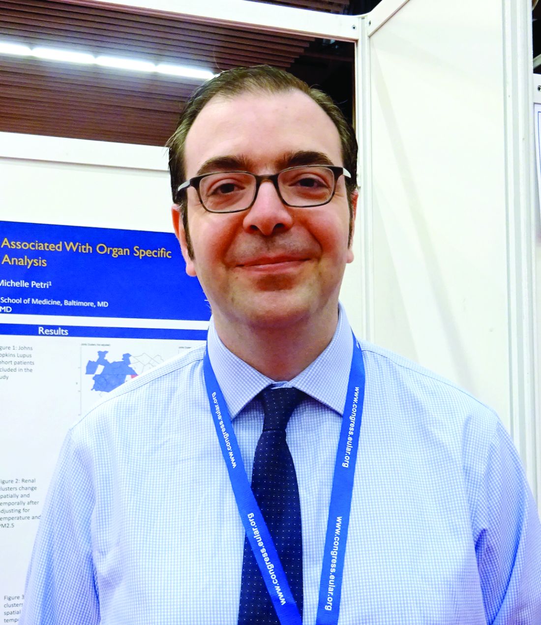
There were two reasons for looking at the role of these environmental factors in relation to SLE flares, said Dr. Stojan, an assistant professor of medicine at Johns Hopkins University, Baltimore. “The first was a clinical observation – I was getting clusters of patients with certain disease manifestations, for example with serositis or joint flares, and there wasn’t a random distribution.”
The second was a patient observation, added Dr. Stojan, who is also codirector of the Johns Hopkins Lupus Center. Every year, patients treated at the Center have the opportunity to meet each other and hear about the research being done by the team, and it was at this meeting that patients said they felt they were experiencing similar disease flares.
To look at the underlying role of environmental exposures in the development of SLE and possible associations with disease activity, Dr. Stojan and associates used a method known as cluster detection, which is commonly used in public health studies.
The investigators used a 350-km radial zone around the Johns Hopkins Lupus Center for the analysis as this was an area where a uniform number of patients treated by the center were living. They obtained data on 1,261 patients in the Hopkins Lupus Cohort, spanning a 10-year period from 1999 to 2009, and used SaTScan software to identify clusters of disease activity occurring during 3 separate monthly time intervals in different counties. The researchers then linked these clusters to average temperature and PM2.5 data obtained from the Environmental Protection Agency for the 10 days prior to patients’ visits.
“The SaTScan system predicts how many flares per organ system you would expect in a county based on the number of patients and based on the total flares we have in our cohort,” Dr. Stojan explained. Previously, the system helped to identify areas that had a higher flare incidence for each organ system that lasted for about 2-3 years, did not overlap, and could not be explained. So, the next step was to look for potential environmental triggers.
“Basically, the SaTScan adjusts the data that’s inputted for temperature and small particulate pollution. If the cluster moves in space and time then these did affect it,” Dr. Stojan said. “It seems that these do affect certain types of organ flares,” even after adjustment for other variables such as patients’ age, gender, income, ethnicity, and living situation (rural or urban).
Flares in skin symptoms during the summer have been identified before, he acknowledged, but the link to joint flares or neurologic flares have not. The latter includes things like seizures, neuropathy, or abnormal brain imaging rather than mood changes or mild cognitive dysfunction.
These data could have an impact on how clinical trials are designed, Dr. Stojan added, suggesting that factoring in where patients live and how close they are to areas of pollution could ensure a uniform population of patients is studied.
From a more practical perspective, these data might help to develop predictive models to help understand when patients are likely to experience a flare and if any action can be taken to ameliorate the effects of exposure.
The next step is a collaboration with patients to develop software or a mobile application where patients could input information about any disease flares. This would enable a finer view of what could be happening, Dr. Stojan said, as while daily readings are available for the environmental factors studied, disease activity data is only available during 3 separate monthly intervals. It would also allow other environmental factors to be considered.
“I think this is an important step in figuring out environmental factors and their influence on lupus,” he said. “There has been an extensive amount of research into viral causes and potential infectious triggers, but spatial-temporal analysis of environmental variables have never been done before in lupus.”
The study received no commercial funding, and Dr. Stojan reported having no disclosures.
SOURCE: Stojan G et al. Ann Rheum Dis. 2018;77(Suppl 2):1191. Abstract SAT0685.
REPORTING FROM THE EULAR 2018 CONGRESS
Key clinical point:
Major finding: For every 1° F increase in temperature, the risk for skin, joint, or neurologic flares increased.
Study details: A spatial-time cluster analysis of 1,261 patients in the Hopkins Lupus Cohort linking disease activity to temperature changes and fine particulate matter pollution.
Disclosures: The study received no commercial funding, and Dr. Stojan reported having no disclosures.
Source: Stojan G et al. Ann Rheum Dis. 2018;77(Suppl 2):1191. Abstract SAT0685.
Prolia tops risedronate at 2 years in steroid-induced osteoporosis
AMSTERDAM – Greater increases in bone strength were achieved with denosumab (Prolia) than with risedronate in patients receiving glucocorticoid treatment in a phase 3 trial, suggesting it may prove to be an effective means of preventing osteoporosis in this at-risk population of patients.
At 2 years, the percentage change in bone mineral density (BMD) from baseline with denosumab compared to risedronate in patients initiating steroid treatment was significantly higher (all P values less than .001) at the lumbar spine (6% vs. 2%), total hip (3% vs. 0%), and the femoral neck (1.5% vs. –1%). Similar results were seen in patients who were continuing steroid treatment when they entered the randomized, multicenter, double blind trial.
These data build on the 1-year data that have just been published (Lancet Diabetes Endocrinol. 2018;6[6]:445-54), Willem F. Lems, MD, the presenting study investigator, said at the European Congress of Rheumatology.
“Denosumab continued to increased BMD significantly more then risedronate through 24 months,” said Dr. Lems of VU University Medical Center, Amsterdam. He added that “the treatment differences at all skeletal sites were larger at 24 months than as 12 months.”
The Food and Drug Administration approved an additional indication in May of 2018 for denosumab to treat glucocorticoid-induced osteoporosis in adults at high risk of fracture. The biologic, a monoclonal antibody that targets the RANK ligand, was already approved to treat postmenopausal women with osteoporosis at high fracture risk, to increase bone mass in men with osteoporosis at high fracture risk, and to increase bone mass in women receiving adjuvant aromatase inhibitor therapy for breast cancer and in men receiving androgen deprivation therapy for nonmetastatic prostate cancer.
Secondary osteoporosis is a well known downside of glucocorticoid treatment, and despite there being approved therapies, many patients do not receive such treatment, Dr. Lems noted.
“This 24-month study assessed the safety and efficacy of denosumab versus risedronate in glucocorticoid-treated individuals, in whom guidelines advocate treatment,” Dr. Lems said.
In all, 795 adults who were taking glucocorticoids (7.5 mg prednisone or more daily, or equivalent) were enrolled at 79 sites in Europe, North America, Latin America, and Asia. Patients were subdivided into two populations based on their duration of glucocorticoid use, with 290 designated “glucocorticoid initiating” as they had been treated for less than 3 months, and 505 as “glucocorticoid continuing” as they had been treated for 3 or more months. Within these groups, approximately half were randomized to receive denosumab at a 60-mg subcutaneous dose every 6 months and half to receive risedronate 5 mg orally every day. All participants received calcium and vitamin D supplementation.
Bone turnover markers also declined “significantly more” with denosumab than with risedronate, with the exception of procollagen type 1 N-terminal telopeptide on day 10 and at 2 years and serum C-telopeptide of type 1 collagen at 2 years.
As for safety, a similar percentage of adverse events, including serious adverse events, was seen, Dr. Lems said. So far, he added, “data seem to be reassuring” regarding the risk for serious infections in high-risk subgroups of patients, such as those also taking biologic or immunosuppressant medications. There were no infections reported in patients taking biologics (vs. 12.1% for risedronate) and fewer infections in those taking immunosuppressant or biologic medication (3.6% vs. 6.8%).
Although the study was not powered to look at the rate of fractures between the two treatments, Dr. Lems noted that there was no significant difference in the rates of either new vertebral (4.1% for denosumab; 5.8% risedronate) or clinical (5.8% vs. 6.3%) fractures at 2 years.
“Additional studies are needed to confirm if denosumab is superior to other bisphosphonates for BMD improvement and fracture reduction,” Dr. Lems said. “Denosumab may offer a useful osteoporosis treatment option for patients receiving glucocorticoids,” he concluded.
Amgen sponsored the study. Dr. Lems acknowledged ties to Amgen, Eli Lilly, and Merck: being on the speakers bureau, receiving honoraria for advisory board meetings, and acting as a consultant. All but one other author disclosed ties to Amgen.
SOURCE: Saag KG et al. Ann Rheum Dis. 2018;77(Suppl 2):218. Abstract OP0345.
REPORTING FROM THE EULAR 2018 CONGRESS
Key clinical point: Denosumab may offer a useful alternative to risedronate in patients at risk for steroid-induced osteoporosis.
Major finding: Treatment differences were greater at 2 years than at 1 year at all skeletal sites assessed.
Study details: A phase 3, randomized, double-blind, active-controlled study to evaluate the efficacy and safety of denosumab versus risedronate in 795 glucocorticoid-treated individuals.
Disclosures: Amgen sponsored the study. Dr. Lems acknowledged ties to Amgen, Eli Lilly, and Merck: being on the speakers bureau, receiving honoraria for advisory board meetings, and acting as a consultant. All but one other author disclosed ties to Amgen.
Source: Saag KG et al. Ann Rheum Dis. 2018;77(Suppl 2):218. Abstract OP0345.
CRISS hailed as transforming systemic sclerosis drug development
AMSTERDAM – A new way to assess clinically meaningful, multiorgan changes in patients receiving treatment for systemic sclerosis has transformed the way new drugs for this disease are judged.
The Combined Response Index for Systemic Sclerosis (CRISS) “will change how we look at drugs” for systemic sclerosis, Daniel E. Furst, MD, said at the European Congress of Rheumatology.
The CRISS was close to 10 years in the making. “First we had to decide what measures were important, then we had to run a prospective study entering all the data, and then we had to do a very sophisticated statistical analysis and put the results in front of experts and ask: Does this make sense?” Dr. Furst recalled in an interview. Now the combined endpoint measure has been “fully validated,” and is under consideration by the Food and Drug Administration as an endpoint for drug trials, he noted.
“I think that now, after 10 years, we finally came up with what will be the equivalent” of the American College of Rheumatology 20% improvement (ACR 20) in core-set measures of rheumatoid arthritis (Arthritis Res Ther. 2014 Jan 3;16[1]:101). “I think CRISS will make a huge difference because when you do a combined measure, like the ACR 20, it becomes more clinically and statistically powerful,” said Dr. Furst, professor of medicine (emeritus) at the University of California, Los Angeles, an adjunct professor at the University of Washington, Seattle, and research professor at the University of Florence (Italy). He is also in part-time practice in Los Angeles and Seattle.
In a talk he gave at the meeting on recent clinical studies of new drugs for treating patients with systemic sclerosis, many of the reports included CRISS as a measure of patient response to treatment.
The CRISS involves a two-step assessment of how a patient has responded to therapy. First, patients are considered not improved by their treatment if they develop any one of these four outcomes following treatment if it appears linked to the disease process:
- A new scleroderma renal crisis.
- A decline in forced vital capacity of 15% or more of predicted.
- A new decline of left ventricular ejection fraction to 45% or less.
- New onset of pulmonary arterial hypertension that requires treatment.
If none of these apply, the next step is to assess treatment response by measuring changes in five parameters and then integrating them into a single number using a mathematical formula. The five elements in the equation are:
- Modified Rodnan skin score.
- Percent of predicted forced vital capacity.
- Health Assessment Questionnaire-Disability Index.
- Patient’s global assessment.
- Physician’s global assessment.
When factored together, changes in these five measures determine the probability that the patient responded to the intervention.
One limitation of the CRISS is that it only measures change from baseline, which makes it similar to the ACR 20, Dr. Furst noted. Another useful score would be one that reflects the status of a patient with systemic sclerosis at a specific point in time, a type of disease activity score. Dr. Furst said that he and others active in the systemic sclerosis field would like to develop a method that provides this type of patient assessment. Another addition would be to develop a “minimally clinically important change” in the score, which would make the CRISS more intuitive to understand.
Dr. Furst has received research support from Amgen, Bristol-Myers Squibb, Celgene, Corbus, Genentech-Roche, GlaxoSmithKline, Pfizer, and Novartis.
SOURCE: Furst DE. EULAR 2018. Abstract SP0012.
REPORTING FROM THE EULAR 2018 CONGRESS
ACR and EULAR to review new criteria for classifying vasculitis
AMSTERDAM – New classification criteria for antineutrophil cytoplasmic antibody (ANCA)–associated vasculitides have been drafted and now need formal review by the American College of Rheumatology and the European League Against Rheumatism before they can be put into practice.
According to Joanna Robson, MBBS, PhD, the chair of the DCVAS steering committee, these new criteria better “reflect current practice by incorporating, but not relying on, ANCA testing and advanced imaging.”
“The old criteria were actually produced in the early 1990s and since then we’ve had further thinking about the different subtypes of systemic vasculitis,” explained Dr. Robson of the University of the West of England in Bristol. There has also been a consensus conference held at Chapel Hill (Arthritis Rheum 2013;65[1]:1-11) which identified MPA as a separate entity, and ANCA testing has become routine practice. Computed tomography and magnetic resonance imaging are also now used to help differentiate between the different vasculitides.
“This really has been a collaborative, multinational effort,” Dr. Robson said at the European Congress of Rheumatology. To develop the draft criteria, data collated from 135 sites in 32 countries on more than 2,000 patients were used. These had been collected as part of the ACR/EULAR–run DCVAS study, which has been coordinated at the University of Oxford since 2011.
Three phases were used to develop these criteria: first an expert panel reviewed all cases in the DCVAS to identify those that they felt were attributable to small vessel vasculitis. Second, variables that might be appropriate to use in the models were examined, with more than 8,000 individual DVCAS items considered and then whittled down to 91 items and then sifted again to form a clear set of 10 or fewer items. Third, statistical analyses combined with expert review were used to develop the criteria and then validate these.
Dr. Robson reported that of 2,871 cases identified as ANCA-associated vasculitis, 2,072 (72%) were agreed upon by the expert review panel. Of these, there were 724 cases of GPA, 291 of MPA, 226 of EGPA, and around 300 cases of other small vessel vasculitis or polyarteritis nodosa. To develop the criteria the GPA cases were used as the “cases” and the other types of vasculitis as the comparators, Dr. Robson explained.
For GPA, MPA, and EGPA a set of items (10, 6, and 7, respectively) were derived and scored, positively or negatively, and a cutoff determined at which a classification of the particular vasculitis could be made. During discussion, Dr. Robson noted that the threshold score for a classification of EGPA (greater than or equal to 6) had been set slightly higher than for GPA or MPA (both greater than or equal to 5) “because of the clinical problem of there being very close comparators which can actually mimic EGPA.” This is where the negative scoring of some items used in these criteria are very important, she said.
The 10-item GPA criteria included three clinical (such as the presence of bloody nasal discharge upon examination) and seven investigational (such as cANCA positivity) items. These criteria were found to have a high sensitivity (92%) and specificity (94%) for identifying GPA.
The six-item MPA criteria included one clinical item (bloody nasal discharge, which was this time attributed a negative score) and five investigational items (with ANCA testing given a higher positive score than for GPA). The sensitivity and specificity of these criteria were a respective 91% and 94%.
Finally, the seven-item EGPA criteria included three clinical items (including obstructive airways disease and nasal polyps) and four investigational items (with ANCA positivity given a negative score). These criteria had an 85% sensitivity and 99% specificity for EGPA.
Dr. Robson emphasized that all of these classification criteria were to be used only after exclusion of other possible causes of vasculitis, such as infection, malignancy, or other autoimmune diseases such as inflammatory bowel disease, and a “diagnosis of small- or medium-vessel vasculitis has been made.”
These criteria are to help classify into the subtypes of vasculitis “primarily for the purpose of clinical trials,” she said. “The next steps are review by the EULAR and ACR committee, and only on final approval will these criteria be ready to use.”
DCVAS is sponsored by the University of Oxford (England) with funding from the European League Against Rheumatism, the American College of Rheumatology, and the Vasculitis Foundation. Dr. Robson had no relevant financial disclosures.
SOURCE: Robson JC et al. EULAR 2018. Abstract OP0021.
REPORTING FROM THE EULAR 2018 CONGRESS
Key clinical point: New classification criteria for ANCA-associated vasculitides have been drafted and now need formal review before they are ready to use.
Major finding: Analysis of the 10-, 6-, and 7-item GPA, MPA, and EGPA criteria showed a respective 92%, 94%, and 91% sensitivity and 94%, 85%, and 99% specificity.
Study details: The Diagnostic and Classification Criteria in Vasculitis (DCVAS) observational study of more than 6,000 cases of vasculitides and comparators.
Disclosures: DCVAS is sponsored by the University of Oxford (England) with funding from the American College of Rheumatology, the European League Against Rheumatism, and the Vasculitis Foundation. Dr. Robson had no relevant financial disclosures.
Source: Robson JC et al. EULAR 2018. Abstract OP0021.
EULAR nears first recommendations for managing Sjögren’s syndrome
AMSTERDAM – , and they divide the treatment targets into sicca syndrome and systemic manifestations of the disease.
“In Sjögren’s, we always have two subtypes of patients: those who have sicca syndrome only, and those with sicca syndrome plus systemic disease,” explained Soledad Retamozo, MD, who presented the current version of the recommendations at the European Congress of Rheumatology. “We wanted to highlight that there are two types of patients,” said Dr. Retamozo, a rheumatologist at the University of Córdoba (Argentina). “It’s hard to treat patients with sicca syndrome plus fatigue and pain because there is no high-level evidence on how to do this; all we have is expert opinion,” Dr. Retamozo said in an interview.
In fact, roughly half of the recommendations have no supporting evidence base, as presented by Dr. Retamozo. That starts with all three general recommendations she presented:
• Patients with Sjögren’s should be managed at a center of expertise using a multidisciplinary approach, which she said should include ophthalmologists and dentists to help address the mouth and ocular manifestations of sicca syndrome.
• Patients with sicca syndrome should receive symptomatic relief with topical treatments.
• Systemic treatments – glucocorticoids, immunosuppressants, and biologicals – can be considered for patients with active systemic disease.
The statement’s specific recommendations start with managing oral dryness, an intervention that should begin by measuring salivary gland (SG) dysfunction. The document next recommends nonpharmacologic interventions for mild SG dysfunction, pharmacological stimulation for moderate SG dysfunction, and a saliva substitute for severe SG dysfunction. All three recommendations are evidence based, relying on results from either randomized trials or controlled studies.
The second target for topical treatments is ocular dryness, which starts with artificial tears, or ocular gels or ointments, recommendations based on randomized trials. Refractory or severe ocular dryness should receive eye drops that contain a nonsteroidal anti-inflammatory drug or a glucocorticoid, based on controlled study results, or autologous serum eye drops, a strategy tested in a randomized trial.
The recommendations then shift to dealing with systemic manifestations, starting with fatigue and pain, offering the expert recommendation to evaluate the contribution of comorbid diseases and assess their severity with tools such as the Eular Sjögren’s Syndrome Patient-Reported Index (ESSPRI) (Ann Rheum Dis. 2011 June;70[6]:968-72), the Profile of Fatigue, and the Brief Pain Inventory.
Using evidence from randomized trials, the recommendations tell clinicians to consider treatment with analgesics or pain-modifying agents for musculoskeletal pain by weighing the potential benefits and adverse effects from this treatment.
For other forms of systemic disease, the recommendations offer the expert opinion to tailor treatment to the organ-specific severity using the ESSPRI definitions. If using glucocorticoids to treat systemic disease, they should be given at the minimum effective dose and for the shortest period of time needed to control active systemic disease, a recommendation based on retrospective or descriptive studies. Expert opinion called for using immunosuppressive treatments as glucocorticoid-sparing options for systemic disease, and this recommendation added that no particular immunosuppressive agent stands out as best compared with all available agents. In more than 95% of reported cases of systemic disease treatment in Sjögren’s patients, clinicians used the immunosuppressive drugs in association with glucocorticoids, Dr. Retamozo noted.
Finally, for systemic disease the recommendations cited evidence from controlled studies that B-cell targeted therapies, such as rituximab (Rituxan) and belimumab (Benlysta), may be considered in patients with severe, refractory systemic disease. An additional expert opinion was that the systemic, organ-specific approach should sequence treatments by using glucocorticoids first, followed by immunosuppressants, and finally biological drugs.
The recommendations finish with an entry that treatment of B-cell lymphoma be individualized based on the specific histopathologic subtype involved and the level of disease extension, an approach based on results from retrospective or descriptive studies.
The recommendations must still undergo final EULAR review and endorsement, with publication on track to occur before the end of 2018, Dr. Retamozo said.
She had no disclosures.
SOURCE: Retamozo S al. Ann Rheum Dis. 2018;77(Suppl 2):42. Abstract SP0159.
REPORTING FROM THE EULAR 2018 CONGRESS
Predicting rituximab responses in lupus remains challenging
AMSTERDAM – Despite some “encouraging signs” seen in a cross-sectional study, it remains difficult to determine whether a patient with systemic lupus erythematosus (SLE) will respond to off-label rituximab therapy, according to David Isenberg, MD.
The presence of constitutional symptoms, which includes fatigue, at baseline were associated with a favorable response to rituximab at 6 months (odds ratio, 7.35; P = .01). Conversely, having more than one anti–extractable nuclear antigen (anti-ENA) antibody was associated with a worse response to rituximab at 12 months (OR, 0.33; P = .032).
The video associated with this article is no longer available on this site. Please view all of our videos on the MDedge YouTube channel
“It’s not a cure for lupus, that’s obvious, but used at the appropriate time in the right sort of patients, it can be very helpful,” Dr. Isenberg said in an interview at the European Congress of Rheumatology. That’s both in terms of improving disease activity and reducing the use of glucocorticosteroids and immunosuppressive drugs, he noted.
“The great mystery, as with all drugs, is: Are there any markers which might tell us that the disease is going to do better with rituximab than perhaps with another form of medication, or is it just saying to us, ‘Whatever you do, it doesn’t make much difference’?” Dr. Isenberg said. He discussed the results of a study involving the first 121 patients treated with rituximab at his institution, University College London Hospital (UCLH); he noted this is one of the largest single-center cohorts of individuals with SLE who have used rituximab.
The aim of the study, which was presented at the congress by Hiurma Sánchez Pérez, MD, was therefore to try to find demographic, clinical, and serological markers that might help in predicting the response to rituximab in SLE patients.
Response to treatment was determined using the British Isles Lupus Assessment Group (BILAG) Index. Patients who initially had markedly (BILAG A) or moderately (BILAG B) active disease but who no longer fell into these categories at 6 or 12 months were designated as responders. Those who remained were designated as nonresponders. In addition, any person moving into category A or B of the BILAG Index was said to have relapsed because of a flare in disease activity.
At 6 and 12 months, a respective 85% and 70% of patients exhibited a response to rituximab. Just under a quarter (24%) relapsed before 1 year, Dr. Sánchez Pérez reported.
Most of the patients in the cohort were white (n = 50) or Afro-Caribbean (n = 38), but ethnicity did not seem to play a role in predicting whether patients would respond to rituximab treatment.
Aside from constitutional symptoms and having more than one anti-ENA antibody, there were no other biological markers of response that remained significant after multivariate analysis.
The mean time to flare after rituximab was nearly 8 months, Dr. Sánchez Pérez said. Arthritis was the main manifestation seen during relapse (41% of patients), and mucocutaneous symptoms occurred in 21%. Biological markers of flare at 12 months after multivariate analysis were musculoskeletal symptoms at baseline (OR, 0.26; P = .039) and being anti-RNP antibody positive (OR, 10.56; P = .03).
“There were one or two encouraging signs,” said Dr. Isenberg, who was the senior author of the study. “Unfortunately, what I think the data show us, it remains pretty hard to know how any individual patient is going to respond to B-cell depletion using rituximab.”
Dr. Sánchez Pérez reported having no disclosures in relation to the study. Dr. Isenberg was one of the first people to use rituximab to treat patients with lupus but reported having no other competing interests.
SOURCE: Sánchez Pérez H and Isenberg D. Ann Rheum Dis. 2018;77(Suppl 2):177. EULAR 2018 Congress, Abstract OP0255.
REPORTING FROM THE EULAR 2018 CONGRESS
Key clinical point: Only the presence of constitutional symptoms at baseline and having more than one anti–extractable nuclear antigen (anti-ENA) antibody were related to rituximab response.
Major finding: Having more than one anti-ENA antibody was related to a worse response to rituximab at 12 months.
Study details: A cross-sectional study of 121 patients with systemic lupus erythematosus treated with rituximab during 2000-2016.
Disclosures: Dr. Isenberg has used rituximab to treat patients with lupus for almost 20 years but reported having no other competing interests. Dr. Sánchez Pérez reported having no disclosures in relation to the study.
Source: Sánchez Pérez H and Isenberg D. Ann Rheum Dis. 2018;77(Suppl 2):177. EULAR 2018 Congress, Abstract OP0255.
Ankylosing spondylitis progression slowed when NSAIDs added to TNFi
AMSTERDAM – When combined with a tumor necrosis factor inhibitor, NSAIDs provide protection against long-term radiographic progression in patients with ankylosing spondylitis, according to an analysis of more than 500 patients presented at the European Congress of Rheumatology.
Relative to TNFi alone, the addition of NSAIDs of any type provided protection at 4 years against radiographic progression as measured with the modified Stoke Ankylosing Spondylitis Spine Score (mSASSS). However, the protection associated with adding celecoxib was significant at 2 years and greater than that of adding nonselective NSAIDs at 4 years.
These data were drawn from 519 patients participating in the Prospective Study of Ankylosing Spondylitis study. All patients in this analysis were followed for at least 4 years. Radiographs were obtained every 6 months.
Although the study was a retrospective analysis of prospectively collected data, Dr. Gensler explained that control of variables such as disease and symptom duration with a technique called causal interference modeling “allows simulation of a randomized, controlled trial with observational data.”
Whether measured at 2 or 4 years, the reductions in mSASSS score for TNFi use versus no TNFi use were modest and did not reach statistical significance. However, exposure to NSAIDs plus TNFi did reach significance at 4 years, and the effect was dose dependent when patients taking a low-dose NSAID, defined as less than 50% of the index dose, were compared with those taking a higher dose. In this study, 70% were on chronic NSAID therapy, and these patients were divided relatively evenly between those on a low-dose or high-dose regimen.
At 2 years, relative radiographic protection for TNFi plus NSAIDs was not significantly greater than with TNFi alone, but at 4 years the median mSASSS score was 1.24 points lower (P less than .001) in those receiving low-dose NSAIDs, and 3.31 points lower (P less than .001) in those receiving high-dose NSAIDs.
In the subgroup of patients taking high-dose NSAIDs, the protection from progression was greatest among those receiving the selective COX2-inhibitor celecoxib. In these, the median 3.98 points lower mSASSS score (P less than .001) was already significant at 2 years. At 4 years, the median mSASSS score in those receiving TNFi plus celecoxib was 4.69 points lower (P less than .001).
Further evaluation suggested that the benefit from celecoxib plus TNFi was not just additive but synergistic, according to Dr. Gensler. She reported that neither TNFi nor celecoxib alone provided radiographic protection at 2 or 4 years.
Despite the modeling strategy employed to reduce the effect of bias, Dr. Gensler acknowledged that residual confounding is still possible. But she contended that “a large effect [from a such a variable] would be required to negate the findings.”
One of the messages from this study is that “not all NSAIDs are alike,” Dr. Gensler said. “Despite this, when I sit with a patient across from me, I will still treat the patient based on symptoms and disease activity first, though perhaps choose to be more NSAID selective if this is warranted and feasible.”
The next steps for research include a randomized, controlled trial combining TNFi and varying NSAIDs or different doses, Dr. Gensler said. In addition, “the development of newer imaging modalities will allow us to answer these questions in a more feasible time frame.”
The study was not industry funded. Dr. Gensler reported financial relationships with Amgen, AbbVie, Janssen, Eli Lilly, Novartis, and UCB.
SOURCE: Gensler L et al. Ann Rheum Dis. 2018;77(Suppl 2):148. Abstract OP0198.
REPORTING FROM THE EULAR 2018 CONGRESS
Key clinical point: Ankylosing spondylitis patients have less progression on a TNFi when they also receive an NSAID, especially celecoxib.
Major finding: At 4 years, the mSASSS score was 4.69 points (P less than .0001) lower on TNFi with celecoxib than on TNFi alone.
Study details: Retrospective study with causal interference modeling.
Disclosures: The study was not industry funded. Dr. Gensler reported financial relationships with Amgen, AbbVie, Janssen, Eli Lilly, Novartis, and UCB.
Source: Gensler L et al. Ann Rheum Dis. 2018;77(Suppl 2):148. Abstract OP0198.
Ultrasound aids treat-to-target approach for gout
AMSTERDAM – Results from the longitudinal NOR-GOUT study show that ultrasound can help visualize decreasing levels of uric acid deposits that occur during a treat-to-target approach with urate-lowering therapy.
“Gout is a really painful disease when there are flares,” said Dr. Hammer, a senior consultant in the rheumatology department at Diakonhjemmet Hospital in Oslo. Ultrasound has been shown to be a sensitive method to detect uric monosodium urate (MSU) deposition and its use is included in the classification criteria for gout.
“MSU depositions are found in many different regions with some predilection sites,” Dr. Hammer noted. This led the OMERACT (Outcome Measures in Rheumatology) Ultrasound Group to develop three key definitions for MSU lesions: the “double contour sign” (DC), which occurs when urate crystals form on the surface of cartilage; tophus, which is where there is a larger, hypoechoic aggregation of crystals that are usually well delineated; and “aggregates,” which are small, hyperechoic deposits.
“There are, up until now, only a few smaller studies that have explored the decrease of depositions during uric acid–lowering treatment,” Dr. Hammer observed.
NOR-GOUT was a prospective, observational study of 161 consecutively-recruited patients with urate crystal–proven gout who needed treatment with urate-lowering therapy. Patients were included if they had a recent gout flare and had serum urate levels of more than 360 micromol/L (6.0 mg/dL) and had no contraindication to urate-lowering therapy.
“We used a treat-to-target approach with the medication,” Dr. Hammer explained. The aim was to get uric acid levels to 360 micromol/L or lower, or to less than 300 micromol/L (5.0 mg/dL) if clinical tophi were present. “The medication was optimized by monthly follow-up by a study nurse until the treatment target was met,” she added.
Patients underwent an extensive ultrasound assessment at study entry and again after 3, 6, and 12 months of urate-lowering therapy. This included bilateral assessment of all relevant joints and the presence of crystals semiquantitatively scored from 0 to 3, the latter signifying many deposits. The sum of scores for the three key OMERACT definitions were calculated each time the patients were assessed, with a total score for all three also calculated.
Mean serum urate levels dropped from a baseline of 487 to 312 micromol/L (from 8.2 to 5.3 mg/dL) at 12 months (P less than .001), Dr. Hammer reported. The percentage of patients achieving a urate target of less than 360 micromol/L increased from 71% at 3 months to 81% at 6 months and to 84% at 12 months, she said.
Ultrasound scores decreased with decreasing urate levels at 3, 6, and 12 months, with the highest numeric difference from baseline seen at 12 months for DC (3.1, 2.3, and 1.2; all P less than .001 vs. baseline of 4.2). The respective values for tophi were 6.3, 5.4, and 4.2 versus a baseline of 6.5; for aggregates, the values were 8.8, 7.9, and 6.7 versus a baseline of 9.1.
Standardized Response Mean values from baseline to 3, 6, and 12 months showed that DC was the most sensitive for change, with a respective 0.73, 1.02, and 1.26 in ultrasound scores. Values for tophi were 0.06, 0.57, and 0.91, and 0.20, 0.51 and 0.66 for aggregates.
“Not all patients had reached 12 months of follow-up when we made these calculations,” Dr. Hammer said, noting the limitations of the study. Nevertheless, these interim findings suggest that ultrasound is a valuable tool that can help see how patients fare on a treat-to-target approach, she concluded.
Dr. Hammer had no conflicts of interest to disclose.
SOURCE: Hammer HB et al. Ann Rheum Dis. 2018;77(Suppl 2):154-5. Abstract OP0211.
REPORTING FROM THE EULAR 2018 CONGRESS
Key clinical point: Deceasing uric acid load during a treat-to-target approach can be seen on ultrasound.
Major finding: Ultrasound scores decreased with decreasing urate levels at 3, 6 and 12 month, with the highest numeric difference from baseline seen at 12 months for double contour sign (3.1, 2.3, and 1.2; all P less than .001 vs. baseline of 4.2).
Study details: A prospective, observational study of 161 patients with urate crystal–proven gout treated with urate-lowering therapy.
Disclosures: Dr. Hammer had no conflicts of interest to disclose.
Sources: Hammer HB et al. Ann Rheum Dis. 2018;77(Suppl 2):154-5. Abstract OP0211.


