User login
Pain remains after rheumatoid arthritis diagnosis
AMSTERDAM – according to data from a large prospective study reported at the European Congress of Rheumatology.
At enrollment, 64% of patients still had remaining pain, and at 1 year, 24% had remaining pain, according to data from the Canadian Early Arthritis Cohort (CATCH).
Dr. Bykerk of the University of Toronto and the Hospital for Special Surgery, New York, presented the research on behalf of Yvonne C. Lee, MD, of Northwestern University, Chicago, and noted that they had previously published data showing the incidence of widespread pain was highest within the first year of an RA diagnosis and then plateaued (Ann Rheum Dis. 2013;72[6];949-54). This plateau could have been related to the control of inflammation, Dr. Bykerk observed.
“We think the first 12 months are really a critical window during which time acute inflammatory pain may transition, if you will, to chronic noninflammatory pain,” Dr. Bykerk said.
The aim of their current study was to look at the evolution of pain in the year following an RA diagnosis and to see whether there were any predictive factors for remaining pain and widespread pain. The latter might previously have been described as fibromyalgia, Dr. Bykerk said, and was defined as a difference between the tender and swollen 28-joint counts of at least seven points. Remaining pain was defined as a score of more than 4 on a pain intensity numerical rating scale.
Data on 1,270 patients who were enrolled in CATCH across 17 sites in Canada during 2007-2017 were used. The majority of the sample was female (73%) and white (82%) and had been experiencing symptoms for a median of 6 months. The mean age of patients was 54 years.
At enrollment, patients had a mean number of two comorbidities, with up to 12% having another condition that might contribute to pain, such as osteoarthritis (12%), a back or spinal problem (10%), or fibromyalgia (9%).
Most (72%) patients were being treated with methotrexate initially, either alone (29%) or in combination with other drugs, and almost two-thirds were using steroids, either orally (29%) or parenterally (32%). A small percentage (2%) were treated with biologics, but this is unusual until almost 12 months, Dr. Bykerk observed.
The baseline predictors for remaining pain at 1 year were high baseline pain (odds ratio, 2.1), sleep difficulties (OR, 2.2), and disability measured by the Health Assessment Questionnaire Disability Index (OR, 1.5). The latter was the strongest predictor for lingering widespread pain. The number of comorbidities were also predictive of lingering pain, though this was not a significant difference.
Assessing and considering treatment for these predictors may be important for improving persisting pain, Dr. Bykerk suggested. “In future we’d like to validate our findings with other validated questionnaires; we have a depression questionnaire now being used in the study, and we want to understand what subgroups of patients have different trajectories of pain.”
CATCH is currently funded via unrestricted research grants from Amgen, Pfizer, Bristol-Myers Squibb, Medexus, Eli Lilly, Merck, and Sandoz Canada Pharmaceuticals. Dr. Bykerk had no personal financial disclosures.
SOURCE: Lee YC et al. EULAR 2018 Congress.Abstract OP0349.
REPORTING FROM THE EULAR 2018 CONGRESS
Key clinical point: A substantial number of patients have pain 1 year after being diagnosed with rheumatoid arthritis.
Major finding: Remaining pain and widespread pain were a respective 64% and 9% at baseline and 9% and 5% at 1 year.
Study details: A prospective, observational, cohort of 1,270 patients with new-onset inflammatory arthritis.
Disclosures: Dr. Bykerk is the principle investigator of the Canadian Early Arthritis Cohort (CATCH). CATCH is currently funded via unrestricted research grants from Amgen, Pfizer, Bristol-Myers Squibb, Medexus, Eli Lilly, Merck, and Sandoz Canada Pharmaceuticals. Dr. Bykerk had no personal financial disclosures.
Source: Lee YC et al. EULAR 2018 Congress, Abstract OP0349.
Limited anakinra course often enough for systemic JIA if used first
AMSTERDAM – Rather than waiting for other drugs or immunotherapies to fail, an immediate up-front but time-limited course of an interleukin-1 receptor (IL-1R) antagonist induced rapid and sustained remissions in most children with systemic juvenile idiopathic arthritis (JIA), according to 5-year data presented at the European Congress of Rheumatology.
In the latest follow-up of a protocol first described in 2014, over 90% of patients still had inactive disease, 75% of whom were completely off therapy, reported Sebastiaan J. Vastert, MD, PhD, of the division of pediatrics at University Medical Center, Utrecht, the Netherlands.
“Translating this into clinical practice, you could say that there might be a window of opportunity early in systemic JIA in which the innate immune system is the major player and perhaps you could down-regulate this to control the inflammation,” Dr. Vastert explained.
Citing a series of experimental studies at his institution that suggest immune mediators change as systemic JIA evolves from an acute to a chronic phase, Dr. Vastert believes that early use of an anti–IL-1R therapy may alter the trajectory of systemic JIA, compared with when it goes untreated or is treated with conventional therapies.
In the original series reported in 2014 (Arthritis Rheumatol. 2014 Apr;66:1034-43), data were presented on 20 patients. All fulfilled the International League of Associations for Rheumatology criteria for systemic JIA. They were treated with anakinra after failing to respond to indomethacin but before receiving any other therapy, including corticosteroids, disease-modifying antirheumatic drugs, or other biologics.
In the protocol described in the initial publication, a stop-therapy strategy permitted treatment discontinuation after 3 months in those who met American College of Rheumatology criteria for 90% improvement (ACR Pedi 90) in JIA.
By 1 year, 73% of the patients had met criteria to stop therapy. Of 11 patients followed for 3 years, 10 met criteria for disease remission, 8 of whom were off medication. The remaining two continued to receive anti–IL-1R or another therapy.
The systemic JIA cohort at Dr. Vastert’s institution has now grown to 50 patients, of whom 42 patients have received first-line anakinra. Among the 25 patients who have been followed for at least 5 years, 72% have inactive disease as defined by ACR Pedi 90 criteria off therapy. Another 20% have inactive disease on therapy, which is anakinra or another biologic in most cases. The majority of patients have avoided corticosteroids completely.
Freedom from corticosteroids has been accompanied by high rates of satisfaction and has allowed patients to avoid adverse events associated with corticosteroids. For example, only one patient in this series has a growth curve more than two standard deviations below normal for age and gender, according to Dr. Vastert.
“This is just a single-center cohort study, but we now have 3 more years of data to be convinced of this concept,” Dr. Vastert said.
Another notable finding from this cohort: 12 patients have been enrolled that did not fulfill International League of Associations for Rheumatology criteria for systemic JIA because of the absence of joint involvement. Strongly suspected of having systemic JIA because of other clinical signs and features, these patients have also responded well to first-line anakinra therapy.
“Our data point to a classification [of systemic JIA] that does not include arthritis as a prerequisite for diagnosis,” said Dr. Vastert, who provided data suggesting that elevated levels of IL-18 might be among biomarkers that could be employed in a revised classification system.
The study was not funded by industry. Dr. Vastert reported receiving consulting fees from Novartis.
REPORTING FROM THE EULAR 2018 CONGRESS
Key clinical point: Based on long-term outcomes, first-line anakinra may change natural history of systemic juvenile idiopathic arthritis.
Major finding: In a 5-year follow-up of 25 patients with systemic JIA, 92% have inactive disease, most of whom are off therapy.
Study details: A prospective, single-center cohort registry.
Disclosures: The study was not funded by industry. Dr. Vastert reported receiving consulting fees from Novartis.
High RA biologic drug levels linked with more infections
AMSTERDAM – Patients with rheumatoid arthritis who had a high serum level of biologic immunomodulatory drugs had a statistically significant 51% higher rate of infection during their first year on the drug, compared with RA patients who maintained usual or low serum levels of the same drugs, according to an analysis of 703 U.K. patients in a national database.
The results suggest that, once patients with rheumatoid arthritis go into remission on a higher dosage of biologic agents that produce a high serum level “dose tapering may lower their risk of infection,” Meghna Jani, MD, said at the European Congress of Rheumatology.
The study used data and specimens collected in two separate, prospective U.K. studies: the British Society for Rheumatology Biologics Register-RA, which had data from more than 20,000 U.K. patients with RA who started treatment with a biologic agent, and BRAGGSS (Biologics in Rheumatoid Arthritis and Genetics and Genomics Study Syndicate), a national prospective cohort of 3,000 RA patients who had serum specimens drawn at 3, 6, and 12 months after starting biologic drug treatment and tested by an immunoassay for the concentration of the drug each patient received.
Dr. Jani and her associates considered serum levels that exceeded the following thresholds to categorize patients as having a high drug level: 8 mcg/mL adalimumab, 25 mcg/mL certolizumab, 4 mcg/mL etanercept, 4 mcg/mL infliximab, and 4 mcg/mL tocilizumab. The patients averaged about 59 years old, about three-quarters were women, and they had been diagnosed with RA for approximately 5-7 years. About 22% were also on treatment with a steroid, and most patients had not received prior treatment with a biologic agent.
The researchers tallied 229 diagnosed infections in the subgroup with high serum levels of their biologic drug, and 63 infections in those with levels below this threshold. After adjustment for age, sex, methotrexate use, and disease activity score, patients with high serum levels of their biologic drug had a 51% higher rate of all infections than did patients with levels that fell below the high-level threshold, reported Dr. Jani, a rheumatologist at Manchester (England) University. Analysis of the accumulation of infections over the course of 1 year of follow-up showed that this difference in infection rates became apparent after about 2 months of exposure and then began to diverge more sharply after about 5 months of exposure.
The results also showed that the rate of serious infections – defined as those needing intravenous antibiotics, hospitalization, or resulting in death – were similar in the two subgroups. The types of infections and their relative frequencies were also roughly similar in the two subgroups. Lower respiratory infections were the most common infection in both subgroups, followed by infections of the upper respiratory tract, urinary tract, and skin as the next three most common infections in both subgroups.
SOURCE: Jani M et al. EULAR 2018 Congress, Abstract OP0229.
REPORTING FROM THE EULAR 2018 CONGRESS
Key clinical point:
Major finding: High serum levels of a biologic drug were linked with a 51% higher infection rate, compared with those who had normal or low levels.
Study details: Prospective data collected from 703 patients throughout the United Kingdom.
Disclosures: Dr. Jani had no relevant disclosures. Dr. Isaacs has been a consultant to several companies that market biologic drugs for treating rheumatoid arthritis.
Source: Jani M et al. EULAR 2018 Congress, Abstract OP0229.
EULAR recommendations on steroids: ‘As necessary, but as little as possible’
AMSTERDAM – Glucocorticosteroids remain an important therapeutic option for many patients with rheumatic and nonrheumatic disease, but careful assessment of their relative benefits and risks needs to be considered when prescribing, according to an expert summary of currently available European League Against Rheumatism (EULAR) recommendations.
The video associated with this article is no longer available on this site. Please view all of our videos on the MDedge YouTube channel
From rheumatoid arthritis (RA) and polymyalgia rheumatica (PMR) to vasculitis, myositis, and even gout, steroids are widely used in the rheumatic diseases, said Frank Buttgereit, MD, during a final plenary session at the European Congress of Rheumatology.
“These are strong-acting, rapidly acting, efficacious drugs,” observed Dr. Buttgereit, who is a professor in the department of rheumatology at Charité–Universitätsmedizin Berlin. While effective at reducing inflammation and providing immunosuppression, they are, of course, not without their well-known risks. Some of the well-documented risks he pointed out were the development of osteoporosis, myopathy, and edema; the disruption of lipid and carbohydrate metabolism; and the risk of developing glaucoma and cataracts.
“This leads to the question on how to optimize the use of these drugs,” Dr. Buttgereit said. “EULAR is constantly working to improve its guidelines,” and updating these in line with the available evidence, he added. “The bottom line is always give as much as necessary but as little as possible.”
Over the past few years, EULAR’s Glucocorticoid Task Force has been reviewing and updating recommendations on the use of these drugs and it has published several important documents clarifying their use in RA and in PMR. The task force has also published a viewpoint article on the long-term use of steroids, defining the conditions where an “acceptably low level of harm” might exist to enable their continued use. There have also been separate recommendations, published in 2010, on how to monitor these drugs (Ann Rheum Dis. 2010;69[11]:1913-9).
Clarifying the role of steroids in rheumatoid arthritis
The latest (2016) EULAR recommendations on the use of glucocorticosteroids were published last year (Ann Rheum Dis. 2017;76[6]:960-77) and included an important adjustment on when they should be initially used in RA, Dr. Buttgereit explained. Previous recommendations had said that steroids could be combined with disease-modifying antirheumatic drugs (DMARDs) but had suggested that they be used at a low dose. Now the wording has changed to focus on short-term use rather than dosing.
“Glucocorticoids can be given initially at different dosages, and using different routes of administration,” he said in a video interview at the EULAR Congress. The practice on what dose to give varies from country to country, he noted, so the recommendations are now being less prescriptive.
“We have made it clear that glucocorticoids should really be used only when initiating conventional synthetic DMARDs, but not necessarily if you switch to biologics or targeted synthetics because usually the onset of their actions is pretty fast,” Dr. Buttgereit said.
One thing that hasn’t changed is that steroid should be tapered down as “rapidly as clinically feasible” until, ideally, their full withdrawal. Although there are cases when that might not be possible, and their long-term use might be warranted. This is when you get into discussion about the benefit-to-risk ratio, he said.
Steroids for polymyalgia rheumatica
Steroids may be used as monotherapy in patients with PMR, Dr. Buttgereit observed, which is in contrast to other conditions such as RA. Although the evidence for use of steroids in PMR is limited, the EULAR Glucocorticoid Task Force and American College of Rheumatology recommended (Ann Rheum Dis. 2015;74[10]:1799-807) using a starting dose of a prednisolone-equivalent dose between 12.5 and 25 mg/day, and if there is an improvement in few weeks, the dose can start to be reduced. Tapering should be rapid at first to bring the dose down to 10 mg/day and followed by a more gradual dose-reduction phase.
“So, you can see we are giving more or less precise recommendations on how to start, how to taper,” Dr. Buttgereit said.
Balancing long-term benefit vs. harm
Balancing the long-term benefits and risks of steroids in rheumatic disease was the focus of a EULAR viewpoint article published 3 years ago in 2015 (Ann Rheum Dis. 2015;75[6]:952-7).
Three main messages can be drawn out of this work, Dr. Buttgereit said.
First, treatment with steroids for 3-6 months is associated with more benefits than risks if doses of 5 mg/day or less are used. There is one important exception to this, however, and that is the use of steroids in patients with comorbid cardiovascular disease.
Second, using doses of 10 mg/day for long periods tips the balance toward more risks than benefits, and “this means you should avoid this.”
Third, doses of 5-10 mg/day may be appropriate, but there are certain patient factors that will influence the benefit-to-harm ratio that need to be considered. These include older age, smoking, high alcohol consumption, and poor nutrition. There are also factors that may help protect the patients from risk, such as early diagnosis, low disease activity, low cumulative dose of steroids, and a shorter duration of treatment.
“It’s not only the dose, it’s also the absence or presence of risk factors and/or preventive measures,” that’s important, Dr. Buttgereit said.
Dr. Buttgereit has received consultancy fees, honoraria, and/or travel expenses from Amgen, Horizon Pharma, Mundipharma, Roche, and Pfizer and grant or study support from Amgen, Mundipharma, and Pfizer.
SOURCE: Buttgereit F. EULAR 2018 Congress, Abstract SP160.
REPORTING FROM THE EULAR 2018 CONGRESS
Key clinical point: When dosing glucocorticoids, give as much as necessary but as little as possible.
Major finding:
Disclosures: Dr. Buttgereit has received consultancy fees, honoraria, and/or travel expenses from Amgen, Horizon Pharma, Mundipharma, Roche, and Pfizer and grant or study support from Amgen, Mundipharma, and Pfizer.
Source: Buttgereit F. EULAR 2018 Congress. Abstract SP160.

In JIA, etanercept associated with lower uveitis risk compared with methotrexate
AMSTERDAM – Etanercept does not appear to increase the risk of uveitis relative to methotrexate in children with juvenile idiopathic arthritis (JIA), according to a large retrospective cohort study presented at the EULAR 2018 Congress.
“When we added in a fully adjusted hazard ratio, it showed a lower risk in the development of uveitis in patients on etanercept [compared with methotrexate],” reported Rebecca Davies, a research assistant at the University of Manchester (England).
These data were characterized as reassuring for JIA patients being considered for etanercept, but Ms. Davies was cautious about suggesting that etanercept has a protective effect. Even though hazard ratios were calculated with propensity-adjusted Cox regression analyses, Ms. Davies believes the best interpretation of these data is that etanercept therapy is not likely to contribute significantly to the risk of uveitis.
The substantial differences between the comparator groups provide one reason to refrain from speculating that etanercept is protective against uveitis. The risk of uveitis is greater both in younger patients and in the first year after diagnosis. Patients in the etanercept group were older than patients in the methotrexate group and they started etanercept a longer time after the JIA diagnosis.
“The initiation of etanercept later in the disease may mean that more of these patients had already passed through the window of greatest risk,” Ms. Davies explained.
The data for this study were drawn from a British Society for Pediatric and Adolescent Rheumatology cohort registry. Confined to patients first starting as opposed to restarting therapy, 1,009 patients initiating etanercept were compared to 508 patients initiating methotrexate.
In addition to an older age (11 vs. 9 years) and disease duration at start of therapy (3 vs. 1 years), a lower proportion of patients in the etanercept group had a persistent oligoarthritis subtype (5% vs. 17%). During follow-up, there were 15 cases (0.15%) of uveitis in the etanercept group and 18 (3.5%) in the methotrexate group.
The crude incidence of uveitis was 0.6 per 100 patient-years for etanercept versus 2.4 per 100 patient-years for methotrexate, according to Ms. Davies. After adjustment for a broad number of variables, including age, gender, disease scores, disease duration, baseline steroid use, and the presence of comorbidities, there was still a 70% lower risk of uveitis among those treated with etanercept (hazard ratio 0.30; 95% confidence interval, 0.1-0.9).
The low relative rate of uveitis after starting etanercept is discordant with several previous studies, according to Ms. Davies. In two retrospective studies conducted in the United States and one in Canada, etanercept treatment was associated with higher rates of uveitis than other tumor necrosis factor inhibitors. In a German study, etanercept was associated with a higher risk of uveitis than that of methotrexate.
Although a highly effective anti-inflammatory agent such as etanercept might be expected to have a protective effect against uveitis, at least relative to methotrexate, Ms. Davies suggested that the previous reports of potential causal association and the limitations of this retrospective analysis require a more cautious interpretation.
“It is possible that those considered to be at high risk of developing uveitis were kept away from etanercept,” said Ms. Davis, providing one of several explanations why skepticism is needed in regard to assuming uveitis protection from etanercept.
With up to 1 in 10 patients with JIA eventually developing sight-threatening uveitis, risk management is a priority, according to Ms. Davies. Current guidelines in the United Kingdom call for an ophthalmologist consult within 6 weeks of a diagnosis. Although several risk factors for uveitis have been published, the goal of this study was to determine whether exposure to etanercept is among these risks. According to Ms. Davies, these data suggest that this is not the case, but prospective studies comparing etanercept to other biologics would be particularly helpful in determining which therapy is most appropriate in order to reduce uveitis risk.
SOURCE: EULAR 2018 Congress. Abstract OP0351.
REPORTING FROM THE EULAR 2018 CONGRESS
Key clinical point:
Major finding: In children on new medication, the incidence of uveitis per 100 patients was 0.6 for etanercept and 2.4 for methotrexate.
Study details: Retrospective cohort registry.
Disclosures: The study was not funded by industry. Ms. Davies reports no potential conflicts of interest.
Source: EULAR 2018 Congress. Abstract OP0351.
Glucocorticosteroid use raises sarcopenia risk in RA
AMSTERDAM – Patients with RA have a higher risk of developing sarcopenia if they are treated with glucocorticosteroids, study findings suggested.
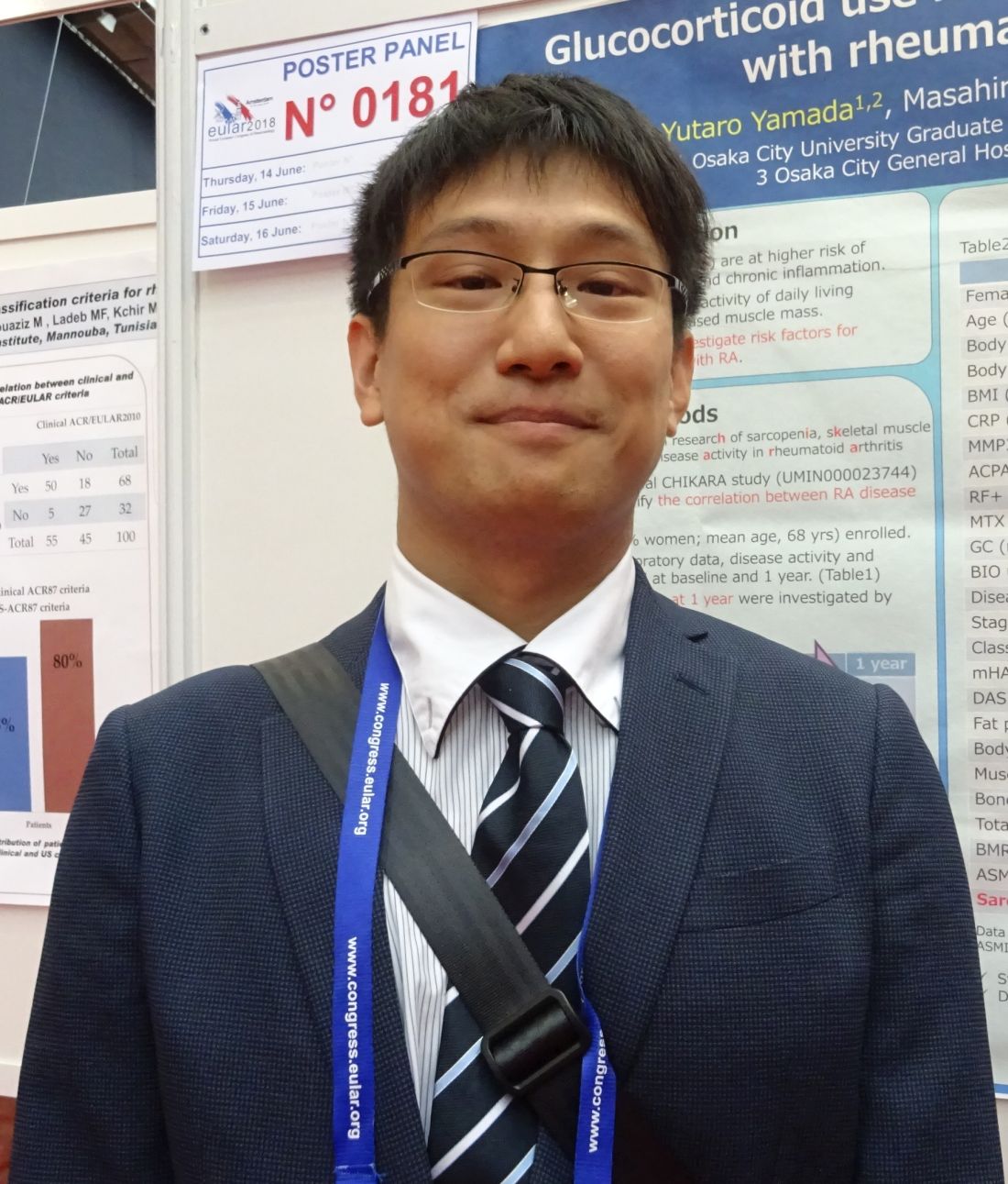
“The strength of our study is that it is a prospective, longitudinal study and this study is the first, to our knowledge, to look at risk factors for sarcopenia limited to RA patients,” Yutaro Yamada, MD, said in an interview at the European Congress of Rheumatology.
Dr. Yamada, of the department of orthopedic surgery at Osaka City University, Japan, said that there is a twofold rationale for looking at risk factors for sarcopenia in RA patients. First, RA causes chronic inflammation and this in turn is thought to lead to a metabolic state that then results in muscle loss. Furthermore, joint dysfunction and subsequent disuse likely contribute to weakening muscles. Second, glucocorticosteroids, which are commonly used to treat patients with RA, have themselves been associated with a low muscle area in prior research.
In 2016, the CHIKARA study was initiated “to clarify the correlation between RA disease activity and sarcopenia,” Dr. Yamada and his associates reported in a poster presentation. They recruited 100 patients, of whom 78% were women, and recorded body weight, muscle mass, fat mass, and predicted bone mass using a body composition analyzer at entry and 1 year later. Laboratory data and disease activity parameters were also assessed, along with radiologic findings and ability to perform activities of daily living. Patients’ treatment was also recorded.
At baseline, the mean age of study participants was 68 years with a mean disease duration of 5.5 years. The majority (86%) had been treated with methotrexate, with 26% also using glucocorticosteroids, and 30% using biologic agents.
Just over one-quarter (28%) were diagnosed with sarcopenia over the course of 1 year. Sarcopenia was defined by criteria agreed by the Asia Working Group on Sarcopenia (Am Med Dir Assoc. 2014;15[2]:95-101) that set thresholds for low muscle mass, low muscle strength, and low physical performance.
Comparing the 9 patients who did develop sarcopenia with the 86 who did not, the researchers found that sarcopenia patients tended to be younger (66.7 vs. 80.2 years), although the difference was not statistically significant. Interestingly, however, more than half (55.6%) of patients who developed sarcopenia were using glucocorticosteroids, compared with 22% of those who were not (P = .029). Of note, the average glucocorticosteroid dose was just 2 mg/day. Patients who developed sarcopenia were also observed to have a lower fat mass (11.7 vs. 15.8, P = .058).
A multiple logistic regression analysis found that using a glucocorticosteroid dose of 2 mg/day or more and lower body fat mass (odds ratio, 0.78; 95% CI 0.61-0.98; P = 0.037) were significant variables for the onset of sarcopenia.
Dr. Yamada noted that 2 mg/day is “a relatively low dose” and so avoiding glucocorticosteroid therapy in those at risk of sarcopenia, particularly those with a low fat mass, may be something to consider.
The study did not receive commercial funding, and Dr. Yamada had no conflicts of interest to disclose.
SOURCE: Yamada Y et al. Ann Rheum Dis. 2018;77(Suppl 2):308-9. Abstract THU0181.
AMSTERDAM – Patients with RA have a higher risk of developing sarcopenia if they are treated with glucocorticosteroids, study findings suggested.
“The strength of our study is that it is a prospective, longitudinal study and this study is the first, to our knowledge, to look at risk factors for sarcopenia limited to RA patients,” Yutaro Yamada, MD, said in an interview at the European Congress of Rheumatology.
Dr. Yamada, of the department of orthopedic surgery at Osaka City University, Japan, said that there is a twofold rationale for looking at risk factors for sarcopenia in RA patients. First, RA causes chronic inflammation and this in turn is thought to lead to a metabolic state that then results in muscle loss. Furthermore, joint dysfunction and subsequent disuse likely contribute to weakening muscles. Second, glucocorticosteroids, which are commonly used to treat patients with RA, have themselves been associated with a low muscle area in prior research.
In 2016, the CHIKARA study was initiated “to clarify the correlation between RA disease activity and sarcopenia,” Dr. Yamada and his associates reported in a poster presentation. They recruited 100 patients, of whom 78% were women, and recorded body weight, muscle mass, fat mass, and predicted bone mass using a body composition analyzer at entry and 1 year later. Laboratory data and disease activity parameters were also assessed, along with radiologic findings and ability to perform activities of daily living. Patients’ treatment was also recorded.
At baseline, the mean age of study participants was 68 years with a mean disease duration of 5.5 years. The majority (86%) had been treated with methotrexate, with 26% also using glucocorticosteroids, and 30% using biologic agents.
Just over one-quarter (28%) were diagnosed with sarcopenia over the course of 1 year. Sarcopenia was defined by criteria agreed by the Asia Working Group on Sarcopenia (Am Med Dir Assoc. 2014;15[2]:95-101) that set thresholds for low muscle mass, low muscle strength, and low physical performance.
Comparing the 9 patients who did develop sarcopenia with the 86 who did not, the researchers found that sarcopenia patients tended to be younger (66.7 vs. 80.2 years), although the difference was not statistically significant. Interestingly, however, more than half (55.6%) of patients who developed sarcopenia were using glucocorticosteroids, compared with 22% of those who were not (P = .029). Of note, the average glucocorticosteroid dose was just 2 mg/day. Patients who developed sarcopenia were also observed to have a lower fat mass (11.7 vs. 15.8, P = .058).
A multiple logistic regression analysis found that using a glucocorticosteroid dose of 2 mg/day or more and lower body fat mass (odds ratio, 0.78; 95% CI 0.61-0.98; P = 0.037) were significant variables for the onset of sarcopenia.
Dr. Yamada noted that 2 mg/day is “a relatively low dose” and so avoiding glucocorticosteroid therapy in those at risk of sarcopenia, particularly those with a low fat mass, may be something to consider.
The study did not receive commercial funding, and Dr. Yamada had no conflicts of interest to disclose.
SOURCE: Yamada Y et al. Ann Rheum Dis. 2018;77(Suppl 2):308-9. Abstract THU0181.
REPORTING FROM THE EULAR 2018 CONGRESS
Key clinical point: Patients with RA and a lean body mass have a heightened risk of developing sarcopenia if they are treated with glucocorticosteroids.
Major finding: Using glucocorticosteroids at doses of 2 mg/day or more significantly increased the odds of developing sarcopenia (odds ratio, 8.0; 95% confidence interval, 1.17-54.8; P = .034).
Study details: The CHIKARA study – a prospective, observational study involving 100 patients with RA.
Disclosures: The study did not receive commercial funding, and Dr. Yamada had no conflicts of interest to disclose.
Source: Yamada Y et al. Ann Rheum Dis. 2018;77(Suppl 2):308-9. Abstract THU0181.
Methotrexate proves largely ineffective for maintaining peripheral SpA remission
AMSTERDAM – Starting patients with newly diagnosed, peripheral spondyloarthritis (SpA) on treatment with a tumor necrosis factor (TNF) inhibitor within 12 weeks of symptom onset produced an “amazing,” long-term, complete remission that resembled cure in more than half of the 40 treated patients, a finding that now needs replication in a larger, multicenter study, Philippe Carron, MD, said at the European Congress of Rheumatology.
Ongoing research on patients in the original study cohort also showed that methotrexate is largely ineffective to help wean patients in remission on a tumor necrosis factor inhibitor off the biologic drug, said Dr. Carron, a rheumatologist at the University of Ghent (Belgium). In his group’s most recent experience with 22 patients in remission on a regimen of golimumab and methotrexate, 5 remained in remission (23%) when golimumab treatment stopped, whereas the other 17 patients had to restart golimumab (Simponi) while continuing on methotrexate after relapsing on methotrexate monotherapy, Dr. Carron reported.
Methotrexate has “overall weak efficacy for maintaining biological-free remission” in patients with peripheral SpA, he concluded.
But Dr. Carron remained very positive about the main finding of the CRESPA (Clinical Remission in Patients with Early Peripheral Spondyloarthritis) study, which has now shown a durable complete remission – free from arthritis, enthesitis, and dactylitis – in 21 of 40 (53%) patients who began golimumab treatment within 12 weeks of their symptom onset and then remained in remission when the golimumab was eventually stopped. These patients have now remained in complete remission for 2.4-5.8 years of follow-up, Dr. Carron said. He attributed this very durable remission while off any treatment to the rapid start of TNF-inhibitor therapy within weeks of their symptom onset.
“This is fantastic; these patients are cured. Early treatment is the most important reason why the result is so good,” but it also poses the biggest challenge for using this approach in routine practice, Dr. Carron said in an interview. “It took us 3 years to find the 60 patients” enrolled in CRESPA. “Most of the time, patients go elsewhere for treatment, and it’s several months until they see us. In many countries there are not enough rheumatologists, and patients wait 3, 4 months before we see them.” Another important feature of the intervention was that golimumab treatment continued until patients presented as completely symptom free on two consecutive clinic visits spaced about 12 weeks apart.
Dr. Carron and his associates published their initial findings from CRESPA in 2017 (Ann Rheum Dis. 2017 Aug;76[8]:1389-95), and they also have reported updates on the main results at meetings. At the 2018 EULAR Congress, Dr. Carron reported the outcomes of 31 patients in the study who entered a 2-year period of extended golimumab treatment either because they never reached complete remission or because they relapsed after stopping golimumab and so restarted the drug. Of these patients, 25 completed the full 2 years of the CRESPA extension phase on golimumab. Of those patients, 22 were in complete remission and agreed to continue; they began a tapering phase that started with receiving concurrent treatment with 15 mg/week of oral methotrexate then, after 12 weeks on methotrexate, discontinued their golimumab but continued on the methotrexate regimen.
After an average follow-up of 80 weeks in this postextension phase, 5 patients (23%) remained in remission on methotrexate monotherapy, while the other 17 patients (77%) had to restart golimumab. Of these 17 patients, 15 because of a relapse, and 2 restarted because they had to discontinue methotrexate after developing adverse effects. The median time to restarting golimumab among these 17 patients was 228 days, Dr. Carron said. In all 17 patients, remission returned within 12 weeks of restarting golimumab treatment.
SOURCE: Carron P et al. EULAR 2018 Congress, Abstract OP0335.
REPORTING FROM THE EULAR 2018 CONGRESS
Key clinical point:
Major finding: Among 22 patients who stopped golimumab and continued methotrexate, 17 (77%) relapsed and had to restart golimumab.
Study details: CRESPA, a single-center, controlled study of 60 patients with recently diagnosed peripheral spondyloarthritis.
Disclosures: CRESPA received partial funding from Janssen, the company that markets golimumab (Simponi). Dr. Carron had no additional disclosures.
Source: Carron P et al. EULAR 2018 Congress, Abstract OP0335.77.
IMAGINE-RA: No need for MRI with treat-to-target strategy
AMSTERDAM – Results of the
“Despite patients achieving a target of clinical remission, we still see erosive progression in about 20%-30%,” study investigator Signe Møller-Bisgaard, MD, PhD, said at the European Congress of Rheumatology. That’s regardless of the definition of remission that you use, she added.
Dr. Møller-Bisgaard, a resident in rheumatology and postdoctoral researcher who works at Rigshospitalet and Frederiksberg Hospital in Copenhagen, observed that both synovial inflammation and bone marrow edema seen on MRI had been shown to predict progression in patients with rheumatoid arthritis.
What was not known, however, was whether there was any value in specifically targeting MRI remission in patients who had already achieved clinical remission. This is what the IMAGINE-RA study set out to address. It was a 2-year trial of 200 patients with rheumatoid arthritis in clinical remission who were recruited and randomized to either an MRI or conventional treat-to-target strategy. The study involved nine rheumatology and eight radiological departments, Dr. Møller-Bisgaard said.
The protocol for the study (Trials. 2015;16:178) defined clinical remission as a DAS28-CRP of 3.2 or lower and no swollen joints. Patients had to have erosions on x-ray, be anti–cyclic citrullinated peptide positive, and be treated only with conventional synthetic disease-modifying antirheumatic drugs (csDMARDs) at the time of entry.
During the study patients were assessed every 4 months via the DAS28 or DAS28 plus MRI of the dominant hand and wrist, with radiographs of the hands and feet performed annually in both groups and MRI also performed yearly in the conventional treat-to-target group.
“Treatment was intensified in both arms if the DAS28-CRP was above 3.2, and there was at least one clinical swollen joint,” Dr. Møller-Bisgaard explained. Treatment was also intensified in the MRI group if bone marrow edema was observed. Treatment intensification involved maximal doses of csDMARDs alone or in combinations, and then addition of biologic treatments, such as a tumor necrosis factor inhibitor.
“Targeting absence of MRI bone marrow edema in addition to a conventional treat-to-target strategy in RA patients in clinical remission had no effect on the probability of achieving DAS28-CRP remission or halting radiographic progression,” she said.
However, there were some positive effects on several predefined secondary endpoints. For instance, more patients in the MRI group than in the conventional treat-to-target group achieved American College of Rheumatology/EULAR remission (49% vs. 32%; P = .017). There was a significant improvement in the number of swollen joints and a patient and physician global assessment. “There was also more improvement in HAQ [Health Assessment Questionnaire], with a difference between the groups of .14 [P less than .001], Dr. Møller-Bisgaard reported.
The IMAGINE-RA study is funded by grants from the Danish Rheumatism Association and the Research Fund of Region Zealand. Funding is also provided by AbbVie via a nonrestricted grant, and adalimumab is provided free of charge. Dr. Møller-Bisgaard and coauthors had no personal conflicts of interest to declare.
SOURCE: Møller-Bisgaard S et al. EULAR 2018 Congress. Abstract OP0018.
REPORTING FROM THE EULAR 2018 CONGRESS
Key clinical point: When a treat-to-target approach is used there is no added benefit of using magnetic resonance imaging.
Major finding: At 2 years, a similar percentage of patients achieved a DAS28-CRP of less than 2.6 and had no radiographic progression with an MRI-guided and conventional treat-to-target strategy.
Study details: The IMAGINE-RA study, a 2-year, prospective, randomized, multicenter trial of 200 patients with rheumatoid arthritis in clinical remission treated at 10 Danish hospitals.
Disclosures: The IMAGINE-RA study is funded by grants from the Danish Rheumatism Association and the Research Fund of Region Zealand. Funding is also provided by AbbVie via a nonrestricted grant and adalimumab is provided free of charge. Dr. Møller-Bisgaard and her coauthors had no personal conflicts of interest to declare.
Source: Møller-Bisgaard S et al. EULAR 2018 Congress. Abstract OP0018.
Heart failure confers poor prognosis in rheumatoid arthritis
AMSTERDAM – The combination of heart failure and rheumatoid arthritis carries a poor prognosis, according to data from the German biologics register.
Of 393 patients enrolled in RABBIT (Rheumatoide Arthritis: Beobachtung der Biologika-Therapie) who had heart failure in addition to their rheumatoid arthritis, 131 (33%) needed hospital treatment or died over a 10-year period. The mean time to hospitalization or death was 2.5-3.0 years.
“We include patients at the start of their DMARD [disease modifying antirheumatic drug] treatment and follow them up for at least 5 years,” said Dr. Meissner, explaining how the German biologics register works. “For this analysis, we selected all the patients with prevalent heart failure and followed them up until either the database was closed, they dropped out, or had an event.” In this case, an event was defined as a composite of deterioration in heart failure that required hospitalization or death from any cause.
Dr. Meissner, of the German Rheumatism Research Center in Berlin, noted that 19 (14.5%) patients experienced a deterioration in their heart failure and 123 deaths were recorded during the study period that started in May 2001 and ended in October 2017. Around one-third of deaths were attributable to infections (34%), and one-third were attributable to cardiovascular causes (31%). Of the CV deaths, more than half (58%) were attributed to patients’ heart failure.
“What impressed us the most is the number of comorbidities at baseline,” Dr. Meissner said. Not including heart failure or rheumatoid arthritis, patients who experienced an event had an average of 6.5 comorbidities versus 5.6 for those who did not have an event. These additional comorbidities included hypertension, coronary artery disease, diabetes mellitus, chronic renal disease, and osteoporosis.
Crude incidence rates (IRs) for heart failure deterioration or death were calculated according to the rheumatoid arthritis treatment being used and found to be highest in those treated with conventional DMARDs, at 18.1/100 patient-years. IRs with biologic treatments were lower, at 10.2/100 patient-years for abatacept, 9.3 for TNF inhibitors, 8.8 for tocilizumab, and 6.0 for rituximab. What this suggests it that better control of inflammation results in a lower risk for hospitalization and death, Dr. Meissner and her associates reported in their poster presentation.
“If patients are not effectively treated for rheumatoid arthritis, there might be other consequences like the deterioration of heart failure or death,” Dr. Meissner said.
She noted that investigators also looked at identifying risk factors for hospitalization or death and found that there was a greater adjusted relative risk if patients were male (RR = 2.4), older (RR = 1.3 per 5-year increase in age), or if they smoked (RR = 1.7). Using higher doses of glucocorticoids also was a risk factor, with a RR of 1.4 for every 5 mg/day increase in dose.
Better physical function was associated with a lower risk of an event (RR = 0.9) and, along with smoking and adjustment of the steroid dose, is a risk factor that could potentially be influenced, Dr. Meissner proposed.
No data on how the heart failure was being managed are available for this cohort and more research is needed.
“There is still not enough known about this topic, and we need the research to determine how to best manage these patients,” Dr. Meissner said. “The time to an event was only 3 years from the time of inclusion in the register, so we need better management of those patients with heart failure as a comorbidity.”
RABBIT is supported by a joint, unconditional grant from AbbVie, Bristol-Myers Squibb, Celltrion, Hexal AG, Lilly, MSD Sharp & Dohme, Pfizer, Roche, Samsung Bioepis, Sanofi-Aventis, and UCB. Dr. Meissner disclosed being on a speakers bureau for Pfizer.
SOURCE: Meissner Y et al. EULAR 2018 Congress. Abstract THU0142 .
REPORTING FROM THE EULAR 2018 CONGRESS
Key clinical point: Comorbid heart failure and rheumatoid arthritis carry an unfavorable prognosis, but risk factors have been identified.
Major finding: One-third of patients were hospitalized or died, with a mean time to deterioration or death of 2.5-3 years.
Study details: 393 patients with both heart failure and rheumatoid arthritis enrolled in the German biologics register RABBIT.
Disclosures: RABBIT is supported by a joint, unconditional grant from AbbVie, Bristol-Myers Squibb, Celltrion, Hexal AG, Lilly, MSD Sharp & Dohme, Pfizer, Roche, Samsung Bioepis, Sanofi-Aventis, and UCB. Dr. Meissner disclosed being part of a speakers bureau for Pfizer.
Source: Meissner Y et al. EULAR 2018 Congress, Abstract THU0142.
Severe OA sparks depression, surgery “ameliorates” depression in RA
AMSTERDAM – Structural severity in OA is related to the onset of depressive symptoms while surgery “ameliorates” depression in RA, according to the results of two separate studies presented at the European Congress of Rheumatology.
Using data on more than 1,600 individuals with knee OA from the Osteoarthritis Initiative, Alan Rathbun, PhD, and his associates looked at the components of disease severity and how they might individually contribute to the development of depression. They found that the odds of having depression more than doubled as joint space width increased (odds ratio, 2.25) and gait speed decreased (OR, 2.08), and rose 60% as pain became more severe (OR, 1.60).
Worsening knee OA could set off depression
“Studies have consistently shown that depressive symptoms are associated with worse osteoarthritis disease severity, however, there is a lack of research focused on identifying the specific components that contribute to the onset of depressive symptoms in nondepressed OA patients,” said Dr. Rathbun in an interview ahead of his presentation.
Dr. Rathbun, a research associate in the departments of epidemiology and of public health and medicine at the University of Maryland, Baltimore, also said that while OA guidelines do advise on treating depression, there is no standardized way to manage comorbid depression in routine clinical practice.
“If OA disease severity contributes to the development and worsening of depressive symptoms, it may be necessary to intervene on both conditions simultaneously in order to successfully manage them,” he suggested.
“Depression is a frequently occurring comorbidity in persons with OA,” Dr. Rathbun later observed during a press conference. Around one in five people with knee OA have depression, he said. This is an important fact if you consider how common symptomatic knee OA is – affecting 10% of men and 13% of women aged over 60 years – and the impact that it has on people’s quality of life, healthcare utilization, and mortality.
Dr. Rathbun and colleagues examined data on 1,652 men and women who were part of the Osteoarthritis Initiative, a multicenter, prospective, longitudinal study of knee health sponsored by the National Institutes of Health. For inclusion in the study, participants had to have radiographic knee OA, no depressive symptoms, and complete data on the disease severity components of interest: minimum joint space width, 20-meter gait speed, and the pain subscale of the Western Ontario and McMaster Universities Osteoarthritis Index. Depression was assessed using the Center for Epidemiological Studies Depression Scale, with no depression ascribed a score of 16 or under. Data from three additional annual follow-up visits was also required.
“All three components of OA disease progression were associated with an increased risk for the onset of depressive symptoms in those with radiographic knee OA,” Dr. Rathbun said in the interview. While pain severity has previously been linked to depressive symptoms in OA, the finding of worsening structural disease is new.
“The clinical implications of our findings are that the onset of depressive symptoms in OA patients is related to worsening pain, physical function, and structural disease severity,” he added. They also mean that these components and depression need to be targeted at the same time.
“Future studies need to ascertain whether depressive symptoms modify clinical response to analgesic medications in OA patients,” Dr. Rathbun suggested. “Considering that analgesics are often the first-line treatment for OA patients and the high prevalence of depressive symptoms in this population, comorbid depression may be an important contributor to ineffective medical management in the many OA patients who undergo total joint replacement.”
Depression “ameliorated” by orthopedic surgery for RA
While the effects of analgesic medications might be something to look at in relation to depression in OA, research presented elsewhere at the congress suggested that appropriate surgical intervention might also be key to dealing with depressive symptoms, at least in patients with RA.
After a 1-year follow-up in patients with RA who underwent orthopedic surgery, the mean BDI-II score improved from 13.0 to 11.5 (P less than .01), and the percentage of patients with a BDI-II score of 14 or more fell from 43% to 35%. Improvements in other health assessments – the Japanese version of the Health Assessment Questionnaire and the EuroQol 5 dimensions instrument – were also seen.
The prospective, observational cohort study included 276 patients with structural damage caused by RA. The most common site of joint damage requiring elective surgery was the wrist (n = 74), followed by the hand (n = 63), knee (n = 50), forefoot (n = 50), elbow (n = 26), hand and wrist (n = 18), hip (n = 13), ankle (n = 12), and shoulder (n = 6).
Looking at the improvement in depression scores by surgical site revealed a significant difference from baseline for the elbow (P less than .001), wrist (P less than .001), and forefoot (P less than .05). The magnitude of decrease in the BDI-II scores was independently related to Steinbrocker stage and pain measured on a visual analog scale.
“Depression was ameliorated by surgical intervention in patients with RA,” Hajime Ishikawa, MD, PhD, of the department of rheumatology at Niigata Rheumatic Center in Shibata, Japan, and associates concluded in their poster presentation. They added that the psychological changes observed were “related to the preoperative severity of joint damage and pain in the affected joint.”
Dr. Rathbun’s work was supported by a Rheumatology Research Foundation Scientist Development Award. Dr. Ishikawa and associates stated they had no disclosures of interest.
SOURCES: Rathbun AM et al. Ann Rheum Dis. 2018;77(Suppl 2):50-1. Abstract OP0003; Ishikawa H et al. Ann Rheum Dis. 2018;77(Suppl 2):297-8. Abstract THU0156.
REPORTING FROM THE EULAR 2018 CONGRESS
Key clinical point: Structural severity is related to the onset of depressive symptoms in OA while surgery “ameliorates” depression in RA.
Major findings: Depression increased the odds of having worse OA (2.25 for greater joint space width, 2.08 for slower gait, and 1.60 for pain severity). The mean Beck Depression Inventory–II score of RA patients who underwent orthopedic surgery was 11.5 postsurgery, an improvement of 1.5 points versus presurgery scores (P less than .01).
Study details: Data on more than 1,600 individuals with knee OA from the Osteoarthritis Initiative and a separate prospective, observational cohort study of 276 patients with structural joint damage caused by RA who underwent elective surgery.
Disclosures: Dr. Rathbun’s work was supported by a Rheumatology Research Foundation Scientist Development Award. Dr. Ishikawa and associates reported no disclosures of interest.
Sources: Rathbun AM et al. Ann Rheum Dis. 2018;77(Suppl 2):50-1. Abstract OP0003; Ishikawa H et al. Ann Rheum Dis. 2018;77(Suppl 2):297-8. Abstract THU0156.