User login
Single-cell genomics drive progress toward human breast cell atlas development
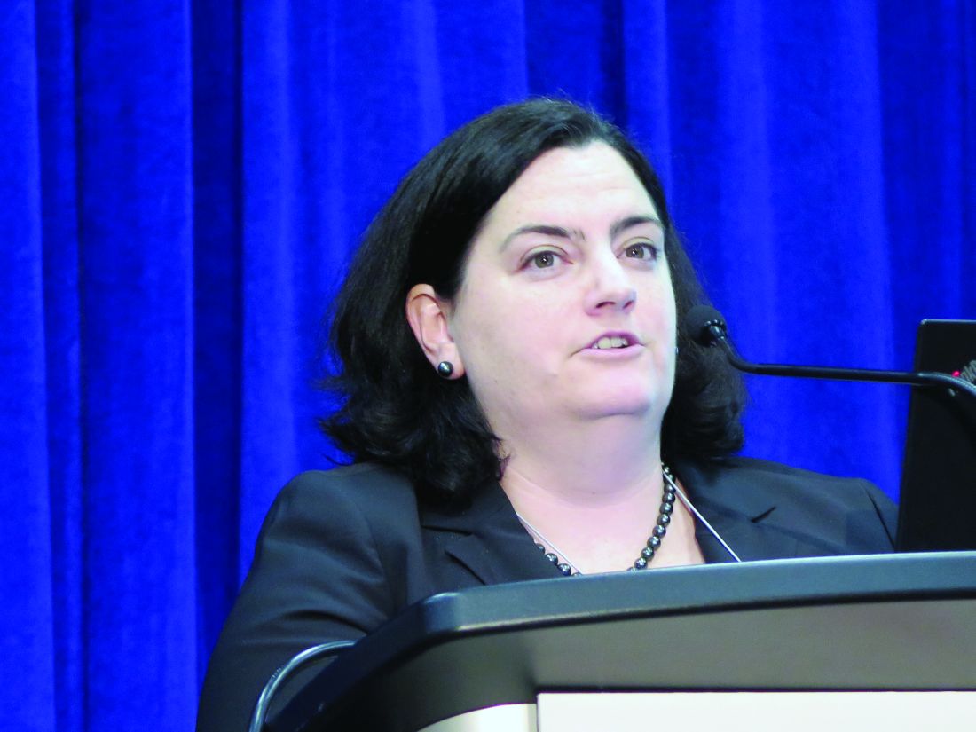
SAN ANTONIO – Researchers at MD Anderson Cancer Center in Houston and the University of New South Wales (UNSW) in Sydney are among teams from around the world working toward human breast cell atlas development using single-cell genomics, and their efforts to date have yielded new understanding of both the normal breast cell ecosystem and the breast cancer tumor microenvironment.
The work at MD Anderson, for example, has led to the identification of a number of new gene markers and multiple cell states within breast cell types, according to Tapsi Kumar Seth, who reported early findings from an analysis of more than 32,000 cells from normal breast tissue during a presentation at the San Antonio Breast Cancer Symposium.
At the UNSW’s Garvan Institute of Medical Research, Alexander Swarbrick, PhD, and his colleagues are working to better define the tumor microenvironment at the single-cell level. At the symposium, Dr. Swarbrick presented interim findings from cellular analyses in the first 23 breast cancer cases of about 200 that will be studied in the course of the project.
Improved understanding of the cellular landscape of both normal breast tissue and breast cancer tissue should lead to new stromal- and immune-based therapies for the treatment of breast cancer, the investigators said.
The normal breast cell ecosystem
The MD Anderson researchers studied 32,148 stromal cells from pathologically normal breast tissues collected from 11 women who underwent mastectomy at the center.
Unbiased expression analysis identified three major cell types, including epithelial cells, fibroblasts, and endothelial cells, as well as several minor cell types such as macrophages, T-cells, apocrine cells, pericytes, and others, said Ms. Seth, a graduate student in the department of genetics at the center and a member of the Navin Laboratory there.
The work is designed to help identify the presence and function of cells and explain how they behave in a normal breast ecosystem, she said.
“We know that a female breast undergoes a lot of changes due to age, pregnancy, or when there is a disease such as cancer, so it’s essential to chart out what a normal cell reference would look like,” she said.
Toward that goal, a protocol was developed to dissociate the tissue samples within 2 hours due to the decline in viability seen in cells and RNA over time. Analysis of the cell states revealed different transcriptional programs in luminal epithelial cells (hormone receptor positive and secretory), basal epithelial cells (myoepithelial or basement-like), endothelial cells (lymphatic or vascular), macrophages (M1 or M2) and fibroblasts (three subgroups) and provided insight into progenitors of each cell types, she said.
A map was created to show gene expression and to identify transcriptomally similar cells.
“We were able to identify most of the major cell types that are present in human breasts,” she said. “What was interesting was that the composition of these cells also varied across women.”
For example, the proportion of fibroblasts was lower in 3 of the 11 patients, and even though the cells were pathologically normal, immune cell populations, including T-cells and macrophages, were also seen.
Adipocytes cannot be evaluated using this technology because they are large and the layer of fat cells must be removed during dissociation to prevent clogging of the machines, she noted, adding that “this is really a limitation of our technology.”
A closer look was taken at each of the major cell types identified.
Epithelial cells
Both canonical and new gene markers were used to identify luminal and basal epithelial cells, Ms. Seth noted.
Among the known markers were KRT18 for luminal epithelial cells and KRT5, KRT6B, KRT14, and KRT15 for basal epithelial cells. Among the new markers were SLC39A6, EFHD1 and HES1 for the luminal epithelial cells, and CITED4, CCK28, MMP7, and MDRG2 for the basal epithelial cells.
“We went on and validated these markers on the tissue section using methods like spatial transcriptomics,” she said, explaining that this “really helps capture the RNA expression spatially,” and can resolve the localization of cell types markers in anatomical structures.
For these cells, the expression of the newly identified gene markers was mostly confined to ducts and lobules.
In addition, an analysis of cell states within the luminal epithelial cells showed four different cell states, each of which have “different kinds of genes that they express, and also different pathways that they express, suggesting that these might be transcriptomally different,” Ms. Seth said.
Of note, these cells and cells states are not biased to a specific condition or patient, suggesting that they are coming from all of the patients, she added.
Two of the four cell states – the secretory and hormone responsive states – have previously been reported, but Ms. Seth and her colleagues identified two additional cell states that may have different biological functions and are present in the different anatomical regions of the breast.
Fibroblasts
Fibroblasts, the cells of the connective tissue, were the most abundant cell type. Like the epithelial cells, both canonical collagen markers (COL6A3, MMP2, FBN1, FBLN2, FBN, and COL1A1) and newly identified gene markers (TNXB, AEBP1, CFH, CTSK, TPPP3, MEG3, HTRA1, LHFP, and OGN) were used to identify them.
Endothelial cells
Breast tissue is highly vascular, so endothelial cells, which line the walls of veins, arteries, and lymphatic vessels, are plentiful.
“Again, for both these cell types, we identified them using the canonical marker CD31, and we identified some new gene markers,” she said, noting that the new markers include CCL21, CLDN5, MMRN1, LYVE1, and PROX1 for lymphatic endothelial cells, and RNASE1 and IFI27 for vascular endothelial cells.
Two different groups – or states – of vascular endothelial cells were identified, with each expressing “very different genes as well as very different pathways, again suggesting that they might have different biological functions, which we are still investigating,” she said.
Additional findings and future directions
In addition to stromal cells, some immune cells were also seen. These included T cells that came mostly from two patients, as well as macrophages and monocytes, which comprised the most abundant immune cell population.
Of note, all of these cells are also found in the tumor microenvironment, but they are in a transformed state. For example cancer-associated fibroblasts, tumor endothelial cells, tumor-associated macrophages, and tumor-associated adipocytes have been seen in that environment, she said.
“So what we are trying to do with this project is ... learn how these cells are, and how these cells behave in the normal ecosystem,” she explained, noting that the hope is to provide a valuable reference for the research community with new insights about how normal cell types are transformed in the tumor microenvironment.
In an effort to overcome the adipocyte-associated limitation of the technology, adipocytes are “now being isolated by single nucleus RNA sequencing.”
“This [sequencing] technology has helped us identify multiple cell states within a cell type; and most of these cell states may have different biological functions, which probably can be investigated by spatial transcriptomic methods,” she said.
Spatial transcriptomics also continue to be used for validation of the new gene markers identified in the course of this research, she noted.
The breast tumor microenvironment
At the Garvan Institute, current work is focusing more on defining the landscape of the breast tumor microenvironment at single-cell resolution, according to Dr. Swarbrick, a senior research fellow and head of the Tumour Progression Laboratory there.
“Breast cancers ... are complex cellular ecosystems, and it’s really the sum of the interactions between the cell types that play major roles in determining the etiology of disease and its response to therapy,” he said. “So I think that going forward toward a new age of diagnostics and therapeutics, there’s wonderful potential in capitalizing on the tumor microenvironment for new developments, but this has to be built on a really deep understanding of the tumor microenvironment, and – I might say – a new taxonomy of the breast cellular environment.”
Therefore, in an effort to address “this limitation in our knowledge base,” his lab is also working toward development of a breast cell atlas.
A fresh tissue collection program was established to collect early breast cancer tissues at the time of surgery, metastatic biopsies, and metastatic lesions from autopsies. The tissues are quickly dissociated into their cellular components and they undergo massively parallel capture and sequencing using the 10x genomics platform, he said.
Thousands of cells per case are analyzed using single-cell RNA sequencing (RNA-seq), as well as “RAGE-seq” and “CITE-seq,” which are performed in parallel to the RNA sequencing to address some of the limitations of the RNA sequencing alone and to “try to gain a multi-omic insight into the cell biology,” he explained.
RAGE-seq, which Dr. Swarbrick and his team developed, “is essentially a method to do targeted long-read sequencing in parallel to the short-read sequencing that we use for RNA-seq,” and CITE-seq is “a really fantastic method developed at the New York Genome Center that essentially allows us to gather proteomic data in parallel to the RNA data,” he said.
Based on findings from the analyses of about 125,000 cells from 25 patients, a map was created that showed the cell clusters identified by both canonical markers and gene expression signatures.
“We find the cell types we would expect to be present in a breast cancer,” he said.
The map shows clusters of myeloid, epithelial-1 and -2, cancer-associated fibroblast (CAF)-1 and -2, endothelial, T Reg, B, and CD8 and CD4 T cells.
Next, each cell type is quantified in each patient, and a graphic representation of the findings shows large variability in the proportions of each cell type in each patient.
“Ultimately, our goal is to be able to relate the frequencies of cell types and molecular features to each other, but also to clinical-pathological features from these patients,” he noted.
A closer look at the findings on an individual case level demonstrates the potential for development of better therapies.
For example, a case involving a high-grade triple-negative invasive ductal carcinoma exhibited each of the cell types found overall.
“One of the things that strikes us early on is we see a number of malignant epithelial populations,” he said, noting that proliferation is one of the drivers of the heterogeneity, but that heterogeneity was also seen for “other clinically relevant features such as basal cytokeratins,” which were heterogeneously expressed in different cell-type clusters.
“This was kind of paralleled in the immunohistochemistry results that we obtained from this patient,” he said. “We could also apply other clinically used tests that we’ve developed on bulk (such as PAM50 intrinsic subtyping) and ask whether they can be applied at the single-cell resolution.
“We think that these are going to be great tools to try to now get in and understand the significance of this heterogeneity and try to identify the lethal cells within this patient, and potentially therapeutic strategies to eradicate those cells,” he added.
Fibroblasts
A notable finding of this project was the presence of “not one, but two populations of fibroblasts,” Dr. Swarbrick said, noting that fibroblasts are typically discussed as a single entity.
“This is arguing that there are at least two major types present within the breast, and almost every case has these populations present at roughly equal amounts,” he said.
This is of particular interest, because it has been shown in prior studies that targeting fibroblasts can have therapeutic outcomes.
“So we think this is a very important population within the tumor microenvironment,” he added.
With respect to gene expression features, CAF-1 is dominated by signatures of extracellular matrix deposition and remodeling, which “look like the classic myofibroblasts that we typically think of when we study cancer-associated fibroblasts.”
“In contrast, the CAF-2 population ... have what appears to be quite a predominant secretory function, so we see a lot of cytokines being produced by these cells, but we also see a very high level of expression of a number immune checkpoint ligands,” he said, adding that his team is actively pursuing whether these cells may be undergoing signaling events with infiltrating lymphocytes in the tumor microenvironment.
The signatures for both CAF types are prognostic within large breast cancer data sets, suggesting that they do actually have an important role in disease, he noted.
Markers for these cells include ACTA2, which was previously known to be a marker, and which is almost exclusively restricted to CAF-1, and the cell surface protein CD34 – a progenitor marker in many different cellular systems, “which is actually beautifully expressed on the CAF-2 population” as demonstrated using CITE-seq.
“So we’re now using this as a way to prospectively identify these cells, pull them out of tumors, and conduct biologic assays to learn more about them,” he said.
The immune milieu
“We’re in the age of immunotherapy, and this is an area of huge interest, but we have a long way to go in making it as effective as possible for breast cancer patients,” Dr. Swarbrick said. “I believe part of that is through a very deep understanding of the taxonomy.”
RNA data alone are useful but insufficient to fully identify subsets of immune cells due to a “relatively low-resolution ability to resolve T cells.”
“But because we’re now using the panel of 125 antibodies in parallel, we can now start to use protein levels to split up these populations and we can start to now identify, with higher resolution, more unique populations within the environment,” he said, noting that the availability of protein data not only helps identify subtypes, but is also therapeutically important as it allows for certainty regarding whether the protein target of therapeutic antibodies is expressed on the surface of cells.
Ultimately the hope is that this effort to build a multi-omic breast cancer atlas will continue to drive new discoveries in personalized medicine for breast cancer, Dr. Swarbrick concluded, adding that the field is moving fast, and it will be very important for labs like his and the Navin Lab to communicate to avoid needlessly duplicating efforts.
“I think it’s going to be really exciting to start to put some of these [findings] together,” he said.
The MD Anderson project is funded by the Chan Zuckerberg Initiative as part of its work in supporting the Human Cell Atlas project. Ms. Seth reported having no disclosures. Dr. Swarbrick’s research is funded by the Australian Government/National Health and Medical Research Council and the National Breast Cancer Foundation. He reported having no relevant disclosures.
SOURCE: Seth T et al. SABCS 2018, Abstract GS1-02; Swarbrick A et al. SABCS 2018, Abstract GS1-01
REPORTING FROM SABCS 2018
Key clinical point: Improved understanding of the cellular landscape of both normal breast tissue and breast cancer could lead to new stromal- and immune-based therapies.
Major finding: From pathologically normal breast tissues expression, investigators identified three major cell types, as well as several minor cell types. In analyses of cells from breast cancer patients, a map was created that showed the cell clusters identified by both canonical markers and gene expression signatures.
Study details: An analysis of 32,138 breast cells from 11 women, and another of about 125,000 cells from 25 patients.
Disclosures: The MD Anderson research is part of the Human Cell Atlas project and is funded by the Chan Zuckerberg Initiative. Ms. Seth reported having no disclosures. Dr. Swarbrick’s research is funded by the Australian Government/National Health and Medical Research Council and the National Breast Cancer Foundation. He reported having no relevant disclosures.
Source: Seth T et al. SABCS 2018: Abstract GS1-02; Swarbrick A et al. SABCS 2018: Abstract GS1-01.
Daily News Special: SABCS
Stories include: uUing low-dose tamoxifen, the latest findings from the KATHERINE trial, results of a meta-analysis of neoadjuvant chemotherapy, and capecitabine in early stage triple negative breast cancer.
Amazon Alexa
Apple Podcasts
Google Podcasts
Spotify
Stories include: uUing low-dose tamoxifen, the latest findings from the KATHERINE trial, results of a meta-analysis of neoadjuvant chemotherapy, and capecitabine in early stage triple negative breast cancer.
Amazon Alexa
Apple Podcasts
Google Podcasts
Spotify
Stories include: uUing low-dose tamoxifen, the latest findings from the KATHERINE trial, results of a meta-analysis of neoadjuvant chemotherapy, and capecitabine in early stage triple negative breast cancer.
Amazon Alexa
Apple Podcasts
Google Podcasts
Spotify
Young women opt for mastectomy even when neoadjuvant chemo works well
SAN ANTONIO – Response to neoadjuvant chemotherapy has little if any influence on the choice of surgery among young women with early-stage breast cancer, suggests a multicenter, prospective cohort study reported at the San Antonio Breast Cancer Symposium.
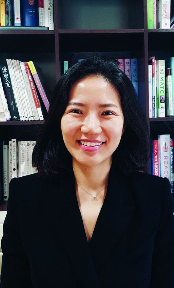
Randomized, controlled trials have found high levels of mastectomy among patients who are eligible for breast-conserving surgery, according to first author Hee Jeong Kim, MD, PhD, a visiting scholar at the Dana-Farber Cancer Institute, Boston, and an associate professor in the division of breast in the department of surgery at the University of Ulsan, Seoul, South Korea.
“Young women are more likely to present with large tumors and particularly benefit from a neoadjuvant systemic approach,” she noted. “Recent data suggest that response rates, including pathological complete response, are higher in women younger than 40 than in older women, but little is known about how response to neoadjuvant chemotherapy influences surgical decisions in young women.”
The investigators studied 315 women aged 40 years or younger at diagnosis of unilateral stage I-III breast cancer who received neoadjuvant chemotherapy. Results showed that the chemotherapy doubled the proportion who were eligible for breast-conserving surgery, but 41% of all women eligible after neoadjuvant chemotherapy opted to undergo mastectomy, and the value was essentially the same (42%) among the subset who achieved a complete clinical response. The leading reason given in the medical record for this choice was personal preference in the absence of any known high-risk predisposition.
“Surgical decisions among young women with breast cancer appear to be driven by factors beyond the extent of disease and response to neoadjuvant chemotherapy,” she commented. “We should focus our efforts to optimize surgical decisions in these patients.”
The study complements another study undertaken in the same cohort, also reported at the symposium, that assessed longer-term quality of life according to which surgery women chose; this quality-of-life study found poorer measures after mastectomy.
Drivers and explanatory factors
Session moderator Fatima Cardoso, MD, director of the Breast Unit at the Champalimaud Clinical Center in Lisbon, asked, “Do you think this is really the patient preference, or is this more the surgeon’s preference that is passed on to the patient? Because there is now data showing that breast conservation with radiation is better, even in terms of survival, than mastectomy.”
“Patient preference includes a variety of things. Maybe it is a real patient preference [driven by] fear of recurrence or their peace of mind, but another important factor is maybe the doctor, especially the surgeon. That’s why we should be aware of surgical overtreatment, especially in these young early breast cancer patients,” Dr. Kim replied. “But the good news from this study is that neoadjuvant chemotherapy can give options to the patients, they can choose mastectomy. I think that it’s totally different when the patient has no option other than mastectomy versus the patient can choose mastectomy.”
Two main groups of patients in the United States are being given neoadjuvant chemotherapy, noted session attendee Steven E. Vogl, MD, an oncologist at Montefiore Medical Center, New York. One group has large tumors, and the goal is to shrink the tumor; the other group is planning to have unilateral or bilateral mastectomy with some type of reconstruction by a plastic surgeon.
“The medical oncologist, having decided the [latter] patient needs chemotherapy, chooses to give the chemotherapy preoperatively, so it’s not delayed by 3-5 months for the wounds to heal,” he elaborated. “How many of your patients were in the second category?”
The study did not tease out that population, Dr. Kim replied.
Study details
The women studied were participants in the Young Women’s Breast Cancer Study (YWS). Some 67% had a clinical complete response (no palpable tumor in the breast) to their neoadjuvant chemotherapy, and 32% had a pathological complete response (no tumor in the breast, with or without ductal carcinoma in situ [DCIS], and no tumor deposit exceeding 0.2 mm in the lymph nodes).
Before neoadjuvant chemotherapy, 26% of the women overall were eligible for breast-conserving surgery, but after neoadjuvant chemotherapy, 42% were eligible, Dr. Kim reported.
However, in the entire cohort, breast-conserving surgery was the initial surgery in just 25% of women and the final (definitive) surgery in just 23%.
Among patients eligible for breast conservation after neoadjuvant chemotherapy, 41% chose mastectomy instead as their initial surgery. Response to the chemotherapy seemingly did not influence this choice given that 42% of the subset with a clinical complete response still chose mastectomy. Furthermore, among those eligible for breast conservation who underwent mastectomy, 35% had a pathologic complete response to the chemotherapy.
Of all patients eligible for breast-conserving surgery who opted for mastectomy (and usually a bilateral procedure), the most common reason for choosing this more extensive surgery was personal preference, documented in 53% of cases, followed by presence of a BRCA or p53 mutation or a strong family history, documented in 40%. Reasons were similar among the breast conservation–eligible women who had a clinical complete response and/or ultimately a pathological complete response but chose mastectomy.
The study did not analyze disease factors that may have influenced choice of surgery, such as multicentricity or presence of DCIS, acknowledged Dr. Kim, who disclosed that she had no relevant conflicts of interest.
In an exploratory analysis, use of neoadjuvant chemotherapy increased over time among YWS participants, from 23% among those with diagnosis in 2006-2007 to 44% among those with diagnosis in 2014-2015. There were concurrent improvements in the proportions who achieved a clinical complete response (from 64% to 77%) and a pathological complete response (from 23% to 34%). Yet the proportion undergoing breast-conserving surgery as their initial surgery fell slightly, from 21% to 19%, during the same period.
SOURCE: Kim HJ et al. SABCS 2018, Abstract GS6-01,
REPORTING FROM SABCS 2018
Key clinical point: Response to neoadjuvant chemotherapy does not alter choice of surgery among young breast cancer patients.
Major finding: Neoadjuvant chemotherapy increased the proportion eligible for breast-conserving surgery from 26% to 42%, but about 40% of those eligible chose mastectomy regardless of chemotherapy response, mainly because of personal preference.
Study details: A multicenter, prospective cohort study of 315 women aged 40 years or younger at diagnosis of early-stage breast cancer who received neoadjuvant chemotherapy (Young Women’s Breast Cancer Study).
Disclosures: Dr. Kim disclosed that she had no relevant conflicts of interest.
Source: Kim HJ et al. SABCS 2018, Abstract GS6-01.
Older breast cancer patients given adjuvant chemo live longer
SAN ANTONIO – captured in the National Cancer Database.
“Data for elderly patients in clinical trials is limited. The NCCN [National Comprehensive Cancer Network] guidelines note that there is limited data to make chemotherapy recommendations for those older than 70 years of age,” commented first author Shreya Sinha, MD, an oncology fellow at the State University of New York, Syracuse. Furthermore, the few studies assessing adjuvant chemotherapy benefit among geriatric breast cancer patients have had conflicting results.
In the new study, reported at the San Antonio Breast Cancer Symposium, older women with stage I-III breast cancer who received adjuvant chemotherapy had a nearly 40% reduction in the adjusted risk of death relative to counterparts who did not receive adjuvant chemotherapy. Benefit was seen across disease stages and across hormone receptor statuses.
Patients’ fitness to receive chemotherapy and their causes of death could not be determined, Dr. Sinha acknowledged. Therefore, chemotherapy’s role in the observed survival difference is not definitive.
“In general, when we are treating our elderly population, we have to take physiologic age into consideration when coming up with a treatment plan,” she said. However, “we also have to use chemotherapy toxicity prediction calculators,” such as the Cancer and Aging Research Group tool and the Chemotherapy Risk Assessment Scale for High-Age Patients.
In addition, gene-based assays, such as Oncotype DX and MammaPrint, which were not taken into account for the study, can be applied to estimate the likely benefit of chemotherapy and further inform the treatment decision.
“It can’t be that chemotherapy is making these patients live longer. It has to be that the doctors know not to give chemotherapy to those who are going to die soon,” speculated symposium attendee Steven E. Vogl, MD, an oncologist at Montefiore Medical Center, New York.
He therefore wondered if the amount of chemotherapy received was related to survival. “If it was a chemotherapy effect, then more is probably better, and if it’s a selection effect at the time of initiation, then more probably wouldn’t be better.”
“These are the issues we run into when we use the National Cancer Database or such large databases,” Dr. Sinha replied. “We don’t necessarily have the information on how much chemotherapy the patients received. It really is based on if they received chemotherapy or not.”
Study details
The 160,676 patients studied were treated for stage I-III breast cancer during 2004-2015 and were included regardless of hormone receptor status and HER2 status.
Overall, 60.45% received adjuvant chemotherapy, Dr. Sinha reported. Mean age was 70.7 years among chemotherapy recipients and 75.5 years among nonrecipients.
Women were more likely to receive adjuvant chemotherapy if they had a tumor grade of 2 or 3 (adjusted odds ratios, 1.88 and 3.51), had a tumor negative for both estrogen and progesterone receptors or just progesterone receptors (aOR, 2.72 and 1.70), had private insurance versus Medicaid or Medicare (aOR, 1.40 and 1.20), or received radiation therapy (aOR, 2.55).
Women were less likely to receive adjuvant chemotherapy if they had stage 1 or 2 disease (aOR, 0.23 and 0.56; P less than .0001 for each), were older than 80 years (aOR, 0.105; P less than .0001), had undergone lumpectomy versus mastectomy (aOR, 0.82; P = .0011), were treated in an academic versus community program (aOR, 0.93; P = .0007), or had a Charlson/Deyo comorbidity score of 3 or higher (aOR, 0.38; P less than .0001).
Median overall survival was 144.9 months with and 112.6 months without adjuvant chemotherapy. The difference translated to a significantly reduced risk of death for the women given adjuvant chemotherapy (adjusted hazard ratio, 0.617; P less than .0001). The corresponding 10-year overall survival rates were 59.5% and 46.7%.
The reduced risk of death with adjuvant chemotherapy was evident in women with stage 1 disease (aHR, 0.801), stage 2 disease (aHR, 0.608), and stage 3 disease (aHR, 0.666) (P less than .0001 for all). It was also evident in those with tumors positive for both estrogen and progesterone receptors (aHR, 0.649), negative for progesterone receptors only (aHR, 0.609), and negative for both (aHR, 0.547) (P less than .0001 for all).
“The HER2/neu patient unfortunately was not well defined since there was no data [on that marker] before 2010,” Dr. Sinha noted
Dr. Sinha reported no relevant conflicts of interest. The study received funding from the Research Foundation of SUNY.
SOURCE: Sinha S et al. SABCS 2018, Abstract GS2-02.
REPORTING FROM SABCS 2018
Key clinical point: Older patients with early breast cancer who are given adjuvant chemotherapy live longer.
Major finding: Receipt of adjuvant chemotherapy was associated with a reduced risk of death after taking into account factors such as age and comorbidity burden (adjusted hazard ratio, 0.617; P less than .0001).
Study details: A retrospective cohort study of 160,676 breast cancer patients aged 65 years and older with stage I-III disease.
Disclosures: Dr. Sinha reported no relevant conflicts of interest. The study received funding from the Research Foundation of SUNY.
Source: Sinha S et al. SABCS 2018, Abstract GS2-02.
QOL is poorer for young women after mastectomy than BCS
SAN ANTONIO – , according to investigators for a multicenter cross-sectional cohort study reported at the San Antonio Breast Cancer Symposium.
Women aged 40 or younger make up about 7% of all newly diagnosed cases of breast cancer in the United States, according to lead author, Laura S. Dominici, MD, of Dana-Farber/Brigham and Women’s Cancer Center and Harvard Medical School, Boston.
“Despite the fact that there is equivalent local-regional control with breast conservation and mastectomy, the rates of mastectomy and particularly bilateral mastectomy are increasing in young women, with a 10-fold increase seen from 1998 to 2011,” she noted in a press conference. “Young women are at particular risk for poorer psychosocial outcomes following a breast cancer diagnosis and in survivorship. However, little is known about the impact of surgery, particularly in the era of increasing bilateral mastectomy, on the quality of life of young survivors.”
Nearly three-fourths of the 560 young breast cancer survivors studied had undergone mastectomy, usually with some kind of reconstruction. Roughly 6 years later, compared with peers who had undergone breast-conserving surgery, women who had undergone unilateral or bilateral mastectomy had significantly poorer adjusted BREAST-Q scores for satisfaction with the appearance and feel of their breasts (beta, –8.7 and –9.3 points) and psychosocial well-being (–8.3 and –10.5 points). The latter also had poorer adjusted scores for sexual well-being (–8.1 points). Physical well-being, which captures aspects such as pain and range of motion, did not differ significantly by type of surgery.
“Local therapy decisions are associated with a persistent impact on quality of life in young breast cancer survivors,” Dr. Dominici concluded. “Knowledge of the potential long-term impact of surgery and quality of life is of critical importance for counseling young women about surgical decisions.”
Moving away from mastectomy
“The data are, to me anyway, more disconcerting when you consider the high mastectomy rate in this country relative to Europe, and this urge to have bilateral mastectomies, which, pardon the expression, is ridiculous in some cases because it doesn’t improve your outcome. And yet, it does have deleterious effects that last for years psychologically,” commented SABCS codirector and press conference moderator C. Kent Osborne, MD, who is director of the Dan L. Duncan Cancer Center at Baylor College of Medicine, Houston. “What can we do about that?” he asked.
“It’s a really challenging problem,” Dr. Dominici replied. “Part of what we are missing in the conversation that we have with our patients is this kind of information. We can certainly tell patients that the outcomes are equivalent, but if they don’t know that the long-term [quality of life] impact is potentially worse, then that may not affect their decision. The more prospective data that we generate to help us figure out which patients are going to have better or worse outcomes with these different types of surgery, the better we will be able to counsel patients with things that will be meaningful to them in the long run.”
The study was not designed to tease out the specific role of anxiety about a recurrence or a new breast cancer, which is a major driver of the decision to have mastectomy and also needs to be addressed during counseling, Dr. Dominici and Dr. Osborne agreed. “I think I spend more time talking patients out of bilateral mastectomy or mastectomy at all than anything,” he commented.
Study details
The women studied were participants in the prospective Young Women’s Breast Cancer Study (YWS) and had a mean age of 37 years at diagnosis. Most (86%) had stage 0-2 breast cancer. (Those with metastatic disease at diagnosis or a recurrence during follow-up were excluded.)
Overall, 52% of the women underwent bilateral mastectomy, 20% underwent unilateral mastectomy, and 28% underwent breast-conserving surgery, Dr. Dominici reported. Within the mastectomy group, most underwent implant-based reconstruction (69%) or flap reconstruction (12%), while some opted for no reconstruction (11%).
Multivariate analyses showed that, in addition to mastectomy, other significant predictors of poorer breast satisfaction were receipt of radiation therapy (beta, –7.5 points) and having a financially uncomfortable status as compared with a comfortable one (–5.4 points).
Additional significant predictors of poorer psychosocial well-being were receiving radiation (beta, –6.0 points), being financially uncomfortable (–7 points), and being overweight or obese (–4.2 points), and additional significant predictors of poorer sexual well-being were being financially uncomfortable (–6.8 points), being overweight or obese (–5.3 points), and having lymphedema a year after diagnosis (–3.8 points).
The only significant predictors of poorer physical health were financially uncomfortable status (beta, –4.8 points) and lymphedema (–6.4 points), whereas longer time since surgery (more than 5 years) predicted better physical health (+6.0 points), according to Dr. Dominici.
Age, race, marital status, work status, education level, disease stage, chemotherapy, and endocrine therapy did not significantly predict any of the outcomes studied.
“This was a one-time survey of women who were enrolled in an observational cohort study, and we know that preoperative quality of life likely drives surgical choices,” she commented, addressing the study’s limitations. “Our findings may have limited generalizability to a more diverse population in that the majority of our participants were white and of high socioeconomic status.”
Dr. Dominici disclosed that she had no conflicts of interest. The study was funded by the Agency for Healthcare Research and Quality, Susan G. Komen, the Breast Cancer Research Foundation, and The Pink Agenda.
SOURCE: Dominici LS et al. SABCS 2018, Abstract GS6-06,
REPORTING FROM SABCS 2018
Key clinical point: More extensive breast surgery has a long-term negative impact on QOL for young breast cancer survivors.
Major finding: Compared with peers who underwent breast-conserving surgery, young women who underwent unilateral or bilateral mastectomy had significantly poorer adjusted scores for breast satisfaction (beta, –8.7 and –9.3 points) and psychosocial well-being (beta, –8.3 and –10.5 points).
Study details: A multicenter cross-sectional cohort study of 560 women with a mean age of 37 years at breast cancer diagnosis who completed the BREAST-Q questionnaire a median of 5.8 years later.
Disclosures: Dr. Dominici disclosed that she had no conflicts of interest. The study was funded by the Agency for Healthcare Research and Quality, Susan G. Komen, the Breast Cancer Research Foundation, and The Pink Agenda.
Source: Dominici LS et al. SABCS 2018, Abstract GS6-06.
Data underscore the importance of lifestyle interventions in breast cancer patients
SAN ANTONIO – Data continue to underscore the benefits of lifestyle interventions in women with breast cancer, but questions remain about their effects on recurrence, according to Jennifer Ligibel, MD.
Findings from the EBBA-II trial as presented at the San Antonio Breast Cancer Symposium, for example, showed that exercise improves cardiorespiratory fitness in women with early breast cancer, and findings from the SUCCESS C study showed that breast cancer patients who completed a weight-loss intervention showed some improvements, compared with those who did not, said Dr. Ligibel of Dana-Farber Cancer Institute in Boston, who was the discussant for those and other lifestyle-intervention studies at the symposium.
SUCCESS C failed to show an overall reduction in breast cancer recurrence or survival, but weight loss among intervention-group participants was modest, and more than half of the participants dropped out of the study, so it’s hard to make any firm conclusions, she said.
Overall, the findings – in the context of what is already known about lifestyle interventions among women with breast cancer – provide “yet another reason to tell women that it’s important to exercise during treatment,” she said.

In this video interview, Dr. Ligibel discussed the studies and the implications of the findings, and also mentioned an ongoing study for which she is an investigator. In that study – the Breast Cancer Weight Loss study (BWEL) – adherence among the approximately 1,700 women enrolled has been high. “So we’re hoping that this study in a few years will give us a bit more information about the power of weight loss to potentially reduce recurrence.”
For now, the available data show that there are “lots of concrete benefits” associated with improving lifestyle in women with breast cancer, she said, noting that she tells all of her patients to live as healthy a lifestyle as possible, and especially to exercise.
SAN ANTONIO – Data continue to underscore the benefits of lifestyle interventions in women with breast cancer, but questions remain about their effects on recurrence, according to Jennifer Ligibel, MD.
Findings from the EBBA-II trial as presented at the San Antonio Breast Cancer Symposium, for example, showed that exercise improves cardiorespiratory fitness in women with early breast cancer, and findings from the SUCCESS C study showed that breast cancer patients who completed a weight-loss intervention showed some improvements, compared with those who did not, said Dr. Ligibel of Dana-Farber Cancer Institute in Boston, who was the discussant for those and other lifestyle-intervention studies at the symposium.
SUCCESS C failed to show an overall reduction in breast cancer recurrence or survival, but weight loss among intervention-group participants was modest, and more than half of the participants dropped out of the study, so it’s hard to make any firm conclusions, she said.
Overall, the findings – in the context of what is already known about lifestyle interventions among women with breast cancer – provide “yet another reason to tell women that it’s important to exercise during treatment,” she said.

In this video interview, Dr. Ligibel discussed the studies and the implications of the findings, and also mentioned an ongoing study for which she is an investigator. In that study – the Breast Cancer Weight Loss study (BWEL) – adherence among the approximately 1,700 women enrolled has been high. “So we’re hoping that this study in a few years will give us a bit more information about the power of weight loss to potentially reduce recurrence.”
For now, the available data show that there are “lots of concrete benefits” associated with improving lifestyle in women with breast cancer, she said, noting that she tells all of her patients to live as healthy a lifestyle as possible, and especially to exercise.
SAN ANTONIO – Data continue to underscore the benefits of lifestyle interventions in women with breast cancer, but questions remain about their effects on recurrence, according to Jennifer Ligibel, MD.
Findings from the EBBA-II trial as presented at the San Antonio Breast Cancer Symposium, for example, showed that exercise improves cardiorespiratory fitness in women with early breast cancer, and findings from the SUCCESS C study showed that breast cancer patients who completed a weight-loss intervention showed some improvements, compared with those who did not, said Dr. Ligibel of Dana-Farber Cancer Institute in Boston, who was the discussant for those and other lifestyle-intervention studies at the symposium.
SUCCESS C failed to show an overall reduction in breast cancer recurrence or survival, but weight loss among intervention-group participants was modest, and more than half of the participants dropped out of the study, so it’s hard to make any firm conclusions, she said.
Overall, the findings – in the context of what is already known about lifestyle interventions among women with breast cancer – provide “yet another reason to tell women that it’s important to exercise during treatment,” she said.

In this video interview, Dr. Ligibel discussed the studies and the implications of the findings, and also mentioned an ongoing study for which she is an investigator. In that study – the Breast Cancer Weight Loss study (BWEL) – adherence among the approximately 1,700 women enrolled has been high. “So we’re hoping that this study in a few years will give us a bit more information about the power of weight loss to potentially reduce recurrence.”
For now, the available data show that there are “lots of concrete benefits” associated with improving lifestyle in women with breast cancer, she said, noting that she tells all of her patients to live as healthy a lifestyle as possible, and especially to exercise.
REPORTING FROM SABCS 2018
Extended AI therapy reduces breast cancer recurrence risk, ups fracture risk
SAN ANTONIO – Extending aromatase inhibitor (AI) therapy an additional 5 years reduces breast cancer recurrence risk, particularly in patients with node involvement, but the benefits vary based on prior treatment and must be weighed against the risk of bone fracture, according to findings from a meta-analysis involving more than 22,000 women.
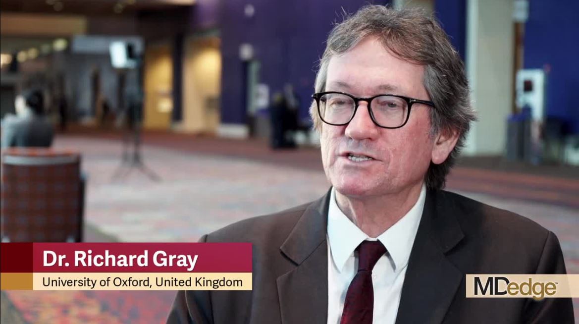
The rate of any recurrence after 10 years in almost 7,500 women treated with 5 years of tamoxifen and then randomized to 5 additional years of AI treatment was reduced by 35%, compared with the rate in those who did not receive 5 additional years of AI therapy (recurrence rate, 10.7% vs. 7.1%, respectively; relative risk, 0.67), and the difference was “very highly significant,” Richard Gray, MD, reported at the San Antonio Breast Cancer Symposium.
The distant recurrence rate and mortality rate were also significantly improved in those who received 5 years of AI therapy (rr, 0.77 for each), but the difference in mortality was of borderline significance, Dr. Gray, professor of medical statistics at the University of Oxford, London, reported on behalf of the Early Breast Cancer Trialists’ Collaborative Group.
However, in many of the trials included in the analysis, control group patients crossed over to the treatment group, which likely reduced the effect, he noted.
In about 12,000 women who were treated with 2-3 years of tamoxifen followed by 2-3 years of an AI and who were then randomized to an additional 3-5 years of AI therapy, the effects were less pronounced, with about a 20% reduced risk of any recurrence after 10 years vs. the rate in those without extended therapy (recurrence rate, 9.2% vs. 7.1%), he said.
The differences in the rates of distant recurrence and breast cancer mortality were not statistically significant in this group, but again, follow-up was short, he said.
Similarly, in about 3,300 women treated with an AI followed by an additional 5 years of AI therapy, recurrence risk was reduced by about 25% vs. the rate in those who did not receive extended therapy, and no difference was seen in the rates of distant recurrence or breast cancer mortality.
Of note, the benefits in those who received tamoxifen were seen immediately, whereas the benefits in those receiving AIs in the first 5 years emerged after about 2 years of extended therapy, Dr. Gray said, explaining that this was likely due to “carry-over benefits” of the earlier AI therapy.
The downside with extended AI therapy was a 25% increase in fracture risk, as well as bone pain, which can reduce quality of life.
Therefore, decisions about extended therapy should involve careful risk-benefit analyses, he said, adding that the findings of this meta-analysis of 12 trials, which included postmenopausal women – 99% of whom had estrogen receptor–positive disease – provide “the most reliable assessment of the available evidence ... [for] guiding decisions about endocrine therapy.”
In this video interview, he further discussed the details and limitations of the study, the effects of nodal status on outcomes, implications of the findings for clinical practice, the need for further follow-up on all of the studies included in the analysis, and plans for incorporating new data from the AERAS trial, which were also presented at the symposium and which complement and reinforce the current findings.
Dr. Gray reported having no disclosures.
SOURCE: Gray R et al., SABCS 2018: Abstract GS3-03.
REPORTING FROM SABCS 2018
Key clinical point: Five extra years of aromatase inhibitor therapy reduces breast cancer recurrence, but increases fracture risk.
Major finding: Extending AI therapy by 5 years reduces breast cancer recurrence by 20% to 35%.
Study details: A meta-analysis of more than 22,000 women from 12 trials.
Disclosures: Dr. Gray reported having no disclosures.
Source: Gray R et al. SABCS 2018: Abstract GS3-03.
Oxybutynin rapidly quells hot flashes
SAN ANTONIO – because of a history of or concern about breast cancer, suggests a phase 3 double-blind randomized controlled trial.
Managing hot flashes in breast cancer survivors is important for ensuring their adherence to endocrine therapy, as about a third fail to complete the recommended 5- to 7-year course, in part because of side effects, Roberto A. Leon-Ferre, MD, of the Mayo Clinic, Rochester, Minn., reported at the San Antonio Breast Cancer Symposium
But many survivors cannot use estrogen because of hormone receptor–positive disease, and currently used nonhormonal alternatives have drawbacks. “Some of these agents interfere with the metabolic activation of tamoxifen, for example. There is also the association, unfortunately, of the taboo of taking antidepressants or anticonvulsants when you don’t have those diagnoses,” he said. In addition, a variety of nonpharmacologic options, such as black cohosh and vitamin E, have not proved any more effective than placebo.
The 150 women enrolled in the trial, ACCRU study SC-1603, were experiencing frequent, bothersome hot flashes and had a history of or concern about breast cancer. The 6-week reduction in a hot flash score capturing both frequency and severity was about 30% with placebo, 65% with oxybutynin 2.5 mg b.i.d., and 80% with oxybutynin 5 mg b.i.d. (P less than .01 across groups and for each dose vs. placebo), with a difference emerging within 2 weeks. “These doses are on the lower end of the currently used doses for urinary incontinence,” Dr. Leon-Ferre noted, with that range extending up to 20 mg daily.
The oxybutynin groups also had significantly greater reductions in hot flash frequency alone and improvements in measures of quality of life such as sleep, work, and relations. The drug was well tolerated, with expected main side effects of dry mouth and difficulty urinating.
Despite the potential pitfalls of cross-trial comparisons, the magnitude of benefit with oxybutynin appeared to exceed that previously reported with clonidine, fluoxetine, citalopram, venlafaxine, and pregabalin, according to Dr. Leon-Ferre.
“Oxybutynin significantly improves hot flash frequency and severity. The use of oxybutynin, more importantly, is associated with a positive impact in several quality of life metrics. And toxicity was acceptable,” he said. “While the two oxybutynin doses were not formally compared, 5 mg twice daily appears to be more effective.”
Treatment considerations
“What is your current strategy for using this variety of drugs?” asked SABCS codirector and press conference moderator C. Kent Osborne, MD, director of the Dan L. Duncan Cancer Center at Baylor College of Medicine, Houston. “Also, acupuncture has been shown to work in several randomized trials,” he noted.
“Before this study, we had been primarily using citalopram or venlafaxine as our first drug intervention. We typically favored venlafaxine for patients who are taking tamoxifen due to the concern about interaction with the CYP2D6 inhibitors,” Dr. Leon-Ferre replied. Oxybutynin is an attractive alternative here because patients can stop it abruptly if they want, whereas venlafaxine may require a lengthy period of tapering and weaning.
His institution doesn’t have a structured acupuncture program. “We do have acupuncturists, but they have to follow a specific program, it’s not any acupuncture. But we often recommend that patients pursue it if they have access to it,” he explained. “With the results of this particular study, we have become more keen on using oxybutynin. As a matter of fact, many of the patients who enrolled in this study decided to continue [or start] it after it had been revealed whether they were taking it or the placebo.”
As with all therapies, it is important to match the therapy to the patient, Dr. Leon-Ferre cautioned. “I can tell you that we have been using oxybutynin, but one has to be cautious about which patients to select for this because this is an anticholinergic drug. We were very careful about not including patients who had taken other potent anticholinergic drugs because these medications can lead to confusion episodes and altered mental status, particularly in more elderly patients and patients who suffer from polypharmacy and take many medications that start interacting with each other.” Another contraindication is urinary retention.
It is also noteworthy that women in the trial received just 6 weeks of oxybutynin therapy, as there has been some concern that extended use of anticholinergics can lead to memory issues.
“With those caveats, I think that if we have an informed decision, we could prescribe oxybutynin to patients,” Dr. Leon-Ferre said. “But ideally, I would say try to use it for a shorter rather than longer period of time.”
Study details
The women randomized in ACCRU study SC-1603 had had hot flashes for at least 30 days and were experiencing at least 28 of them each week. Concurrent stable-dose antidepressants, gabapentin, and pregabalin were allowed, whereas concurrent potent anticholinergics were not. Two-thirds of the women were on tamoxifen or an aromatase inhibitor.
In addition to the dramatic reduction in hot flash scores seen with oxybutynin, the drug was associated with marked reductions in hot flash frequency: 30% with placebo versus 60% with oxybutynin 2.5 mg b.i.d. and 75% with oxybutynin 5 mg b.i.d. (P less than .01 across groups and for each dose compared with placebo), Dr. Leon-Ferre reported.
Most of the 10 domains on the Hot Flash-Related Daily Interference Scale were significantly more improved with both doses of oxybutynin relative to placebo. The exceptions were mood and life enjoyment, which were significantly more improved only with the higher dose, and concentration and sexuality, which were not significantly more improved with either dose.
Both doses of oxybutynin were overall well tolerated, according to Dr. Leon-Ferre. Each was associated with higher incidence of dry mouth, abdominal pain, and difficulty urinating relative to placebo, as expected from what is known about the drug. The higher dose had a greater incidence of dry eyes, episodes of confusion, diarrhea, and headache.
Dr. Leon-Ferre disclosed that he had no conflicts of interest. The study was funded by the Breast Cancer Research Foundation.
SOURCE: Leon-Ferre RA et al. SABCS 2018 Abstract GS6-02.
REPORTING FROM SABCS 2018
Partial- and whole-breast irradiation very close in efficacy
SAN ANTONIO – , suggests a phase 3, randomized, controlled trial conducted by NRG Oncology.
At a median follow-up of 10.2 years, the trial was unable to refute the hypothesis that the partial technique was inferior in terms of ipsilateral breast tumor recurrences; however, the difference between techniques in this outcome was an absolute 0.7%, lead investigator Frank Vicini, MD, principal investigator at the MHP Radiation Oncology Institute/21st Century Oncology in Pontiac, Mich., reported in a session and press conference at the San Antonio Breast Cancer Symposium. The difference in recurrence-free interval was significant but likewise small, at 1.6%, and other efficacy outcomes were similar.
Meanwhile, the groups had low, statistically indistinguishable rates of grade 3-5 toxicities and second cancers. The investigators are still analyzing quality of life and cosmesis outcomes.
“This was the largest trial ever looking at partial-breast [irradiation] in a very diverse group of patients. Even though we weren’t able to demonstrate equivalence, it’s nice to see that in this large population of patients with extended follow-up, the differences are quite small,” Dr. Vicini said. “Because the differences for both ipsilateral breast tumor recurrence and recurrence-free interval were very small, partial-breast irradiation may be an acceptable alternative to whole-breast irradiation for a proportion of women who are undergoing breast-conserving surgery.”
Implications for practice and research
SABCS codirector and press conference moderator Virginia Kaklamani, MD, leader of the breast cancer program at UT Health San Antonio, asked how the findings have influenced his practice.
“This trial is over 15 years old now, and a lot of these techniques have been refined. But we are offering partial-breast irradiation to our patients,” Dr. Vicini replied. “There are a lot of competing ways to do radiation now; probably the biggest competing way is to do 3 weeks of whole-breast irradiation. But for those patients who have transportation issues and more elderly patients, we try to offer partial-breast irradiation, within the guidelines of ASTRO [American Society for Therapeutic Radiology and Oncology].”
Some of the women enrolled had risk factors that fall outside those guidelines, for example, higher-grade tumors or axillary node involvement, he acknowledged. “We tried to do exploratory analyses to look at whether certain patients did better with whole-breast irradiation or not, and we weren’t able to really pick out any group of patients that had better or worse outcome based on those criteria,” he said. “We have yet to look at the quality indices for radiation therapy, in other words, how much the breast actually needs to be treated. But at the present time, I would just suggest sticking with the ASTRO guidelines.”
It is noteworthy that likely the most important endpoints, disease-free and overall survival, did not differ between groups, according to Dr. Vicini. “Certainly, a recurrence is still an important event for a patient, so our goal is to always limit that as much as possible,” he said. “But putting it into perspective, does a 0.7% higher risk of recurrence [matter] when you know the survival rates are the same? That’s what patients and doctors need to take into consideration. This is a pretty dramatic difference in treatment [duration], 6 weeks, down to 1 week or less. There have been many studies looking at quality of life and, as you can imagine, quality of life is better” with the shorter therapy.
The trial’s results can also inform statistical planning of future trials, according to Dr. Kaklamani. “It’s important when we design the trials to look at clinically meaningful differences because we don’t want to harm our patients, but at the same time, we are also harming them by giving them more treatment. So if you are designing a trial where a 0.7% difference is statistically significant, you probably would have been able to get away with many fewer patients and a difference of 1.5% or 2% not being significant, and I think everybody would be happy with that.”
Study details
Dr. Vicini and colleagues enrolled in their trial 4,216 women with stage 0-II breast cancer who had undergone lumpectomy. They were randomized to whole-breast irradiation (5-6 weeks of radiation therapy at that time) or partial-breast irradiation using one of three techniques (3D conformal external beam radiation completed in 5 days, interstitial brachytherapy completed in 5 days, or device-based brachytherapy).
The hazard ratio for ipsilateral breast tumor recurrence (invasive or DCIS) as a first recurrence with partial-breast irradiation versus whole-breast irradiation was 1.22, with the upper bound of the 90% confidence interval (0.94-1.58) falling just outside the predefined range to declare the two modalities equivalent (0.667-1.5), Dr. Vicini reported. However, the absolute difference in the 10-year cumulative incidence of ipsilateral breast tumor recurrences was merely 0.7% (4.6% vs. 3.9%).
The 10-year recurrence-free interval was inferior with partial-breast irradiation (hazard ratio, 1.33; P = .02), but the absolute difference was again small at 1.6% (91.8% vs 93.4%). The partial- and whole-breast irradiation groups were statistically indistinguishable on distant disease-free interval (96.7% vs 97.1%; HR, 1.31; P = .15) and overall survival (90.6% vs. 91.3%; HR, 1.10; P = .35).
Dr. Vicini disclosed that he is a research advisor for ImpediMed. The study was sponsored by the National Cancer Institute.
SOURCE: Vicini FA et al. SABCS 2018, Abstract GS4-04,
REPORTING FROM SABCS 2018
Key clinical point: Partial- and whole-breast irradiation yield outcomes that are statistically nonequivalent but very similar.
Major finding: The hazard ratio for ipsilateral recurrences with partial- vs. whole-breast irradiation was 1.22, with the confidence interval falling just outside the range for equivalence, but the absolute difference in 10-year rate was just 0.7% (4.6% vs. 3.9%).
Study details: A phase 3, randomized, controlled trial among women who underwent lumpectomy for stage 0-II breast cancer, conducted by NRG Oncology (NSABP B-39/RTOG 0413).
Disclosures: Dr. Vicini disclosed that he is a research advisor for ImpediMed. The study was sponsored by the National Cancer Institute.
Source: Vicini FA et al. SABCS 2018, Abstract GS4-04.
CTC counts signal treatment choice in ER-positive/HER2-negative metastatic breast cancer
SAN ANTONIO – Circulating tumor cell (CTC) counts could serve as a standalone biomarker for determining which patients with newly diagnosed estrogen receptor–positive, HER2-negative metastatic breast cancer are at high risk and should receive first-line chemotherapy and which are at low risk and could safely receive upfront hormonal therapy.
In the phase 3 STIC CTC trial, there were no significant differences in the primary endpoint of progression-free survival (PFS) or a secondary endpoint of overall survival (OS) between patients whose treatments were assigned according to the clinicians’ judgment and those whose treatments were chosen based on CTC count, reported Francois-Clement Bidard, MD, PhD, from Institut Curie in St. Cloud, France.
“CTC count should or may be included in the decision algorithm for hormone receptor–positive, HER2-negative metastatic breast cancer patients,” he said at the San Antonio Breast Cancer Symposium.
The CTC investigators sought to compare CTC-driven clinical decisions with the clinicians choice for first-line therapy in 778 patients with hormone receptor–positive, HER2-negative breast cancer.
Patients were stratified by performance status (0-3), treatment center, and disease-free interval and were randomly assigned to receive therapy based on either the clinician’s judgment of the best course of therapy for each patient or to CTC count, with a cutoff of less than 5 CTC/7.5 mL indicating hormonal therapy and 5 CTC/7.5 mL or above indicating higher-risk disease requiring chemotherapy. In the clinician’s choice arm, the CTC reading was recorded but not implemented, and in the CTC arm, the clinician’s choice was dismissed.
The trial protocol did not specify a chemotherapy regimen, and patients initially assigned to chemotherapy were allowed to have maintenance hormonal therapy.
At 42 months of follow-up, median PFS in the CTC arm was 15.6 months, compared with 14.0 months in the clinician choice arm. The hazard ratio for PFS was 0.92 (90% confidence interval, 0.80-1.06), and the trial met its primary noninferiority endpoint with a prespecified noninferiority margin of 1.25.
The OS rate at 24 months in the CTC group was 82.1%, and in the clinician choice arm was 81.4% (nonsignificant).
Planned subgroup analyses in which the two decision methods were in agreement on whether a patient was at either low or high risk found no significant differences in either PFS or OS, showing that the CTC count complemented the prognostic estimate and isolated patients with either excellent or poor outcomes.
However, when the clinician rated the risk as low but the CTC count rated it as high (196 patients), PFS was significantly higher with CTC (HR, 0.62; P = .002). OS did not differ in this circumstance.
In an exploratory analysis combining all patients with discordant findings (clinician low/CTC high or vice versa), the investigators determined that chemotherapy would offer a significant advantage for both PFS (HR, 0.66; adjusted P = .001) and OS (HR, 0.65; adjusted P = .04).
Virginia Kaklamani, MD, leader of the breast cancer program at the University of Texas, San Antonio, who moderated a briefing where Dr. Bidard discussed the findings prior to presentation in a general session, said that she is not fully convinced that CTC counts can substitute for the clinician’s discretion. “I would probably want to see another study or some more data.”
She noted that for the patient population in this study clinicians today often prescribe cyclin-dependent kinase (CDK) 4/6 inhibitors in the first-line setting over conventional chemotherapy.
“But it is true that CDK 4/6 inhibitors, besides their cost, $10,000 a month, also are toxic to our patients. They cause neutropenias, increase infections, some diarrhea, so if there is a group I can potentially save from taking a CDK 4/6 inhibitor in the first-line setting I’d love to do that. In the metastatic setting what we’re trying to do for years is deescalate therapy, so the idea of giving chemotherapy instead of endocrine therapy is a little foreign to us,” Dr. Kaklamani said.
The strength of the study, however, is that it’s the first to show a survival benefit using CTCs as a diagnostic aid, she added.
Lisa A. Carey, MD, from the University of North Carolina at Chapel Hill, who was not involved in the study, said in an interview that the technology is promising, but not ready for prime time.
“To be honest, it didn’t appear to me that it helped very much, so that particular approach I don’t think is likely to have much benefit for patients. But the concept is an excellent one, and I do think that’s something we need to take home, that this is an area of an unmet need,” she said.
The study was funded by the National Cancer Institute of France, Institut Curie, and Menarini. Dr. Bidard reported receiving research funding and travel grants from Menarini. Dr. Kaklamani and Dr. Carey reported having no relevant conflicts of interest.
SOURCE: Bidard F-C et al. SABCS 2018, Abstract GS3-07.
REPORTING FROM SABCS 2018
Key clinical point: Patients assigned to therapy based on circulating tumor cell (CTC) counts or clinician choice had no significant differences in progression-free survival or overall survival.
Major finding: At 42 months of follow-up, median progression-free survival in the circulating tumor cell arm was 15.6 months versus 14.0 months in the clinician choice arm.
Study details: A randomized, phase 3 trial in 778 patients with previously untreated estrogen receptor–positive/HER2-negative metastatic breast cancer.
Disclosures: The study was funded by the National Cancer Institute of France, Institut Curie, and Menarini Silicon Biosystems. Dr. Bidard disclosed research funding and travel grants from Menarini. Dr. Kaklamani and Dr. Carey reported having no relevant conflicts of interest.
Source: Bidard F-C et al. SABCS 2018, Abstract GS3-07.

