User login
Percutaneous procedure gives alternative to anticoagulation for portal vein thrombosis
WASHINGTON – Catheter-directed clot lysis and thrombectomy with creation of a bypass shunt is a reasonable alternative to prolonged anticoagulation for treating patients with portal vein thrombosis (PVT) based on the accumulated reported experience since 1993 using this percutaneous treatment.
” Nelson Valentin, MD, said at the annual Digestive Disease Week.® “TIPS should be considered a viable treatment option for patients with PVT,” said Dr. Valentin, a gastroenterology fellow at Mount Sinai Beth Israel hospital in New York.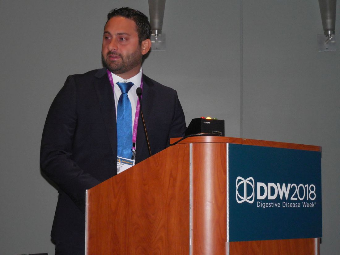
“There is sufficient evidence from these reports to at least consider TIPS as an adjunct to anticoagulation or perhaps as primary therapy,” especially for patients with PVT who have a contraindication for anticoagulation, Dr. Valentin said in an interview. Standard anticoagulation for PVT would today involve acute treatment with a low-molecular-weight heparin followed by oral anticoagulation for a total treatment time of at least 6 months and continued for a year or longer in some patients. A recently published review of reported experience using anticoagulation to treat PVT found a complete recanalization rate of 41% and a complete or partial rate of 66%, which suggests that TIPS is at least as effective, although Dr. Valentin cautioned that no reported study has directly compared the two alternative approaches. A study designed to make this direct comparison is warranted by the reported results using TIPS, Dr. Valentin said. And the experience with TIPS positions it as an option for patients who do not respond to anticoagulation or would prefer an alternative to prolonged anticoagulation.
One factor currently limiting use of TIPS, which is usually performed by an interventional radiologist, is that the procedure is technically demanding, with a limited number of operators with the expertise to perform it. If TIPS became more widely accepted as an option for treating PVT, then the pool of interventionalists experienced with performing the procedure would grow, Dr. Valentin noted.
mzoler@mdedge.com
On Twitter @mitchelzoler
SOURCE: Valentin N et al. Digestive Disease Week, Presentation 361.
REPORTING FROM DDW 2018
Key clinical point: Reported worldwide experience with TIPS in 439 patients shows it works and is relatively safe.
Major finding: TIPS was technically successful in 87% of reported patients and achieved complete portal recanalization in 74% of patients.
Study details: Systematic review of 18 published case series from 1993 to 2016 with 439 total patients.
Disclosures: Dr. Valentin had no disclosures.
Source: Valentin N et al. Digestive Disease Week, Presentation 361.
Barrett’s segment length, low-grade dysplasia tied to increased risk of neoplastic progression
WASHINGTON – , investigators here reported, but risk of esophageal progression from Barrett’s esophagus to adenocarcinoma remains low.
Tracking neoplastic progression is of prime importance in patients with Barrett’s esophagus (BE) because it can lead to the formation of esophageal adenocarcinoma (EAD), Esther Klaver of the Academic Medical Center of the University of Amsterdam noted at the annual Digestive Disease Week. By the time many patients present with symptoms, they are at an incurable stage of the disease and have 5-year survival rates below 20%. Endoscopic surveillance of patients with BE can detect neoplastic progression and EAD when it is still curable.
Ms. Klaver and her colleagues attempted to conduct the “perfect study” by observing patients with BE to identify endoscopic and clinical factors associated with increased risk of neoplastic progression. They did this by establishing a surveillance program to track disease progression that enrolled 987 patients from 2003 to 2017 at six community-based hospitals. The patients who enrolled had been diagnosed with BE and identified via a Dutch pathology registry or were newly diagnosed BE patients. Those with any history of EAD or high-grade dysplasia (HGD) were not included.
Ms. Klaver and her colleagues found that after a 7-year follow-up period the annual risk of progression to HGD or EAD was 0.79% per patient year, with 68 of the 987 patients progressing. Of the patients who progressed, 27 progressed to HGD (40%), and 41 progressed to EAD (60%). An overwhelming majority of patients received endoscopic management (59 patients, 87%), while some patients required surgery (9, 13%). Only 32 (3%) patients in the entire study population were lost to follow-up.
Low-grade dysplasia at baseline was the factor with the highest risk for esophageal progression, with a hazard ratio (HR) of 2.33 (95% CI, 1.27-4.29). Longer BE length (HR 1.07, 95% CI 1.04-1.10) and age at baseline (HR 1.17, 95% CI 1.12-1.24) were less associated with risk of HGD or EAD, but still significant.
Ms. Klaver pointed out that this study is unique in its design. The long-term follow-up and the focus on strict adherence to guidelines and optimal surveillance set this study apart from many BE studies.
“We tried to perform the perfect, optimal, prospective Barrett’s surveillance study in a large cohort with almost 1,000 patients with a median follow-up of almost 8 years.” Ms. Klaver said. “We have done this in a community, nonacademic setting, with the average Barrett’s patient. We have showed you that even with perfect surveillance that progression risk is low, with only 68 of almost 1,000 patients showing progression.”
The study was managed by tertiary referral centers that had two research nurses who attended surveillance endoscopies to ensure that guidelines were followed. Additionally, all endoscopies were performed by a dedicated endoscopist. As part of the endoscopy visit, patients filled out questionnaires containing demographic and clinical data. Researchers also retrospectively collected any prior surveillance data for patients who had previously been under histologic and endoscopic surveillance.
Ms. Klaver and her colleagues had no financial conflicts of interest to report.
SOURCE: Klaver E. et al. Gastroenterology. 154 (6). Abstract 10. doi: 10.1016/S0016-5085(18)30500-6.
REPORTING FROM DDW 2018
Key clinical point: Barrett’s segment length and low-grade dysplasia are associated with neoplastic progression.
Major finding: Low-grade dysplasia at baseline is associated with an increased risk of high-grade dysplasia or esophageal adenocarcinoma, hazard ratio of 2.38 (1.30 - 4.36).
Study details: This study was a prospective, multi-center cohort study involving 986 patients receiving treatment in six Dutch community-based hospitals from 2003 to 2017.
Disclosures: The study author did not report any financial disclosures.
Source: Klaver E et al. Gastroenterology. 154 (6). Abstract 10. doi: 10.1016/S0016-5085(18)30500-6.
App monitoring improves quality of IBD care
WASHINGTON – in a single-center randomized study with 320 patients.
The video associated with this article is no longer available on this site. Please view all of our videos on the MDedge YouTube channel
Based on this success, the app will soon be made available to all of the roughly 5,000 inflammatory bowel disease (IBD) patients managed at Mount Sinai Medical Center in New York as well as IBD patients at several other North American centers that plan to adopt the app, Ashish Atreja, MD, said at the annual Digestive Disease Week.®
Home monitoring of IBD patients “is feasible with high adoption,” said Dr. Atreja, a gastroenterologist at Mount Sinai who directs the Sinai AppLab. The 162 IBD patients randomized to regularly use the HealthPROMISE app had their quality-of-care metric rise from 50% at baseline to 84% after an average follow-up of 575 days (19 months), a statistically significant improvement over the 158 control patients whose metric rose from 50% to 65% for the study’s primary endpoint, he reported. The results also showed a trend toward improved quality of life among the patients using the HealthPROMISE app, compared with the controls, who used an IBD educational app that produced less patient engagement than did the HealthPROMISE app, Dr Atreja said.
Dr. Atreja and his associates modeled the app on remote monitoring methods developed for patients with other types of chronic disease, such as diabetes and heart failure.
“You can’t provide proactive IBD care without remote monitoring,” Dr. Atreja explained in a video interview. “Reactive care is not best practice anymore. The only way to do treat-to-target is with remote monitoring.”
Care coordinators monitor the entries that IBD patients send in via the app. Dr. Atreja estimated that about five care coordinators will be able to track the inputs from the roughly 5,000 IBD patients at Mount Sinai who will soon begin using the app. The financial feasibility of this approach depends in part on the $45/patient per month reimbursement that U.S. health insurers now provide to centers that run remote monitoring programs, he said.
“The direction for managing chronic diseases is increasingly looking at home monitoring as a way to streamline costs and improve patient care,” commented Gil Y. Melmed, MD, director of Clinical Inflammatory Bowel Disease at Cedars-Sinai Medical Center in Los Angeles. The results that Dr. Atreja reported came from “a highly selected population that was well educated and largely white.” The study needs replication in different patient groups to establish its reproducibility and generalizability, Dr. Melmed said in an interview.
Dr. Melmed had no relevant disclosures.
mzoler@mdedge.com
On Twitter @mitchelzoler
SOURCE: Atreja A et al. Digestive Disease Week 2018 abstract 17.
*This story was updated on June 7, 2018.
REPORTING FROM DDW 2018
Key clinical point: Regular remote monitoring of IBD patients improved the medical care they received.
Major finding: Quality of care rose from 50% at baseline to 84% in app-monitored patients and to 65% in controls.
Study details: A single-center randomized study with 320 IBD patients.
Disclosures: The study had no commercial funding. Dr. Atreja had no disclosures.
Source: Atreja A et al. Digestive Disease Week 2018 abstract 17.

Liver transplantation is on the rise for patients with severe alcoholic hepatitis
More transplant centers are offering liver transplantation as a viable therapeutic option for patients with severe alcoholic hepatitis who do not respond to steroid treatment.
“Alcoholic hepatitis is a disease caused by drinking alcohol. Excessive alcohol consumption causes fat to build up in your liver cells, as well as inflammation and scarring of the liver,” stated Saroja Bangaru, MD, chief resident at the University of Texas, Dallas. “Severe alcoholic hepatitis has an extremely high mortality and steroids are really the mainstay of therapy. Some alcoholic hepatitis patients do not respond to steroids and a significant percentage of them will die within 3 months. For these patients, liver transplantation is a therapeutic option.”
Dr. Bangaru and her colleagues conducted a survey that gathered data from 45 transplant centers in the United States and found that an increasing number have changed this practice and now offer liver transplantation to patients with severe alcoholic hepatitis.
The survey revealed that 51.1% of the 45 clinics offered liver transplantation to patients who had not yet been sober for 6 months, and 47.8% of transplant centers reported performing at least one liver transplant for severe alcoholic hepatitis. Just over a third (34.8%) of these centers had conducted three to five liver transplants, while only 8.9% of clinics performed at least six transplants. It is of note that most clinics have transplanted livers in fewer than five patients with severe alcoholic hepatitis, Dr. Bangaru said at the annual Digestive Disease Week®.
Patients experienced positive outcomes from these transplants, with almost 75% of surveyed clinics reporting 1-year survival rates of more than 90%, and 15% reporting 1-year survival rates of 80%-90%.
A factor that may have contributed to such positive outcomes was good patient selection based on liver transplant criteria for severe alcoholic hepatitis. More than 85% of center directors believed that liver transplant candidates should have a strong social support system, absence of severe psychiatric disorders, and a completed psychosocial evaluation, among other criteria.
Dr. Bangaru pointed out that the change in treating patients who have not abstained from alcohol is a break from traditional medical practice. “Historically, transplant centers would not consider a liver transplantation as an option unless a patient had abstained from drinking alcohol for 6 months. This rule was due to a concern that the patient would return to drinking after transplant as well as a perceived high risk that patients who continued drinking would miss medical appointments, fail to take their immunosuppressants and medications, and that this would lead to eventual graft failure.”
Another compounding issue was that patients were not counseled on their alcohol consumption habits, leading to further issues with transplantation. “Not infrequently, patients receive a diagnosis of severe alcoholic hepatitis during their initial visit and no one had previously told them to stop drinking. Since their presentation was preceded by active alcohol consumption, they would essentially be rendered ineligible for a transplant at that time,” she said.
Despite the history surrounding liver transplants in patients with severe alcoholic hepatitis, Dr. Bangaru hopes the shift in practice will improve the lives of more patients. “Because this practice of transplantation is being increasingly accepted and demonstrating positive outcomes, the hope is that more patients will be evaluated for transplantation and that transplant centers will improve their posttransplant support to ensure patients have great success after transplantation.”
Digestive Disease Week® is jointly sponsored by the American Association for the Study of Liver Diseases (AASLD), the American Gastroenterological Association (AGA) Institute, the American Society for Gastrointestinal Endoscopy (ASGE), and the Society for Surgery of the Alimentary Tract (SSAT).
REPORTING FROM DDW
A methylene blue dye pill sheds light on hard-to-see polyps
A methylene blue dye pill increases the adenoma detection rate in patients undergoing colonoscopy screenings.
“An oral, delayed-release, methylene blue tablet taken during the standard bowel preparation for colonoscopy has the potential to increase the adenoma detection rate and could assist in the early detection and prevention of colorectal cancer,” according to Michael B. Wallace, MD, professor of medicine and director of digestive disease research at the Mayo Clinic in Jacksonville, Fla.
“The number of polyps that providers find during a screening colonoscopy and remove is referred to as the adenoma detection rate. This is the most widely accepted national benchmark on quality for screening colonoscopy,” Dr. Wallace said in a media briefing in advance of the annual Digestive Disease Week®.
Dr. Wallace pointed out that missed adenomas tend to go on and develop into cancer.
“The ones that are most frequently missed are typically called ‘flat’ or ‘subtle’ polyps, making them very difficult to identify,” he said. “If they cannot be identified, they cannot be removed.”
The study involved more than 1,200 patients scheduled for colonoscopies at 20 centers around the world. Patients were randomly assigned to receive a full dose of the methylene blue dye, a half dose, or placebo. Patients receiving a half dose were not included in the statistical analysis, but served the purpose of not allowing doctors to know which patients were in the study group. Dr. Wallace noted that the nature of this study made it impossible to perform a truly double-blind study because the blue dye is visible during colonoscopy.
For the patients who took the full dose of methylene blue, the adenoma detection rate increased by nearly 9%, compared with the placebo group, with detection rates of 56.29% and 47.81%, respectively.
“While 9% may not actually seem like a large number, it is actually very clinically significant,” Dr. Wallace said. For every 1% increase in the absolute adenoma detection rate, there was a corresponding 3% decline in the incidence of colorectal cancer and a 5% decline in colorectal cancer deaths.
While the results of this study are overwhelmingly positive, Dr. Wallace pointed out that utilizing blue dye to increase adenoma detection rate is not a new concept, with a number of clinical trials showing its benefit.
Apart from improving adenoma detection rates, the tablet formulation of methylene blue dye no longer requires on-site mixing by providers. This was a limiting factor because the dye itself is difficult to mix and hard to obtain for many health care centers around the world. The mixed solution was also not as effective because it was sprayed during colonoscopies. Dr. Wallace asserted that spraying through the colonoscope could be imprecise, time consuming, and localized. For these reasons, this technique was never widely adopted. The primary advantage of the tablet formulation is that it releases the dye in the colon at or near the time of the colonoscopy.
Dr. Wallace emphasized that there is no substitute for good colonoscopy practice and that methylene blue is just another tool to help improve the practice.
“The oral, delayed-release methylene blue provides gastroenterologists with a new and supplemental method to improve their adenoma detection rate on top of what is otherwise a high-quality examination.
New ‘immune checkpoint’ vaccine shows promise in treating colorectal cancer
A novel vaccine may provide a breakthrough treatment option in colorectal and other cancers.
“We [researchers] at the Peter MacCallum Cancer Centre in Melbourne, Australia, have developed a new, DNA-based vaccine, which we call TetMYB, for the treatment of MYB-overexpressing cancers such as colorectal, adenoid cystic carcinoma, and breast cancer, to name a few,” Toan Pham, MD, of the Peter MacCallum Cancer Centre, said in a media briefing in advance of the annual Digestive Disease Week® conference.
The immunotherapy approach described by Dr. Pham involves combining the TetMYB vaccine, which boosts the immune system, and an antibody, BGB-A317, that helps enhance the effectiveness of the vaccine.
“The research we are presenting at DDW involves testing this approach in mouse models, which we had previously published. In our studies, tumors in the mice responded very well to the treatment and cancer was cured in about half of them,” stated Dr. Pham.
The study is really composed of two smaller studies that looked at colonic adenoma, induced using tamoxifen-enriched feed, in a total of 22 mice. The mice were split into two study groups: 15 in a prophylactic study and 7 in a therapeutic pilot study.
In the prophylactic study, eight treatment mice received three TetMYB vaccinations at weeks 7, 9, and 11. Tamoxifen was given to the treatment and control mice at week 13.
The seven mice in the therapeutic pilot study were given tamoxifen at week 9 and subsequently monitored via colonoscopy weekly. Once the presence of adenoma was identified, mice received six doses of TetMYB and four doses of anti-PD1 antibody.
In both the prophylactic and therapeutic study, the mice survival rates were higher than expected. In the prophylactic study, the treatment groups’ median survival time was 356 days, nearly double the control group (183 days). More impressively, all mice in the therapeutic group were alive at 235 days.
“The mice were only expected to live for a couple of days or weeks. But, about 50% of them, they lived for more than 2 years,” said Dr. Pham. “Additionally, when the cured mice from the original study were later rechallenged with the same treatment, it was immediately rejected, thus proving there is an immune memory induced by the vaccine.”
With the positive results of the mouse trial, Dr. Pham spoke to the ongoing human clinical trial and the future of this vaccine.
“Currently, we are conducting a first-in-human phase 1 clinical trial,” stated Dr. Pham. “The next phase of evolution for our vaccine, which I will also be presenting at DDW, is testing our vaccine as an anti-adenoma vaccine.” Adenomas account for nearly 80% of bowel cancers, according to Dr. Pham.
Should the safety data from the phase 1 trial prove the vaccine to be safe, a follow-up clinical trial looking at high-risk populations would be the next step.
REPORTING FROM DDW
Experimental drug may help those living with celiac disease
“Contamination with gluten happens during food processing, food packaging, cooking, and [because of] inadequate labeling,” Francisco Leon, MD, a consultant for Amgen and former CEO of Celimmune, said in a media briefing in advance of the annual Digestive Disease Week® (DDW). “Gluten is [also] present in unsuspected places like lipstick, toothpaste, even medicines. It is virtually impossible for celiac patients to completely avoid gluten.”
The new drug, AMG 714, works by blocking interleukin 15, a known intestinal inflammatory mediator, and lowers inflammation, resulting in fewer symptoms for patients.
In a 12-week study, patients received either 150- or 300-mg doses of subcutaneous AMG 714, or placebo. All doses of AMG 714 were administered six times over the course of the study. From weeks 2 to 12, patients were challenged with a daily dose of 2.5 grams of gluten, which is considered a high dose.
The results showed that AMG 714 significantly reduced Celiac Disease Patient Reported Outcomes at the 300-mg dose (P = .02) compared with placebo. Similarly, AMG 714 decreased Celiac Disease Gastrointestinal Symptom Rating Scale scores at the 150-mg (P = .17) and 300-mg (P = .07) doses, compared with placebo.
Dr. Leon noted that there was another subset of patients who did not receive the gluten challenge because they were shown to have intestinal atrophy – marked by the presence of flat mucosa – because of contamination in their gluten-free diets. This was measured by way of a novel stool and urine detection test. This information will be presented at DDW.
AMG 714 also improved gluten-triggered diarrhea and physician assessments, according to Dr. Leon. In fact, using the Bristol Stool Form Scale, patients who received both the 150-mg (P = .01) and 300-mg (P = .0002) doses had substantially lower rates of diarrhea, compared with placebo.
These positive results were not isolated to the Bristol Stool Form Scale scores, but extended to the Physician Global Assessment as well.
“The proportion of subjects whom physicians evaluated as clinically active at week 12 was 0 in the high dose of AMG 714 while it was 33% in the placebo group,” stated Dr. Leon.
While AMG 714 has shown promise in alleviating celiac disease symptoms for patients inadvertently exposed to gluten, it is not intended to treat exposure to high amounts of gluten.
“It is important to note that AMG 714 is intended to protect against modest contamination [with] gluten and not against the effects of regularly eaten large amounts of gluten, like bread or pasta,” Dr. Leon cautioned. “But we know that people with celiac disease are inadvertently exposed to gluten. So it is our hope that AMG 714 will allow these patients to experience fewer gluten-triggered events.”
The information presented by Dr. Leon was only a taste of what will be presented at DDW®.
“At DDW, we will show you a reduction in inflammation, and we will also show you the safety profile. There were no serious adverse events.
“We are hopeful that this will become one of the tools to combat celiac disease.”
REPORTING FROM DDW 2018