User login
Length of stay, complications predict readmission for cirrhosis patients
PHILADELPHIA – Patients with cirrhosis have a higher risk of hospital readmission if their length of stay is less than 4 days, if they have cirrhosis-related complications, and if they are discharged to an extended-care facility or to home health care, according to a recent presentation at the annual meeting of the American College of Gastroenterology.
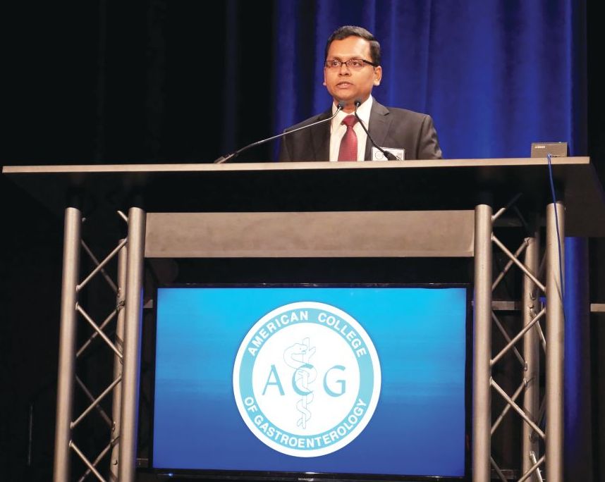
“The presence of cirrhosis-related complications is very strongly associated with readmissions,” Chandraprakash Umapathy, MD, MS, from the University of California, San Francisco, Fresno, said during his presentation. “Quality improvement efforts should focus on optimizing the management of complications of cirrhosis in the outpatient setting to reduce readmissions.”
In a retrospective cohort study, Dr. Umapathy and colleagues identified 230,036 patients from the Healthcare Cost and Utilization Project National Readmission Database for 2014 who had been discharged with a diagnosis of cirrhosis; of these patients, there were 185,737 index cases after excluding readmissions. Included patients had a mean age of 60.2 years and mean length of stay of 6.4 days, with 46% of patients having a length of stay longer than 4 days and mean total charges of $56,519. With regard to cirrhosis, 55% of patients displayed cirrhosis complications and 6.7% had more than three cirrhosis-related complications; the most common complication was ascites, in 32% of patients.
Overall, 11.09% of patients were readmitted at 30 days and 18.74% of patients were readmitted at 90 days, Dr. Umapathy said. Patients were more likely to be readmitted at 30 days if they were originally admitted on a weekend (adjusted prevalence ratio, 1.06; P = .001); into a medium (1.09; P = .009) or large (1.11; P less than .001) hospital; were admitted at a metropolitan teaching hospital (1.07; P less than .001); were insured by Medicaid (1.07; P less than .001); or were transferred to an extended care (1.51; P less than .001) facility or discharged to home health care (1.43; P less than .001).
Compared with patients who were not readmitted at 30 days, patients with 30-day readmission had a higher rate of alcoholic liver disease (43% vs. 46%; P less than .001), hepatitis C (28% vs. 32%; P less than .001), ascites (31% vs. 43%; P less than .001), hepatic encephalopathy (15% vs. 22%; P less than .001), hepatorenal syndrome (2.3% vs. 4.9%; P less than .001), hepatocellular cancer (5.1% vs. 5.7%; P = .001), presence of any cirrhosis complications (54% vs. 65%; P less than .001), and presence of more than three cirrhosis-related complications (6.3% vs. 10%; P less than .001). When adjusted in a multivariate analysis, association with readmission at 30 days for patients with cirrhosis-related complications such as ascites (1.42; P less than .001), hepatic encephalopathy (1.44; P less than .001), and hepatorenal syndrome (1.34; P less than .001) remained, Dr. Umapathy noted.
Length of stay longer than 4 days (0.84; P less than .001) and variceal hemorrhage (0.74; P = .002) were associated with reduced risk of readmissions at 30 days. “Focus on length of stay may result in patients being discharged prematurely, leading to higher early readmission,” Dr. Umapathy said.
Dr. Umapathy reports no relevant conflicts of interest.
SOURCE: Umapathy C et al. ACG 2018, Presentation 60
REPORTING FROM ACG 2018
Key clinical point: Cirrhosis-related complications and shorter length of stay were associated with higher rates of readmissions for patients with cirrhosis.
Major finding: 11.09% of patients were readmitted at 30 days and 18.74% of patients at 90 days, with the most common reasons for readmission including presence of cirrhosis complications and length of stay less than 4 days.
Study details: A retrospective cohort study of 185,737 index cases in the Healthcare Cost and Utilization Project National Readmission Database.
Disclosures: Dr. Umapathy reports no relevant conflicts of interest.
Source: Umapathy C et al. ACG 2018, Presentation 60.
Child-Pugh class does not affect HE recurrence in patients taking rifaximin
PHILADELPHIA – according to a recent presentation at the annual meeting of the American College of Gastroenterology.
Steven L. Flamm, MD, from Northwestern University, Chicago, and his colleagues examined results from a previous randomized, double-blinded trial of 140 patients receiving twice-daily rifaximin at 550 mg for 6 months in which the results showed rifaximin successfully maintained remission of hepatic encephalopathy (HE), compared with 159 patients receiving placebo.
“This pivotal study was published in March of 2010, but one of the post hoc assessments that was not performed was looking at various different phases of hepatic impairment as dictated by [Child-Pugh] class and each of those responses to this product,” Dr. Flamm said in his presentation.
Patients in the study were included if they had a Model For End-Stage Liver Disease score of 25 or less and two or more overt HE within 6 months (Conn score 1 or less) but were currently in remission. The researchers allowed the use of concomitant lactulose during the study period, which was used in 94.1% of rifaximin and 91.2% of placebo patients.
In the post hoc analysis, Dr. Flamm and his colleagues divided rifaximin and placebo patients into Child-Pugh class A (46 patients vs. 56 patients), class B (65 patients vs. 72 patients), and class C (17 patients vs. 14 patients) groups. For rifaximin and placebo patients, the mean age was 57.3 years and 57.2 years in the class A group, 54.4 years and 57.0 years in the class B group, and 56.1 years and 57.6 years in the class C group, respectively.
Overall, 8 of 46 rifaximin patients (17.4%) in the Child-Pugh class A and 15 of 65 rifaximin patients (23.1%) in the class B groups experienced an overt HE event during the 6-month study period, compared with 26 of 56 patients in the class A (46.4%) and 32 of 72 patients (44.4%) in the class B placebo groups; 5 of 17 rifaximin patients (29.4%) in the Child-Pugh class C group experienced their first overt HE event, compared with 9 of 14 (64.3%) patients in the placebo group.
With regard to first HE-related hospitalization, 5 of 46 patients (10.9%) in the rifaximin Child-Pugh class A group, 8 of 65 rifaximin patients (12.3%) in the class B group, and 4 of 17 rifaximin patients (23.5%) in the class C group experienced hospitalization because of HE, compared with 15 of 56 patients (23.2%) in the Child-Pugh class A group, 15 of 72 patients (20.8%) in the class B group, and 5 of 14 patients (35.7%) in the class C placebo group. The researchers noted lactulose use in the majority of patient cases in the study “provided further benefit” in reducing overt HE events.
“Although numeric differences were observed favoring rifaximin for the incidence of HE-related hospitalizations, a risk reduction versus placebo could not be firmly established, and presumably this was largely due to a lack of adequate power because of small sample size,” Dr. Flamm said.
This study and its analysis were supported by Salix Pharmaceuticals. Dr. Flamm and other coauthors report advisory committee membership, board membership, employment, or consultancy with AbbVie, Bristol-Myers Squibb, Gilead Sciences, Intercept Pharmaceuticals, Merck and Salix Pharmaceuticals. One coauthor reported no relevant conflicts of interest.
SOURCE: Flamm SL et al ACG 2018, Presentation 58.
REPORTING FROM ACG 2018
Key clinical point: Child-Pugh class does not significantly affect the overt hepatic encephalopathy recurrence rate in patients taking rifaximin, compared with placebo.
Major finding: A total of 17.4% of Child-Pugh class A, 23.1% of class B, and 29.4% class C patients taking rifaximin experienced overt hepatic encephalopathy, compared with 46.4% of Child-Pugh class A, 44.4% of class B, and 64.3% of class C patients receiving placebo.
Study details: A post hoc analysis of 299 patients receiving twice-daily rifaximin or placebo for 6 months.
Disclosures: This study and its analysis were supported by Salix Pharmaceuticals. Dr. Flamm and other coauthors reported advisory committee membership, board memberships, employment, or consultancy with AbbVie, Bristol-Myers Squibb, Gilead Sciences, Intercept Pharmaceuticals, Merck, and Salix Pharmaceuticals. One coauthor reported no relevant conflicts of interest.
Source: Flamm SL et al. ACG 2018, Presentation 58.
Endocuff decreases withdrawal time but not detection rate during colonoscopy
PHILADELPHIA – Use of a device on the distal end of a colonoscope to expand the view of the colon lowered the mean inspection time during colonoscopy without significantly reducing adenoma or sessile serrated polyp detection rate, according to a presentation at the annual meeting of the American College of Gastroenterology.
“The finger projections on the tip of the Endocuff can engage the colonic folds, and that allows us to see the proximal sides of these folds,” Seth A. Gross, MD, chief of gastroenterology at NYU Langone Health Tisch Hospital in New York, said in his presentation. “It also changes topography and temporarily stretches different segments of the colon depending on where you are to help expose more surface area and ultimately identify more polyps.”
Dr. Gross and his colleagues analyzed the withdrawal time of colonoscopy with the Endocuff Vision (Olympus, Center Valley, Penn.) in 101 patients, compared with withdrawal time during a standard colonoscopy in 99 patients as measured by stopwatch. Other endpoints in the study included insertion time, adenoma detection rate (ADR), sessile serrated polyp detection (SSPD), and number of adenomas and sessile serrated polyps per colonoscopy. Patients were included if they were at least 40 years old with a screening, surveillance, or diagnostic indication for colonoscopy; they were excluded if they had inflammatory bowel disease, polyposis syndrome, prior colon resection, prior colorectal polyp or cancer, previous incomplete colonoscopy or severe diverticular disease.
Inspection time in the Endocuff group was 6.3 minutes, compared with 8.2 minutes in the standard colonoscopy group (P less than .001), and insertion time was 9.9 minutes in the Endocuff group, compared with 11.3 minutes in the standard colonoscopy group. A multivariate analysis showed the shorter inspection times in the Endocuff group remained significant (P less than .0001).
In the Endocuff group, ADR was 61.4% with 1.43 adenomas per colonoscopy, while the standard colonoscopy group had an ADR of 52.5% with an adenoma detection rate of 1.07 per colonoscopy. SSPD was 19.8% in the Endocuff group and 11.1% in the standard group with a SSPD per colonoscopy of 0.27 and 0.21, respectively.
The study was unblinded and there were two endoscopists performing the procedures, which raises the question of whether the results could be generalized to other gastroenterologists, Dr. Gross noted.
“We recommend that future studies that are meant to be powered for adenoma detection rate and sessile serrated lesions be done to sort of validate this, and probably have more endoscopists involved in a study like this,” Dr. Gross said. “But this is the start of an interesting conversation where one could be more efficient without sacrificing our detection rate for both adenomas and sessile serrated lesions.”
Dr. Gross reports a consultancy with Olympus.
SOURCE: Gross SA et al. ACG 2018, Presentation 37.
REPORTING FROM ACG 2018
Key clinical point: Inspection times were lower during colonoscopy for patients who underwent the procedure using Endocuff, but there was no significant decrease in adenoma or sessile serrated polyp detection rate.
Major finding: Endocuff reduced inspection time to 6.3 minutes, compared with 8.2 minutes in the standard colonoscopy group.
Study details: An analysis of 200 patients who underwent standard colonoscopy or colonoscopy with Endocuff Vision.
Disclosures: Dr. Gross reports a consultancy with Olympus.
Source: Gross SA et al. ACG 2018, Presentation 37.
Longer withdrawal time in right colon increases adenoma detection rate
PHILADELPHIA – There was a significantly higher adenoma detection rate when the withdrawal rate in the right colon was more than 3 minutes in patients undergoing colonoscopy, according to a recent presentation at the annual meeting of the American College of Gastroenterology.
Although adenomas precede colon cancer in approximately 70% of cases, and detection of adenomas is associated with 5% risk of dying from colorectal cancer, miss rates of adenomas are high in both sides of the colon and ideal withdrawal times are not known, Fahad F. Mir, MD, MSc, from the University of Missouri-Kansas City, said.
“Miss rates are high, especially in the right side of the colon. A colonoscopy offers protection in up to 80% of the left side of the patients but only up to 40%-60% in the right side of patients,” Dr. Mir stated in his presentation. “The quality standard now [for withdrawal time] is 6 minutes, so we hypothesized that adenoma detection rate is not the same if the right colon withdrawal time is equal to or more than 3 minutes, compared to less than 3 minutes.”
The abstract received an ACG Governor’s Award for Excellence in Clinical Research.
Dr. Mir and his colleagues performed a prospective, randomized, case-controlled study of 226 patients undergoing colonoscopy at three endoscopy centers in St. Luke’s Health System, Kansas City, who were aged at least 50 years and had not undergone colonic resections, emergent procedures, or were unable to consent because of mental status or language barrier. Patients were randomized to a control group (113 patients) in whom right colon withdrawal time was under 3 minutes and an intervention group (113 patients) in whom withdrawal time was 3 minutes or more.
There was a 33% adenoma detection rate in the 3 minute or more group, compared with 14% in the less than 3 minutes group (odds ratio, 3.0; 95% confidence interval, 1.62-5.64; P less than .001). Polyp detection rates were 49% in the 3 minutes or more group and 14% in the less than 3 minutes group (OR 5.1; 95% CI, 2.84-9.32; P less than .001). The optimal cut-off point was 3 minutes and 1 second with optimal sensitivity and specificity with an area under the curve of 0.73 (95% CI, 0.65-0.81; P less than .001) for optimal cut-off time for withdrawal from the right colon.
“There was a difference in fellow involvement, where fellows were more likely to be involved when the withdrawal time was more than 3 minutes as opposed to less than 3 minutes; the ADR [adenoma detection rate] was not different based upon fellow involvement,” Dr. Mir said.
The researchers noted similar rates of retroflexion between both groups and said there were no adverse events related to the study in either group. Limitations of the study included its unblinded design, data collection from multiple centers, and a higher rate of previous polyps in patients in the withdrawal in more than 3 minutes group.
Dr. Mir report no relevant conflicts of interest.
SOURCE: Mir FF et al. ACG 2018, Presentation 5.
REPORTING FROM ACG 2018
Key clinical point: Spending more than 3 minutes in the right colon during withdrawal was associated with a greater adenoma detection rate during colonoscopy.
Major finding: There was a 33% rate of adenoma detection in patients in whom withdrawal time was greater than 3 minutes compared with a 14% detection rate when withdrawal time was under 3 minutes.
Study details: A prospective, randomized, case-controlled study of 226 patients undergoing colonoscopy.
Disclosures: Dr. Mir reports no relevant conflicts of interest.
Source: Mir FF et al. ACG 2018. Presentation 5.
Etrasimod improves clinical, endoscopic outcomes in patients with UC
PHILADELPHIA – Use of etrasimod was associated with improved clinical and endoscopic results, and was generally safe and well tolerated compared with placebo in patients with moderate to severe ulcerative colitis, according to a recent award-winning presentation at the annual meeting of the American College of Gastroenterology.
“Patients with moderate to severe ulcerative colitis receiving etrasimod 2 mg per day achieved statistically significant and clinically meaningful differences in all of the primary and secondary endpoints, and most exploratory endpoints were also significantly proved,” William J. Sandborn, MD, AGAF, FACG, professor of clinical medicine at the University of California, San Diego, stated in his presentation at the meeting. “A dose-response relationship was observed in virtually all of the measures of treatment efficacy.”
The abstract received the ACG Auxiliary Award (Member), which is given to ACG members each year with outstanding abstract submissions.
Dr. Sandborn and his colleagues enrolled 156 patients with ulcerative colitis (UC) into the OASIS study, a randomized, double-blind, parallel-group, phase 2 study of etrasimod, an oral sphingosine 1-phosphate (S1P) receptor modulator, compared with placebo. Patients were aged 18-80 years, with moderate to severe UC as defined by a three-component Mayo Clinic Score (MCS) comprising rectal bleeding, frequency of stool, and endoscopy.
Those patients who achieved an MCS score between 4 and 9 points with an endoscopic subscore of at least 2 and rectal bleeding (RB) subscore of at least 1 were included. Patients were divided into once-daily etrasimod 1 mg (52 patients), once-daily etrasimod 2 mg (50 patients) and placebo (54 patients) groups and treated over a 12-week period.
At 12 weeks, the least-squares mean difference for change in baseline in three-component MCS was 1.94 in the 1-mg etrasimod group and 2.49 in the 2-mg etrasimod group compared with placebo (1.50). Endoscopic improvement was greater in the 1-mg etrasimod (22.5%) and 2-mg (41.8%) groups compared with placebo (17.8%); endoscopic remission rates also improved in the 1-mg etrasimod (13.7%) and 2-mg (15.3%) groups compared with placebo (5.3%). Lymphocyte count circulation significantly decreased in the 1-mg etrasimod (37.2%) and 2-mg (57.3%) groups compared with the placebo group. With regard to rectal bleeding, the rectal bleeding subscore also decreased in the 1-mg etrasimod and 2-mg groups compared with placebo at 12 weeks from baseline.
The researchers noted no significant differences in adverse events among groups, with the placebo group showing a higher rate of major adverse events (11.1%) compared with the 1-mg etrasimod (5.8%) and 2-mg etrasimod (0%) groups.
“The OASIS trial results for etrasimod would support proceeding to a phase 3 program for this drug in patients with moderate to severe ulcerative colitis,” Dr. Sandborn concluded.
Dr. Sandborn reports consultancies, speaker bureau memberships, and research support from AbbVie, Biogen, Celgene, Ferring, Genentech, Gilead Sciences, Immune Pharmaceuticals, Janssen, Lilly, MedImmune, Novartis, Pfizer, Regeneron, Ritter Pharmaceuticals, Salix, Theradiag, UCB Pharma, and Vascular Biogenics, among others.
SOURCE: Sandborn WJ et al. ACG 2018. Presentation 11.
REPORTING FROM ACG 2018
Key clinical point: Compared with placebo, etrasimod was associated with endoscopic and clinical improvement in patients with ulcerative colitis.
Major finding: Patients taking etrasimod 2 mg (41.8%) and etrasimod 1 mg (22.5%) saw greater endoscopic improvement compared with placebo (17.8%).
Study details: A randomized, double-blind, parallel-group, phase 2 study of 156 patients with ulcerative colitis receiving etrasimod or placebo.
Disclosures: Dr. Sandborn reports consultancies, speaker bureau memberships, and research support from AbbVie, Biogen, Celgene, Ferring, Genentech, Gilead Sciences, Immune Pharmaceuticals, Janssen, Lilly, MedImmune, Novartis, Pfizer, Regeneron, Ritter Pharmaceuticals, Salix, Theradiag, UCB Pharma, and Vascular Biogenics, among others.
Source: Sandborn WJ et al. ACG 2018. Presentation 11.
Novel score predicts esophageal varices in patients with cirrhosis prior to EGD
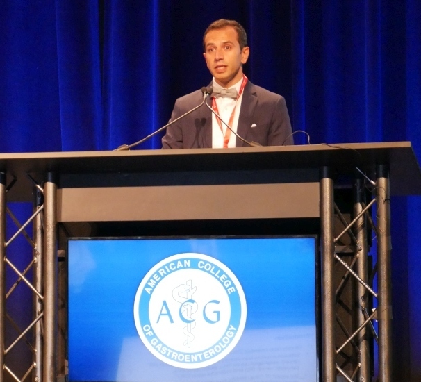
PHILADELPHIA – A novel score accurately predicted the size and presence of esophageal varices in a noninvasive manner, which may help clinicians avoid unnecessary esophagogastroduodenoscopy in patients with cirrhosis, according to a recent award-winning presentation at the annual meeting of the American College of Gastroenterology.
Although there are two validated scores for predicting esophageal varices (EV), platelet count to spleen diameter ratio and Baveno VI criteria, they have drawbacks, Tien Dong, MD, from the University of California, Los Angeles said.
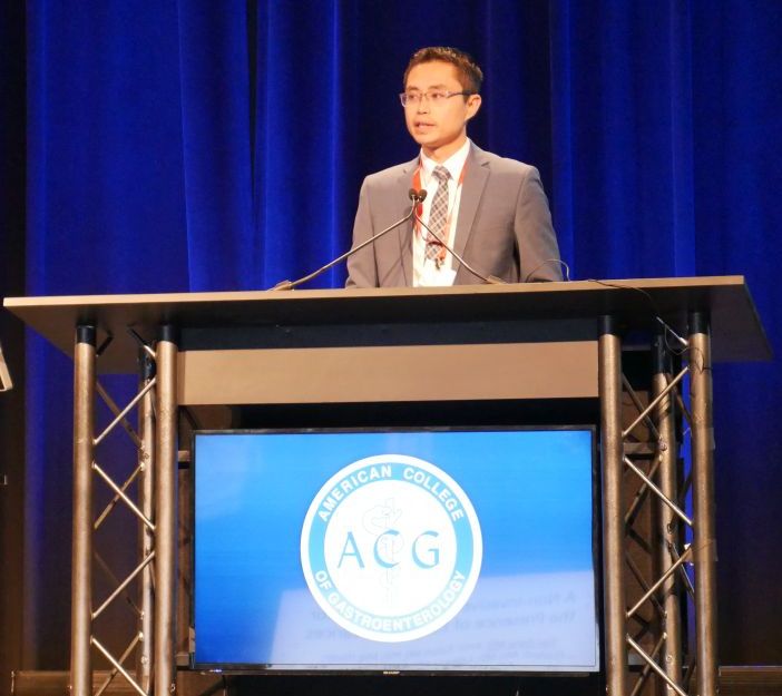
“The limitations to these existing scores and criteria are both of them require imaging to do, so because of that, they oftentimes are limited in national clinical use,” Dr. Dong said in his presentation of his team’s abstract, which won the ACG Governors Award for Excellence in Clinical Research. “The other thing is that, even though it’s recommended, sometimes spleen diameter on a normal ultrasound is not reported. Furthermore, elastography – even though it’s becoming more and more common – is still not yet readily available across the country.”
Dr. Dong and his colleagues sought to identify noninvasive clinical predictors of EV to create a predictive score to identify EV to overcome these drawbacks. They gathered endoscopy data from the Olive View–UCLA and West Los Angeles Veterans Affairs Hospital to create a discovery cohort (165 patients) and tested the score on patients from Ronald Reagan UCLA Medical Center in a validation cohort (73 patients).
They used a random forest classifier machine learning algorithm “to create a forest of decision trees so that it can tell us which variables it believes to be the most predictive of our outcomes of interest,” Dr. Dong said.
The algorithm identified several variables that appeared to be predictive of EV presence, such as international normalized ratio, aminotransferase, platelet mean, hemoglobin, albumin and blood urea nitrogen less than or equal to 3, which the researchers used to create an EV-endoscopy (EV-Endo) score.
In the discovery cohort, area under the curve (AUC) for the presence of EV was 0.81, compared with an AUC of 0.82 in the validation cohort, while there was an AUC of 0.77 in the discovery cohort and an AUC of 0.79 for small/absent vs. medium/large EV. Patients with Child-Pugh class A cirrhosis had an AUC of 0.81 for EV presence and an AUC of 0.77 for EV size. The researchers then created a cutoff score of 3.48 or less and 7.70 or more for EV presence, which had a sensitivity and specificity of 93.9% and 97.5%, respectively. EV-Endo score EV size cutoff scores were also 3.48 or less and 7.70 or more, with a sensitivity of 95.8% and specificity of 95.0%.
Dr. Dong reports no conflicts of interest.
SOURCE: Hauer M et al. ACG 2018. Presentation 31.
REPORTING FROM ACG 2018
Black patients present as sicker, more likely to receive liver transplant
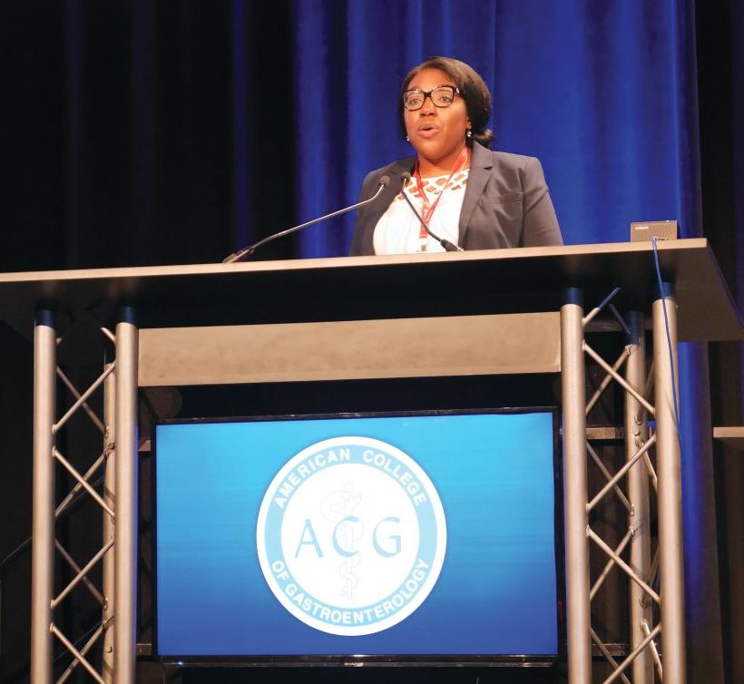
PHILADELPHIA – Black patients are more likely to be put on a transplant list because of acute liver failure, be listed as status 1, and receive a liver transplant, compared with white patients, according to a recent presentation at the annual meeting of the American College of Gastroenterology.
Lauren D. Nephew, MD, MSCE, of Indiana University in Indianapolis, and her colleagues performed a retrospective cohort study of black and white patients with a minimum age of 18 years in the United Network of Organ Sharing database who were wait-listed for a liver transplantation during 2002-2016. They examined patient clinical characteristics, acute liver failure (ALF) etiologies, wait-list status, and posttransplant survival outcomes through Kaplan Meier analysis.
“We really wanted to explore this topic in patients with acute liver failure, some of the sickest patients that we see,” Dr. Nephew said in her presentation. “We wanted to really determine whether or not there were differences in clinical characteristics and etiologies of acute liver failure in patients by race who are listed for liver transplantation.”
“Then, we wanted to compare wait-list outcomes,” she added, such as “differences by race in liver transplantation or wait-list removal because of death or becoming too sick for transplant.”
There were 11,289 patients in the white ALF group and 2,112 patients in the black ALF group; 2,876 (25.5%) of patients in the white ALF and 790 (37.4%) in the black ALF group were listed as status 1, which indicated an expected survival of 7 days or less. There were similar clinical characteristics for the white and black ALF status 1 patients regarding age (34.2 years vs. 36.3 years), Model for End-Stage Liver Disease (MELD) score (34 vs. 36; P less than .001), international normalized ratio (INR) test (mean 4.5 vs. mean 5.0; P = .001), creatinine levels (2.1 mg/dL vs. 1.9 mg/dL; P less than .001), and percentage of patients who were hepatic encephalopathy grade 3 or 4 (60.0% vs. 63.2%; P = .10). However, Dr. Nephew noted significantly higher bilirubin levels in the black ALF status 1 cohort (17.9 mg/dL), compared with the white ALF status 1 cohort (11.3 mg/dL; P less than .001).
The causes for ALF in each group included drug-induced liver failure (white status 1 cohort, 34.1%; black status 1 cohort, 20.6%), autoimmune hepatitis (2.7% vs. 9.4%), Wilson’s disease (0.58% vs. 0.13%), unknown etiology (34.5% vs. 42.5%), and other etiology (22.9% vs. 17%). For patients who underwent liver transplant and wait-list removal, there were no significant differences in wait-list removal “despite black patients being sicker at presentation,” Dr. Nephew said. Black patients were more likely to be listed to status 1 and transplanted at 62% (490 patients), compared with white patients at 53% (1,524 patients). There were 713 white patients (24.8%) removed from the transplant list, compared with 114 (13.8%) of black patients.
“If you are transplanted and you don’t die, then you are likely removed from the list for other reasons, and the most common reason is that you improved and became well, and so white patients were significantly more likely to be removed from the wait-list because of improvement, compared with black patients,” Dr. Nephew said.
In a competing risk analysis, the researchers found the hazard ratio for white patients who were status 1 and removed from the wait-list because of death or becoming too sick was 1.04 (95% confidence interval, 0.89-1.21) and those white patients who were listed as status 1 and then transplanted was 1.2 (95% CI, 1.08-1.30). In a multivariate analysis, the hazard ratio for white patients who were listed as status 1 and transplanted, which contained bilirubin at transplant, was 1.08 (95% CI, 0.98-1.19). Kaplan Meier 1-year survival post-transplant was 82.8% in white patients and 79.6% in black patients (P = .09).
“I think the question that we’ve been asking ourselves is, is this because black patients are presenting later with their acute liver disease and are sicker at presentation, or do they just have worse liver disease inherently on presentation that drove these findings?” Dr. Nephew said.
Dr. Nephew reports no relevant conflicts of interest.
SOURCE: Nephew L et al. ACG 2018, Presentation 59.
REPORTING FROM ACG 2018
Key clinical point: Black patients are sicker than white patients when they present with acute liver failure and are awaiting liver transplantation.
Major finding: Black patients with acute liver failure were more likely to be wait-listed, listed as status 1, and have higher Model for End-Stage Liver Disease (MELD) scores, creatinine levels, and INR tests, compared with white patients.
Study details: A retrospective cohort analysis of patients with acute liver failure awaiting a liver transplant in the United Network of Organ Sharing database.
Disclosures: Dr. Nephew reports no relevant conflicts of interest.
Source: Nephew L et al. ACG 2018, Presentation 59.
Increased incidence of GI, colorectal cancers seen in young, obese patients
PHILADELPHIA – Researchers have identified a link between obesity and an increased incidence rate of gastrointestinal (GI) cancers in younger patients as well as an increased rate of colorectal, esophageal, and pancreatic cancer resections in obese patients of various ages, according to an award-winning presentation at the annual meeting of the American College of Gastroenterology.
“These findings strengthen a contributing role of obesity in etiology as well as increasing incidence of these cancers, and call for more efforts targeting obesity,” Hisham Hussan, MD, assistant professor at The Ohio State University Wexner Medical Center in Columbus, stated in his presentation.
The abstract, presented by Dr. Hussan, received an American College of Gastroenterology Category Award in the subject of obesity. He noted in his presentation that the obesity rate in U.S. adults exceeded 37% in 2014. In addition, the temporal changes of obesity-related GI cancers with regard to age-specific groups are not known, he said.
“There’s sufficient evidence linking obesity to certain [GI cancers], such as esophagus, colon, pancreas, and gastric,” Dr. Hussan said. “However, the impact of rising obesity prevalence on the incidence of these obesity-related GI cancers is unknown.”
Dr. Hussan and his colleagues sought to investigate the incidence of obesity-related GI cancers by age group as well as whether there was an association between obesity-related GI cancers in both obese and nonobese patients.
“Our hypothesis is that the incidence of some obesity-related GI cancers is rising in some age groups, and we suspect that this corresponds with increasing rates of obese patients undergoing these cancerous resections,” he said in his presentation.
The researchers evaluated cancer incidence trends in the Surveillance, Epidemiology and End Results (SEER) database between 2002 and 2013 as well as obesity trends from 91,116 obese patients (7.16%) and 1,181,127 nonobese patients (92.84%) in the National Inpatient Sample (NIS) database who underwent cancer resection surgeries. Of these, 93.1% of patients underwent colorectal and 4.4% of patients underwent gastric cancer resections. Patients were considered obese if they had a body mass index of at least 30 kg/m2. The researchers examined annual trends for incidence rates of obesity-related GI cancers by age group and obesity-related GI cancer resection by age and obesity, using a joinpoint regression analysis to determine the percentage change per year.
In patients aged between 20 and 49 years, the incidence of colorectal cancer increased by 1.5% compared with a decrease of 1.5% in patients aged between 50 and 64 years old, a 3.8% decrease in patients aged 65-74 years, and a 3.9% decrease in patients who were a minimum of 75 years old. Gastric cancer incidence also increased by 0.7% in patients aged between 20 and 49 years compared with a 0.5%, 1.1%, and 1.8% decrease among patients who were aged 50-64 years, 65-74 years, and at least 75 years, respectively. There was an increased cancer incidence among patients in the 20- to 49-year-old age group (0.8%), 50- to 64-year-old age group (1.0%), 65- to 74-year-old age group (0.7%), and the 75-and-older group (1.0%). Esophageal cancer was associated with a decreased incidence in the 20- to 49-year-old group (1.8%), 50- to 64-year-old group (1.1%), 65- to 74-year-old age group (1.2%), and the 75-and-older group (0.7%)
For obese patients who underwent colorectal cancer resection, there was a 13.1% increase in the 18- to 49-year-old group, a 10.3% increase in the 50- to 64-year-old group, an 11.3% increase in the 65- to 74-year-old group, and a 12.8% increase in the 75-year-or-older group, compared with an overall decreased incidence in the nonobese group. There was an increased rate of pancreatic cancer resections for obese patients in the 50-to 64-year-old group (26.9%) and 65- to 74-year-old group (27.6%), compared with nonobese patients. Patients in the 18- to 49-year-old group (11.2%), 50- to 64-year-old group (14.6%), and 65- to 74-year-old group (25.7%) also had a higher incidence of esophageal cancer resections.
The limitations of the study included defining BMI at the time of surgery, which does not account for weight loss due to cachexia, and relying on ICD-9 codes for obesity, which “may not be reliable in some cases.
“However, we [saw] an increase in obese patients who come for resection, so it could have probably been more pronounced if we had accounted for obesity at the earlier age before diagnosis,” he added.
Dr. Hussan reports no relevant conflicts of interest.
SOURCE: Hussan H. ACG 2018, Presentation 34.
REPORTING FROM ACG 2018
Key clinical point: There is a higher incidence of colorectal and gastric cancer among obese patients under 50 years old.
Major finding: Obese patients of all age ranges had a higher rate of colorectal cancer resections, obese patients 75 and younger had a significantly higher incidence of esophageal cancer resections, and obese patients aged between 50 and 74 years had a higher rate of pancreatic resections, compared with nonobese patients.
Study details: A database analysis of cancer incidence trends in the Surveillance, Epidemiology, and End Results database and patient cancer incidence and obesity trends in the National Inpatient Sample database between 2002 and 2013.
Disclosures: Dr. Hussan reports no conflicts of interest.
Source: Hussan H. ACG 2018, Presentation 34.
Low-FODMAP diet improves fecal incontinence in patients with loose stool
PHILADELPHIA – A low fermentable oligo-, di-, and monosaccharides and polyols (FODMAP) diet was associated with improvement in fecal incontinence in 64.6% of patients who received dietary training, according to recent research presented at the American College of Gastroenterology annual meeting.
“In this case series, the low-FODMAP diet improved fecal incontinence in two-thirds of patients with fecal incontinence and loose stools,” Stacy Menees, MD, MS, assistant professor at the University of Michigan in Ann Arbor, said in her presentation.
Dr. Menees and her colleagues performed a retrospective chart review of 65 patients in the Michigan Bowel Control clinic with fecal incontinence (FI) “without alarming features” who were recommended a low FODMAP diet and received formal dietary teaching between August 2012 and December 2017. Patients received the teaching from a Michigan Medicine gastrointestinal dietitian. The dietitians performed a semi-quantitative analysis of patient response, including complete response, Dr. Menees said.
If we are going to help people with fecal incontinence, the key is to concentrate on the area of their stool consistency,” she explained. “We know that foods high in fermentable oligo-, di-, and monosaccharides and polyols, otherwise known as FODMAPS, can cause diarrhea and urgency.”
These FODMAP foods can include fruits with fructose exceeding glucose, fructan-containing vegetables, wheat-based products, sorbitol- and lactose-containing foods, and raffinose-containing foods, Dr. Menees said.
“We know from observations in data and research from the Monash University and our own group that patients with irritable bowel syndrome who are on a [low-]FODMAP diet [get relief from] their symptoms,” she added.
Most of the patients were female, and most were white. The mean age was 61.9 years, and mean body mass index was 26.7 kg/m2. Among the patients, 27.7% had irritable bowel syndrome (IBS), 9.2% had inflammatory bowel disease (IBD), 16.9% had diabetes mellitus, and 20% had a prior cholecystectomy.
With regard to reports of loose stool and FI, 46.1% of patients said they had loose stool daily with a mean of 2.8 loose bowel movements daily. More than half of the patients (60.5%) reported FI daily; 37.8% said they experienced weekly FI, and 4.6% reported monthly FI. The number of patients reporting FI was higher than 100% because some patients reported in the chart that they had FI daily or weekly, Dr. Menees said.
FI improvement was reported by 64.6% of patients, with 88.1% of responders having a greater than 50% reduction in FI and 35.7% of patients experiencing complete resolution of FI. There were no serious adverse events in the study, Dr. Menees noted.
“The three patients with postinfection IBS and fecal incontinence had no response to a low-FODMAP diet,” she said. “Our dietitians noted that onion and garlic were the triggers that were most often identified.”
Limitations include the retrospective chart review design and small number of patients, which may overestimate or underestimate the study results, and the fact that the study was done at a tertiary care center, Dr. Menees said.
“We are nearing the completion of a confirmatory, prospective, pilot randomized clinical trial to confirm the efficacy of a low-FODMAP diet in patients who suffer from fecal incontinence and loose stools,” she concluded.
Dr. Menees reports being a consultant for Synergy.
Use AGA’s patient education materials to help your patients better understand the low-FODMAP diet, which can be found at http://ow.ly/Xfqj30maODu
SOURCE: Menees SB et al. ACG 2018. Presentation 9.
REPORTING FROM ACG 2018
Key clinical point: Patients who underwent formal dietary teaching for a fermentable oligo-, di-, and monosaccharides and polyols (FODMAP) diet showed associated improvements in fecal incontinence.
Major finding: Among patients with fecal incontinence, 64.6% experienced improvement; of these, 88.1% of responders had a greater than 50% reduction in fecal incontinence and 35.7% experienced complete resolution of fecal incontinence.
Study details: A retrospective chart review of 65 patients with fecal incontinence who received formal dietary teaching at the Michigan Bowel Control clinic.
Disclosures: Dr. Menees reports being a consultant for Synergy.
Source: Menees SB et al. ACG 2018. Presentation 9.
Low-FODMAP diet improves fecal incontinence in patients with loose stool
PHILADELPHIA – A low fermentable oligo-, di-, and monosaccharides and polyols (FODMAP) diet was associated with improvement in fecal incontinence in 64.6% of patients who received dietary training, according to recent research presented at the American College of Gastroenterology annual meeting.
“In this case series, the low-FODMAP diet improved fecal incontinence in two-thirds of patients with fecal incontinence and loose stools,” Stacy Menees, MD, MS, assistant professor at the University of Michigan in Ann Arbor, said in her presentation.
Dr. Menees and her colleagues performed a retrospective chart review of 65 patients in the Michigan Bowel Control clinic with fecal incontinence (FI) “without alarming features” who were recommended a low FODMAP diet and received formal dietary teaching between August 2012 and December 2017. Patients received the teaching from a Michigan Medicine gastrointestinal dietitian. The dietitians performed a semi-quantitative analysis of patient response, including complete response, Dr. Menees said.
If we are going to help people with fecal incontinence, the key is to concentrate on the area of their stool consistency,” she explained. “We know that foods high in fermentable oligo-, di-, and monosaccharides and polyols, otherwise known as FODMAPS, can cause diarrhea and urgency.”
These FODMAP foods can include fruits with fructose exceeding glucose, fructan-containing vegetables, wheat-based products, sorbitol- and lactose-containing foods, and raffinose-containing foods, Dr. Menees said.
“We know from observations in data and research from the Monash University and our own group that patients with irritable bowel syndrome who are on a [low-]FODMAP diet [get relief from] their symptoms,” she added.
Most of the patients were female, and most were white. The mean age was 61.9 years, and mean body mass index was 26.7 kg/m2. Among the patients, 27.7% had irritable bowel syndrome (IBS), 9.2% had inflammatory bowel disease (IBD), 16.9% had diabetes mellitus, and 20% had a prior cholecystectomy.
With regard to reports of loose stool and FI, 46.1% of patients said they had loose stool daily with a mean of 2.8 loose bowel movements daily. More than half of the patients (60.5%) reported FI daily; 37.8% said they experienced weekly FI, and 4.6% reported monthly FI. The number of patients reporting FI was higher than 100% because some patients reported in the chart that they had FI daily or weekly, Dr. Menees said.
FI improvement was reported by 64.6% of patients, with 88.1% of responders having a greater than 50% reduction in FI and 35.7% of patients experiencing complete resolution of FI. There were no serious adverse events in the study, Dr. Menees noted.
“The three patients with postinfection IBS and fecal incontinence had no response to a low-FODMAP diet,” she said. “Our dietitians noted that onion and garlic were the triggers that were most often identified.”
Limitations include the retrospective chart review design and small number of patients, which may overestimate or underestimate the study results, and the fact that the study was done at a tertiary care center, Dr. Menees said.
“We are nearing the completion of a confirmatory, prospective, pilot randomized clinical trial to confirm the efficacy of a low-FODMAP diet in patients who suffer from fecal incontinence and loose stools,” she concluded.
Dr. Menees reports being a consultant for Synergy.
SOURCE: Menees SB et al. ACG 2018. Presentation 9.
REPORTING FROM ACG 2018
Key clinical point: Patients who underwent formal dietary teaching for a fermentable oligo-, di-, and monosaccharides and polyols (FODMAP) diet showed associated improvements in fecal incontinence.
Major finding: Among patients with fecal incontinence, 64.6% experienced improvement; of these, 88.1% of responders had a greater than 50% reduction in fecal incontinence and 35.7% experienced complete resolution of fecal incontinence.
Study details: A retrospective chart review of 65 patients with fecal incontinence who received formal dietary teaching at the Michigan Bowel Control clinic.
Disclosures: Dr. Menees reports being a consultant for Synergy.
Source: Menees SB et al. ACG 2018. Presentation 9.
