User login
Anti-Ro52 autoantibodies signal interstitial lung disease in juvenile dermatomyositis teaser
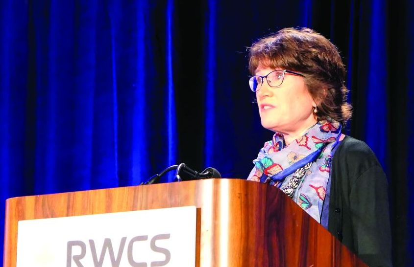
MAUI, HAWAII – , Anne M. Stevens, MD, PhD, said at the 2020 Rheumatology Winter Clinical Symposium.
And in a recent potential treatment advance, Janus kinase inhibition shows promise as a novel therapy for ILD in patients with juvenile dermatomyositis (JDM), added Dr. Stevens, a pediatric rheumatologist at the University of Washington, Seattle, and senior director for the adaptive immunity research program at Janssen Pharmaceuticals.
Autoantibodies predict ILD in JDM
Dr. Stevens highlighted recent work by Sara Sabbagh, DO, of the National Institute of Arthritis and Musculoskeletal and Skin Diseases and coinvestigators in the Childhood Myositis Heterogeneity Collaborative Study Group. They reported the presence of anti-Ro52 autoantibodies in 14% of a cohort of 302 patients with JDM as well as in 3 (12%) of 25 patients with juvenile polymyositis and in 8 (18%) of 44 youths with juvenile connective tissue disease–myositis overlap. In addition, 13% of patients were positive for autoantibodies previously identified as being associated with ILD in these forms of juvenile myositis: namely, 9% of the cohort were positive for anti–melanoma differentiation–associated protein 5 (anti-MDA5) autoantibodies, and antiaminoacyl-tRNA synthetase (anti-Jo-1) autoantibodies were present in 4%.
Thirty-three of the 371 juvenile myositis patients had ILD based upon CT imaging, chest x-ray, dyspnea on exertion, and/or biopsy. Most patients with anti-Ro52 also had other autoantibodies associated with ILD. Indeed, 31% of patients with anti-MDA5 autoantibodies also had anti-Ro52, as did 64% of those with anti-Jo1. After controlling for the presence of these other myositis-specific autoantibodies, anti-Ro52 autoantibodies were independently associated with ILD, which was present in 36% of those with and just 4% of those without anti-Ro52 autoantibodies.
Importantly, if a patient with JDM or another form of juvenile myositis had both anti-Ro52 and another myositis-specific autoantibody, the risk for ILD rose dramatically, climbing to 70% in patients with anti-Ro52 and anti-MDA5 autoantibodies, and to 100% in those who were both anti-Ro52 and anti-Jo1 positive (Ann Rheum Dis. 2019 Jul;78[7]:988-95).
Patients with anti-Ro autoantibodies had a worse prognosis, with more severe and chronic disease, Dr. Stevens noted.
Potential treatment for ILD in JDM: JAK inhibitors
Standard treatment of ILD in JDM in all cases includes high-dose pulsed corticosteroids, IVIG, and either methotrexate or mycophenolate mofetil. Consideration should be given to adding cyclosporine, particularly when a macrophage activation–syndrome component is present. In addition, several exciting recent lines of evidence suggest a potential role for Janus kinase (JAK) inhibitors in the subset of JDM patients with anti-MDA5 autoantibody–positive disease, according to Dr. Stevens.
For one, Dr. Sabbagh and colleagues have reported impressive success with the use of the JAK 1/3 inhibitor tofacitinib (Xeljanz) in two patients with anti-MDA5 autoantibody–positive refractory JDM with ILD. Both patients experienced moderate clinical improvement in disease activity in their skin, muscles, and other target organs. But particularly striking was what the investigators termed the “remarkable” improvement in ILD, including near resolution of abnormal findings on high-resolution CT imaging and a more robust performance on pulmonary function testing.
Both of these hitherto treatment-refractory patients were able to wean or discontinue their immunosuppressive medications. The patients’ elevated blood interferon-response gene signature improved significantly in response to tofacitinib, and their problematic upregulation of STAT1 phosphorylation of CD4+ T cells and monocytes stimulated with interferon-gamma was tamed, dropping to levels typically seen in healthy individuals (Brain. 2019 Nov 1;142[11]:e59).
Also, French pediatric rheumatologists have identified key phenotypic and cytokine differences between 13 patients with JDM or juvenile overlap myositis who were anti-MDA5 autoantibody–positive at presentation and 51 others who were not. The anti-MDA5 autoantibody–positive group had a higher frequency of ILD, arthritis, skin ulcerations, and lupus features, but milder muscle involvement than the anti-MDA5 autoantibody–negative group. The anti-MDA5 autoantibody–positive patients demonstrated enhanced interferon-alpha signaling based upon their significantly higher serum interferon-alpha levels, compared with the anti-MDA5-negative group, and those levels decreased following treatment with improvement in symptoms (Rheumatology [Oxford]. 2019 Nov 22. doi: 10.1093/rheumatology/kez525. [Epub ahead of print]).
The French investigators proposed that interferon-alpha may constitute a novel therapeutic target in the subgroup of patients with severe, refractory juvenile myositis and anti-MDA5 autoantibodies – and, as it happens, it’s known that JAK inhibitors modulate the interferon pathway.
Dr. Stevens reported research collaborations with Kineta and Seattle Genetics in addition to her employment at Janssen Pharmaceuticals.
REPORTING FROM RWCS 2020
Expert discusses red flags for interstitial lung disease in pediatric rheumatology
MAUI, HAWAII – Anti-Ro52 autoantibodies are the latest and most potent of the autoantibody predictors of interstitial lung disease (ILD) discovered in patients with juvenile dermatomyositis, Anne M. Stevens, MD, PhD, said at the 2020 Rheumatology Winter Clinical Symposium.
In addition to detailing the autoantibody red flags for ILD in juvenile dermatomyositis (JDM), she called for “hypervigilance” in patients with systemic juvenile idiopathic arthritis (SJIA) who exhibit any of a series of risk factors for ILD.
“Most of the lung disease in kids with systemic JIA is asymptomatic until very late, but it can be reversible if we treat it. So it’s worth finding and monitoring and giving everyone PCP [pneumocystis pneumonia] prophylaxis, because they have a high incidence of PCP if they have any of those risk factors,” observed Dr. Stevens, a pediatric rheumatologist at the University of Washington, Seattle, and senior director for the adaptive immunity research program at Janssen Pharmaceuticals.
Autoantibodies predict ILD in JDM
Dr. Stevens highlighted recent work by Sara Sabbagh, DO, of the National Institute of Arthritis and Musculoskeletal and Skin Diseases and coinvestigators in the Childhood Myositis Heterogeneity Collaborative Study Group. They reported the presence of anti-Ro52 autoantibodies in 14% of a cohort of 302 patients with JDM as well as in 12% of 25 patients with juvenile polymyositis and in 18% of 44 youths with an overlap of juvenile connective tissue disease and myositis. In addition, 13% of patients were positive for autoantibodies previously identified as being associated with ILD in these forms of juvenile myositis: Namely, 9% of the cohort were positive for antimelanoma differentiation–associated protein 5 (anti-MDA5) autoantibodies, and antiaminoacyl tRNA synthestase (anti-Jo-1) autoantibodies were present in 4%.
A total of 33 of the 371 juvenile myositis patients had ILD based upon CT imaging, chest X-ray, dyspnea on exertion, and/or biopsy. Most patients with anti-Ro52 also had other autoantibodies associated with ILD. Indeed, 31% of patients with anti-MDA5 autoantibodies also had anti-Ro52, as did 64% of those with anti-Jo-1. After controlling for the presence of these other myositis-specific autoantibodies, auto-Ro52 autoantibodies were independently associated with ILD, which was present in 36% of those with and just 4% of those without anti-Ro52 autoantibodies.
Importantly, if a patient with JDM or another form of juvenile myositis had both anti-Ro52 and another myositis-specific autoantibody, the risk for ILD rose dramatically, climbing to 70% in patients with anti-Ro52 and anti-MDA5 autoantibodies, and to 100% in those who were both anti-Ro52- and anti-Jo-1 positive.
Patients with anti-Ro52 autoantibodies had a worse prognosis, with more severe and chronic disease, Dr. Stevens noted.
Novel potential treatment for ILD in JDM: JAK inhibitors
Standard treatment of ILD in JDM in all cases includes high-dose pulsed corticosteroids, intravenous immunoglobulin (IVIG), and either methotrexate or mycophenolate mofetil. Consideration should be given to adding cyclosporine, particularly when a macrophage activation syndrome component is present. In addition, several exciting recent lines of evidence suggest a potential role for Janus kinase (JAK) inhibitors in the subset of JDM patients with anti-MDA5 autoantibody-positive disease, according to Dr. Stevens.
For one, Dr. Sabbagh and colleagues have reported impressive success with the use of the JAK 1/3 inhibitor tofacitinib (Xeljanz) in two patients with anti-MDA5 autoantibody-positive refractory JDM with ILD. Both patients experienced moderate clinical improvement in disease activity in their skin, muscles, and other target organs. But particularly striking was what the investigators termed the “remarkable” improvement in ILD, including near-resolution of abnormal findings on high-resolution CT imaging and a more robust performance on pulmonary function testing.
Both of these hitherto treatment-refractory patients were able to wean or discontinue their immunosuppressive medications. The patients’ elevated blood interferon-response gene signature improved significantly in response to tofacitinib, and their problematic upregulation of STAT1 phosphorylation of CD4+ T cells and monocytes stimulated with interferon-gamma was tamed, dropping to levels typically seen in healthy individuals.
Also, French pediatric rheumatologists have identified key phenotypic and cytokine differences between 13 patients with JDM or juvenile overlap myositis who were anti-MDA5 autoantibody positive at presentation and 51 others who were not. The anti-MDA5 autoantibody–positive group had a higher frequency of ILD, arthritis, skin ulcerations, and lupus features, but milder muscle involvement than did the anti-MDA5 autoantibody–negative group. The anti-MDA5 autoantibody–positive patients demonstrated enhanced interferon-alpha signaling based upon their significantly higher serum interferon-alpha levels, compared with the anti-MDA5-negative group, and those levels decreased following treatment with improvement in symptoms.
The French investigators proposed that interferon-alpha may constitute a novel therapeutic target in the subgroup of patients with severe, refractory juvenile myositis and anti-MDA5 autoantibodies – and, as it happens, it’s known that JAK inhibitors modulate the interferon pathway.
Risk factors for ILD in SJIA
In the past half-dozen years or so, pediatric rheumatologists have become increasingly aware of and concerned about a new development in SJIA: the occurrence of comorbid ILD. This is a poor-prognosis disease: In a cohort from the United Kingdom, 5-year mortality from the time of diagnosis was 41%, fully 40-fold higher than in patients with SJIA only.
Patient cohorts with SJIA and ILD have unusual clinical and laboratory features that aren’t part of the typical picture in SJIA. These include acute clubbing, lymphopenia, a fixed pruritic rash, unexplained abdominal pain, peripheral eosinophilia, facial swelling, and an increased ferritin level, a hallmark of acute macrophage activation syndrome. Onset of SJIA before 2 years of age is another red flag associated with increased risk for ILD. So is trisomy 21, which is up to 50 times more prevalent in patients with SJIA and ILD than in the general population or in patients with SJIA only. Another clue is an adverse reaction to tocilizumab (Actemra).
Any of these findings warrant hypervigilance: “Be on high alert and monitor these patients for ILD much more closely,” Dr. Stevens advised.
This means ordering a CT scan, prescribing PCP prophylaxis, and regularly measuring pulmonary function, admittedly a challenge in children under 7 years old. In these younger kids, practical solutions include measuring their oxygen saturation before and after running around the room to see if it drops. A 6-minute walk test and sleep oximetry are other options.
The explanation for the abrupt arrival of ILD as part of the picture in SJIA during the past decade remains unclear. The timing coincides with a major advance in the treatment of SJIA: the arrival of biologic agents blocking interleukin-1 and -6. Could this be a serious treatment side effect?
“It’s all association so far, and we’re not really sure why we’re seeing this association. Is it because we’re using a lot [fewer] corticosteroids now, and maybe those were preventing lung disease in the past?” Dr. Stevens speculated.
At this point, she and her fellow pediatric rheumatologists are awaiting further evidence before discussing a curb in their use of IL-1 or -6 inhibitors in patients with SJIA.
“These drugs have turned around the lives of kids with SJIA. They used to suffer through all our ineffective treatments for years, with terrible joint destruction and a pretty high mortality rate. These are great drugs for this disease, and we certainly don’t want to limit them,” she said.
Dr. Stevens reported research collaborations with Kineta and Seattle Genetics in addition to her employment at Janssen Pharmaceuticals.
REPORTING FROM RWCS 2020
Methotrexate adherence: It’s worse than you think
MAUI, HAWAII – Results of a carefully conducted real-world study of adherence to oral methotrexate in patients with RA were “kind of scary,” Arthur Kavanaugh, MD, said at the 2020 Rheumatology Winter Clinical Symposium.
“At 24 weeks, adherence was only 75%. And these were people who knew they were being monitored, so this is the best of the best. And yet less than 20% took the drug perfectly, meaning they took every dose as it was supposed to be,” noted Dr. Kavanaugh, professor of medicine at the University of California, San Diego, and RWCS program director.
“Adherence to methotrexate is really not very good. This is our cornerstone drug – methotrexate – and I think it certainly applies to other medications that we’re using,” he added.
He and his fellow panelist John J. Cush, MD, discussed the implications of this recent study, led by Kaleb Michaud, PhD, of the University of Nebraska Medical Center, Omaha.
The methotrexate adherence study included 60 patients with RA whose use of the disease-modifying antirheumatic drug (DMARD) over 24 weeks was monitored via the electronic Medication Event Monitoring System. These were motivated patients seen in routine clinical practice: They were participants in Forward, the National Databank for Rheumatic Diseases, and they understood that their use of methotrexate was being monitored.
Among the key findings: Patients on average took their weekly dose as directed for a total of 18 of the 24 weeks, although adherence decreased over time. Overall, 13% of participants missed 1 week, and 68% skipped 2 or more weeks. There was no significant difference in methotrexate adherence between biologic-naive and -experienced patients, nor between those on methotrexate monotherapy versus those on additional medication. Patient demographics and RA severity were similar between patients who missed taking their methotrexate for 2 weeks or more and those who missed fewer or no doses.
Higher Patient Global Assessment of Disease Activity scores were associated with correct dosing. So was being unemployed, having no prior conventional DMARD experience, and having less disability. A higher baseline score on the Beliefs about Medicines Questionnaire Specific-Necessity subscale, which indicates stronger belief in the necessity of the medication, were associated with greater likelihood of appropriate dosing, while lower scores were linked with more weeks of early dosing. However, the other elements of the Beliefs about Medicines Questionnaire weren’t significantly associated with adherence.
“This is a big problem. A lot of factors go into medication nonadherence. The solution has to begin with your relationship with the patient. If you want people to trust you, you’re going to have to work at that,” observed Dr. Cush, who is professor of medicine and rheumatology at Baylor University Medical Center, Dallas, and director of clinical rheumatology at the Baylor Research Institute.
Roy Fleischmann, MD, a rheumatologist and medical director of the Metroplex Clinical Research Center, Dallas, said that widespread suboptimal adherence to oral methotrexate has important implications for clinical trials. Often the placebo response rate in a randomized trial where the control group is on background methotrexate is so unexpectedly high that the potential efficacy of the active drug is masked; in such situations, it’s quite possible that patients who previously weren’t taking their methotrexate consistently start doing so when they join a closely supervised study, with a resultant inflated placebo response rate, he said.
One audience member who sees lots of medication adherence issues in his practice suggested that it might be time to become more aggressive in using intravenous infusion therapy instead of subcutaneously administered agents in patients with active RA and adherence problems.
“Maybe that’s why rituximab does so well in the clinical trials,” he said.
Dr. Cush and Dr. Kavanaugh reported receiving research funding from and/or serving as consultants to numerous pharmaceutical companies.
REPORTING FROM RWCS 2020
Differentiating hypersensitivity reactions to monoclonal antibodies
MAUI, HAWAII – Desensitization is a powerful and effective tool in patients with certain types of hypersensitivity reactions to therapeutic monoclonal antibodies, but it’s best considered a last resort reserved for individuals with no options left other than the offending biologic, Anna Postolova, MD, said at the 2020 Rheumatology Winter Clinical Symposium.
Why so selective? Desensitization is considered a high-risk intervention. It’s typically done as an inpatient procedure involving an overnight hospital stay followed by an elaborate 12-step protocol involving administration of small quantities of the culprit biologic in ascending concentrations over a 5- to 6-hour period.
Moreover, for an intravenous agent, such as infliximab (Remicade), desensitization has to be repeated prior to giving every dose of the biologic. So it makes sense to skip desensitization and simply switch to an alternative tumor necrosis factor inhibitor or a different class of biologic unless experience has shown that the culprit monoclonal antibody is the only one that works for that patient. It’s known, for example, that infliximab has no crossreactivity with adalimumab (Humira), explained Dr. Postolova, a dual rheumatologist and allergist/immunologist at Stanford (Calif.) University.
Defining type and severity of the hypersensitivity reaction
Dr. Postolova favors the hypersensitivity reaction classification system developed by Mariana Castells, MD, PhD, and coworkers at Brigham and Women’s Hospital, Boston.
They divide the field into immediate and delayed hypersensitivity reactions. Immediate hypersensitivity reactions arise rapidly, between minutes and a few hours. They can be categorized as infusion reactions, cytokine-release reactions, and IgE-mediated reactions. Phenotypically, infusion reactions and cytokine-release reactions are typically characterized by various combinations of chills, fever, flushing, hypertension, tachycardia, nausea, vomiting, syncope, and shortness of breath.
IgE-mediated reactions can also involve flushing and shortness of breath, and in addition itch, urticaria, and hypotension. These are anaphylactic reactions. Neither hypertension nor fever is part of the anaphylactic picture; those findings point instead to an infusion reaction or cytokine-release reaction.
Most allergists grade reaction severity on a 1-3 scale. Grade 1 reactions are considered mild and involve symptoms limited to the skin, such as flushing, or a single other organ system.
“That being said, if my patient is having a reaction with bronchospasm, I consider that a moderate, grade 2 reaction, and I stop the infusion. There’s only so much you can do for bronchospasm. It’s a very serious reaction,” Dr. Postolova observed.
Grade 2 reactions ordinarily involve two or more organ systems, but without hypotension or cyanosis. Grade 3 reactions are severe anaphylactic reactions with cardiovascular and/or neurologic compromise.
Delayed hypersensitivity reactions are of two types: serum sickness–like reactions and type IV cell-mediated mucocutaneous reactions.
Type IV reactions can range from a mild maculopapular rash to erythema multiforme, Stevens-Johnson syndrome, toxic epidermal necrolysis, and DRESS (drug reaction with eosinophilia and systemic symptoms). Onset of type IV reactions can occur after 12 hours up to several weeks after exposure.
Serum sickness–like reactions typically begin 5-7 days after the infusion. These reactions are marked by evidence of immune overactivation: fever, arthralgia, arthritis, malaise, purpura, skin rash, and even renal failure.
Management of reactions
A patient with a grade 3 reaction who needs to continue using the culprit monoclonal antibody should be referred to an allergist for skin testing in an effort to identify an IgE-mediated reaction.
The timing of the referral for skin testing is important: The allergist wants to test roughly 4-6 weeks after the hypersensitivity reaction. Test too early and the results will be uninformative because the patient will still be anergic. On the other hand, after 7-8 weeks the patient will have lost the allergy. So there is a sweet spot.
If the patient is skin test positive – with the caveat that skin testing in this setting is not well validated – then the allergist will suggest desensitization, usually as an inpatient.
In contrast, infusion reactions can be handled in the rheumatologist’s infusion center. They are self-limited upon repeat exposure with premedication using antihistamines, NSAIDs, oral or injectable steroids, and perhaps montelukast (Singulair).
If a patient initially thought to have an infusion reaction continues to experience reactions even after the biologic is being delivered more slowly and under the protection of premedication, it’s time to consider the possibility that what’s really going on is a cytokine-release reaction or an IgE-mediated reaction. Diagnostic skin testing is in order.
For a skin test–negative patient with a suspected cytokine-release reaction, the allergist may propose a therapeutic challenge. This is reserved for patients who the allergist believe will not experience an immediate reaction, and unlike desensitization it’s not an intervention intended to induce drug tolerance. The challenge involves giving 10% of a full dose of the biologic, waiting in the allergist’s office for 30-60 minutes, then giving the other 90% of the medication, followed by an hour of in-office observation.
The solution to severe type IV delayed hypersensitivity reactions is strict medication avoidance, not desensitization, according to Dr. Postolova.
Top offending monoclonal antibodies
Infliximab and rituximab (Rituxan) are the most common culprits when it comes to immediate hypersensitivity reactions. About 10% of infliximab-treated patients develop these reactions. Although the reaction can occur with the first dose, the peak incidence is with the seventh infusion. Patients with anti-infliximab IgG antibodies are at 140%-300% increased risk; however, concomitant disease-modifying antirheumatic drug therapy lessens that risk.
Infusion reactions or cytokine-release reactions occur upon the first infusion of rituximab in about 25% of treated rheumatology patients and in a higher proportion of cancer patients. Most of these reactions are mild and don’t recur when the biologic is administered more slowly and with premedication. Severe recurrent reactions upon subsequent exposure are much more likely to be an IgE-mediated hypersensitivity reaction.
“Stop the medication, send the patient to your local allergist for skin testing, and they’ll use a desensitization protocol if rituximab is the best drug for your patient,” Dr. Postolova advised.
She reported having no financial conflicts regarding her presentation.
REPORTING FROM RWCS 2020
Experts review recent winners and losers in the RA pipeline
MAUI, HAWAII – Filgotinib, the oral Janus kinase (JAK) inhibitor now under Food and Drug Administration review for the treatment of RA, has a better safety profile than some of the approved oral JAK inhibitors, but that’s unlikely to save it from being saddled with a black-box safety warning label, experts agreed at the 2020 Rheumatology Winter Clinical Symposium.

“There’s probably a class label out there,” according to Mark C. Genovese, MD, professor of medicine and clinical chief of the division of immunology and rheumatology at Stanford (Calif.) University.
He cited the example of upadacitinib (Rinvoq), approved last year as the third oral JAK inhibitor for RA. Even though venous thromboembolic (VTE) events weren’t seen with any significantly increased frequency, compared with placebo, in the upadacitinib development program – unlike for the earlier-approved tofacitinib (Xeljanz) and baricitinib (Olumiant) –the FDA nevertheless required that upadacitinib’s product labeling include this black-box warning: “Thrombosis, including deep vein thrombosis, pulmonary embolism, and arterial thrombosis, have occurred in patients treated with Janus kinase inhibitors used to treat inflammatory conditions.”
“I would fully expect that there’ll be a similar label in the filgotinib package insert saying that VTEs have been seen in other members of the class,” he predicted.
His copanelist Roy Fleischmann, MD, noted that filgotinib displayed a clean long-term safety profile in a pooled analysis of the 24-week results in the 2,088 filgotinib-treated participants in all phase 3 clinical trials for RA. For example, the incidence of herpes zoster in that large treated population was a mere 0.1%.
“Herpes zoster is almost nonexistent across the program,” observed Dr. Fleischmann, a rheumatologist at the University of Texas and medical director of the Metroplex Clinical Research Center, both in Dallas.
That’s consistent with what he’s heard from Japanese investigators about their experience. They tell him that in their studies the incidence of herpes zoster with filgotinib is five times less than with other JAK inhibitors.
The long-term pooled phase 3 filgotinib safety data also show less than a 0.1% incidence of adjudicated VTE/pulmonary embolism through 24 weeks. That’s a substantially lower rate than with tofacitinib or baricitinib, he noted.
The two rheumatologists, long-time observers of the FDA regulatory scene, stressed that they have no inside information regarding what the agency will do about filgotinib. It seems beyond doubt that the JAK inhibitor will be approved. But an open question of practical importance to clinicians is whether the agency will approve only the 100-mg dose or the 200-mg dose as well. The panelists agreed that having access to both would be advantageous since the clinical trials data indicate the higher dose is more effective and this greater efficacy doesn’t come at a cost of additional safety issues.
“If the 100 mg is sufficient, that’s great, but the reality is if you want to push to low disease activity or remission, the 200 mg seems to work better, particularly in patients who’ve already failed TNF [tumor necrosis factor] inhibitors or other biologics,” Dr. Genovese said. “If you’re not having additional safety concerns and you can get additional efficacy, I love the idea of having flexibility.”
Dr. Fleischmann is skeptical that the regulators will see things that way.
“There is a real risk that the FDA will do what it’s done before and say: ‘Well, the 200 works and the 100 works, so we’re going to approve the lower dose.’ But there doesn’t appear to be a big safety difference between 100 and 200. So I can see why they would approve the two doses, but I think that’d be unusual,” according to the rheumatologist.
The RA pipeline
The two speakers also highlighted several agents with novel mechanisms of action moving through the RA developmental pipeline, including olokizumab, otilimab, fenebrutinib, and a promising oral selective interleukin-1 receptor–associated kinase 4 inhibitor (IRAK4).
Olokizumab: This humanized monoclonal antibody targets IL-6. It has a different mechanism of action than the two commercially available IL-6 inhibitors approved for RA, tocilizumab (Actemra) and sarilumab (Kevzara), in that olokizumab uniquely targets the IL-6 ligand.
At the 2019 annual meeting of the American College of Rheumatology, Dr. Genovese presented the positive findings of a double-blind, placebo-controlled, randomized, phase 3 clinical trial of olokizumab in 428 RA patients with an inadequate response to methotrexate. The primary outcome, an ACR 20 response at 12 weeks, occurred in 25.9% of patients on placebo, 63.6% with 64 mg of olokizumab given subcutaneously every 2 weeks, and 70.4% with 64 mg every 4 weeks, with all participants on background methotrexate. An ACR 50 response at week 24 occurred in 7.7%, 42.7%, and 48.6%, respectively, with an acceptable side effect profile.
This was the first phase 3 trial to be presented from a large, ongoing phase 3 olokizumab developmental program for a variety of diseases.
“The results certainly support the idea that a 4-week regimen would probably be quite useful with this medication, although we’ll have to see what happens with the remaining phase 3 trials,” Dr. Genovese said.
Dr. Fleischmann posed a question: Do we really need a third IL-6 inhibitor?
That would make for a crowded field, Dr. Genovese agreed, adding that grabbing a reasonable market share for olokizumab may come down to cost, formulary access, and the convenience factor of once-monthly dosing. Whether the biologic’s unique mechanism of action in blocking the IL-6 ligand makes any practical difference in outcomes is unknown.
IRAK4 inhibitor: PF-06650833 is an oral selective reversible inhibitor of IRAK4, a key signaling kinase for IL-1 and toll-like receptors.
“This should be a really good drug for IL-1-mediated diseases,” according to Dr. Fleischmann.
In a phase 2b, double-blind, randomized, placebo-controlled, 12-week study featuring tofacitinib at 5 mg twice daily as an active comparator, the IRAK4 inhibitor exhibited dose-dependent efficacy for the primary endpoint of improvement from baseline in Simple Disease Activity Index score, compared with placebo. The same was true for the secondary endpoint, change over time in 28-joint Disease Activity Score using C-reactive protein (DAS28-CRP). The trial results were also presented at the 2019 ACR annual meeting.
“This is a drug they should probably take forward and see how far it goes in RA,” he said.
Dr. Genovese concurred.
“We’re still trying to figure out how we can put together rational combinations in RA, and this might be something that could be considered as a combination play. In fact, Pfizer has already teed up a study looking at a JAK inhibitor/IRAK4 combination. The question will be whether this is a standalone or has an opportunity to be part of a combination approach,” the rheumatologist said.
Otilimab: This monoclonal antibody is a granulocyte-macrophage colony-stimulating factor inhibitor. In a secondary analysis of the BAROQUE trial, a phase 2b, 50-week study in RA inadequately responsive to methotrexate, otilimab demonstrated an impressive effect in terms of pain reduction. This new analysis, which was first presented at the 2019 ACR annual meeting, showed that, at week 12, a minimum clinically important difference in pain was achieved in 29% of placebo-treated controls, compared with 65%-75% of patients on low to higher doses of otilimab.
“The question is: Is this pain effect unique to this molecule, this pathway, or is it a simple reflection of the treated patient population?” Dr. Genovese commented. “It’s an interesting molecule. It’s being developed in RA, and it might have unique benefits on the pain side.”
A tale of two BTK inhibitors: Bruton tyrosine kinase (BTK) is an intracellular kinase viewed as a promising target in autoimmune disease. Fenebrutinib is an oral, noncovalent, reversible, and highly selective BTK inhibitor that performed well in a phase 2, randomized, double-blind, placebo-controlled clinical trial with adalimumab as an active comparator in 480 patients with an inadequate response to methotrexate in one branch, and in 98 patients with an inadequate response to TNF inhibitor therapy in the other. All subjects were on background methotrexate. (The study results were published April 9 in Arthritis & Rheumatology.)
In the group with a prior inadequate response to TNF inhibitors, the ACR 50 response rate at 12 weeks was 25% in the group on fenebrutinib at 200 mg twice daily, significantly better than the 12% rate in placebo. And there were favorable effects on biomarkers: The reduction in erythrocyte sedimentation rate from baseline was nearly twice as great with fenebrutinib, the drop in CRP was nearly three times greater than with placebo, and there was also a significantly greater decrease over time in DAS28-CRP.
In the methotrexate-inadequate responders, the ACR50 rates were 28% and 35% with the BTK inhibitor at 150 and 200 mg once daily, respectively, compared with 15% in controls.
The safety picture was encouraging, with similarly low adverse event rates across all treatment arms.
In contrast to the fenebrutinib experience, Dr. Genovese was lead investigator in a 250-patient, phase 2 study of another oral BTK inhibitor, poseltinib, which differs from fenebrutinib in that it is an irreversible covalent inhibitor. It was a failed study, with no significant difference between poseltinib and placebo in ACR 20 response at 12 weeks. It’s unclear whether the problem was insufficient dosing or that poseltinib is a failed molecule, perhaps because of its irreversible covalent binding to BTK, he said.
Other notable failures
The spleen tyrosine kinase (Syk) inhibitor known as GS-9876 showed no clinical efficacy in a phase 2, double-blind, randomized trial in 83 RA patients with an inadequate response to methotrexate or a biologic DMARD.
“This is like the fourth Syk inhibitor that’s failed. Syk inhibition is not sick, Syk is dead,” Dr. Fleischmann declared.
Cadherin-11 is an inflammatory cytokine expressed on fibroblasts in RA joints. In a phase 2, double-blind, randomized trial in 109 patients with RA inadequately responsive to TNF inhibitors, RG6125, a humanized monoclonal antibody directed against cadherin-11, failed to outperform placebo.
“It should have worked. It didn’t. So the question is whether this pathway is not an appropriate pathway, or the molecule was not quite the right molecule. I have a feeling it was possibly not the right molecule and the pathway may be viable,” according to Dr. Fleischmann.
He reported receiving clinical trial research grants from and serving as a consultant to more than a dozen pharmaceutical companies. Dr. Genovese also reported financial relationships with more than a dozen pharmaceutical companies.
MAUI, HAWAII – Filgotinib, the oral Janus kinase (JAK) inhibitor now under Food and Drug Administration review for the treatment of RA, has a better safety profile than some of the approved oral JAK inhibitors, but that’s unlikely to save it from being saddled with a black-box safety warning label, experts agreed at the 2020 Rheumatology Winter Clinical Symposium.
“There’s probably a class label out there,” according to Mark C. Genovese, MD, professor of medicine and clinical chief of the division of immunology and rheumatology at Stanford (Calif.) University.
He cited the example of upadacitinib (Rinvoq), approved last year as the third oral JAK inhibitor for RA. Even though venous thromboembolic (VTE) events weren’t seen with any significantly increased frequency, compared with placebo, in the upadacitinib development program – unlike for the earlier-approved tofacitinib (Xeljanz) and baricitinib (Olumiant) –the FDA nevertheless required that upadacitinib’s product labeling include this black-box warning: “Thrombosis, including deep vein thrombosis, pulmonary embolism, and arterial thrombosis, have occurred in patients treated with Janus kinase inhibitors used to treat inflammatory conditions.”
“I would fully expect that there’ll be a similar label in the filgotinib package insert saying that VTEs have been seen in other members of the class,” he predicted.
His copanelist Roy Fleischmann, MD, noted that filgotinib displayed a clean long-term safety profile in a pooled analysis of the 24-week results in the 2,088 filgotinib-treated participants in all phase 3 clinical trials for RA. For example, the incidence of herpes zoster in that large treated population was a mere 0.1%.
“Herpes zoster is almost nonexistent across the program,” observed Dr. Fleischmann, a rheumatologist at the University of Texas and medical director of the Metroplex Clinical Research Center, both in Dallas.
That’s consistent with what he’s heard from Japanese investigators about their experience. They tell him that in their studies the incidence of herpes zoster with filgotinib is five times less than with other JAK inhibitors.
The long-term pooled phase 3 filgotinib safety data also show less than a 0.1% incidence of adjudicated VTE/pulmonary embolism through 24 weeks. That’s a substantially lower rate than with tofacitinib or baricitinib, he noted.
The two rheumatologists, long-time observers of the FDA regulatory scene, stressed that they have no inside information regarding what the agency will do about filgotinib. It seems beyond doubt that the JAK inhibitor will be approved. But an open question of practical importance to clinicians is whether the agency will approve only the 100-mg dose or the 200-mg dose as well. The panelists agreed that having access to both would be advantageous since the clinical trials data indicate the higher dose is more effective and this greater efficacy doesn’t come at a cost of additional safety issues.
“If the 100 mg is sufficient, that’s great, but the reality is if you want to push to low disease activity or remission, the 200 mg seems to work better, particularly in patients who’ve already failed TNF [tumor necrosis factor] inhibitors or other biologics,” Dr. Genovese said. “If you’re not having additional safety concerns and you can get additional efficacy, I love the idea of having flexibility.”
Dr. Fleischmann is skeptical that the regulators will see things that way.
“There is a real risk that the FDA will do what it’s done before and say: ‘Well, the 200 works and the 100 works, so we’re going to approve the lower dose.’ But there doesn’t appear to be a big safety difference between 100 and 200. So I can see why they would approve the two doses, but I think that’d be unusual,” according to the rheumatologist.
The RA pipeline
The two speakers also highlighted several agents with novel mechanisms of action moving through the RA developmental pipeline, including olokizumab, otilimab, fenebrutinib, and a promising oral selective interleukin-1 receptor–associated kinase 4 inhibitor (IRAK4).
Olokizumab: This humanized monoclonal antibody targets IL-6. It has a different mechanism of action than the two commercially available IL-6 inhibitors approved for RA, tocilizumab (Actemra) and sarilumab (Kevzara), in that olokizumab uniquely targets the IL-6 ligand.
At the 2019 annual meeting of the American College of Rheumatology, Dr. Genovese presented the positive findings of a double-blind, placebo-controlled, randomized, phase 3 clinical trial of olokizumab in 428 RA patients with an inadequate response to methotrexate. The primary outcome, an ACR 20 response at 12 weeks, occurred in 25.9% of patients on placebo, 63.6% with 64 mg of olokizumab given subcutaneously every 2 weeks, and 70.4% with 64 mg every 4 weeks, with all participants on background methotrexate. An ACR 50 response at week 24 occurred in 7.7%, 42.7%, and 48.6%, respectively, with an acceptable side effect profile.
This was the first phase 3 trial to be presented from a large, ongoing phase 3 olokizumab developmental program for a variety of diseases.
“The results certainly support the idea that a 4-week regimen would probably be quite useful with this medication, although we’ll have to see what happens with the remaining phase 3 trials,” Dr. Genovese said.
Dr. Fleischmann posed a question: Do we really need a third IL-6 inhibitor?
That would make for a crowded field, Dr. Genovese agreed, adding that grabbing a reasonable market share for olokizumab may come down to cost, formulary access, and the convenience factor of once-monthly dosing. Whether the biologic’s unique mechanism of action in blocking the IL-6 ligand makes any practical difference in outcomes is unknown.
IRAK4 inhibitor: PF-06650833 is an oral selective reversible inhibitor of IRAK4, a key signaling kinase for IL-1 and toll-like receptors.
“This should be a really good drug for IL-1-mediated diseases,” according to Dr. Fleischmann.
In a phase 2b, double-blind, randomized, placebo-controlled, 12-week study featuring tofacitinib at 5 mg twice daily as an active comparator, the IRAK4 inhibitor exhibited dose-dependent efficacy for the primary endpoint of improvement from baseline in Simple Disease Activity Index score, compared with placebo. The same was true for the secondary endpoint, change over time in 28-joint Disease Activity Score using C-reactive protein (DAS28-CRP). The trial results were also presented at the 2019 ACR annual meeting.
“This is a drug they should probably take forward and see how far it goes in RA,” he said.
Dr. Genovese concurred.
“We’re still trying to figure out how we can put together rational combinations in RA, and this might be something that could be considered as a combination play. In fact, Pfizer has already teed up a study looking at a JAK inhibitor/IRAK4 combination. The question will be whether this is a standalone or has an opportunity to be part of a combination approach,” the rheumatologist said.
Otilimab: This monoclonal antibody is a granulocyte-macrophage colony-stimulating factor inhibitor. In a secondary analysis of the BAROQUE trial, a phase 2b, 50-week study in RA inadequately responsive to methotrexate, otilimab demonstrated an impressive effect in terms of pain reduction. This new analysis, which was first presented at the 2019 ACR annual meeting, showed that, at week 12, a minimum clinically important difference in pain was achieved in 29% of placebo-treated controls, compared with 65%-75% of patients on low to higher doses of otilimab.
“The question is: Is this pain effect unique to this molecule, this pathway, or is it a simple reflection of the treated patient population?” Dr. Genovese commented. “It’s an interesting molecule. It’s being developed in RA, and it might have unique benefits on the pain side.”
A tale of two BTK inhibitors: Bruton tyrosine kinase (BTK) is an intracellular kinase viewed as a promising target in autoimmune disease. Fenebrutinib is an oral, noncovalent, reversible, and highly selective BTK inhibitor that performed well in a phase 2, randomized, double-blind, placebo-controlled clinical trial with adalimumab as an active comparator in 480 patients with an inadequate response to methotrexate in one branch, and in 98 patients with an inadequate response to TNF inhibitor therapy in the other. All subjects were on background methotrexate. (The study results were published April 9 in Arthritis & Rheumatology.)
In the group with a prior inadequate response to TNF inhibitors, the ACR 50 response rate at 12 weeks was 25% in the group on fenebrutinib at 200 mg twice daily, significantly better than the 12% rate in placebo. And there were favorable effects on biomarkers: The reduction in erythrocyte sedimentation rate from baseline was nearly twice as great with fenebrutinib, the drop in CRP was nearly three times greater than with placebo, and there was also a significantly greater decrease over time in DAS28-CRP.
In the methotrexate-inadequate responders, the ACR50 rates were 28% and 35% with the BTK inhibitor at 150 and 200 mg once daily, respectively, compared with 15% in controls.
The safety picture was encouraging, with similarly low adverse event rates across all treatment arms.
In contrast to the fenebrutinib experience, Dr. Genovese was lead investigator in a 250-patient, phase 2 study of another oral BTK inhibitor, poseltinib, which differs from fenebrutinib in that it is an irreversible covalent inhibitor. It was a failed study, with no significant difference between poseltinib and placebo in ACR 20 response at 12 weeks. It’s unclear whether the problem was insufficient dosing or that poseltinib is a failed molecule, perhaps because of its irreversible covalent binding to BTK, he said.
Other notable failures
The spleen tyrosine kinase (Syk) inhibitor known as GS-9876 showed no clinical efficacy in a phase 2, double-blind, randomized trial in 83 RA patients with an inadequate response to methotrexate or a biologic DMARD.
“This is like the fourth Syk inhibitor that’s failed. Syk inhibition is not sick, Syk is dead,” Dr. Fleischmann declared.
Cadherin-11 is an inflammatory cytokine expressed on fibroblasts in RA joints. In a phase 2, double-blind, randomized trial in 109 patients with RA inadequately responsive to TNF inhibitors, RG6125, a humanized monoclonal antibody directed against cadherin-11, failed to outperform placebo.
“It should have worked. It didn’t. So the question is whether this pathway is not an appropriate pathway, or the molecule was not quite the right molecule. I have a feeling it was possibly not the right molecule and the pathway may be viable,” according to Dr. Fleischmann.
He reported receiving clinical trial research grants from and serving as a consultant to more than a dozen pharmaceutical companies. Dr. Genovese also reported financial relationships with more than a dozen pharmaceutical companies.
REPORTING FROM RWCS 2020
When the going gets tough, ophthalmologists call the rheumatologist
MAUI, HAWAII – When a rheumatologist gets a call from an ophthalmologist regarding a patient with an inflamed eye and elevated intraocular pressure unresponsive to the eye specialist’s customary array of topical, systemic, and intraocular implanted corticosteroids, that’s a patient who needs to be seen immediately, Alvin F. Wells, MD, PhD, said at the 2020 Rheumatology Winter Clinical Symposium.

Elevated intraocular pressure due to uveitis or scleritis can result in blindness. Eye specialists call upon rheumatologists here because of their expertise in step-up therapy with methotrexate and other traditional oral disease-modifying antirheumatic drugs as well as biologic agents.
“Here’s my treatment approach to inflammatory eye disease: We’re pulling out all the guns,” declared Dr. Wells, a rheumatologist with a special interest in eye disease. He is director of the Rheumatology and Immunotherapy Center in Franklin, Wisc., with academic appointments to the Karolinska Institute in Stockholm, Duke University, and Marquette University.
Uveitis involves inflammation of the iris, choroid, and ciliary body. A straightforward case of noninfectious anterior uveitis will typically respond to 2 weeks of topical steroid drops, or sometimes even topical NSAID drops.
However, noninfectious posterior, intermediate, or panuveitis is another matter. In those circumstances, he gives the patient 125 mg of methylprednisolone by intramuscular injection and a 20-mg dose of oral methotrexate at that first clinic visit. The patient is sent home with a prescription for oral prednisone, tapering over 2-3 weeks, and another for methotrexate at 15-25 mg/week plus 1-2 mg/day of folic acid. Dr. Wells also gives consideration to add-on azathioprine or mycophenolate mofetil. He views multidrug therapy as having a sound rationale because multiple inflammatory pathways are involved in noninfectious uveitis.
“Ophthalmologists like to push for cyclophosphamide, but there’s no controlled data out there showing it’s effective in inflammatory eye disorders. It’s a pretty toxic regimen, and when you think about all the complications we see in using this drug to treat patients with lupus, I’d rather hold it in reserve for severe cases where we can go to it if we need to,” the rheumatologist explained.
He conducted a literature review to rank rheumatologic medications in terms of their evidence base for treatment of inflammatory ocular disorders. Among oral agents, at the top of the heap is methotrexate, whose efficacy for both noninfectious uveitis and scleritis is supported by multiple randomized, controlled studies. But mycophenolate mofetil is a reasonable alternative first-line corticosteroid-sparing agent, as demonstrated in the 265-patient multicenter FAST (First-line Antimetabolites as Steroid-sparing Treatment) trial sponsored by the National Eye Institute. That trial demonstrated no significant difference in treatment success at 6 months between methotrexate and mycophenolate mofetil.
Oral apremilast (Otezla) is approved for treatment of the oral ulcers of Behçet’s disease, but not for Behçet’s eye disease, where the experience is anecdotal.
Dr. Wells is quick to turn to adalimumab (Humira) when he deems a biologic to be warranted; indeed, it’s the only biologic approved for noninfectious uveitis. Of course, not everyone is a responder.
“Can we extrapolate that high-quality evidence of benefit for adalimumab to other drugs? Probably yes, and if you did that it would be for the IgG monoclonal antibodies that can cross the blood/aqueous barrier,” he said.
Infliximab (Remicade) is the biologic with the second-strongest supporting evidence in noninfectious uveitis. For the uveitis of Behçet’s disease, one of the most common rheumatic causes of inflammatory eye disease, Spanish investigators who conducted a nationwide nonrandomized study reported that both adalimumab and infliximab were effective, although adalimumab had superior outcomes at 1 year.
Uveitis is the most common extra-articular expression of axial spondyloarthritis (axSpA). In the open-label extension of the randomized RAPID-axSpA trial, patients randomized to certolizumab pegol (Cimzia) had a significantly lower incidence of uveitis flares than with placebo through 204 weeks of follow-up.
“The take-home message is we have some post hoc data here to say, ‘Hey, this could work in those patients who have inflammatory eye diseases in the setting of axSpA,’ ” Dr. Wells said.
The interleukin-6 receptor inhibitor tocilizumab (Actemra) “definitely works” for noninfectious uveitis, according to Dr. Wells, pointing to the positive results of the multicenter U.S. STOP-Uveitis study.
“The caveat here is tocilizumab has only been studied in the IV formulation. It’s too bad they didn’t use the [subcutaneous formulation]; you can’t get IV tocilizumab approved by payers in the U.S.,” according to the rheumatologist.
Based upon positive anecdotal case reports, Dr. Wells has a few patients on rituximab (Rituxan) for uveitis, with favorable results. The same for abatacept (Orencia).
It’s imperative that a patient on a biologic for uveitis undergo weekly ophthalmologic examinations. Only after the intraocular pressure is normal and inflammatory cells in the anterior chamber have waned is it appropriate to discontinue the biologic and slowly taper the methotrexate and any other oral disease-modifying antirheumatic drugs. Some experts argue for lifelong therapy in patients who’ve experienced uveitis. Dr. Wells disagrees, preferring to treat acute uveitis flares as they arise, although if underlying disease such as psoriatic arthritis or axSpA is present, some form of background therapy will probably be necessary.
Get to know teprotumumab
Rheumatologists who operate an infusion center are likely to increasingly be called upon by endocrinologists and ophthalmologists to administer intravenous teprotumumab-trbw (Tepezza), a human monoclonal antibody directed against the insulin-like growth factor 1 receptor that was approved earlier this year by the Food and Drug Administration as the first-ever drug for thyroid eye disease, a disfiguring and potentially blinding condition.
“This is really exciting,” Dr. Wells said. “The disease has an acute inflammatory stage, and that’s when you’ll be called on to give this drug. It makes a dramatic difference. Once a patient gets to the scarring phase there’s not a whole lot they can do other than surgery.”
In the pivotal phase 3 randomized trial, 83% of the teprotumumab group achieved the primary endpoint, a reduction in proptosis, or eye bulging, of at least 2 mm at week 24, compared with 10% of placebo-treated controls. The number needed to treat was 1.4. The chief side effects were muscle spasms, hair loss, fatigue, and nausea.
“You might say, ‘two millimeters, that’s nothing.’ But the primary drug used before teprotumumab was IV steroids, and there a 0.6-mm reduction in proptosis was considered improvement,” Dr. Wells observed.
Obtaining payer approval
“I’ve found over the last 10 years that when it comes to eye disease, insurance companies have a little more wiggle room,” he said. “They’re not going to let somebody go blind. You can get the references I’ve mentioned and show them the data. After all, we only have one biologic drug that’s been approved, and not everybody responds to it.
“Titrate your therapy based upon the intraocular pressure, the number of inflammatory cells in the anterior chamber, and any visual changes. You’ve got to be very aggressive with therapy, and don’t take no for an answer from the insurance companies,” he advised.
Dr. Wells reported serving as a member of an advisory board and/or speakers bureau for more than a dozen pharmaceutical companies.
MAUI, HAWAII – When a rheumatologist gets a call from an ophthalmologist regarding a patient with an inflamed eye and elevated intraocular pressure unresponsive to the eye specialist’s customary array of topical, systemic, and intraocular implanted corticosteroids, that’s a patient who needs to be seen immediately, Alvin F. Wells, MD, PhD, said at the 2020 Rheumatology Winter Clinical Symposium.
Elevated intraocular pressure due to uveitis or scleritis can result in blindness. Eye specialists call upon rheumatologists here because of their expertise in step-up therapy with methotrexate and other traditional oral disease-modifying antirheumatic drugs as well as biologic agents.
“Here’s my treatment approach to inflammatory eye disease: We’re pulling out all the guns,” declared Dr. Wells, a rheumatologist with a special interest in eye disease. He is director of the Rheumatology and Immunotherapy Center in Franklin, Wisc., with academic appointments to the Karolinska Institute in Stockholm, Duke University, and Marquette University.
Uveitis involves inflammation of the iris, choroid, and ciliary body. A straightforward case of noninfectious anterior uveitis will typically respond to 2 weeks of topical steroid drops, or sometimes even topical NSAID drops.
However, noninfectious posterior, intermediate, or panuveitis is another matter. In those circumstances, he gives the patient 125 mg of methylprednisolone by intramuscular injection and a 20-mg dose of oral methotrexate at that first clinic visit. The patient is sent home with a prescription for oral prednisone, tapering over 2-3 weeks, and another for methotrexate at 15-25 mg/week plus 1-2 mg/day of folic acid. Dr. Wells also gives consideration to add-on azathioprine or mycophenolate mofetil. He views multidrug therapy as having a sound rationale because multiple inflammatory pathways are involved in noninfectious uveitis.
“Ophthalmologists like to push for cyclophosphamide, but there’s no controlled data out there showing it’s effective in inflammatory eye disorders. It’s a pretty toxic regimen, and when you think about all the complications we see in using this drug to treat patients with lupus, I’d rather hold it in reserve for severe cases where we can go to it if we need to,” the rheumatologist explained.
He conducted a literature review to rank rheumatologic medications in terms of their evidence base for treatment of inflammatory ocular disorders. Among oral agents, at the top of the heap is methotrexate, whose efficacy for both noninfectious uveitis and scleritis is supported by multiple randomized, controlled studies. But mycophenolate mofetil is a reasonable alternative first-line corticosteroid-sparing agent, as demonstrated in the 265-patient multicenter FAST (First-line Antimetabolites as Steroid-sparing Treatment) trial sponsored by the National Eye Institute. That trial demonstrated no significant difference in treatment success at 6 months between methotrexate and mycophenolate mofetil.
Oral apremilast (Otezla) is approved for treatment of the oral ulcers of Behçet’s disease, but not for Behçet’s eye disease, where the experience is anecdotal.
Dr. Wells is quick to turn to adalimumab (Humira) when he deems a biologic to be warranted; indeed, it’s the only biologic approved for noninfectious uveitis. Of course, not everyone is a responder.
“Can we extrapolate that high-quality evidence of benefit for adalimumab to other drugs? Probably yes, and if you did that it would be for the IgG monoclonal antibodies that can cross the blood/aqueous barrier,” he said.
Infliximab (Remicade) is the biologic with the second-strongest supporting evidence in noninfectious uveitis. For the uveitis of Behçet’s disease, one of the most common rheumatic causes of inflammatory eye disease, Spanish investigators who conducted a nationwide nonrandomized study reported that both adalimumab and infliximab were effective, although adalimumab had superior outcomes at 1 year.
Uveitis is the most common extra-articular expression of axial spondyloarthritis (axSpA). In the open-label extension of the randomized RAPID-axSpA trial, patients randomized to certolizumab pegol (Cimzia) had a significantly lower incidence of uveitis flares than with placebo through 204 weeks of follow-up.
“The take-home message is we have some post hoc data here to say, ‘Hey, this could work in those patients who have inflammatory eye diseases in the setting of axSpA,’ ” Dr. Wells said.
The interleukin-6 receptor inhibitor tocilizumab (Actemra) “definitely works” for noninfectious uveitis, according to Dr. Wells, pointing to the positive results of the multicenter U.S. STOP-Uveitis study.
“The caveat here is tocilizumab has only been studied in the IV formulation. It’s too bad they didn’t use the [subcutaneous formulation]; you can’t get IV tocilizumab approved by payers in the U.S.,” according to the rheumatologist.
Based upon positive anecdotal case reports, Dr. Wells has a few patients on rituximab (Rituxan) for uveitis, with favorable results. The same for abatacept (Orencia).
It’s imperative that a patient on a biologic for uveitis undergo weekly ophthalmologic examinations. Only after the intraocular pressure is normal and inflammatory cells in the anterior chamber have waned is it appropriate to discontinue the biologic and slowly taper the methotrexate and any other oral disease-modifying antirheumatic drugs. Some experts argue for lifelong therapy in patients who’ve experienced uveitis. Dr. Wells disagrees, preferring to treat acute uveitis flares as they arise, although if underlying disease such as psoriatic arthritis or axSpA is present, some form of background therapy will probably be necessary.
Get to know teprotumumab
Rheumatologists who operate an infusion center are likely to increasingly be called upon by endocrinologists and ophthalmologists to administer intravenous teprotumumab-trbw (Tepezza), a human monoclonal antibody directed against the insulin-like growth factor 1 receptor that was approved earlier this year by the Food and Drug Administration as the first-ever drug for thyroid eye disease, a disfiguring and potentially blinding condition.
“This is really exciting,” Dr. Wells said. “The disease has an acute inflammatory stage, and that’s when you’ll be called on to give this drug. It makes a dramatic difference. Once a patient gets to the scarring phase there’s not a whole lot they can do other than surgery.”
In the pivotal phase 3 randomized trial, 83% of the teprotumumab group achieved the primary endpoint, a reduction in proptosis, or eye bulging, of at least 2 mm at week 24, compared with 10% of placebo-treated controls. The number needed to treat was 1.4. The chief side effects were muscle spasms, hair loss, fatigue, and nausea.
“You might say, ‘two millimeters, that’s nothing.’ But the primary drug used before teprotumumab was IV steroids, and there a 0.6-mm reduction in proptosis was considered improvement,” Dr. Wells observed.
Obtaining payer approval
“I’ve found over the last 10 years that when it comes to eye disease, insurance companies have a little more wiggle room,” he said. “They’re not going to let somebody go blind. You can get the references I’ve mentioned and show them the data. After all, we only have one biologic drug that’s been approved, and not everybody responds to it.
“Titrate your therapy based upon the intraocular pressure, the number of inflammatory cells in the anterior chamber, and any visual changes. You’ve got to be very aggressive with therapy, and don’t take no for an answer from the insurance companies,” he advised.
Dr. Wells reported serving as a member of an advisory board and/or speakers bureau for more than a dozen pharmaceutical companies.
REPORTING FROM RWCS 2020
Belimumab may improve skin in scleroderma
MAUI, HAWAII – Belimumab shows promise as a novel biologic treatment for skin involvement in early diffuse cutaneous systemic sclerosis, Janet E. Pope, MD, said at the 2020 Rheumatology Winter Clinical Symposium.

She highlighted a single-center, double-blind, placebo-controlled, New York pilot study including 20 patients with early diffuse cutaneous systemic sclerosis and moderate skin involvement. Participants had recently started on background mycophenolate mofetil (MMF) at 1,000 mg twice daily and were then randomized to add-on belimumab (Benlysta) at the dosing approved for systemic lupus erythematosus or to infusions of normal saline.
At 52 weeks, the modified Rodnan skin thickness score (mRSS) decreased by a median of 10 points from a baseline of 27 in the belimumab group, compared with just a 3-point reduction in controls on MMF plus placebo.
This small study raises several key points. It definitely warrants confirmation in a large phase 3 trial, according to Dr. Pope, professor of medicine at the University of Western Ontario and head of the division of rheumatology at St. Joseph’s Health Care, both in London.
For one thing, the pilot study makes a good case for multidrug therapy in scleroderma. “In rheumatoid arthritis, if in general one drug is not as good as two, why would we ever think, in our most difficult-to-treat disease, one drug would be okay?” the rheumatologist observed.
The belimumab study also highlights the role of abnormalities in B-cell function in the pathogenesis of skin involvement in early diffuse cutaneous systemic sclerosis. Belimumab is a fully human monoclonal antibody which binds to soluble B-lymphocyte stimulator and inhibits autoantibody production.
Belimumab’s mechanism of benefit was as expected: The improvement in skin scores in the belimumab group was accompanied by decreased expression of profibrotic genes and B-cell signaling, changes that didn’t occur in the controls on MMF alone.
The belimumab study makes another important point: MMF, despite its growing popularity for treatment of skin manifestations of scleroderma, is actually a wimpy drug for that purpose, achieving a mere 3-point reduction in mRSS.
“To be quite honest, mycophenolate mofetil is not all that great on skin,” Dr. Pope said.
Nonetheless, when she and her coworkers recently polled 170 scleroderma experts as to their favored treatments directed at various target organs impaired by the disease, as she had previously done in 2012, a clear trend was evident. “There’s a shift in that mycophenolate mofetil is moving to first-line treatment across the board for skin,” Dr. Pope observed.
Indeed, in the more recent survey, 71% of the experts agreed upon a scleroderma skin involvement treatment algorithm in which the first-line treatment for severe skin disease as defined by an mRSS of 32 was MMF, with methotrexate as second line, intravenous cyclophosphamide third, and autologous stem cell transplantation as fourth line for the small number of patients who qualify for it.
For moderate skin involvement, with an mRSS of 24, methotrexate was endorsed as first line, although by the narrowest of margins, over MMF, with intravenous cyclophosphamide as third line. For mild disease, with an mRSS of 10, methotrexate again narrowly beat out MMF by expert consensus as the preferred first-line therapy.
When asked about concomitant use of corticosteroids for treatment of skin involvement, 35% of experts said they never prescribe them for that indication, 33% do so occasionally, 19% sometimes, and 13% routinely. There was an even split on dosing among those who prescribe steroids: 49% suggested using prednisone at less than 7.5 mg/day, and 51% recommended 7.5-20 mg/day.
The purpose in polling the experts, who were drawn from the Scleroderma Clinical Trials Consortium and the Canadian Scleroderma Research Group, was to provide treatment guidance to general rheumatologists and dermatologists who may not see many patients with scleroderma. In contrast, the great majority of the polled experts see more than 50 scleroderma patients per year. And they had a high level of total agreement for treatment algorithms addressing not only skin disease, but also pulmonary arterial hypertension, interstitial lung disease, Raynaud’s phenomenon, renal crisis, digital ulcers, inflammatory arthritis, cardiac involvement, and gastrointestinal disease, Dr. Pope noted.
She attributed the experts’ rising enthusiasm for MMF for scleroderma skin involvement to the results of the Scleroderma Lung Study II, the first randomized, controlled trial to compare MMF and cyclophosphamide for the treatment of symptomatic scleroderma interstitial lung disease. Two years of MMF improved forced vital capacity as much as 1 year of oral cyclophosphamide. At 2 years of follow-up, the mRSS dropped modestly from baseline by an average of 6.1 points in the cyclophosphamide group and 2.9 points with MMF, a nonsignificant difference. But the incidence of serious adverse events was roughly three times higher and deaths were twice as frequent in the cyclophosphamide group.
“I think mycophenolate mofetil is surging for treatment of skin because of the lung protection and it was safer, but it’s hard for me to know if the deaths were more common in the cyclophosphamide group because of the cyclophosphamide or because of no treatment in year 2,” Dr. Pope commented.
She reported receiving research grants from Bristol-Myers Squibb, Merck, Roche, Seattle Genetics, and UCB, and serving as a consultant to more than a dozen pharmaceutical companies.
MAUI, HAWAII – Belimumab shows promise as a novel biologic treatment for skin involvement in early diffuse cutaneous systemic sclerosis, Janet E. Pope, MD, said at the 2020 Rheumatology Winter Clinical Symposium.
She highlighted a single-center, double-blind, placebo-controlled, New York pilot study including 20 patients with early diffuse cutaneous systemic sclerosis and moderate skin involvement. Participants had recently started on background mycophenolate mofetil (MMF) at 1,000 mg twice daily and were then randomized to add-on belimumab (Benlysta) at the dosing approved for systemic lupus erythematosus or to infusions of normal saline.
At 52 weeks, the modified Rodnan skin thickness score (mRSS) decreased by a median of 10 points from a baseline of 27 in the belimumab group, compared with just a 3-point reduction in controls on MMF plus placebo.
This small study raises several key points. It definitely warrants confirmation in a large phase 3 trial, according to Dr. Pope, professor of medicine at the University of Western Ontario and head of the division of rheumatology at St. Joseph’s Health Care, both in London.
For one thing, the pilot study makes a good case for multidrug therapy in scleroderma. “In rheumatoid arthritis, if in general one drug is not as good as two, why would we ever think, in our most difficult-to-treat disease, one drug would be okay?” the rheumatologist observed.
The belimumab study also highlights the role of abnormalities in B-cell function in the pathogenesis of skin involvement in early diffuse cutaneous systemic sclerosis. Belimumab is a fully human monoclonal antibody which binds to soluble B-lymphocyte stimulator and inhibits autoantibody production.
Belimumab’s mechanism of benefit was as expected: The improvement in skin scores in the belimumab group was accompanied by decreased expression of profibrotic genes and B-cell signaling, changes that didn’t occur in the controls on MMF alone.
The belimumab study makes another important point: MMF, despite its growing popularity for treatment of skin manifestations of scleroderma, is actually a wimpy drug for that purpose, achieving a mere 3-point reduction in mRSS.
“To be quite honest, mycophenolate mofetil is not all that great on skin,” Dr. Pope said.
Nonetheless, when she and her coworkers recently polled 170 scleroderma experts as to their favored treatments directed at various target organs impaired by the disease, as she had previously done in 2012, a clear trend was evident. “There’s a shift in that mycophenolate mofetil is moving to first-line treatment across the board for skin,” Dr. Pope observed.
Indeed, in the more recent survey, 71% of the experts agreed upon a scleroderma skin involvement treatment algorithm in which the first-line treatment for severe skin disease as defined by an mRSS of 32 was MMF, with methotrexate as second line, intravenous cyclophosphamide third, and autologous stem cell transplantation as fourth line for the small number of patients who qualify for it.
For moderate skin involvement, with an mRSS of 24, methotrexate was endorsed as first line, although by the narrowest of margins, over MMF, with intravenous cyclophosphamide as third line. For mild disease, with an mRSS of 10, methotrexate again narrowly beat out MMF by expert consensus as the preferred first-line therapy.
When asked about concomitant use of corticosteroids for treatment of skin involvement, 35% of experts said they never prescribe them for that indication, 33% do so occasionally, 19% sometimes, and 13% routinely. There was an even split on dosing among those who prescribe steroids: 49% suggested using prednisone at less than 7.5 mg/day, and 51% recommended 7.5-20 mg/day.
The purpose in polling the experts, who were drawn from the Scleroderma Clinical Trials Consortium and the Canadian Scleroderma Research Group, was to provide treatment guidance to general rheumatologists and dermatologists who may not see many patients with scleroderma. In contrast, the great majority of the polled experts see more than 50 scleroderma patients per year. And they had a high level of total agreement for treatment algorithms addressing not only skin disease, but also pulmonary arterial hypertension, interstitial lung disease, Raynaud’s phenomenon, renal crisis, digital ulcers, inflammatory arthritis, cardiac involvement, and gastrointestinal disease, Dr. Pope noted.
She attributed the experts’ rising enthusiasm for MMF for scleroderma skin involvement to the results of the Scleroderma Lung Study II, the first randomized, controlled trial to compare MMF and cyclophosphamide for the treatment of symptomatic scleroderma interstitial lung disease. Two years of MMF improved forced vital capacity as much as 1 year of oral cyclophosphamide. At 2 years of follow-up, the mRSS dropped modestly from baseline by an average of 6.1 points in the cyclophosphamide group and 2.9 points with MMF, a nonsignificant difference. But the incidence of serious adverse events was roughly three times higher and deaths were twice as frequent in the cyclophosphamide group.
“I think mycophenolate mofetil is surging for treatment of skin because of the lung protection and it was safer, but it’s hard for me to know if the deaths were more common in the cyclophosphamide group because of the cyclophosphamide or because of no treatment in year 2,” Dr. Pope commented.
She reported receiving research grants from Bristol-Myers Squibb, Merck, Roche, Seattle Genetics, and UCB, and serving as a consultant to more than a dozen pharmaceutical companies.
REPORTING FROM RWCS 2020
What is seronegative rheumatoid arthritis, anyway?
MAUI, HAWAII – Viewing seronegative rheumatoid arthritis as something akin to RA-lite would be a big mistake, John J. Cush, MD, asserted at the 2020 Rheumatology Winter Clinical Symposium.

“It’s not a benign subtype of RA. And then again, it may not be RA,” Dr. Cush observed,
“Seronegative RA means that either you need to get serious about what is probably badass disease or you need to reevaluate whether this really is RA and your need for DMARDs [disease-modifying antirheumatic drugs] in an ongoing fashion,” the rheumatologist said. “Always reconsider whether they need less therapy or maybe no therapy at all. Maybe they had inflammation at one point and now they’re left with degenerative and mechanical changes that don’t require a DMARD or biologic.”
He highlighted a Finnish 10-year, prospective, observational study that sheds light on the subject. The study demonstrated that seronegative RA is seldom what it at first seems. The Finnish rheumatologists followed 435 consecutive patients initially diagnosed as having seronegative early RA. The structured follow-up entailed four or five interdisciplinary clinic visits within the first 2 years after diagnosis and again at 5 and 10 years.
By the 10-year mark only 4 of the 435 initially seronegative RA patients had been reclassified as having seropositive RA, while another 9 were reclassified as having erosive RA based upon the development of pathognomonic joint lesions. That’s a paltry 3% reclassification rate to classic RA.
Nearly two-thirds of patients were ultimately reclassified within 10 years as they evolved into diagnoses other than their original seronegative RA. The most common included nonerosive polymyalgia rheumatica in 16% of participants, psoriatic arthritis in 11%, osteoarthritis in 10%, spondyloarthritis in 8.7%, gout in 2.3%, and pseudogout in 3.9%.
“I think that’s sobering for you if you’re taking care of these patients, that maybe you need to rethink the diagnosis at every visit or at periodic intervals, especially if you’re going to change therapy,” advised Dr. Cush, who is professor of medicine and rheumatology at Baylor University Medical Center, Dallas, and director of clinical rheumatology at the Baylor Research Institute.
The Finnish rheumatologists observed that their findings have important implications both for clinical practice and for research, since RA clinical trials typically include a substantial proportion of seronegative patients.
“If seronegative patients are treated according to the treatment guidelines for progressive RA, a substantial proportion of patients is exposed to unnecessary long-term medication,” the investigators wrote, adding that their “results suggest that it may not be reasonable to study seronegative arthritis patients as a homogeneous entity in RA studies.”
The best recent data suggests about 15% of RA patients are seronegative, Dr. Cush said.
Delay in diagnosis is common in seronegative RA, as highlighted in a recent population-based study by Mayo Clinic rheumatologists. They reported that the median time from first joint swelling to diagnosis of seronegative RA using the 2010 American College of Rheumatology/European League Against Rheumatism criteria was 187 days, compared with a mere 11 days for seropositive RA. The median time to DMARD initiation was longer, too. Half of seropositive RA patients achieved remission within 5 years, as did 28% of seronegative patients, prompting the investigators to conclude “the window of opportunity for intervention may be more frequently missed in this group.”
Choosing the best treatment
Several medications appear to have greater efficacy in seropositive than seronegative RA patients. For example, a meta-analysis of four randomized trials including a collective 2,177 RA patients assigned 2:1 to rituximab (Rituxan) or placebo concluded that 75% of seropositive RA patients had a EULAR moderate or good response at week 24 on the biologic, compared with 44% of seronegative patients.
“Would you not use rituximab in someone who’s seronegative? No, I actually would use it. I may not rush to use it as much, maybe give it earlier in someone who’s seropositive, but I’ve used rituximab in seronegative patients who’ve done just fine,” according to Dr. Cush.
The published experience with abatacept (Orencia) is mixed, most of it coming from European observational datasets. On balance though, 80% of the articles addressing the issue have concluded that response rates to the biologic are better in seropositive RA, he continued.
Australian investigators who pooled data from five phase 3 randomized clinical trials of tofacitinib (Xeljanz) in RA concluded that double-positive patients – that is, those who were seropositive for both rheumatoid factor and anti–citrullinated protein antibody (ACPA) – were roughly twice as likely to achieve ACR20 and ACR50 responses to the oral Janus kinase inhibitor at either 5 or 10 mg twice daily than patients who were double negative.
“Double positivity is very important in prognosis and severity, compared to single positivity,” the rheumatologist observed. “I think you should worry most about the patients who have the highest titers of rheumatoid factor and ACPA.”
Asked about the merits of supplemental laboratory testing for serum 14-3-3 eta, a proposed novel biomarker in RA, as well as for anti–carbamylated protein antibodies (anti-CarP), Dr. Cush replied that it’s unclear that the additional testing is really worthwhile.
“Ordering more tests doesn’t make us smarter,” he commented. “Quite simply, with rheumatoid factor and ACPA, adding one on top of the other, you just gain maybe 10% more certainty in the diagnosis. Adding anti-CarP antibodies or serum 14-3-3 eta doesn’t add more than a few percentage points, but now you’ve quadrupled the cost of testing.”
Dr. Cush reported receiving research funding from and/or serving as a consultant to numerous pharmaceutical companies.
MAUI, HAWAII – Viewing seronegative rheumatoid arthritis as something akin to RA-lite would be a big mistake, John J. Cush, MD, asserted at the 2020 Rheumatology Winter Clinical Symposium.
“It’s not a benign subtype of RA. And then again, it may not be RA,” Dr. Cush observed,
“Seronegative RA means that either you need to get serious about what is probably badass disease or you need to reevaluate whether this really is RA and your need for DMARDs [disease-modifying antirheumatic drugs] in an ongoing fashion,” the rheumatologist said. “Always reconsider whether they need less therapy or maybe no therapy at all. Maybe they had inflammation at one point and now they’re left with degenerative and mechanical changes that don’t require a DMARD or biologic.”
He highlighted a Finnish 10-year, prospective, observational study that sheds light on the subject. The study demonstrated that seronegative RA is seldom what it at first seems. The Finnish rheumatologists followed 435 consecutive patients initially diagnosed as having seronegative early RA. The structured follow-up entailed four or five interdisciplinary clinic visits within the first 2 years after diagnosis and again at 5 and 10 years.
By the 10-year mark only 4 of the 435 initially seronegative RA patients had been reclassified as having seropositive RA, while another 9 were reclassified as having erosive RA based upon the development of pathognomonic joint lesions. That’s a paltry 3% reclassification rate to classic RA.
Nearly two-thirds of patients were ultimately reclassified within 10 years as they evolved into diagnoses other than their original seronegative RA. The most common included nonerosive polymyalgia rheumatica in 16% of participants, psoriatic arthritis in 11%, osteoarthritis in 10%, spondyloarthritis in 8.7%, gout in 2.3%, and pseudogout in 3.9%.
“I think that’s sobering for you if you’re taking care of these patients, that maybe you need to rethink the diagnosis at every visit or at periodic intervals, especially if you’re going to change therapy,” advised Dr. Cush, who is professor of medicine and rheumatology at Baylor University Medical Center, Dallas, and director of clinical rheumatology at the Baylor Research Institute.
The Finnish rheumatologists observed that their findings have important implications both for clinical practice and for research, since RA clinical trials typically include a substantial proportion of seronegative patients.
“If seronegative patients are treated according to the treatment guidelines for progressive RA, a substantial proportion of patients is exposed to unnecessary long-term medication,” the investigators wrote, adding that their “results suggest that it may not be reasonable to study seronegative arthritis patients as a homogeneous entity in RA studies.”
The best recent data suggests about 15% of RA patients are seronegative, Dr. Cush said.
Delay in diagnosis is common in seronegative RA, as highlighted in a recent population-based study by Mayo Clinic rheumatologists. They reported that the median time from first joint swelling to diagnosis of seronegative RA using the 2010 American College of Rheumatology/European League Against Rheumatism criteria was 187 days, compared with a mere 11 days for seropositive RA. The median time to DMARD initiation was longer, too. Half of seropositive RA patients achieved remission within 5 years, as did 28% of seronegative patients, prompting the investigators to conclude “the window of opportunity for intervention may be more frequently missed in this group.”
Choosing the best treatment
Several medications appear to have greater efficacy in seropositive than seronegative RA patients. For example, a meta-analysis of four randomized trials including a collective 2,177 RA patients assigned 2:1 to rituximab (Rituxan) or placebo concluded that 75% of seropositive RA patients had a EULAR moderate or good response at week 24 on the biologic, compared with 44% of seronegative patients.
“Would you not use rituximab in someone who’s seronegative? No, I actually would use it. I may not rush to use it as much, maybe give it earlier in someone who’s seropositive, but I’ve used rituximab in seronegative patients who’ve done just fine,” according to Dr. Cush.
The published experience with abatacept (Orencia) is mixed, most of it coming from European observational datasets. On balance though, 80% of the articles addressing the issue have concluded that response rates to the biologic are better in seropositive RA, he continued.
Australian investigators who pooled data from five phase 3 randomized clinical trials of tofacitinib (Xeljanz) in RA concluded that double-positive patients – that is, those who were seropositive for both rheumatoid factor and anti–citrullinated protein antibody (ACPA) – were roughly twice as likely to achieve ACR20 and ACR50 responses to the oral Janus kinase inhibitor at either 5 or 10 mg twice daily than patients who were double negative.
“Double positivity is very important in prognosis and severity, compared to single positivity,” the rheumatologist observed. “I think you should worry most about the patients who have the highest titers of rheumatoid factor and ACPA.”
Asked about the merits of supplemental laboratory testing for serum 14-3-3 eta, a proposed novel biomarker in RA, as well as for anti–carbamylated protein antibodies (anti-CarP), Dr. Cush replied that it’s unclear that the additional testing is really worthwhile.
“Ordering more tests doesn’t make us smarter,” he commented. “Quite simply, with rheumatoid factor and ACPA, adding one on top of the other, you just gain maybe 10% more certainty in the diagnosis. Adding anti-CarP antibodies or serum 14-3-3 eta doesn’t add more than a few percentage points, but now you’ve quadrupled the cost of testing.”
Dr. Cush reported receiving research funding from and/or serving as a consultant to numerous pharmaceutical companies.
EXPERT ANALYSIS FROM RWCS 2020
Are CRMO and SAPHO syndrome one and the same?
MAUI, HAWAII – Chronic recurrent multifocal osteomyelitis (CRMO) in children and SAPHO syndrome in adults may well be a single clinical syndrome.
That contention, recently put forth by Austrian investigators, resonates with Anne M. Stevens, MD, PhD, a pediatric rheumatologist at the University of Washington, Seattle, and senior director for the adaptive immunity research program at Janssen Pharmaceuticals.
“Is CRMO just for kids? No,” she asserted at the 2020 Rheumatology Winter Clinical Symposium.
First off, she noted that the nomenclature is shifting: The more familiar acronym CRMO is giving way to CNO (chronic nonbacterial osteomyelitis) in light of evidence that roughly 30% of patients with CRMO start out with a single characteristic bone lesion, with the disease turning multifocal in the subsequent 4 years in the great majority of cases.
SAPHO syndrome – an acronym for synovitis, acne, pustulosis, hyperostosis, and osteitis – a formerly obscure disease entity first described in 1987 in France, has suddenly become a trendy research topic, with three small studies presented at the 2019 annual meeting of the American College of Rheumatology.
CNO is a pediatric autoinflammatory bone disease characterized by sterile bone lesions, most often on the clavicle, spine, mandible, and lower extremities. It is marked by prominent focal bone and/or joint pain, worse at night, with or without swelling. With no agreed-upon diagnostic criteria or biomarkers, CNO is a diagnosis of exclusion. Two-thirds of the time the condition is initially misdiagnosed as bacterial osteomyelitis or a malignant tumor.
Austrian investigators at the University of Graz recently conducted a retrospective comparison of 24 pediatric patients diagnosed with CNO and 10 adults with SAPHO syndrome. The median age at diagnosis of CNO was 12.3 years versus 32.5 years for SAPHO syndrome. The two groups shared compelling similarities in mean number of bone lesions, prevalence of skin involvement, and other aspects of initial clinical presentation, as well as laboratory and histopathologic findings on bone biopsy.
There were, however, several notable clinical differences in this small dataset: CNO bone lesions affected mainly the lower extremities, clavicle, spine, and mandible, while SAPHO syndrome more commonly involved the sternum (50% vs. 8%) and vertebrae (50% vs. 21%). Also, the most frequent cutaneous manifestation was palmoplantar pustulosis in adults with SAPHO syndrome, while severe acne predominated in children with CNO. In both children and adults, the skin lesions most often arose after the bone symptoms, making early diagnosis a challenge.
Another similarity: Although there have been no randomized treatment trials in either CNO or SAPHO syndrome, case series suggest the same treatments are effective for both, with NSAIDs as first line, followed by nonbiologic disease-modifying antirheumatic drugs, tumor necrosis factor (TNF) inhibitors, or bisphosphonates.
CNO diagnosis, treatment, and follow-up
Various investigators have pegged the sensitivity of physical examination for diagnosis of CNO at 31%, radiographs at a lowly 13%, and bone scintigraphy at 74%, all in comparison with MRI.
“Our go-to now is MRI with STIR [short tau inversion recovery],” according to Dr. Stevens. “There’s no contrast – so no IV – no radiation, and it’s fast, 20 minutes for a whole body MRI in a little kid, 45 minutes in a big one.”
Insurers are reluctant to pay for serial whole-body MRIs for patient follow-up, so it’s often necessary to order a series of images covering different body parts.
Her University of Washington colleague Dan Zhao, MD, PhD, is developing infrared thermal imaging as an inexpensive, convenient alternative to MRI which could theoretically be done at home. In a pilot study in 30 children with CNO and 31 controls, inflamed leg segments showed significantly higher temperatures. Larger studies are planned.
Dr. Stevens advised leaning towards a diagnosis of CNO with avoidance of bone biopsy in a patient with multifocal osteomyelitis at the typical sites, a normal CBC, the typical extraosseous manifestations, and normal or only mildly elevated erythrocyte sedimentation rate and C-reactive protein in an otherwise well-appearing child. In contrast, strongly consider a bone biopsy to rule out malignancy or infection if the child has unexplained highly elevated C-reactive protein and erythrocyte sedimentation rate, cytopenia, high fever, excessive pain, lymphadenopathy, hepatosplenomegaly, or suspicious imaging findings.
German rheumatologists have developed a clinical score for diagnosis of CNO. A normal blood cell count gets 13 points; symmetric bone lesions 10; lesions with marginal sclerosis 10; a normal body temperature 9; two or more radiologically proven lesions 7; a C-reactive protein of 1 mg/dL or greater 6; and vertebral, clavicular, or sternal lesions 8. A score of 39 points or more out of a possible 63 had a 97% positive predictive value for CNO in a retrospective study of 224 children with CNO, proven bacterial osteomyelitis, or malignant bone tumors. A score of 28 points or less had a 97% negative predictive value for CNO. An indeterminate score of 29-38 warrants close monitoring.
The scoring system hasn’t been validated, but most pediatric rheumatologists agree that it’s useful, according to Dr. Stevens.
The Childhood Arthritis and Rheumatology Research Alliance (CARRA) is in the process of developing standardized diagnostic and classification criteria and treatment plans for CNO. Dr. Zhao was first author of a CARRA consensus treatment plan for CNO refractory to NSAID monotherapy. The plan for the first 12 months includes three options: methotrexate or sulfasalazine, TNF inhibitors with or without methotrexate, and bisphosphonates.
“The main point of this is you try a medicine and then wait 3 months. If they’re not responding then, switch medicines or add another drug. Monitor every 3 months based upon pain,” she said.
Dr. Stevens reported research collaborations with Kineta and Seattle Genetics in addition to her employment at Janssen Pharmaceuticals.
EXPERT ANALYSIS FROM RWCS 2020
Expect to encounter more rheumatology/primary immunodeficiency overlap
MAUI, HAWAII – An underlying primary immunodeficiency is far more common among patients in rheumatologists’ offices than is generally appreciated, Anna Postolova, MD, said at the 2020 Rheumatology Winter Clinical Symposium.

“Autoimmunity and immunodeficiency are often incorrectly thought to be mutually exclusive, but I hope to convey to you that they actually run together more than we think, and more than you realize. In the immunodeficiency community, we’re now realizing that autoimmunity can be the first presentation of an immunodeficiency disease,” according to Dr. Postolova, a dual rheumatologist and allergist/immunologist at Stanford (Calif.) University.
She cited a major retrospective study of 2,183 consecutive patients with primary immunodeficiencies (PIDs), mean age 25.8 years, enrolled in a French national PID registry. Of these patients, 26% had at least one autoimmune or inflammatory condition. The French investigators, all immunologists, categorized 12.8% of the PID patients as having rheumatologic disorders, but Dr. Postolova is convinced this figure would have been substantially higher had a rheumatologist been on the research team. That’s because vasculitis and inflammatory eye disorders weren’t included in the rheumatologic disease category, and psoriatic arthritis was probably present but undiagnosed in some psoriasis patients grouped in the skin disease category.
“Our results demonstrate that autoimmune and inflammatory diseases are much more frequent by a factor of at least 10 in patients with PIDs than in the general population,” the French investigators observed.
Indeed, the relative risks of vasculitis, inflammatory eye disease, and skin disease in patients with PID were increased 13-, 7-, and 10-fold, respectively. French children with PID were at 40-fold increased risk for rheumatoid arthritis and 80-fold elevated risk for inflammatory bowel disease.
Among the PID patients with at least one autoimmune or inflammatory condition, 31.6% had more than one. And therein lies a clue for rheumatologists.
“You might have some patients in your panel who have both arthritis and vasculitis, for example. It’s not that common for our patients to have two of our diseases, but those are the ones who might have an immune dysregulatory inborn error of metabolism mutation that’s driving their disease – and we’re now able to look into that,” Dr. Postolova explained.
At present, more than 300 identified single-gene inborn errors of immunity have been identified, some of which can manifest variously as systemic lupus erythematosus (SLE), arthritis, vasculitis, and/or cytopenias, she noted.
In the French study, onset of autoimmune or inflammatory manifestations of PID occurred across the age spectrum. The age of onset was earlier with T-cell and innate immunity deficiencies than in patients with B-cell deficiencies. The prevalence of an autoimmune or inflammatory disorder was greater than 40% in PID patients age 50 years or older.
“What I want to hammer home is these patients with PID are no longer succumbing to their infections. They’re being identified as having an immunodeficiency, they’re going on antibiotics, they’re strictly monitored, and they’re growing older. And as their aberrant immune system fights an infection, they have more and more time to dysregulate and develop one of our diseases. So in the coming years I think you’re going to see a lot more of these patients show up in your rheumatology clinic because they are getting older,” Dr. Postolova said.
Red flags for underlying PID
Recurrent infections are a hallmark of PIDs. And mutations that cause increased infections can alter central and peripheral tolerance, affecting cell growth, signaling, and survival, which in turn affects immunity.
“As we use biologics in our patients with rheumatologic diseases, I think there’s a cohort of patients we’re starting to identify who are getting very serious recurrent infections. It’s not every patient. But that patient who’s had three or four serious infections, that’s the patient who I think we’ll be able to identify in our clinics through an immunodeficiency evaluation. Likewise, the patients who are not responding to multiple different drugs, that’s where I’d stop and think about an underlying immune deficiency,” she said.
A show of hands indicated only about 25% of her audience of rheumatologists routinely ask new patients if they have a personal or family history of recurrent infections. That should be routine practice, in Dr. Postolova’s view. The 10 warning signs of PID for adults put forth by the Jeffrey Modell Foundation focus on a family or personal history of recurrent ear or sinus infections, deep skin abscesses, pneumonia, viral infections, persistent thrush, or chronic diarrhea with weight loss.
Testing for PID
It’s “absolutely appropriate” to start a work-up for suspected immunodeficiency in a rheumatology clinic, according to Dr. Postolova.
“I think every allergist/immunologist would be grateful if you can just order a quantitative immunoglobulin panel as well as specific antibody titer responses to tetanus, diphtheria, pneumococcal vaccine, and an IgG subclass analysis. That’s half of an immunologist’s initial assessment,” she said.
Corticosteroids and other disease-modifying antirheumatic drugs (DMARDs) affect flow cytometry test results. It’s best to hold off on testing until after a patient has been off therapy for 2-3 months. Similarly, treatment with intravenous immunoglobulin (IVIg) will confound measurement of vaccine antibody titers, so it’s recommended to wait for 3-6 months off IVIg before testing.
Genetic testing for PID has become a lot simpler and more affordable. Numerous companies have developed test kits featuring relatively small panels of selected genes of interest. Dr. Postolova often uses Invitae, a Bay Area company that has a 207-gene panel covered by most insurers with an out-of-pocket cost to the patient of $250.
“Upfront when I have a complicated patient where I am concerned about the possibility of immunodeficiency, I will ask if that’s something they can afford and if they want to move forward with it. Also, Invitae has a patient assistance program. I’ve found this helpful in some of my very complicated patients,” she said.
Get to know your local allergist/immunologist
Many allergist/immunologists have overcome their traditional reservations about immunosuppressing an immunosuppressed individual and are now treating PID/autoimmune disease–overlap patients with the very drugs rheumatologists use every day, including standard DMARDs, rituximab, abatacept, anakinra, and tumor necrosis factor inhibitors.
“I encourage you to work with your allergist/immunologist for these patients because they’re going to need help,” according to Dr. Postolova. “The people in this room are the most equipped to treat the overlap patients because allergists and immunologists don’t really have the training to use these drugs. A lot of them do use them, but you have a better handle on these drugs than other people.”
Dr. Postolova reported having no financial conflicts regarding her presentation.
MAUI, HAWAII – An underlying primary immunodeficiency is far more common among patients in rheumatologists’ offices than is generally appreciated, Anna Postolova, MD, said at the 2020 Rheumatology Winter Clinical Symposium.
“Autoimmunity and immunodeficiency are often incorrectly thought to be mutually exclusive, but I hope to convey to you that they actually run together more than we think, and more than you realize. In the immunodeficiency community, we’re now realizing that autoimmunity can be the first presentation of an immunodeficiency disease,” according to Dr. Postolova, a dual rheumatologist and allergist/immunologist at Stanford (Calif.) University.
She cited a major retrospective study of 2,183 consecutive patients with primary immunodeficiencies (PIDs), mean age 25.8 years, enrolled in a French national PID registry. Of these patients, 26% had at least one autoimmune or inflammatory condition. The French investigators, all immunologists, categorized 12.8% of the PID patients as having rheumatologic disorders, but Dr. Postolova is convinced this figure would have been substantially higher had a rheumatologist been on the research team. That’s because vasculitis and inflammatory eye disorders weren’t included in the rheumatologic disease category, and psoriatic arthritis was probably present but undiagnosed in some psoriasis patients grouped in the skin disease category.
“Our results demonstrate that autoimmune and inflammatory diseases are much more frequent by a factor of at least 10 in patients with PIDs than in the general population,” the French investigators observed.
Indeed, the relative risks of vasculitis, inflammatory eye disease, and skin disease in patients with PID were increased 13-, 7-, and 10-fold, respectively. French children with PID were at 40-fold increased risk for rheumatoid arthritis and 80-fold elevated risk for inflammatory bowel disease.
Among the PID patients with at least one autoimmune or inflammatory condition, 31.6% had more than one. And therein lies a clue for rheumatologists.
“You might have some patients in your panel who have both arthritis and vasculitis, for example. It’s not that common for our patients to have two of our diseases, but those are the ones who might have an immune dysregulatory inborn error of metabolism mutation that’s driving their disease – and we’re now able to look into that,” Dr. Postolova explained.
At present, more than 300 identified single-gene inborn errors of immunity have been identified, some of which can manifest variously as systemic lupus erythematosus (SLE), arthritis, vasculitis, and/or cytopenias, she noted.
In the French study, onset of autoimmune or inflammatory manifestations of PID occurred across the age spectrum. The age of onset was earlier with T-cell and innate immunity deficiencies than in patients with B-cell deficiencies. The prevalence of an autoimmune or inflammatory disorder was greater than 40% in PID patients age 50 years or older.
“What I want to hammer home is these patients with PID are no longer succumbing to their infections. They’re being identified as having an immunodeficiency, they’re going on antibiotics, they’re strictly monitored, and they’re growing older. And as their aberrant immune system fights an infection, they have more and more time to dysregulate and develop one of our diseases. So in the coming years I think you’re going to see a lot more of these patients show up in your rheumatology clinic because they are getting older,” Dr. Postolova said.
Red flags for underlying PID
Recurrent infections are a hallmark of PIDs. And mutations that cause increased infections can alter central and peripheral tolerance, affecting cell growth, signaling, and survival, which in turn affects immunity.
“As we use biologics in our patients with rheumatologic diseases, I think there’s a cohort of patients we’re starting to identify who are getting very serious recurrent infections. It’s not every patient. But that patient who’s had three or four serious infections, that’s the patient who I think we’ll be able to identify in our clinics through an immunodeficiency evaluation. Likewise, the patients who are not responding to multiple different drugs, that’s where I’d stop and think about an underlying immune deficiency,” she said.
A show of hands indicated only about 25% of her audience of rheumatologists routinely ask new patients if they have a personal or family history of recurrent infections. That should be routine practice, in Dr. Postolova’s view. The 10 warning signs of PID for adults put forth by the Jeffrey Modell Foundation focus on a family or personal history of recurrent ear or sinus infections, deep skin abscesses, pneumonia, viral infections, persistent thrush, or chronic diarrhea with weight loss.
Testing for PID
It’s “absolutely appropriate” to start a work-up for suspected immunodeficiency in a rheumatology clinic, according to Dr. Postolova.
“I think every allergist/immunologist would be grateful if you can just order a quantitative immunoglobulin panel as well as specific antibody titer responses to tetanus, diphtheria, pneumococcal vaccine, and an IgG subclass analysis. That’s half of an immunologist’s initial assessment,” she said.
Corticosteroids and other disease-modifying antirheumatic drugs (DMARDs) affect flow cytometry test results. It’s best to hold off on testing until after a patient has been off therapy for 2-3 months. Similarly, treatment with intravenous immunoglobulin (IVIg) will confound measurement of vaccine antibody titers, so it’s recommended to wait for 3-6 months off IVIg before testing.
Genetic testing for PID has become a lot simpler and more affordable. Numerous companies have developed test kits featuring relatively small panels of selected genes of interest. Dr. Postolova often uses Invitae, a Bay Area company that has a 207-gene panel covered by most insurers with an out-of-pocket cost to the patient of $250.
“Upfront when I have a complicated patient where I am concerned about the possibility of immunodeficiency, I will ask if that’s something they can afford and if they want to move forward with it. Also, Invitae has a patient assistance program. I’ve found this helpful in some of my very complicated patients,” she said.
Get to know your local allergist/immunologist
Many allergist/immunologists have overcome their traditional reservations about immunosuppressing an immunosuppressed individual and are now treating PID/autoimmune disease–overlap patients with the very drugs rheumatologists use every day, including standard DMARDs, rituximab, abatacept, anakinra, and tumor necrosis factor inhibitors.
“I encourage you to work with your allergist/immunologist for these patients because they’re going to need help,” according to Dr. Postolova. “The people in this room are the most equipped to treat the overlap patients because allergists and immunologists don’t really have the training to use these drugs. A lot of them do use them, but you have a better handle on these drugs than other people.”
Dr. Postolova reported having no financial conflicts regarding her presentation.
EXPERT ANALYSIS FROM RWCS 2020