User login
Coffee could be the secret weapon against NAFLD
Treatment of obesity through exercise and diet is unquestionably the foundation of care for patients with nonalcoholic fatty liver disease (NAFLD)/nonalcoholic steatohepatitis (NASH). But drinking at least several cups of coffee a day makes for additional powerful medicine, said Manal F. Abdelmalek, MD, MPH, at the Gastroenterology Updates, IBD, Liver Disease Conference.

“I do recommend at least two to three cups of coffee per day for my patients with NAFLD,” said Dr. Abdelmalek, professor of medicine and a gastroenterologist at Duke University, Durham, N.C.
Her thinking on this recommendation has been influenced by a meta-analysis of 16 studies including more than 3,000 coffee drinkers and 132,000 nonconsumers; the meta-analysis concluded that coffee drinkers were 39% less likely to develop cirrhosis. There was evidence of a dose-response effect: Consumers of two or more cups daily had a 47% reduction in the risk of cirrhosis, compared with the nondrinkers, while more modest consumption was associated with a 34% reduction. Moreover, the investigators found that coffee consumption was also associated with a 27% reduction in the likelihood of developing advanced hepatic fibrosis, compared with that of non–coffee drinkers.
“What’s even more provocative is the evidence that coffee decreases risk of hepatocellular carcinoma,” the gastroenterologist said.
She highlighted a U.K. meta-analysis of 18 cohort studies with 2.27 million participants and 2,905 cases, along with 8 case-control studies featuring a collective 1,825 cases and 4,652 controls. The investigators reported that drinking at least two cups of coffee per day was associated with a 35% reduction in the risk of hepatocellular carcinoma independent of a patient’s stage of liver disease or the presence or absence of high alcohol consumption, smoking, obesity, type 2 diabetes, or hepatitis B or C infection.
“This is very impressive data and certainly not something you should ignore,” according to Dr. Abdelmalek.
There is also “fairly strong” data that coffee reduces the risk of developing type 2 diabetes, she continued. The mechanism of these benefits is unclear.
“It’s not known if it’s caffeine or some other constituent of the bean; a phenol, for example. The story behind tea is not as compelling as for coffee, so it may be something beyond caffeine,” according to Dr. Abdelmalek.
Session moderator Norah A. Terrault, MD, MPH, noted that drinking at least two cups of coffee per day has also been associated with reduced risk of cirrhosis in patients with hepatitis B or hepatitis C infection. So she too is on board the coffee express.
“I’m also a big proponent of recommending coffee. We take so much away from the patients, it’s nice to give them back something, right?” said Dr. Terrault, professor of medicine and chief of gastroenterology and liver diseases at the University of Southern California, Los Angeles.
Diet and exercise
Most of the major gastroenterology professional societies emphasize in their practice guidelines for NAFLD that diet and routine physical activity are mandatory. If sustained, these lifestyle modifications can improve NASH and hepatic fibrosis, as well as reduce the risk of portal hypertension and liver cancer. Dr. Abdelmalek counsels her patients to aim for at least 150 minutes per week of moderate or vigorous aerobic and/or resistance exercise. She doesn’t care about the exercise intensity or type, noting that what she considers to be “a beautifully done intervention trial” in 220 patients over the course of 12 months concluded that both moderate and vigorous exercise achieved a significant reduction in intrahepatic triglyceride content.
“Tailor exercise to what patients can do, what they enjoy, and what they can sustain,” she advised.
She identifies and addresses all modifiable risk factors for NAFLD, including hypertension, diabetes, abdominal obesity, smoking, excessive alcohol intake, obstructive sleep apnea, and an unhealthy diet high in fat, red meat, and fructose.
“The primary message I tell my patients interested in dieting is: I want you to find the right approach for you. There is no right or wrong answer. For some of my patients, it’s intermittent fasting and having their first meal at 2 or 3 o’clock in the afternoon. For others it’s a Weight Watchers approach, or a Mediterranean diet, or it’s high protein. The bottom line of my approach is a gravitation away from excess carbohydrates and fats, and beyond that if I can achieve weight loss through caloric restriction or intermittent fasting, I try to tailor that to my patients’ preferences. I do send them to nutritionists,” the gastroenterologist said.
A 7%-10% weight loss has been shown to result in resolution of NASH in 64%-90% of patients. However, only about 10% of patients who achieve clinically meaningful weight loss short term are able to maintain it at 1 year, so ongoing follow-up is essential.
At present there is no FDA-approved therapy for NAFLD/NASH. Beyond diet and exercise – and coffee – there is the option of antiobesity weight-loss drug therapy, which is about as effective as successful lifestyle modification, and bariatric surgery, which is dramatically effective. French surgeons recently reported in a prospective single-center study of 180 severely obese patients with NASH who underwent bariatric surgery that, at 5 years’ follow-up, 84% of them had resolution of NASH with no worsening of liver fibrosis. Indeed, 63% of patients with mild fibrosis at baseline experienced complete resolution of their fibrosis at follow-up, as did 46% of those with more severe baseline bridging fibrosis.
Dr. Abdelmalek reported having no financial conflicts of interest regarding her presentation.
Treatment of obesity through exercise and diet is unquestionably the foundation of care for patients with nonalcoholic fatty liver disease (NAFLD)/nonalcoholic steatohepatitis (NASH). But drinking at least several cups of coffee a day makes for additional powerful medicine, said Manal F. Abdelmalek, MD, MPH, at the Gastroenterology Updates, IBD, Liver Disease Conference.
“I do recommend at least two to three cups of coffee per day for my patients with NAFLD,” said Dr. Abdelmalek, professor of medicine and a gastroenterologist at Duke University, Durham, N.C.
Her thinking on this recommendation has been influenced by a meta-analysis of 16 studies including more than 3,000 coffee drinkers and 132,000 nonconsumers; the meta-analysis concluded that coffee drinkers were 39% less likely to develop cirrhosis. There was evidence of a dose-response effect: Consumers of two or more cups daily had a 47% reduction in the risk of cirrhosis, compared with the nondrinkers, while more modest consumption was associated with a 34% reduction. Moreover, the investigators found that coffee consumption was also associated with a 27% reduction in the likelihood of developing advanced hepatic fibrosis, compared with that of non–coffee drinkers.
“What’s even more provocative is the evidence that coffee decreases risk of hepatocellular carcinoma,” the gastroenterologist said.
She highlighted a U.K. meta-analysis of 18 cohort studies with 2.27 million participants and 2,905 cases, along with 8 case-control studies featuring a collective 1,825 cases and 4,652 controls. The investigators reported that drinking at least two cups of coffee per day was associated with a 35% reduction in the risk of hepatocellular carcinoma independent of a patient’s stage of liver disease or the presence or absence of high alcohol consumption, smoking, obesity, type 2 diabetes, or hepatitis B or C infection.
“This is very impressive data and certainly not something you should ignore,” according to Dr. Abdelmalek.
There is also “fairly strong” data that coffee reduces the risk of developing type 2 diabetes, she continued. The mechanism of these benefits is unclear.
“It’s not known if it’s caffeine or some other constituent of the bean; a phenol, for example. The story behind tea is not as compelling as for coffee, so it may be something beyond caffeine,” according to Dr. Abdelmalek.
Session moderator Norah A. Terrault, MD, MPH, noted that drinking at least two cups of coffee per day has also been associated with reduced risk of cirrhosis in patients with hepatitis B or hepatitis C infection. So she too is on board the coffee express.
“I’m also a big proponent of recommending coffee. We take so much away from the patients, it’s nice to give them back something, right?” said Dr. Terrault, professor of medicine and chief of gastroenterology and liver diseases at the University of Southern California, Los Angeles.
Diet and exercise
Most of the major gastroenterology professional societies emphasize in their practice guidelines for NAFLD that diet and routine physical activity are mandatory. If sustained, these lifestyle modifications can improve NASH and hepatic fibrosis, as well as reduce the risk of portal hypertension and liver cancer. Dr. Abdelmalek counsels her patients to aim for at least 150 minutes per week of moderate or vigorous aerobic and/or resistance exercise. She doesn’t care about the exercise intensity or type, noting that what she considers to be “a beautifully done intervention trial” in 220 patients over the course of 12 months concluded that both moderate and vigorous exercise achieved a significant reduction in intrahepatic triglyceride content.
“Tailor exercise to what patients can do, what they enjoy, and what they can sustain,” she advised.
She identifies and addresses all modifiable risk factors for NAFLD, including hypertension, diabetes, abdominal obesity, smoking, excessive alcohol intake, obstructive sleep apnea, and an unhealthy diet high in fat, red meat, and fructose.
“The primary message I tell my patients interested in dieting is: I want you to find the right approach for you. There is no right or wrong answer. For some of my patients, it’s intermittent fasting and having their first meal at 2 or 3 o’clock in the afternoon. For others it’s a Weight Watchers approach, or a Mediterranean diet, or it’s high protein. The bottom line of my approach is a gravitation away from excess carbohydrates and fats, and beyond that if I can achieve weight loss through caloric restriction or intermittent fasting, I try to tailor that to my patients’ preferences. I do send them to nutritionists,” the gastroenterologist said.
A 7%-10% weight loss has been shown to result in resolution of NASH in 64%-90% of patients. However, only about 10% of patients who achieve clinically meaningful weight loss short term are able to maintain it at 1 year, so ongoing follow-up is essential.
At present there is no FDA-approved therapy for NAFLD/NASH. Beyond diet and exercise – and coffee – there is the option of antiobesity weight-loss drug therapy, which is about as effective as successful lifestyle modification, and bariatric surgery, which is dramatically effective. French surgeons recently reported in a prospective single-center study of 180 severely obese patients with NASH who underwent bariatric surgery that, at 5 years’ follow-up, 84% of them had resolution of NASH with no worsening of liver fibrosis. Indeed, 63% of patients with mild fibrosis at baseline experienced complete resolution of their fibrosis at follow-up, as did 46% of those with more severe baseline bridging fibrosis.
Dr. Abdelmalek reported having no financial conflicts of interest regarding her presentation.
FROM GUILD 2021
AI system beats endoscopists for detecting early neoplasia in Barrett’s
One of the top publications in gastroenterology in 2020 was a Dutch study demonstrating that a computer-aided system suitable for real-time use in clinical practice detected early neoplasia in patients with Barrett’s esophagus with impressively greater accuracy than did a group of general endoscopists, according to Douglas A. Corley, MD, PhD.

It’s not just his personal opinion that this was one of the major studies of the past year, either. Analytic tools showed the Dutch report was one of the most frequently downloaded studies in 2020 by both clinical gastroenterologists and researchers, said Dr. Corley, director of delivery science and applied research at Kaiser Permanente of Northern California, Oakland, and a faculty gastroenterologist at the University of California, San Francisco.
The deep-learning system developed, evaluated, and externally validated by the Dutch investigators is designed to reduce the rate of failed detection of high-grade dysplasia and early adenocarcinoma in patients undergoing surveillance by general practice gastrointestinal endoscopists. The false-negative rate in looking for the sometimes subtle mucosal surface abnormalities indicative of early neoplasia is known to be higher among these general endoscopists than that among expert endoscopists, and yet it’s the general endoscopists who perform the majority of cancer surveillance in patients with Barrett’s esophagus.
The Dutch group developed the computer-aided detection system by applying artificial intelligence methods to analyze nearly one half-million endoscopic images of confirmed early neoplasia. Once the system was ready to go, they compared its diagnostic accuracy in 80 patients to that of 53 general, nonexpert endoscopists. The deep-learning system had 93% sensitivity and 83% specificity for identification of early neoplasia, significantly better than the 72% sensitivity and 74% specificity for the general endoscopists. The overall accuracy of the computer-assisted detection system was 88%, compared to 73% for the general endoscopists. Moreover, the deep-learning system achieved greater accuracy than did any single one of the endoscopists.
“I think this will be a really helpful addition, the equivalent of a second endoscopist raising a yellow flag to take a closer look at a particular area. It’ll be complementary,” Dr. Corley said at the Gastroenterology Updates, IBD, Liver Disease Conference.
An audience member said he’s aware that other computer-assisted detection systems have also shown outstanding performance for the detection of early neoplasia in Barrett’s esophagus. He asked, why aren’t these being deployed yet in routine clinical practice?
Two reasons, Dr. Corley replied. One is that some of those systems aren’t capable of working during real-time endoscopy. Also, industry seems to be taking a wait-and-see approach. The field of applied artificial intelligence is moving incredibly rapidly, and none of the endoscopic equipment manufacturers wants to incorporate a computer-assisted detection system into their gear when rumor has it that an even better system is going to be announced 6 months later. The manufacturers want to make sure they’re operationalizing the right one.
He suspects the major players in the endoscopic imaging industry are waiting to find a computer-assisted detection system that’s been published and widely accepted as clearly a winner. Then they’ll introduce it into their equipment.
“I do think we’re probably going to be seeing these increasingly. Some computer-assisted detection systems for colon cancer are starting to be put into equipment,” he observed.
Dr. Corley reported having no financial conflicts regarding his presentation.
Help your patients better understand the risks, testing, and treatment options for Barrett’s esophagus by sharing education from the AGA GI Patient Center: www.gastro.org/BE.
One of the top publications in gastroenterology in 2020 was a Dutch study demonstrating that a computer-aided system suitable for real-time use in clinical practice detected early neoplasia in patients with Barrett’s esophagus with impressively greater accuracy than did a group of general endoscopists, according to Douglas A. Corley, MD, PhD.
It’s not just his personal opinion that this was one of the major studies of the past year, either. Analytic tools showed the Dutch report was one of the most frequently downloaded studies in 2020 by both clinical gastroenterologists and researchers, said Dr. Corley, director of delivery science and applied research at Kaiser Permanente of Northern California, Oakland, and a faculty gastroenterologist at the University of California, San Francisco.
The deep-learning system developed, evaluated, and externally validated by the Dutch investigators is designed to reduce the rate of failed detection of high-grade dysplasia and early adenocarcinoma in patients undergoing surveillance by general practice gastrointestinal endoscopists. The false-negative rate in looking for the sometimes subtle mucosal surface abnormalities indicative of early neoplasia is known to be higher among these general endoscopists than that among expert endoscopists, and yet it’s the general endoscopists who perform the majority of cancer surveillance in patients with Barrett’s esophagus.
The Dutch group developed the computer-aided detection system by applying artificial intelligence methods to analyze nearly one half-million endoscopic images of confirmed early neoplasia. Once the system was ready to go, they compared its diagnostic accuracy in 80 patients to that of 53 general, nonexpert endoscopists. The deep-learning system had 93% sensitivity and 83% specificity for identification of early neoplasia, significantly better than the 72% sensitivity and 74% specificity for the general endoscopists. The overall accuracy of the computer-assisted detection system was 88%, compared to 73% for the general endoscopists. Moreover, the deep-learning system achieved greater accuracy than did any single one of the endoscopists.
“I think this will be a really helpful addition, the equivalent of a second endoscopist raising a yellow flag to take a closer look at a particular area. It’ll be complementary,” Dr. Corley said at the Gastroenterology Updates, IBD, Liver Disease Conference.
An audience member said he’s aware that other computer-assisted detection systems have also shown outstanding performance for the detection of early neoplasia in Barrett’s esophagus. He asked, why aren’t these being deployed yet in routine clinical practice?
Two reasons, Dr. Corley replied. One is that some of those systems aren’t capable of working during real-time endoscopy. Also, industry seems to be taking a wait-and-see approach. The field of applied artificial intelligence is moving incredibly rapidly, and none of the endoscopic equipment manufacturers wants to incorporate a computer-assisted detection system into their gear when rumor has it that an even better system is going to be announced 6 months later. The manufacturers want to make sure they’re operationalizing the right one.
He suspects the major players in the endoscopic imaging industry are waiting to find a computer-assisted detection system that’s been published and widely accepted as clearly a winner. Then they’ll introduce it into their equipment.
“I do think we’re probably going to be seeing these increasingly. Some computer-assisted detection systems for colon cancer are starting to be put into equipment,” he observed.
Dr. Corley reported having no financial conflicts regarding his presentation.
Help your patients better understand the risks, testing, and treatment options for Barrett’s esophagus by sharing education from the AGA GI Patient Center: www.gastro.org/BE.
FROM GUILD 2021
Treatment paradigm for chronic HBV in flux
These days deciding when to stop targeted treatment for chronic hepatitis B is a bigger challenge than knowing when to start, Norah A. Terrault, MD, MPH, observed at the Gastroenterology Updates, IBD, Liver Disease Conference.

That’s because the treatment paradigm is in flux. The strategy is shifting from achieving hepatitis B virus (HBV) DNA suppression through indefinite use of nucleoside analogues to striving for functional cure, which means eliminating hepatitis B surface antigen (HBsAg) and sustained inactive chronic hepatitis B off therapy. It’s a goal that recognizes that, while suppression is worthwhile because it reduces a patient’s risk of hepatocellular carcinoma, HBsAg clearance is better because it’s associated with an even lower risk of the malignancy, explained Dr. Terrault, professor of medicine and chief of gastroenterology and liver diseases at the University of Southern California, Los Angeles.
The current strategy in patients who are hepatitis B e antigen (HBeAg) positive at the outset is to treat with a nucleoside analogue until seroconversion, followed by a further year or more of consolidation therapy then treatment withdrawal. It’s a rational approach whose primary benefit is it allows identification of the roughly 50% of patients who can remain off treatment with inactive chronic hepatitis B. The other 50% – those who experience clinical relapse – will need retreatment.
Factors predictive of increased likelihood of a sustained off-treatment response include age younger than 40 years at the time of seroconversion, more than 1 year of consolidation therapy, and undetectable HBV DNA at cessation of treatment.
“In my own practice now, I actually extend the consolidation period for 2 years before I consider stopping, and I really favor doing a trial of stopping treatment in those who are younger,” Dr. Terrault said.
The biggest change in thinking involves the duration of therapy in patients who are HBeAg negative. The strategy has been to treat indefinitely unless there is a compelling reason to stop, such as toxicity, cost, or patient preference. However, it has now been demonstrated in at least nine published studies that withdrawal of therapy has a favorable immunologic effect in noncirrhotic patients with HBeAg-negative chronic hepatitis B who have been HBV DNA negative on nucleoside analogues for at least 3 years. This trial off therapy can bring major benefits because roughly 50% of patients will have sustained inactive chronic hepatitis B off-treatment and 20% of patients will become HbsAg negative with functional cure at 3-5 years of follow-up.
“This is what’s impressive: that 20% of patients have lost surface antigen, because if you continue HbeAg-negative patients on nucleoside analogue therapy, essentially none of them lose surface antigen. This is an impressive number, and you’re also able to identify about 50% of patients who didn’t need to be on treatment because they now have immune control and can remain inactive carriers off treatment,” the gastroenterologist commented.
Treatment withdrawal in HBeAg-negative patients usually is followed by disease flares 8-12 weeks later because of host immune clearance, and therein lies a problem.
“The challenge with the withdrawal strategy is these flares that appear to be necessary and important, can be good or bad, and we’re really not very good at predicting what the flare is going to look like and how severe it’s going to be,” according to Dr. Terrault, first author of the current American Association for the Study of Liver Diseases guidance on prevention, diagnosis, and treatment of chronic hepatitis B.
The good flares are accompanied by a reductions in HBV DNA and viral proteins, loss of HbsAg, and preserved liver function. The bad flares entail excessive host immune clearance leading to liver dysfunction or failure, with no reduction in viral proteins. The search is on for predictors of response to treatment withdrawal in HbeAg-negative patients. Potential differences in outcomes with the three available nucleoside analogues are being looked at, as are duration of viral suppression on treatment and differences in patient characteristics. A low quantitative HbsAg level at the time of drug withdrawal may also be important as a predictor of a higher likelihood of HBsAg loss over time off treatment.
“The studies that have been done are basically withdrawing everyone and then seeing what happens. I think we want to have a more refined approach,” she said.
This is an unfolding story. The encouraging news is that the drug development pipeline is rich with agents with a variety of mechanisms aimed at achieving HbsAg loss with finite therapy. Some of the studies are now in phase 2 and 3.
“We should be extremely excited,” Dr. Terrault said. “I think in the future we’re very likely to have curative therapies in a much greater proportion of our patients.”
When to start nucleoside analogues
Three antiviral oral nucleoside analogues are available as preferred therapies for chronic HBV: entecavir (Baraclude), tenofovir alafenamide (Vemlidy), and tenofovir disoproxil (Viread). All three provide high antiviral efficacy and low risk for resistance. The treatment goal is to prevent disease progression and HBV complications, including hepatocellular carcinoma, in individuals with active chronic hepatitis B.
The major liver disease medical societies differ only slightly on the criteria for starting treatment. Broadly, they recommend starting therapy in all patients with cirrhosis, as well as in patients without cirrhosis who have both a serum ALT level more than twice the upper limit of normal and elevated HBV DNA levels. The treatment threshold for HBV DNA levels is higher in patients who are HBeAg positive than it is for patients who are HBeAg negative; for example, the American Association for the Study of Liver Diseases recommends that an HbeAg-positive patient should have a HBV DNA titer greater than 20,000 IU/mL, which is a level 10 times higher than the group’s treatment threshold in HBeAg-negative patients. However, these thresholds are intended as guidance, not absolute rules, Dr. Terrault emphasized. Nearly 40% of patients don’t meet the dual ALT and HBV DNA thresholds, and serial monitoring of such patients for 6-12 months is recommended because they may be in transition.
The choice of nucleoside analogue is largely based on comorbidities. Any of the three preferred antivirals can be used when there are none. Tenofovir disoproxil is preferred in pregnancy because of its safety profile in that setting. In patients who are aged over 60 years or have bone disease or renal impairment, tenofovir alafenamide and entecavir are preferred. Entecavir should be avoided in favor of either form of tenofovir in patients who are HIV positive or have prior exposure to lamivudine.
Regarding treatment with these drugs, the recommendations target those whose liver disease is being driven by active HBV rather than fatty liver disease or some other cause. That’s the reason for the reserving treatment for patients with both high HBV DNA and high serum ALT.
“There’s definitely a camp that feels these are safe drugs, easy to use, and we should treat more people. I have to say I’m not hanging out in that camp. I still feel we should do targeted treatment, especially since there are many new drugs coming where we’re going to be able to offer cure to more people. So I feel like putting everybody on suppressive therapy isn’t the answer,” she said.
Dr. Terrault receives research grants from and/or serves as a consultant to numerous pharmaceutical companies.
These days deciding when to stop targeted treatment for chronic hepatitis B is a bigger challenge than knowing when to start, Norah A. Terrault, MD, MPH, observed at the Gastroenterology Updates, IBD, Liver Disease Conference.
That’s because the treatment paradigm is in flux. The strategy is shifting from achieving hepatitis B virus (HBV) DNA suppression through indefinite use of nucleoside analogues to striving for functional cure, which means eliminating hepatitis B surface antigen (HBsAg) and sustained inactive chronic hepatitis B off therapy. It’s a goal that recognizes that, while suppression is worthwhile because it reduces a patient’s risk of hepatocellular carcinoma, HBsAg clearance is better because it’s associated with an even lower risk of the malignancy, explained Dr. Terrault, professor of medicine and chief of gastroenterology and liver diseases at the University of Southern California, Los Angeles.
The current strategy in patients who are hepatitis B e antigen (HBeAg) positive at the outset is to treat with a nucleoside analogue until seroconversion, followed by a further year or more of consolidation therapy then treatment withdrawal. It’s a rational approach whose primary benefit is it allows identification of the roughly 50% of patients who can remain off treatment with inactive chronic hepatitis B. The other 50% – those who experience clinical relapse – will need retreatment.
Factors predictive of increased likelihood of a sustained off-treatment response include age younger than 40 years at the time of seroconversion, more than 1 year of consolidation therapy, and undetectable HBV DNA at cessation of treatment.
“In my own practice now, I actually extend the consolidation period for 2 years before I consider stopping, and I really favor doing a trial of stopping treatment in those who are younger,” Dr. Terrault said.
The biggest change in thinking involves the duration of therapy in patients who are HBeAg negative. The strategy has been to treat indefinitely unless there is a compelling reason to stop, such as toxicity, cost, or patient preference. However, it has now been demonstrated in at least nine published studies that withdrawal of therapy has a favorable immunologic effect in noncirrhotic patients with HBeAg-negative chronic hepatitis B who have been HBV DNA negative on nucleoside analogues for at least 3 years. This trial off therapy can bring major benefits because roughly 50% of patients will have sustained inactive chronic hepatitis B off-treatment and 20% of patients will become HbsAg negative with functional cure at 3-5 years of follow-up.
“This is what’s impressive: that 20% of patients have lost surface antigen, because if you continue HbeAg-negative patients on nucleoside analogue therapy, essentially none of them lose surface antigen. This is an impressive number, and you’re also able to identify about 50% of patients who didn’t need to be on treatment because they now have immune control and can remain inactive carriers off treatment,” the gastroenterologist commented.
Treatment withdrawal in HBeAg-negative patients usually is followed by disease flares 8-12 weeks later because of host immune clearance, and therein lies a problem.
“The challenge with the withdrawal strategy is these flares that appear to be necessary and important, can be good or bad, and we’re really not very good at predicting what the flare is going to look like and how severe it’s going to be,” according to Dr. Terrault, first author of the current American Association for the Study of Liver Diseases guidance on prevention, diagnosis, and treatment of chronic hepatitis B.
The good flares are accompanied by a reductions in HBV DNA and viral proteins, loss of HbsAg, and preserved liver function. The bad flares entail excessive host immune clearance leading to liver dysfunction or failure, with no reduction in viral proteins. The search is on for predictors of response to treatment withdrawal in HbeAg-negative patients. Potential differences in outcomes with the three available nucleoside analogues are being looked at, as are duration of viral suppression on treatment and differences in patient characteristics. A low quantitative HbsAg level at the time of drug withdrawal may also be important as a predictor of a higher likelihood of HBsAg loss over time off treatment.
“The studies that have been done are basically withdrawing everyone and then seeing what happens. I think we want to have a more refined approach,” she said.
This is an unfolding story. The encouraging news is that the drug development pipeline is rich with agents with a variety of mechanisms aimed at achieving HbsAg loss with finite therapy. Some of the studies are now in phase 2 and 3.
“We should be extremely excited,” Dr. Terrault said. “I think in the future we’re very likely to have curative therapies in a much greater proportion of our patients.”
When to start nucleoside analogues
Three antiviral oral nucleoside analogues are available as preferred therapies for chronic HBV: entecavir (Baraclude), tenofovir alafenamide (Vemlidy), and tenofovir disoproxil (Viread). All three provide high antiviral efficacy and low risk for resistance. The treatment goal is to prevent disease progression and HBV complications, including hepatocellular carcinoma, in individuals with active chronic hepatitis B.
The major liver disease medical societies differ only slightly on the criteria for starting treatment. Broadly, they recommend starting therapy in all patients with cirrhosis, as well as in patients without cirrhosis who have both a serum ALT level more than twice the upper limit of normal and elevated HBV DNA levels. The treatment threshold for HBV DNA levels is higher in patients who are HBeAg positive than it is for patients who are HBeAg negative; for example, the American Association for the Study of Liver Diseases recommends that an HbeAg-positive patient should have a HBV DNA titer greater than 20,000 IU/mL, which is a level 10 times higher than the group’s treatment threshold in HBeAg-negative patients. However, these thresholds are intended as guidance, not absolute rules, Dr. Terrault emphasized. Nearly 40% of patients don’t meet the dual ALT and HBV DNA thresholds, and serial monitoring of such patients for 6-12 months is recommended because they may be in transition.
The choice of nucleoside analogue is largely based on comorbidities. Any of the three preferred antivirals can be used when there are none. Tenofovir disoproxil is preferred in pregnancy because of its safety profile in that setting. In patients who are aged over 60 years or have bone disease or renal impairment, tenofovir alafenamide and entecavir are preferred. Entecavir should be avoided in favor of either form of tenofovir in patients who are HIV positive or have prior exposure to lamivudine.
Regarding treatment with these drugs, the recommendations target those whose liver disease is being driven by active HBV rather than fatty liver disease or some other cause. That’s the reason for the reserving treatment for patients with both high HBV DNA and high serum ALT.
“There’s definitely a camp that feels these are safe drugs, easy to use, and we should treat more people. I have to say I’m not hanging out in that camp. I still feel we should do targeted treatment, especially since there are many new drugs coming where we’re going to be able to offer cure to more people. So I feel like putting everybody on suppressive therapy isn’t the answer,” she said.
Dr. Terrault receives research grants from and/or serves as a consultant to numerous pharmaceutical companies.
FROM GUILD 2021

AI system beats endoscopists for detecting early neoplasia in Barrett’s
One of the top publications in gastroenterology in 2020 was a Dutch study demonstrating that a computer-aided system suitable for real-time use in clinical practice detected early neoplasia in patients with Barrett’s esophagus with impressively greater accuracy than did a group of general endoscopists, according to Douglas A. Corley, MD, PhD.
It’s not just his personal opinion that this was one of the major studies of the past year, either. Analytic tools showed the Dutch report was one of the most frequently downloaded studies in 2020 by both clinical gastroenterologists and researchers, said Dr. Corley, director of delivery science and applied research at Kaiser Permanente of Northern California, Oakland, and a faculty gastroenterologist at the University of California, San Francisco.
The deep-learning system developed, evaluated, and externally validated by the Dutch investigators is designed to reduce the rate of failed detection of high-grade dysplasia and early adenocarcinoma in patients undergoing surveillance by general practice gastrointestinal endoscopists. The false-negative rate in looking for the sometimes subtle mucosal surface abnormalities indicative of early neoplasia is known to be higher among these general endoscopists than that among expert endoscopists, and yet it’s the general endoscopists who perform the majority of cancer surveillance in patients with Barrett’s esophagus.
The Dutch group developed the computer-aided detection system by applying artificial intelligence methods to analyze nearly one half-million endoscopic images of confirmed early neoplasia. Once the system was ready to go, they compared its diagnostic accuracy in 80 patients to that of 53 general, nonexpert endoscopists. The deep-learning system had 93% sensitivity and 83% specificity for identification of early neoplasia, significantly better than the 72% sensitivity and 74% specificity for the general endoscopists. The overall accuracy of the computer-assisted detection system was 88%, compared to 73% for the general endoscopists. Moreover, the deep-learning system achieved greater accuracy than did any single one of the endoscopists.
“I think this will be a really helpful addition, the equivalent of a second endoscopist raising a yellow flag to take a closer look at a particular area. It’ll be complementary,” Dr. Corley said at the Gastroenterology Updates, IBD, Liver Disease Conference.
An audience member said he’s aware that other computer-assisted detection systems have also shown outstanding performance for the detection of early neoplasia in Barrett’s esophagus. He asked, why aren’t these being deployed yet in routine clinical practice?
Two reasons, Dr. Corley replied. One is that some of those systems aren’t capable of working during real-time endoscopy. Also, industry seems to be taking a wait-and-see approach. The field of applied artificial intelligence is moving incredibly rapidly, and none of the endoscopic equipment manufacturers wants to incorporate a computer-assisted detection system into their gear when rumor has it that an even better system is going to be announced 6 months later. The manufacturers want to make sure they’re operationalizing the right one.
He suspects the major players in the endoscopic imaging industry are waiting to find a computer-assisted detection system that’s been published and widely accepted as clearly a winner. Then they’ll introduce it into their equipment.
“I do think we’re probably going to be seeing these increasingly. Some computer-assisted detection systems for colon cancer are starting to be put into equipment,” he observed.
Dr. Corley reported having no financial conflicts regarding his presentation.
This article was updated March 31, 2021.
FROM GUILD 2021
Cumulative inflammatory burden predicts cancer risk in ulcerative colitis
The cumulative burden of histologic inflammation is a strong predictor of colorectal neoplasia risk in ulcerative colitis, according to a recent case-control study.
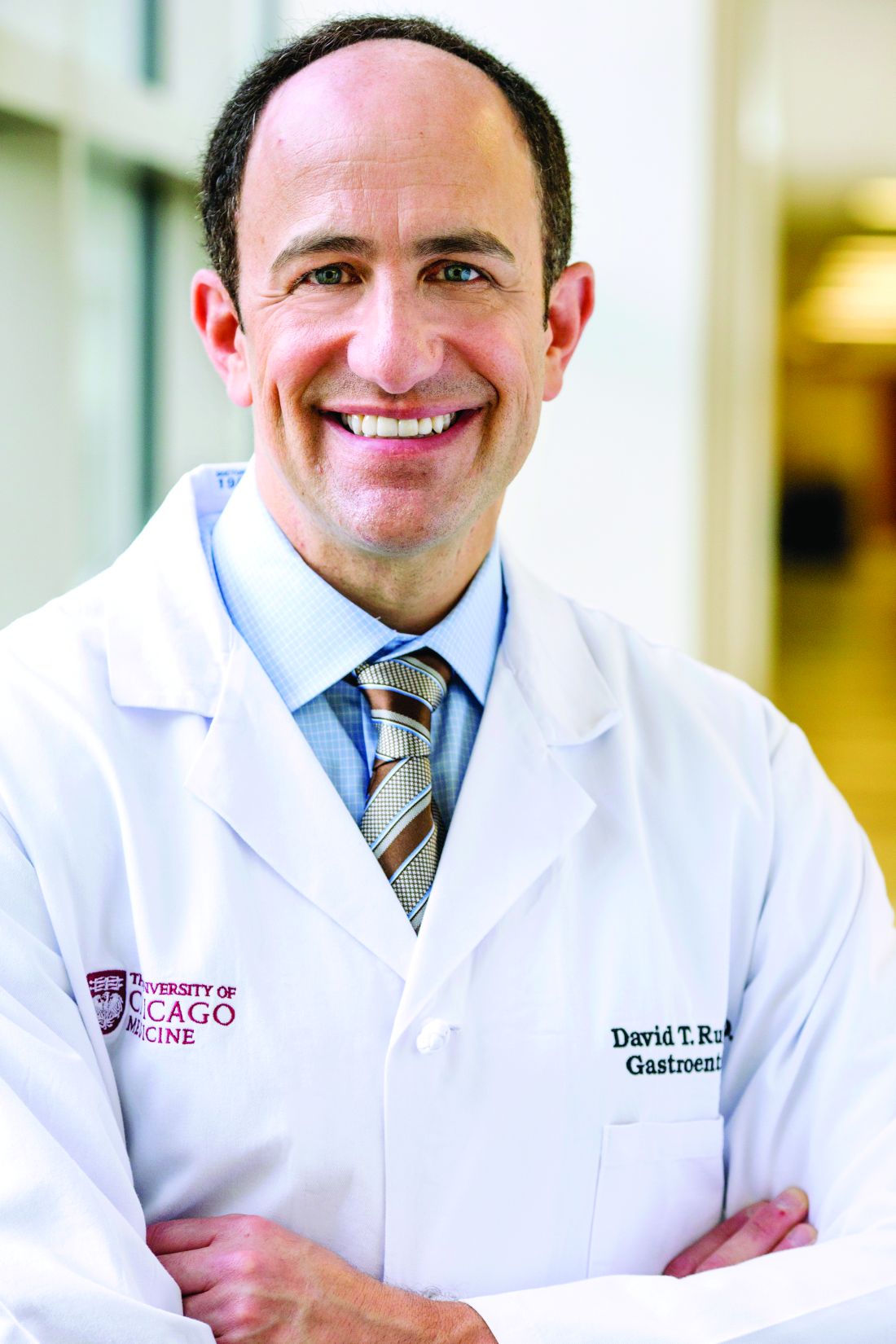
David T. Rubin, MD, was the senior author of the study, which provided independent validation of a metric for cumulative burden of inflammation as a risk stratification tool in ulcerative colitis and presented these findings at the Gastroenterology Updates, IBD, Liver Disease Conference. The metric was developed at St. Mark’s Hospital, London, which he called “a leader in the field.”
“The implication of demonstrating this is that, if you control inflammation and keep it controlled over time, it would imply that you can reduce the overall risk for cancer and dysplasia,” explained Dr. Rubin, professor of medicine and chief of the section of gastroenterology, hepatology, and nutrition at the University of Chicago.
The original retrospective St. Mark’s study included 987 patients with extensive ulcerative colitis followed with colonoscopic surveillance for a median of 13 years. Each colonoscopy was scored for severity of microscopic inflammation on a 0-3 scale. The investigators calculated a patient’s cumulative inflammatory burden by adding each histologic inflammatory activity score and multiplying that figure by the surveillance interval in years.
In a multivariate analysis, the London investigators demonstrated that the risk of colorectal neoplasia jumped by 2.1-fold for each 10-unit increase in cumulative inflammatory burden, defined as the equivalent of either 10 years of continuous mild active histologic inflammation, 5 years of continuous moderate inflammation, or 3.3 years of continuous severe inflammation.
The University of Chicago retrospective external validation study included 26 ulcerative colitis patients with colorectal neoplasia and 36 others without cancer. The mean cumulative histologic inflammatory activity score in the group with colorectal neoplasia was 12.63, compared with 7.98 in controls. For each 1-unit increase in cumulative inflammatory burden the risk of developing colorectal neoplasia increased by 8%, consistent with the magnitude of the hazard previously reported at St. Mark’s.
“The way you could take this back to your practice is by thinking carefully about what is the degree of inflammation each time you’ve done a colonoscopy and considering whether the patient who is in deep remission and doing well might deserve a longer interval between their next exam and the one you just completed,” according to the gastroenterologist.
“The most interval I give a patient is 3 years – for somebody in deep remission with no inflammation on the last exam. And when they’ve had prior inflammation but are now doing well, I keep in mind what that prior inflammation was. We’re now working on using that cumulative histologic inflammation score to guide intervals, but we don’t have prospective data to validate this approach. So when you’re not sure, the conservative approach is surveillance colonoscopy every 1-2 years after you’ve had 10 years of disease. That’s probably overutilization of our resources, but we don’t have a better way to do it yet,” Dr. Rubin said.
The novel metric for calculating cumulative histologic inflammation burden as a means of predicting colorectal cancer in ulcerative colitis dovetails with the current emphasis upon individualized risk assessment as recommended in the latest American College of Gastroenterology practice guidelines, for which Dr. Rubin was first author.
“Like we individualize our treatments, we should individualize our colorectal cancer screening and prevention strategies,” he emphasized.
Risk factors for colorectal cancer and dysplasia in patients with ulcerative colitis can be grouped as either potentially modifiable or immutable. Potentially modifiable risk factors include backwash ileitis, pseudopolyps, prior dysplasia, and mass or stricture, as well as the degree of histologic inflammation. Immutable risk factors include younger age at diagnosis, male gender, duration and extent of disease, family history of colorectal cancer, and primary sclerosing cholangitis, Dr. Rubin noted.
He reported receiving grant support from and/or serving as a consultant to more than two dozen medical companies.
The cumulative burden of histologic inflammation is a strong predictor of colorectal neoplasia risk in ulcerative colitis, according to a recent case-control study.
David T. Rubin, MD, was the senior author of the study, which provided independent validation of a metric for cumulative burden of inflammation as a risk stratification tool in ulcerative colitis and presented these findings at the Gastroenterology Updates, IBD, Liver Disease Conference. The metric was developed at St. Mark’s Hospital, London, which he called “a leader in the field.”
“The implication of demonstrating this is that, if you control inflammation and keep it controlled over time, it would imply that you can reduce the overall risk for cancer and dysplasia,” explained Dr. Rubin, professor of medicine and chief of the section of gastroenterology, hepatology, and nutrition at the University of Chicago.
The original retrospective St. Mark’s study included 987 patients with extensive ulcerative colitis followed with colonoscopic surveillance for a median of 13 years. Each colonoscopy was scored for severity of microscopic inflammation on a 0-3 scale. The investigators calculated a patient’s cumulative inflammatory burden by adding each histologic inflammatory activity score and multiplying that figure by the surveillance interval in years.
In a multivariate analysis, the London investigators demonstrated that the risk of colorectal neoplasia jumped by 2.1-fold for each 10-unit increase in cumulative inflammatory burden, defined as the equivalent of either 10 years of continuous mild active histologic inflammation, 5 years of continuous moderate inflammation, or 3.3 years of continuous severe inflammation.
The University of Chicago retrospective external validation study included 26 ulcerative colitis patients with colorectal neoplasia and 36 others without cancer. The mean cumulative histologic inflammatory activity score in the group with colorectal neoplasia was 12.63, compared with 7.98 in controls. For each 1-unit increase in cumulative inflammatory burden the risk of developing colorectal neoplasia increased by 8%, consistent with the magnitude of the hazard previously reported at St. Mark’s.
“The way you could take this back to your practice is by thinking carefully about what is the degree of inflammation each time you’ve done a colonoscopy and considering whether the patient who is in deep remission and doing well might deserve a longer interval between their next exam and the one you just completed,” according to the gastroenterologist.
“The most interval I give a patient is 3 years – for somebody in deep remission with no inflammation on the last exam. And when they’ve had prior inflammation but are now doing well, I keep in mind what that prior inflammation was. We’re now working on using that cumulative histologic inflammation score to guide intervals, but we don’t have prospective data to validate this approach. So when you’re not sure, the conservative approach is surveillance colonoscopy every 1-2 years after you’ve had 10 years of disease. That’s probably overutilization of our resources, but we don’t have a better way to do it yet,” Dr. Rubin said.
The novel metric for calculating cumulative histologic inflammation burden as a means of predicting colorectal cancer in ulcerative colitis dovetails with the current emphasis upon individualized risk assessment as recommended in the latest American College of Gastroenterology practice guidelines, for which Dr. Rubin was first author.
“Like we individualize our treatments, we should individualize our colorectal cancer screening and prevention strategies,” he emphasized.
Risk factors for colorectal cancer and dysplasia in patients with ulcerative colitis can be grouped as either potentially modifiable or immutable. Potentially modifiable risk factors include backwash ileitis, pseudopolyps, prior dysplasia, and mass or stricture, as well as the degree of histologic inflammation. Immutable risk factors include younger age at diagnosis, male gender, duration and extent of disease, family history of colorectal cancer, and primary sclerosing cholangitis, Dr. Rubin noted.
He reported receiving grant support from and/or serving as a consultant to more than two dozen medical companies.
FROM GUILD 2021