User login
Fremanezumab cut headache days in migraine patients vs. placebo
PHILADELPHIA – , according to a poster presented at the annual meeting of the American Academy of Neurology.

To assess the efficacy of fremanezumab in patients with migraine who had not received relief from trying at least one prior preventive migraine medication, Peter McAllister, MD and colleagues analyzed data from 2 phase 3 trials (HALO EM and HALO CM). Trial participants had either episodic or chronic migraine, confirmed during a 28-day pretreatment baseline period, then received subcutaneous fremanezumab quarterly (675 mg at baseline and placebo at weeks 4 and 8), monthly (for chronic migraine: 675 mg at baseline and 225 mg at weeks 4 and 8; for episodic migraine: 225 mg at baseline and weeks 4 and 8), or placebo (at baseline and weeks 4 and 8).
The present analysis included data from 186 patients with episodic migraine and 407 patients with chronic migraine, which represents the subgroup of study participants in the larger HALO trials who had failed at least one prior preventive migraine medication. Dr. McAllister, who is cofounder and chief medical officer at the New England Institute for Clinical Research in Stamford, Connecticut, and his colleagues, assessed mean changes from baseline in the monthly average number of headache days of at least moderate severity or the monthly average number of migraine days during the 12-week treatment period.
In patients with chronic migraine, fremanezumab yielded greater reductions in the number of headache days of at least moderate severity (quarterly [least-squares mean change]: –4.0, P less than 0.0001; monthly: –4.5, P less than 0.0001) compared with placebo (–1.8). There were similar reductions in the number of migraine days (quarterly: –4.1, P = 0.0027; monthly: –4.8, P less than 0.0001) compared with placebo (–2.3).
In patients with episodic migraine, fremanezumab yielded greater reductions in the number of headache days of at least moderate severity (quarterly: –3.1, P less than 0.0001; monthly: –3.2, P less than 0.0001) compared with placebo (–0.8). There were similar reductions in the number of migraine days (quarterly: –3.3, P = 0.0015; monthly: –3.7, P less than 0.0001) compared with placebo (–1.3).
“The phase 3 HALO CM and HALO EM trials showed that fremanezumab is efficacious in patients who failed one or more prior preventive medication, a potentially difficult-to-treat population,” Dr. McAllister and colleagues said in their poster.
“Effect sizes in this subgroup were greater than those in the overall trial population,” they said. In addition, “both quarterly and monthly fremanezumab were well-tolerated in this subgroup.”
This study was funded by Teva Pharmaceuticals, Petach Tikva, Israel.
SOURCE: McAllister P et al. AAN 2019. P1.10-011.
PHILADELPHIA – , according to a poster presented at the annual meeting of the American Academy of Neurology.
To assess the efficacy of fremanezumab in patients with migraine who had not received relief from trying at least one prior preventive migraine medication, Peter McAllister, MD and colleagues analyzed data from 2 phase 3 trials (HALO EM and HALO CM). Trial participants had either episodic or chronic migraine, confirmed during a 28-day pretreatment baseline period, then received subcutaneous fremanezumab quarterly (675 mg at baseline and placebo at weeks 4 and 8), monthly (for chronic migraine: 675 mg at baseline and 225 mg at weeks 4 and 8; for episodic migraine: 225 mg at baseline and weeks 4 and 8), or placebo (at baseline and weeks 4 and 8).
The present analysis included data from 186 patients with episodic migraine and 407 patients with chronic migraine, which represents the subgroup of study participants in the larger HALO trials who had failed at least one prior preventive migraine medication. Dr. McAllister, who is cofounder and chief medical officer at the New England Institute for Clinical Research in Stamford, Connecticut, and his colleagues, assessed mean changes from baseline in the monthly average number of headache days of at least moderate severity or the monthly average number of migraine days during the 12-week treatment period.
In patients with chronic migraine, fremanezumab yielded greater reductions in the number of headache days of at least moderate severity (quarterly [least-squares mean change]: –4.0, P less than 0.0001; monthly: –4.5, P less than 0.0001) compared with placebo (–1.8). There were similar reductions in the number of migraine days (quarterly: –4.1, P = 0.0027; monthly: –4.8, P less than 0.0001) compared with placebo (–2.3).
In patients with episodic migraine, fremanezumab yielded greater reductions in the number of headache days of at least moderate severity (quarterly: –3.1, P less than 0.0001; monthly: –3.2, P less than 0.0001) compared with placebo (–0.8). There were similar reductions in the number of migraine days (quarterly: –3.3, P = 0.0015; monthly: –3.7, P less than 0.0001) compared with placebo (–1.3).
“The phase 3 HALO CM and HALO EM trials showed that fremanezumab is efficacious in patients who failed one or more prior preventive medication, a potentially difficult-to-treat population,” Dr. McAllister and colleagues said in their poster.
“Effect sizes in this subgroup were greater than those in the overall trial population,” they said. In addition, “both quarterly and monthly fremanezumab were well-tolerated in this subgroup.”
This study was funded by Teva Pharmaceuticals, Petach Tikva, Israel.
SOURCE: McAllister P et al. AAN 2019. P1.10-011.
REPORTING FROM AAN 2019
Key clinical point: Fremanezumab reduced headache days in patients with chronic or episodic migraine.
Major finding: In patients with chronic migraine, fremanezumab reduced the number of headache days (least-squares mean change = -4.0) compared with placebo (-1.8).
Study details: Subgroup analysis of data from two phase 3 studies - HALO EM and HALO CM - including 186 patients with episodic migraine and 407 patients with chronic migraine.
Disclosures: This study was funded by Teva Pharmaceuticals, Petach Tikva, Israel.
Source: McAllister P et al. AAN 2019. P1.10-011.
Liletta IUD efficacy extends to 6 years
NASHVILLE, TENN. – The 52 mg levonorgestrel intrauterine system Liletta, which is currently approved as a contraceptive for up to 5 years of use, remains highly effective and safe for an additional year of use, according to findings from an ongoing 10-year trial.

Of 1,714 women enrolled in the multicenter phase 3 trial and for whom demographic information is available, 379 have completed 6 years of use. Nine pregnancies have occurred to date, including 2 in year 1, 4 in year 2, and 1 each in years 3-5, for a cumulative life-table pregnancy rate through year 6 of 0.87, Carolyn L. Westhoff, MD, MSc, reported during a poster session at the annual clinical and scientific meeting of the American College of Obstetricians and Gynecologists.
Six (67%) of the pregnancies were ectopic, noted Dr. Westhoff of Columbia University, N.Y.
Study subjects include 1,568 women aged 16-35 years at enrollment (986 were nulliparous and 433 were obese) and 146 aged 36 to 45 years. All were followed after device placement, with 138 having completed 8 years of use.
Two perforations occurred following device placement–both within the first year, and none have occurred since, Dr. Westhoff noted.
Other adverse events included expulsion in 68 women (4.0%), with 50 of those (73.5%) occurring in the first year of use, and pelvic infection in 15 women (0.9%), with 11 (73.3%) of those occurring after 6 or more months of use.
Only 40 women (2.3%) discontinued the study due to bleeding, and 30 of those (75%) did so in the first 2 years.
in both nulliparous and parous women, and in both non-obese and obese women, Dr. Westhoff concluded. She added that “there were no additional pregnancies in 6 years, and this study will be continuing to look all the way to 10-year effectiveness.”
This study is funded by Medicines360, a non-profit pharmaceutical company. Dr. Westhoff is a consultant or advisory board member for Agile and Cooper Surgical, and a Data and Safety Monitoring Board member for phase 4 studies for Bayer and Merck.
SOURCE: Westhoff C et al., ACOG 2019: Abstract 13M.
NASHVILLE, TENN. – The 52 mg levonorgestrel intrauterine system Liletta, which is currently approved as a contraceptive for up to 5 years of use, remains highly effective and safe for an additional year of use, according to findings from an ongoing 10-year trial.
Of 1,714 women enrolled in the multicenter phase 3 trial and for whom demographic information is available, 379 have completed 6 years of use. Nine pregnancies have occurred to date, including 2 in year 1, 4 in year 2, and 1 each in years 3-5, for a cumulative life-table pregnancy rate through year 6 of 0.87, Carolyn L. Westhoff, MD, MSc, reported during a poster session at the annual clinical and scientific meeting of the American College of Obstetricians and Gynecologists.
Six (67%) of the pregnancies were ectopic, noted Dr. Westhoff of Columbia University, N.Y.
Study subjects include 1,568 women aged 16-35 years at enrollment (986 were nulliparous and 433 were obese) and 146 aged 36 to 45 years. All were followed after device placement, with 138 having completed 8 years of use.
Two perforations occurred following device placement–both within the first year, and none have occurred since, Dr. Westhoff noted.
Other adverse events included expulsion in 68 women (4.0%), with 50 of those (73.5%) occurring in the first year of use, and pelvic infection in 15 women (0.9%), with 11 (73.3%) of those occurring after 6 or more months of use.
Only 40 women (2.3%) discontinued the study due to bleeding, and 30 of those (75%) did so in the first 2 years.
in both nulliparous and parous women, and in both non-obese and obese women, Dr. Westhoff concluded. She added that “there were no additional pregnancies in 6 years, and this study will be continuing to look all the way to 10-year effectiveness.”
This study is funded by Medicines360, a non-profit pharmaceutical company. Dr. Westhoff is a consultant or advisory board member for Agile and Cooper Surgical, and a Data and Safety Monitoring Board member for phase 4 studies for Bayer and Merck.
SOURCE: Westhoff C et al., ACOG 2019: Abstract 13M.
REPORTING FROM ACOG 2019
Survey: Palliative care blocked by many barriers in end-stage liver disease
results of a recent survey show.

Cultural factors, unrealistic expectations of the patient, lack of reimbursement, and competing demands for physicians’ time were some of the barriers to palliative care cited most frequently in the survey, said the researchers, in their report on the survey results that appears in Clinical Gastroenterology and Hepatology.
Moreover, most responding physicians said they felt end-of-life advance care planning discussions take place too late in the course of illness, according to Nneka N. Ufere, MD, of the Gastrointestinal Unit, Department of Medicine, Massachusetts General Hospital, Boston, and co-authors of the report.
“Multiple interventions targeted at patients, caregivers, institutions, and clinicians are needed to overcome barriers to improve the delivery of high-quality palliative and end-of-life care for patients with end-stage liver disease,” the researchers said.
Specialty palliative care can improve quality of life for patients with life-limiting conditions such as end-stage liver disease, which is associated with poor quality of life and a median survival of just two years without liver transplant, the authors said.
Advance care planning, in which patients discuss goals and care preferences in light of the expected course of illness, was a “critical component” of palliative care that can improve the quality of end-of-life care, Dr. Ufere and co-authors said.
Unfortunately, palliative care planning services are underutilized in end-stage liver disease, studies show, while rates of timely advance care planning discussions are low.
To find out why, Dr. Ufere and colleagues asked 1,238 physicians to fill out a web-based questionnaire designed to assess their perceptions of barriers to use of palliative care and barriers to timely advance care planning discussions. A total of 396 physicians (32%) completed the survey between February and April 2018.
Sixty percent were transplant hepatologists, and 79% of the survey participants said they worked in a teaching hospital, according to Dr. Ufere and co-authors, who added that no respondents had formal palliative care training.
Almost all respondents (95%) agreed that centers providing care for end-stage liver disease patients should have palliative care services, and most (86%) said they thought such patients would benefit from palliative care earlier in the course of disease.
While most (84%) agreed that a hepatologist was the best provider to discuss advance care planning with the patient, only about one-quarter (27%) said the hepatologist was best suited to provide palliative care, while most (88%) said the palliative care specialist was best for that role.
When asked about patient and caregiver barriers, nearly all respondents (95%) agreed that cultural factors that influenced palliative care perception was an issue, while 93% said patients’ unrealistic expectations was an issue.
Clinician barriers that respondents perceived included competing demands for clinicians’ time, cited by 91%, fear that palliative care might destroy the patient’s hope, cited by 82%, and the misperception that palliative care starts when active treatment ends, cited by 81%.
One potential solution to the competing demands on clinicians’ time would be development of “collaborative care models” between palliative care and hepatology services, according to Dr. Ufere and co-authors.
“Outpatient specialty palliative care visits, ideally temporally coordinated with the hepatology visits, can play a role not only in attending to symptom assessment and ACP, but also in addressing important psychosocial aspects of care, such as patient coping and well-being,” they said in their report on the survey.
Institutional barriers of note included limited reimbursement for time spent providing palliative care, cited by 76% and lack of a palliative care service, cited by nearly half (46%).
Some of the most commonly affirmed barriers to timely advance care planning discussions included insufficient training in end-of-life communication issues, and insufficient training in cultural competency issues related to the discussions.
In terms of timeliness, only 17% said advance care planning discussions happen at the right time, while 81% said they happen too late, investigators found.
Funding for the research came from the National Institutes of Health. The authors had no disclosures or conflicts of interest related to the report.
SOURCE: Ufere NN, et al. Clin Gastroenterol Hepatol. 2019 Mar 15. doi: 10.1016/j.cgh.2019.03.022.
results of a recent survey show.
Cultural factors, unrealistic expectations of the patient, lack of reimbursement, and competing demands for physicians’ time were some of the barriers to palliative care cited most frequently in the survey, said the researchers, in their report on the survey results that appears in Clinical Gastroenterology and Hepatology.
Moreover, most responding physicians said they felt end-of-life advance care planning discussions take place too late in the course of illness, according to Nneka N. Ufere, MD, of the Gastrointestinal Unit, Department of Medicine, Massachusetts General Hospital, Boston, and co-authors of the report.
“Multiple interventions targeted at patients, caregivers, institutions, and clinicians are needed to overcome barriers to improve the delivery of high-quality palliative and end-of-life care for patients with end-stage liver disease,” the researchers said.
Specialty palliative care can improve quality of life for patients with life-limiting conditions such as end-stage liver disease, which is associated with poor quality of life and a median survival of just two years without liver transplant, the authors said.
Advance care planning, in which patients discuss goals and care preferences in light of the expected course of illness, was a “critical component” of palliative care that can improve the quality of end-of-life care, Dr. Ufere and co-authors said.
Unfortunately, palliative care planning services are underutilized in end-stage liver disease, studies show, while rates of timely advance care planning discussions are low.
To find out why, Dr. Ufere and colleagues asked 1,238 physicians to fill out a web-based questionnaire designed to assess their perceptions of barriers to use of palliative care and barriers to timely advance care planning discussions. A total of 396 physicians (32%) completed the survey between February and April 2018.
Sixty percent were transplant hepatologists, and 79% of the survey participants said they worked in a teaching hospital, according to Dr. Ufere and co-authors, who added that no respondents had formal palliative care training.
Almost all respondents (95%) agreed that centers providing care for end-stage liver disease patients should have palliative care services, and most (86%) said they thought such patients would benefit from palliative care earlier in the course of disease.
While most (84%) agreed that a hepatologist was the best provider to discuss advance care planning with the patient, only about one-quarter (27%) said the hepatologist was best suited to provide palliative care, while most (88%) said the palliative care specialist was best for that role.
When asked about patient and caregiver barriers, nearly all respondents (95%) agreed that cultural factors that influenced palliative care perception was an issue, while 93% said patients’ unrealistic expectations was an issue.
Clinician barriers that respondents perceived included competing demands for clinicians’ time, cited by 91%, fear that palliative care might destroy the patient’s hope, cited by 82%, and the misperception that palliative care starts when active treatment ends, cited by 81%.
One potential solution to the competing demands on clinicians’ time would be development of “collaborative care models” between palliative care and hepatology services, according to Dr. Ufere and co-authors.
“Outpatient specialty palliative care visits, ideally temporally coordinated with the hepatology visits, can play a role not only in attending to symptom assessment and ACP, but also in addressing important psychosocial aspects of care, such as patient coping and well-being,” they said in their report on the survey.
Institutional barriers of note included limited reimbursement for time spent providing palliative care, cited by 76% and lack of a palliative care service, cited by nearly half (46%).
Some of the most commonly affirmed barriers to timely advance care planning discussions included insufficient training in end-of-life communication issues, and insufficient training in cultural competency issues related to the discussions.
In terms of timeliness, only 17% said advance care planning discussions happen at the right time, while 81% said they happen too late, investigators found.
Funding for the research came from the National Institutes of Health. The authors had no disclosures or conflicts of interest related to the report.
SOURCE: Ufere NN, et al. Clin Gastroenterol Hepatol. 2019 Mar 15. doi: 10.1016/j.cgh.2019.03.022.
FROM CLINICAL GASTROENTEROLOGY AND HEPATOLOGY
Multiple sclerosis may not flare up after pregnancy
PHILADELPHIA – according to a study to be presented at the annual meeting of the American Academy of Neurology.
“We did not observe any rebound disease activity,” said Annette Langer-Gould, MD, PhD, and her research colleagues in their report.
The findings contrast with those of 20-year-old studies that first identified a lower risk of relapse during pregnancy but signficant rebound disease activity in the early postpartum period. The initial studies were conducted before disease-modifying treatments (DMTs) were available and before neurologists used MRI to help diagnose MS after one attack, noted Dr. Langer-Gould in a statement.
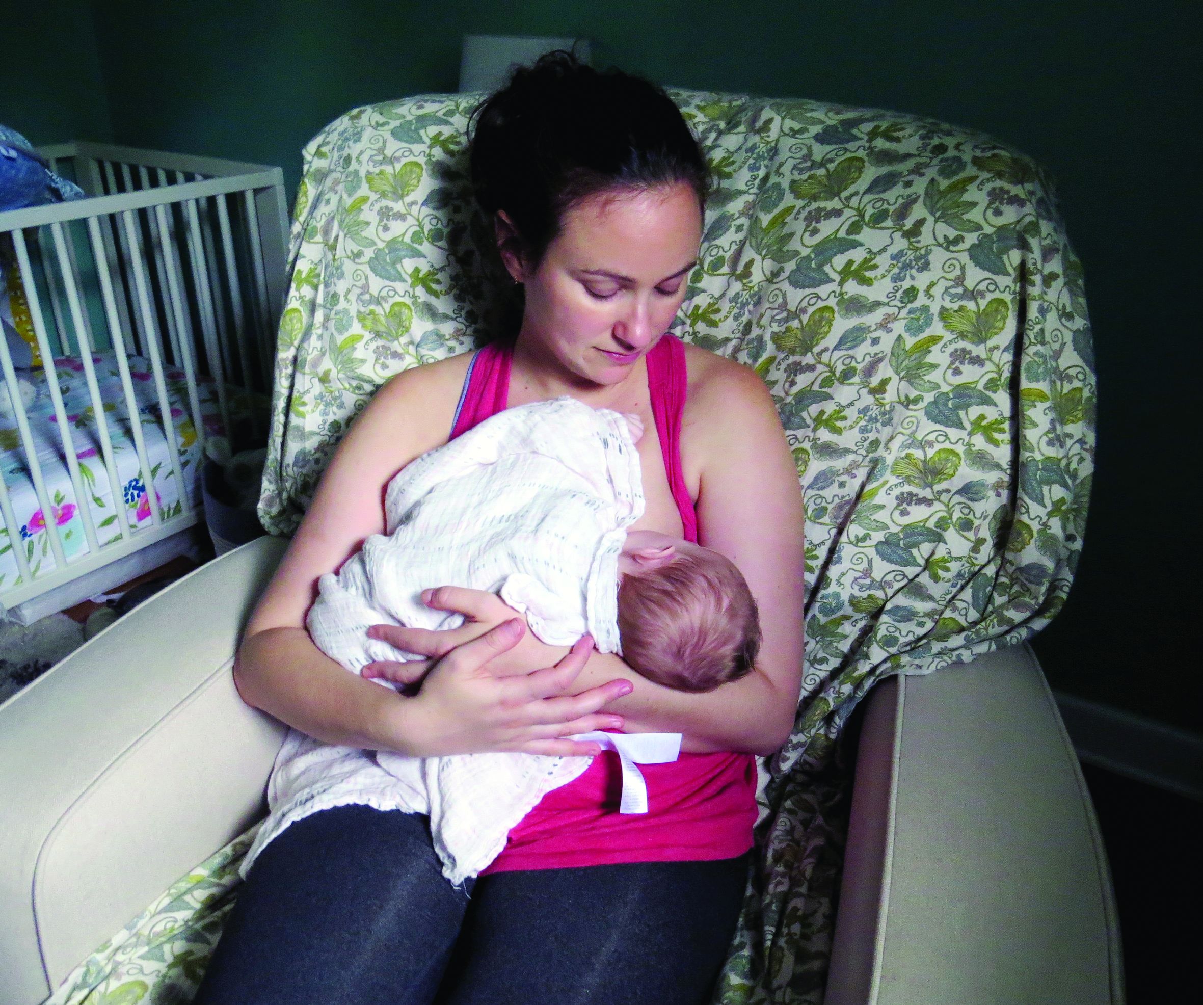
In the large, contemporary cohort of patients with MS, the annualized relapse rate was 0.39 pre-pregnancy, 0.07-0.14 during pregnancy, 0.27 in the first 3 months postpartum, and 0.37 at 4-6 months postpartum. Exclusive breastfeeding significantly reduced the risk of postpartum relapses by 42% (adjusted hazard ratio = 0.58). Women who supplemented breast milk with formula within 2 months of delivery had the same risk of relapse as women who did not breastfeed, however.
“These results are exciting, as MS is more common among women of childbearing age than in any other group,” said Dr. Langer-Gould, who is regional lead for clinical and translational neuroscience at Kaiser Permanente Southern California in Pasadena, in the statement. “This shows us that women with MS today can have children, breastfeed, and resume their treatment without experiencing an increased risk of relapses during the postpartum period.”
To describe the risk of postpartum relapses and identify potential risk factors for relapse the investigators analyzed prospectively collected data from 466 pregnancies among 375 women with MS from the complete electronic health record at Kaiser Permanente Southern and Northern California between 2008 and 2016. The researchers also used surveys to collect information about treatment history, breastfeeding, and relapses. They used multivariable models to account for intraclass clustering and disease severity.
In 38% of the pregnancies, the mother had not received treatment in the year before conception. In 14.6%, the mother had a clinically isolated syndrome; in 8.4%, the mother had a relapse during pregnancy.
Resuming modestly effective DMTs such as interferon-betas and glatiramer acetate did not affect relapse risk.
In the postpartum year, 26.4% of mothers relapsed, 87% breastfed, 35% breastfed exclusively, and 41.2% resumed using DMT.
The lack of rebound disease activity in this cohort could be related to the high rate of exclusive breastfeeding, as well as the inclusion of women from a population-based setting and the inclusion of women who had incorrectly been diagnosed with MS after a single relapse. Few patients in this cohort had been treated with natalizumab or fingolimod prior to pregnancy, so the study does not address the potential harms of stopping these drugs or the potential benefits of breastfeeding among patients treated with these drugs.
The study was supported by the National Multiple Sclerosis Society. The researchers had no disclosures.
SOURCE: Langer-Gould A et al. AAN 2019, Abstract S6.007.
PHILADELPHIA – according to a study to be presented at the annual meeting of the American Academy of Neurology.
“We did not observe any rebound disease activity,” said Annette Langer-Gould, MD, PhD, and her research colleagues in their report.
The findings contrast with those of 20-year-old studies that first identified a lower risk of relapse during pregnancy but signficant rebound disease activity in the early postpartum period. The initial studies were conducted before disease-modifying treatments (DMTs) were available and before neurologists used MRI to help diagnose MS after one attack, noted Dr. Langer-Gould in a statement.
In the large, contemporary cohort of patients with MS, the annualized relapse rate was 0.39 pre-pregnancy, 0.07-0.14 during pregnancy, 0.27 in the first 3 months postpartum, and 0.37 at 4-6 months postpartum. Exclusive breastfeeding significantly reduced the risk of postpartum relapses by 42% (adjusted hazard ratio = 0.58). Women who supplemented breast milk with formula within 2 months of delivery had the same risk of relapse as women who did not breastfeed, however.
“These results are exciting, as MS is more common among women of childbearing age than in any other group,” said Dr. Langer-Gould, who is regional lead for clinical and translational neuroscience at Kaiser Permanente Southern California in Pasadena, in the statement. “This shows us that women with MS today can have children, breastfeed, and resume their treatment without experiencing an increased risk of relapses during the postpartum period.”
To describe the risk of postpartum relapses and identify potential risk factors for relapse the investigators analyzed prospectively collected data from 466 pregnancies among 375 women with MS from the complete electronic health record at Kaiser Permanente Southern and Northern California between 2008 and 2016. The researchers also used surveys to collect information about treatment history, breastfeeding, and relapses. They used multivariable models to account for intraclass clustering and disease severity.
In 38% of the pregnancies, the mother had not received treatment in the year before conception. In 14.6%, the mother had a clinically isolated syndrome; in 8.4%, the mother had a relapse during pregnancy.
Resuming modestly effective DMTs such as interferon-betas and glatiramer acetate did not affect relapse risk.
In the postpartum year, 26.4% of mothers relapsed, 87% breastfed, 35% breastfed exclusively, and 41.2% resumed using DMT.
The lack of rebound disease activity in this cohort could be related to the high rate of exclusive breastfeeding, as well as the inclusion of women from a population-based setting and the inclusion of women who had incorrectly been diagnosed with MS after a single relapse. Few patients in this cohort had been treated with natalizumab or fingolimod prior to pregnancy, so the study does not address the potential harms of stopping these drugs or the potential benefits of breastfeeding among patients treated with these drugs.
The study was supported by the National Multiple Sclerosis Society. The researchers had no disclosures.
SOURCE: Langer-Gould A et al. AAN 2019, Abstract S6.007.
FROM AAN 2019
ACOG guidance addresses cardiac contributors to maternal mortality
NASHVILLE, TENN. – according to new comprehensive guidance from the American College of Obstetricians and Gynecologists.
The toolkit algorithm is called the California Improving Health Care Response to Cardiovascular Disease in Pregnancy and Postpartum Toolkit. It was developed by the Cardiovascular Disease in Pregnancy Postpartum Task Force to serve as a resource for obstetrics, primary care and emergency medicine providers who provide prenatal care or interact with women during the postpartum period. It incldues an overview of clinical assessment and comprehensive management strategies for cardiovascular disease based on risk factors and presenting symptoms.
The guidance also calls for all pregnant and postpartum women with known or suspected CVD to undergo further evaluation by a “Pregnancy Heart Team that includes a cardiologist and maternal–fetal medicine subspecialist, or both, and other subspecialists as necessary.” The guidance was issued in Practice Bulletin 212, Pregnancy and Heart Disease, which is published in the May edition of Obstetrics & Gynecology (Obstet Gynecol. 2019 May;133[5]:e320-e356).
In all, 27 specific recommendations and conclusions relating to screening, diagnosis, and management of CVD for women during the prepregnancy period through the postpartum period are included in the guidance.
ACOG president Lisa Hollier, MD, convened the task force that developed this guidance to address cardiac contributors to maternal mortality, she said during a press briefing at the ACOG annual clinical and scientific meeting.
“When I began my presidency a year ago, my goal was to bring together a multidisciplinary group of clinicians ... to create clinical guidance that would make a difference in the lives of women," said Dr. Hollier, who is also a professor of obstetrics and gynecology at Baylor College of Medicine, Houston. 
Part of her presidential initiative was centered on eliminating preventable maternal death, and this guidance has the potential to make strides toward that goal, she said. When it comes to CVD in pregnancy, “there is so much we can do to prevent negative outcomes and ensure that moms go home with their babies and are around to see them grow up,” she noted.
CVD is the leading cause of death in pregnant women and women in the postpartum period, accounting for 26.5% of U.S. pregnancy-related deaths.
“It’s critical that we as physicians and health care professionals develop expertise in recognizing the signs and symptoms so that we can save women’s lives,” she said in the press breifing. Dr. Hollier also implored her colleagues to “start using this guidance immediately and prevent more women from dying from cardiovascular complications of pregnancy.”
In this video interview, Dr. Hollier further explains the need for the guidance and its potential for improving maternal mortality rates.
Dr. Hollier reported having no relevant disclosures.
SOURCE: Hollier L et al., Obstet Gynecol. 2019 May;133[5]:e320-56.
NASHVILLE, TENN. – according to new comprehensive guidance from the American College of Obstetricians and Gynecologists.
The toolkit algorithm is called the California Improving Health Care Response to Cardiovascular Disease in Pregnancy and Postpartum Toolkit. It was developed by the Cardiovascular Disease in Pregnancy Postpartum Task Force to serve as a resource for obstetrics, primary care and emergency medicine providers who provide prenatal care or interact with women during the postpartum period. It incldues an overview of clinical assessment and comprehensive management strategies for cardiovascular disease based on risk factors and presenting symptoms.
The guidance also calls for all pregnant and postpartum women with known or suspected CVD to undergo further evaluation by a “Pregnancy Heart Team that includes a cardiologist and maternal–fetal medicine subspecialist, or both, and other subspecialists as necessary.” The guidance was issued in Practice Bulletin 212, Pregnancy and Heart Disease, which is published in the May edition of Obstetrics & Gynecology (Obstet Gynecol. 2019 May;133[5]:e320-e356).
In all, 27 specific recommendations and conclusions relating to screening, diagnosis, and management of CVD for women during the prepregnancy period through the postpartum period are included in the guidance.
ACOG president Lisa Hollier, MD, convened the task force that developed this guidance to address cardiac contributors to maternal mortality, she said during a press briefing at the ACOG annual clinical and scientific meeting.
“When I began my presidency a year ago, my goal was to bring together a multidisciplinary group of clinicians ... to create clinical guidance that would make a difference in the lives of women," said Dr. Hollier, who is also a professor of obstetrics and gynecology at Baylor College of Medicine, Houston.
Part of her presidential initiative was centered on eliminating preventable maternal death, and this guidance has the potential to make strides toward that goal, she said. When it comes to CVD in pregnancy, “there is so much we can do to prevent negative outcomes and ensure that moms go home with their babies and are around to see them grow up,” she noted.
CVD is the leading cause of death in pregnant women and women in the postpartum period, accounting for 26.5% of U.S. pregnancy-related deaths.
“It’s critical that we as physicians and health care professionals develop expertise in recognizing the signs and symptoms so that we can save women’s lives,” she said in the press breifing. Dr. Hollier also implored her colleagues to “start using this guidance immediately and prevent more women from dying from cardiovascular complications of pregnancy.”
In this video interview, Dr. Hollier further explains the need for the guidance and its potential for improving maternal mortality rates.
Dr. Hollier reported having no relevant disclosures.
SOURCE: Hollier L et al., Obstet Gynecol. 2019 May;133[5]:e320-56.
REPORTING FROM ACOG 2019

What’s Happening on Connect?
Do you allow your patients to eat during thrombolysis? Do you have any difficulties with your EMR? Have questions about the upcoming Vascular Annual Meeting? Sign in to SVSConnect and participate in discussions surrounding all things vascular. Your SVS credentials will get you into the community, and from there you’ll be able to ask questions, reply to discussions, post resources and network with other members. Log in to SVSConnect here. If you encounter sign-in difficulties, email communications@vascularsociety.orgor call 312-334-2300.
Do you allow your patients to eat during thrombolysis? Do you have any difficulties with your EMR? Have questions about the upcoming Vascular Annual Meeting? Sign in to SVSConnect and participate in discussions surrounding all things vascular. Your SVS credentials will get you into the community, and from there you’ll be able to ask questions, reply to discussions, post resources and network with other members. Log in to SVSConnect here. If you encounter sign-in difficulties, email communications@vascularsociety.orgor call 312-334-2300.
Do you allow your patients to eat during thrombolysis? Do you have any difficulties with your EMR? Have questions about the upcoming Vascular Annual Meeting? Sign in to SVSConnect and participate in discussions surrounding all things vascular. Your SVS credentials will get you into the community, and from there you’ll be able to ask questions, reply to discussions, post resources and network with other members. Log in to SVSConnect here. If you encounter sign-in difficulties, email communications@vascularsociety.orgor call 312-334-2300.
Purchase Raffle Tickets for a Chance to Win
The ‘Vascular Spectacular’ Gala has officially sold out, but there are still ways for non-attendees to be a part of the fun. Raffle tickets are available for $20, and those who purchase will have a chance to win a $500, $250 or $100 cash prize. All proceeds benefit the SVS Foundation’s Greatest Need Fund and tickets are available through the evening of the event. Online raffle tickets can be purchased here.
The ‘Vascular Spectacular’ Gala has officially sold out, but there are still ways for non-attendees to be a part of the fun. Raffle tickets are available for $20, and those who purchase will have a chance to win a $500, $250 or $100 cash prize. All proceeds benefit the SVS Foundation’s Greatest Need Fund and tickets are available through the evening of the event. Online raffle tickets can be purchased here.
The ‘Vascular Spectacular’ Gala has officially sold out, but there are still ways for non-attendees to be a part of the fun. Raffle tickets are available for $20, and those who purchase will have a chance to win a $500, $250 or $100 cash prize. All proceeds benefit the SVS Foundation’s Greatest Need Fund and tickets are available through the evening of the event. Online raffle tickets can be purchased here.
Program at VAM set for Vascular Residents and Fellows
The SVS is introducing a dedicated program for vascular residents and fellows at the 2019 Vascular Annual Meeting. The new program is set for 9:30 p.m. Thursday, June 13, and will provide a forum to those nearing the end of their training that will allow them to explore several topics with leaders in vascular surgery. Discussion topics will focus on transition to practice, which will include presentations on business, career development, leadership and innovations on the horizon for vascular surgery. Attendance is open to all vascular residents and fellows enrolled in vascular fellowship programs or 0+5 residency programs. Participants must pre-register to participate. Register today and contact education@vascularsociety.org for more information.
The SVS is introducing a dedicated program for vascular residents and fellows at the 2019 Vascular Annual Meeting. The new program is set for 9:30 p.m. Thursday, June 13, and will provide a forum to those nearing the end of their training that will allow them to explore several topics with leaders in vascular surgery. Discussion topics will focus on transition to practice, which will include presentations on business, career development, leadership and innovations on the horizon for vascular surgery. Attendance is open to all vascular residents and fellows enrolled in vascular fellowship programs or 0+5 residency programs. Participants must pre-register to participate. Register today and contact education@vascularsociety.org for more information.
The SVS is introducing a dedicated program for vascular residents and fellows at the 2019 Vascular Annual Meeting. The new program is set for 9:30 p.m. Thursday, June 13, and will provide a forum to those nearing the end of their training that will allow them to explore several topics with leaders in vascular surgery. Discussion topics will focus on transition to practice, which will include presentations on business, career development, leadership and innovations on the horizon for vascular surgery. Attendance is open to all vascular residents and fellows enrolled in vascular fellowship programs or 0+5 residency programs. Participants must pre-register to participate. Register today and contact education@vascularsociety.org for more information.
FDA opts not to ban textured breast implants
The Food and Drug Administration decided to continue to allow U.S. sales of textured breast implants, which have been identified as the cause of a rare but significant cancer, breast implant–associated anaplastic large cell lymphoma.

A statement the agency released on May 2 said “The FDA does not believe that, on the basis of available data and information, the device [textured implants] meets the banning standard set forth in the Federal Food and Drug Cosmetic Act.” roughly half of them in the United States.
In coming to this decision, following 2 days of public testimony and discussions by an advisory committee in late March, the FDA is bucking the path taken by regulatory bodies of the European Union as well as several other counties. The EU acted in December 2018 to produce the equivalent of a ban on sales of textured breast implants marketed by Allergan. Then in April 2019, the French drug and device regulatory agency expanded this ban to textured breast implants sold by five other companies.
During the FDA advisory committee meeting in March, one of the world’s experts on BIA-ALCL, Mark W. Clemens, MD, a plastic surgeon at MD Anderson Cancer Center in Houston, said that of about 500 case reports received by the FDA, not one had involved a confirmed and “pure” episode of BIA-ALCL linked with a smooth breast implant. A team of experts recently reached the same conclusion when reviewing the reported worldwide incidence of BIA-ALCL in a published review (Plast Reconstr Surg. 2019 March;143[3S]:30S-40S).
Despite these reports, the FDA said in its new statement that “While the majority of women who develop BIA-ALCL have had textured implants, there are known cases in women with smooth-surface breast implants, and many reports do not include the surface texture of the implant at the time of diagnosis.” The agency added that it is “focused on strengthening the evidence generated to help inform future regulatory action.” During the March advisory committee meeting, some members of the panel spoke against a marketing ban on textured implants for reasons such as the modest number of reported cases and because of the importance of having a textured implant option available.
The FDA took several other notable steps in its May 2 statement:
The agency formally acknowledged that many breast implant recipients have reported experiencing adverse effects that include chronic fatigue, cognitive issues, and joint and muscle pain. “While the FDA doesn’t have definitive evidence demonstrating breast implants cause these symptoms, the current evidence supports that some women experience systemic symptoms that may resolve when their breast implants are removed.” The agency also cited the term that patients have coined for these symptoms: Breast Implant Illness.
The FDA made a commitment to “take steps to improve the information available to women and health care professionals about the risks of breast implants,” including the risk for BIA-ALCL, the increased risk for this cancer with textured implants, and the risk for systemic symptoms. The agency said it would work with stakeholders on possible changes to breast implant labeling, including a possible boxed warning, and a patient-decision checklist.
The FDA announced a change in how manufacturers will file medical device reports for breast implants. The agency will no longer allow these filings to be “summary” reports and will instead require manufacturers to file full individual medical device reports for each case that will be publicly available, with the intent to make reporting more transparent and complete.
Finally, the FDA announced that it would partner with two U.S. breast implant registries, the PROFILE registry of BIA-ALCL cases, and the National Breast Implant Registry, both run by the Plastic Surgery Foundation.
The FDA did not address in its statement other issues that came up during the March advisory committee hearings, including a panel recommendation to change follow-up imaging from MRI to ultrasound for monitoring women with implants for rupture, and the extensive, off-label use of surgical mesh during breast implant surgery.
The Food and Drug Administration decided to continue to allow U.S. sales of textured breast implants, which have been identified as the cause of a rare but significant cancer, breast implant–associated anaplastic large cell lymphoma.
A statement the agency released on May 2 said “The FDA does not believe that, on the basis of available data and information, the device [textured implants] meets the banning standard set forth in the Federal Food and Drug Cosmetic Act.” roughly half of them in the United States.
In coming to this decision, following 2 days of public testimony and discussions by an advisory committee in late March, the FDA is bucking the path taken by regulatory bodies of the European Union as well as several other counties. The EU acted in December 2018 to produce the equivalent of a ban on sales of textured breast implants marketed by Allergan. Then in April 2019, the French drug and device regulatory agency expanded this ban to textured breast implants sold by five other companies.
During the FDA advisory committee meeting in March, one of the world’s experts on BIA-ALCL, Mark W. Clemens, MD, a plastic surgeon at MD Anderson Cancer Center in Houston, said that of about 500 case reports received by the FDA, not one had involved a confirmed and “pure” episode of BIA-ALCL linked with a smooth breast implant. A team of experts recently reached the same conclusion when reviewing the reported worldwide incidence of BIA-ALCL in a published review (Plast Reconstr Surg. 2019 March;143[3S]:30S-40S).
Despite these reports, the FDA said in its new statement that “While the majority of women who develop BIA-ALCL have had textured implants, there are known cases in women with smooth-surface breast implants, and many reports do not include the surface texture of the implant at the time of diagnosis.” The agency added that it is “focused on strengthening the evidence generated to help inform future regulatory action.” During the March advisory committee meeting, some members of the panel spoke against a marketing ban on textured implants for reasons such as the modest number of reported cases and because of the importance of having a textured implant option available.
The FDA took several other notable steps in its May 2 statement:
The agency formally acknowledged that many breast implant recipients have reported experiencing adverse effects that include chronic fatigue, cognitive issues, and joint and muscle pain. “While the FDA doesn’t have definitive evidence demonstrating breast implants cause these symptoms, the current evidence supports that some women experience systemic symptoms that may resolve when their breast implants are removed.” The agency also cited the term that patients have coined for these symptoms: Breast Implant Illness.
The FDA made a commitment to “take steps to improve the information available to women and health care professionals about the risks of breast implants,” including the risk for BIA-ALCL, the increased risk for this cancer with textured implants, and the risk for systemic symptoms. The agency said it would work with stakeholders on possible changes to breast implant labeling, including a possible boxed warning, and a patient-decision checklist.
The FDA announced a change in how manufacturers will file medical device reports for breast implants. The agency will no longer allow these filings to be “summary” reports and will instead require manufacturers to file full individual medical device reports for each case that will be publicly available, with the intent to make reporting more transparent and complete.
Finally, the FDA announced that it would partner with two U.S. breast implant registries, the PROFILE registry of BIA-ALCL cases, and the National Breast Implant Registry, both run by the Plastic Surgery Foundation.
The FDA did not address in its statement other issues that came up during the March advisory committee hearings, including a panel recommendation to change follow-up imaging from MRI to ultrasound for monitoring women with implants for rupture, and the extensive, off-label use of surgical mesh during breast implant surgery.
Experts propose new definition and recommendations for Alzheimer’s-like disorder
An international group of experts has proposed a new name, staging criteria, and recommendations for a recently recognized brain disorder that mimics Alzheimer’s disease and is marked by a proteinopathy caused by malformed transactive response DNA-binding protein of 43 kDa (TDP-43).
The term limbic-predominant age-related TDP-43 encephalopathy (LATE) was coined in an effort to raise awareness and kick-start research into this “pathway to dementia,” the experts wrote in a report appearing in Brain.
“As there is currently no universally agreed-upon terminology or staging system for common age-related TDP-43 proteinopathy, this condition is understudied and not well recognized, even among investigators in the field of dementia research,” wrote the authors of the report, led by Peter T. Nelson, MD, PhD, of the University of Kentucky, Lexington.
LATE neuropathologic changes, associated with a progressive amnesia syndrome that mimics Alzheimer’s, are seen in more than 20% of individuals past the age of 80 years, according to large, community-based autopsy series. It coexists with Alzheimer’s disease in many patients, lowering the threshold for developing dementia, authors said.
The term LATE is designed to encompass several other terms related to TDP-43 pathology, including hippocampal sclerosis and cerebral age-related TDP-43 with sclerosis, Dr. Nelson and coauthors noted in their report.
The TDP-43 protein is encoded by the TARDBP gene and provides several functions related to the regulation of gene expression, the authors wrote.
Misfolded TDP-43 was known to play a causative role in amyotrophic lateral sclerosis and frontotemporal lobar degeneration, the authors noted, and then was also identified in the brains of older individuals with hippocampal sclerosis or Alzheimer’s disease neuropathologic changes.
The authors proposed a three-stage classification system for LATE neuropathologic change based on TDP-43 immunohistochemistry performed during routine autopsy evaluation of the amygdala, hippocampus, and middle frontal gyrus.
The amygdala is an area affected early in the course of the disease (Stage 1), whereas involvement of the hippocampus represents a more intermediate stage (Stage 2), and the middle frontal gyrus is more affected in advanced stages of the disease (Stage 3), according to the schema.
Five genes have been identified with risk alleles for LATE neuropathologic changes, authors said. Of note, several groups have found that the apolipoprotein E epsilon 4 (APOE4) allele, known to be a risk factor for Alzheimer’s disease neuropathologic changes and Lewy body disease, is also linked to increased risk of TDP-43 proteinopathy.
There are no established biomarkers specific to TDP-43 proteinopathy yet, which hampers development of clinical trials designed to test interventions to treat or prevent LATE, Dr. Nelson and colleagues said in their report.
LATE could also obscure the effects of potentially disease-modifying agents being tested in Alzheimer’s disease clinical trials, which can complicate the interpretation of study results, they added.
“Until there are biomarkers for LATE, clinical trials should be powered to account for TDP-43 proteinopathy,” they wrote.
Dr. Nelson and coauthors of the report in Brain reported no competing interests.
SOURCE: Nelson PT, et al. Brain. 2019 Apr 30. doi: 10.1093/brain/awz099
Alois Alzheimer’s original patient was 51 years old, and for roughly 70 years Alzheimer’s disease was considered a rare disease that caused presenile dementia. In the 1970s, Robert Katzman, MD, and Robert D. Terry, MD, equated the neuropathologic features of Alzheimer’s disease with the more common senile dementia, and since then we have recognized Alzheimer’s disease as the most common form of dementia. Autopsy studies of patients dying in their 80s and 90s, however, has revealed that far more common than pure Alzheimer’s disease is a mixed neuropathologic picture. In addition, with the advent of biomarker studies a substantial number of individuals have “suspected non-Alzheimer pathology.”

Interestingly, the authors identify the apolipoprotein E epsilon 4 (APOE4) allele as a predisposing factor for LATE, although given the advanced age of the LATE patient population, one could argue that a certain degree of resilience extended their lives into the LATE age range.
In contrast, in the Alzheimer’s Disease Sequencing Project, among those with autopsy confirmation, the prevalence of APOE4 in Braak stage 5-6 declines with succeeding decades so that, by the 80s and 90s, the prevalence of APOE2 is actually higher at 7.3% vs. 4.1% with APOE4 for ages 80 to younger than 85 years, 9.3% with APOE2 vs. 8.6% with APOE4 for 85 to younger than 90 years, and 16.7% with APOE2 vs. 6.9% with APOE4 for ages 90 years and above.
Our understanding of age-related cognitive decline, from the normal to the pathological ends of the spectrum, continues to evolve, and LATE is simply the latest addition to our growing knowledge base that will further inform clinical diagnosis, research, and experimental therapeutics.
Richard J. Caselli, MD, is professor of neurology at the Mayo Clinic Arizona in Scottsdale and associate director and clinical core director of the Arizona Alzheimer’s Disease Center.
Alois Alzheimer’s original patient was 51 years old, and for roughly 70 years Alzheimer’s disease was considered a rare disease that caused presenile dementia. In the 1970s, Robert Katzman, MD, and Robert D. Terry, MD, equated the neuropathologic features of Alzheimer’s disease with the more common senile dementia, and since then we have recognized Alzheimer’s disease as the most common form of dementia. Autopsy studies of patients dying in their 80s and 90s, however, has revealed that far more common than pure Alzheimer’s disease is a mixed neuropathologic picture. In addition, with the advent of biomarker studies a substantial number of individuals have “suspected non-Alzheimer pathology.”
Interestingly, the authors identify the apolipoprotein E epsilon 4 (APOE4) allele as a predisposing factor for LATE, although given the advanced age of the LATE patient population, one could argue that a certain degree of resilience extended their lives into the LATE age range.
In contrast, in the Alzheimer’s Disease Sequencing Project, among those with autopsy confirmation, the prevalence of APOE4 in Braak stage 5-6 declines with succeeding decades so that, by the 80s and 90s, the prevalence of APOE2 is actually higher at 7.3% vs. 4.1% with APOE4 for ages 80 to younger than 85 years, 9.3% with APOE2 vs. 8.6% with APOE4 for 85 to younger than 90 years, and 16.7% with APOE2 vs. 6.9% with APOE4 for ages 90 years and above.
Our understanding of age-related cognitive decline, from the normal to the pathological ends of the spectrum, continues to evolve, and LATE is simply the latest addition to our growing knowledge base that will further inform clinical diagnosis, research, and experimental therapeutics.
Richard J. Caselli, MD, is professor of neurology at the Mayo Clinic Arizona in Scottsdale and associate director and clinical core director of the Arizona Alzheimer’s Disease Center.
An international group of experts has proposed a new name, staging criteria, and recommendations for a recently recognized brain disorder that mimics Alzheimer’s disease and is marked by a proteinopathy caused by malformed transactive response DNA-binding protein of 43 kDa (TDP-43).
The term limbic-predominant age-related TDP-43 encephalopathy (LATE) was coined in an effort to raise awareness and kick-start research into this “pathway to dementia,” the experts wrote in a report appearing in Brain.
“As there is currently no universally agreed-upon terminology or staging system for common age-related TDP-43 proteinopathy, this condition is understudied and not well recognized, even among investigators in the field of dementia research,” wrote the authors of the report, led by Peter T. Nelson, MD, PhD, of the University of Kentucky, Lexington.
LATE neuropathologic changes, associated with a progressive amnesia syndrome that mimics Alzheimer’s, are seen in more than 20% of individuals past the age of 80 years, according to large, community-based autopsy series. It coexists with Alzheimer’s disease in many patients, lowering the threshold for developing dementia, authors said.
The term LATE is designed to encompass several other terms related to TDP-43 pathology, including hippocampal sclerosis and cerebral age-related TDP-43 with sclerosis, Dr. Nelson and coauthors noted in their report.
The TDP-43 protein is encoded by the TARDBP gene and provides several functions related to the regulation of gene expression, the authors wrote.
Misfolded TDP-43 was known to play a causative role in amyotrophic lateral sclerosis and frontotemporal lobar degeneration, the authors noted, and then was also identified in the brains of older individuals with hippocampal sclerosis or Alzheimer’s disease neuropathologic changes.
The authors proposed a three-stage classification system for LATE neuropathologic change based on TDP-43 immunohistochemistry performed during routine autopsy evaluation of the amygdala, hippocampus, and middle frontal gyrus.
The amygdala is an area affected early in the course of the disease (Stage 1), whereas involvement of the hippocampus represents a more intermediate stage (Stage 2), and the middle frontal gyrus is more affected in advanced stages of the disease (Stage 3), according to the schema.
Five genes have been identified with risk alleles for LATE neuropathologic changes, authors said. Of note, several groups have found that the apolipoprotein E epsilon 4 (APOE4) allele, known to be a risk factor for Alzheimer’s disease neuropathologic changes and Lewy body disease, is also linked to increased risk of TDP-43 proteinopathy.
There are no established biomarkers specific to TDP-43 proteinopathy yet, which hampers development of clinical trials designed to test interventions to treat or prevent LATE, Dr. Nelson and colleagues said in their report.
LATE could also obscure the effects of potentially disease-modifying agents being tested in Alzheimer’s disease clinical trials, which can complicate the interpretation of study results, they added.
“Until there are biomarkers for LATE, clinical trials should be powered to account for TDP-43 proteinopathy,” they wrote.
Dr. Nelson and coauthors of the report in Brain reported no competing interests.
SOURCE: Nelson PT, et al. Brain. 2019 Apr 30. doi: 10.1093/brain/awz099
FROM BRAIN