User login
New Treatment Option for Thick AKs Emerges
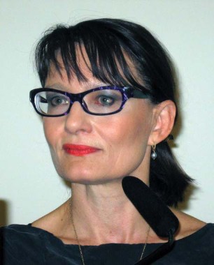
PRAGUE – Intensified photodynamic therapy assisted by ablative fractional laser resurfacing is a new and more effective way to treat thick actinic keratoses, according to Dr. Merete Haedersdal.
At 3 months’ follow-up, cure rates were significantly better with fractional CO2 laser-assisted photodynamic therapy (PDT) than with standard PDT in a randomized trial, she reported at the annual congress of the European Academy of Dermatology and Venereology.

"As a side benefit, the combined therapy gives a nice decrease in photoaging. There is photorejuvenation of the skin," noted Dr. Haedersdal of the University of Copenhagen.
Stand-alone PDT gets good results in thinner AKs, Bowen’s lesions, and basal cell carcinomas, both superficial and nodular. But effectiveness drops off considerably for thicker lesions.
That’s why Dr. Haedersdal and her Copenhagen colleagues, together with researchers at the Wellman Center for Photomedicine at Massachusetts General Hospital, Boston, where she has been a visiting scientist, are developing intensified PDT.
Basically, the dermatologists are using the fractional CO2 laser at 10,600 nm to drill tiny vertical channels surrounded by areas of unexposed skin. These channels facilitate uptake of the photosensitizing agent, rendering PDT more effective at greater depths. The enhanced uptake of photosensitizer is not merely a hypothesis; it has been documented via a marked increase in fluorescence intensity during illumination, she explained.
Dr. Haedersdal reported on 15 patients with a total of 212 AKs on severely photodamaged skin of the scalp and face. Two symmetrical randomly selected areas were chosen on each patient to receive one fractional CO2 laser-assisted PDT treatment and one standard PDT treatment. First, however, both treatment areas underwent curettage. Then one site was treated with the UltraPulse laser using the DeepFx handpiece set to 10 mJ per pulse and a single pulse density of 5%. The photosensitizing agent, methyl aminolevulinate cream, was then applied under occlusion for 3 hours at both sites. This was followed by illumination using a red light–emitting diode at 37 J/cm2.
At 3 months’ follow-up, the complete response rate of thicker grade II-III AKs was 88% with intensified PDT, compared with 59% with conventional PDT. For thinner grade I lesions, the complete response rates were 100% and 79%, respectively.
Only 3 new lesions arose at the intensified PDT-treated sites within 3 months, compared with 11 new lesions in areas that received standard PDT.
"So there might – in terms of avoiding future treatment procedures – be a benefit in combining the photothermal efficacy of the laser with the photochemical response from the PDT procedure," Dr. Haedersdal noted.
Pain scores were significantly higher during illumination in the intensified PDT areas, with a mean of 6.5 on a 1-10 scale compared with 5.4 on skin sites that got standard PDT. Erythema and crusting were also more intense at intensified PDT sites, and long-term pigmentary changes were more frequent at these sites as well.
"We have to be aware that the clinical reactions that we see from this new procedure are more intense than with conventional PDT. So for now we have to take care that we’re not using it for really large treatment areas because then the patients will have really intense phototoxic reactions," the dermatologist cautioned.
She and her coinvestigators are conducting an ongoing clinical trial combining mild daylight PDT and intensified PDT in organ transplant recipients, who are highly prone to the development of numerous skin cancers.
In pig models, the investigators are able to get the photosensitizing agent to a depth of 1.8 mm with the help of the fractional CO2 laser. This makes intensified PDT an attractive proposition for the treatment of basal cell carcinomas. Indeed, Dr. Haedersdal and her coinvestigators are now in the middle of a clinical trial of fractional CO2 laser-assisted PDT in patients with difficult-to-treat basal cell carcinomas.
"It seems very promising so far. We don’t have an evidence base yet, but I believe in it," she said.
The dermatologist reported serving on the advisory boards of Lumenis and Galderma, which are providing financial support for the development of intensified PDT.
European Academy of Dermatology and Venereology
PRAGUE – Intensified photodynamic therapy assisted by ablative fractional laser resurfacing is a new and more effective way to treat thick actinic keratoses, according to Dr. Merete Haedersdal.
At 3 months’ follow-up, cure rates were significantly better with fractional CO2 laser-assisted photodynamic therapy (PDT) than with standard PDT in a randomized trial, she reported at the annual congress of the European Academy of Dermatology and Venereology.
"As a side benefit, the combined therapy gives a nice decrease in photoaging. There is photorejuvenation of the skin," noted Dr. Haedersdal of the University of Copenhagen.
Stand-alone PDT gets good results in thinner AKs, Bowen’s lesions, and basal cell carcinomas, both superficial and nodular. But effectiveness drops off considerably for thicker lesions.
That’s why Dr. Haedersdal and her Copenhagen colleagues, together with researchers at the Wellman Center for Photomedicine at Massachusetts General Hospital, Boston, where she has been a visiting scientist, are developing intensified PDT.
Basically, the dermatologists are using the fractional CO2 laser at 10,600 nm to drill tiny vertical channels surrounded by areas of unexposed skin. These channels facilitate uptake of the photosensitizing agent, rendering PDT more effective at greater depths. The enhanced uptake of photosensitizer is not merely a hypothesis; it has been documented via a marked increase in fluorescence intensity during illumination, she explained.
Dr. Haedersdal reported on 15 patients with a total of 212 AKs on severely photodamaged skin of the scalp and face. Two symmetrical randomly selected areas were chosen on each patient to receive one fractional CO2 laser-assisted PDT treatment and one standard PDT treatment. First, however, both treatment areas underwent curettage. Then one site was treated with the UltraPulse laser using the DeepFx handpiece set to 10 mJ per pulse and a single pulse density of 5%. The photosensitizing agent, methyl aminolevulinate cream, was then applied under occlusion for 3 hours at both sites. This was followed by illumination using a red light–emitting diode at 37 J/cm2.
At 3 months’ follow-up, the complete response rate of thicker grade II-III AKs was 88% with intensified PDT, compared with 59% with conventional PDT. For thinner grade I lesions, the complete response rates were 100% and 79%, respectively.
Only 3 new lesions arose at the intensified PDT-treated sites within 3 months, compared with 11 new lesions in areas that received standard PDT.
"So there might – in terms of avoiding future treatment procedures – be a benefit in combining the photothermal efficacy of the laser with the photochemical response from the PDT procedure," Dr. Haedersdal noted.
Pain scores were significantly higher during illumination in the intensified PDT areas, with a mean of 6.5 on a 1-10 scale compared with 5.4 on skin sites that got standard PDT. Erythema and crusting were also more intense at intensified PDT sites, and long-term pigmentary changes were more frequent at these sites as well.
"We have to be aware that the clinical reactions that we see from this new procedure are more intense than with conventional PDT. So for now we have to take care that we’re not using it for really large treatment areas because then the patients will have really intense phototoxic reactions," the dermatologist cautioned.
She and her coinvestigators are conducting an ongoing clinical trial combining mild daylight PDT and intensified PDT in organ transplant recipients, who are highly prone to the development of numerous skin cancers.
In pig models, the investigators are able to get the photosensitizing agent to a depth of 1.8 mm with the help of the fractional CO2 laser. This makes intensified PDT an attractive proposition for the treatment of basal cell carcinomas. Indeed, Dr. Haedersdal and her coinvestigators are now in the middle of a clinical trial of fractional CO2 laser-assisted PDT in patients with difficult-to-treat basal cell carcinomas.
"It seems very promising so far. We don’t have an evidence base yet, but I believe in it," she said.
The dermatologist reported serving on the advisory boards of Lumenis and Galderma, which are providing financial support for the development of intensified PDT.
European Academy of Dermatology and Venereology
European Academy of Dermatology and Venereology
AT THE ANNUAL CONGRESS OF THE EUROPEAN ACADEMY OF DERMATOLOGY AND VENEREOLOGY
Major Finding: The complete cure rate was 88% for thick grade II-III actinic keratoses at 3 months after ablative fractional laser resurfacing that was followed immediately by photodynamic therapy. The cure rate was 59% with photodynamic therapy alone.
Data Source: This randomized trial included 15 patients with a total of 212 actinic keratoses on severely photodamaged skin on the scalp and face. Each patient underwent treatment using standard PDT on one affected area and intensified PDT with fractional CO2 laser therapy on another.
Disclosures: Dr. Haedersdal has received research grants from, and is an adviser to, Lumenis and Galderma, which are involved in the development of this novel therapy.
Reexamination of Field-Directed Therapy for Actinic Keratosis [editorial]
Let Lip Defect Size Drive Treatment
SAN DIEGO – Principles for lip repair are based on size and location of the defect, etiology of the lesions, and patient age and gender, said Dr. Michael A. Keefe.
Surgical goals of lip reconstruction are to cover the skin and oral lining, leave a semblance of a vermilion and an adequate stomal diameter, make sure sensation is intact, and ensure that the patient has a competent oral sphincter. "The vermilion is the most visible component of the lips, and it’s also the sensory unit of the lip," Dr. Keefe said at a meeting on superficial anatomy and cutaneous surgery. The meeting was sponsored by the University of California, San Diego, School of Medicine and the Scripps Clinic.

"Scars are very well hidden at the vermilion-cutaneous border. If you have to cross the vermilion-cutaneous junction, cross at 90 degrees."
Lower Lip
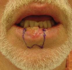
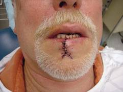
The lower vermilion is the most affected target of solar radiation injury. In cases of premalignant lesions such as actinic cheilitis or leukoplakia, Dr. Keefe, a plastic surgeon with the division of head and neck surgery at Sharp Rees-Stealy Medical Group in San Diego, said he often performs a total vermilionectomy (lip shave). This involves resection from the white roll to the contact area with opposite lip. "Primary closure is possible," he said. "You can get tension and dehiscence and flattening of the lip, but generally it heals up pretty well. An option for vermilion reconstruction of larger defects is the buccal mucosal advancement flap, which involves elevating the mucosa deep to salivary glands and superficial to the orbicularis oris muscle."
An advantage of treating the lower lip is that there is increased soft tissue laxity and there is no Cupid’s bow, philtrum, or nose, "so it’s nice that there are no dominant central structures," he said. "The downside is that you have to be mindful of the effect of gravity on the repair, so there is a greater need for tone to prevent drooling and incompetence."
He recommends a staged approach based on the extent of the defect and the age of the patient. For small defects (those less than one-third of the lip size) he uses primary closure. Options for medium defects (those that involve one-third to two-thirds of the lip size) include the Estlander flap, the Abbe flap, Bernard Burow’s procedure, the Karapandzic flap, and the stairstep repair, while the options for large defects (those that involve more than two-thirds of the lip size) include Bernard Burow’s procedure, the Karapandzic flap, and the free flap. "You have a lot of tools, depending on what you feel comfortable with," Dr. Keefe said.
Upper Lip
Cancerous tumors of the upper lip are less common, "but there are some unique structures to pay attention to, including the nose, columella, Cupid’s bow, and the philtrum," he said. "In men there’s a hair-bearing skin issue, but scars can be disguised in a mustache."
The aesthetic subunits to keep in mind, he continued, are the medial subunit, which is one-half of the philtrum, and the lateral subunit, which consists of the philtral column, the nostril sill, the alar base, and the nasolabial crease. Primary closure is used for upper lip defects that involve less than one-third of the lip size. "You can make some perialar crescentic skin excisions, which can help advance things," Dr. Keefe said.
For centrally located medium-sized defects of the upper lip, he often uses primary closure with perialar crescentic skin excisions. "If it’s greater than one-half of the lip size, you can add an Abbe flap," he said. "That’s nice because that recreates the philtrum area."
For medially located medium-sized defects of the upper lip, "you can use the Abbe flap if the commissure is not involved and the Estlander flap if the commissure is involved."
Options for cases with large defects and adequate cheek tissue, he said, include the reverse Karapandzic flap, the reverse fan flap, inverted Bernard Burow’s procedure, superiorly based cheek flaps, and the bilateral levator anguli oris flap combined with the Abbe flap. Options for cases with inadequate cheek tissue include the distal pedicle flap and the free flap.
Repair Risks
A lot of these patients have medical problems," he said. "When you do your first injection to resect the tumor or put the lip back together, make sure you don’t cause a myocardial infarction. Generally you should be comfortable with patients who have an INR [International Normalized Ratio] of 2.5 and below."
For patients with large cancerous tumors of the lip, be mindful of lymphatic drainage, because larger tumors have an increased risk of metastases, said Dr. Keefe. For tumors larger than 2 cm in length or 6 mm in spread, or if there is perineural spread, the patient should be referred for radiation therapy.
Dr. Keefe said that he had no relevant financial conflicts to disclose.
SAN DIEGO – Principles for lip repair are based on size and location of the defect, etiology of the lesions, and patient age and gender, said Dr. Michael A. Keefe.
Surgical goals of lip reconstruction are to cover the skin and oral lining, leave a semblance of a vermilion and an adequate stomal diameter, make sure sensation is intact, and ensure that the patient has a competent oral sphincter. "The vermilion is the most visible component of the lips, and it’s also the sensory unit of the lip," Dr. Keefe said at a meeting on superficial anatomy and cutaneous surgery. The meeting was sponsored by the University of California, San Diego, School of Medicine and the Scripps Clinic.
"Scars are very well hidden at the vermilion-cutaneous border. If you have to cross the vermilion-cutaneous junction, cross at 90 degrees."
Lower Lip
The lower vermilion is the most affected target of solar radiation injury. In cases of premalignant lesions such as actinic cheilitis or leukoplakia, Dr. Keefe, a plastic surgeon with the division of head and neck surgery at Sharp Rees-Stealy Medical Group in San Diego, said he often performs a total vermilionectomy (lip shave). This involves resection from the white roll to the contact area with opposite lip. "Primary closure is possible," he said. "You can get tension and dehiscence and flattening of the lip, but generally it heals up pretty well. An option for vermilion reconstruction of larger defects is the buccal mucosal advancement flap, which involves elevating the mucosa deep to salivary glands and superficial to the orbicularis oris muscle."
An advantage of treating the lower lip is that there is increased soft tissue laxity and there is no Cupid’s bow, philtrum, or nose, "so it’s nice that there are no dominant central structures," he said. "The downside is that you have to be mindful of the effect of gravity on the repair, so there is a greater need for tone to prevent drooling and incompetence."
He recommends a staged approach based on the extent of the defect and the age of the patient. For small defects (those less than one-third of the lip size) he uses primary closure. Options for medium defects (those that involve one-third to two-thirds of the lip size) include the Estlander flap, the Abbe flap, Bernard Burow’s procedure, the Karapandzic flap, and the stairstep repair, while the options for large defects (those that involve more than two-thirds of the lip size) include Bernard Burow’s procedure, the Karapandzic flap, and the free flap. "You have a lot of tools, depending on what you feel comfortable with," Dr. Keefe said.
Upper Lip
Cancerous tumors of the upper lip are less common, "but there are some unique structures to pay attention to, including the nose, columella, Cupid’s bow, and the philtrum," he said. "In men there’s a hair-bearing skin issue, but scars can be disguised in a mustache."
The aesthetic subunits to keep in mind, he continued, are the medial subunit, which is one-half of the philtrum, and the lateral subunit, which consists of the philtral column, the nostril sill, the alar base, and the nasolabial crease. Primary closure is used for upper lip defects that involve less than one-third of the lip size. "You can make some perialar crescentic skin excisions, which can help advance things," Dr. Keefe said.
For centrally located medium-sized defects of the upper lip, he often uses primary closure with perialar crescentic skin excisions. "If it’s greater than one-half of the lip size, you can add an Abbe flap," he said. "That’s nice because that recreates the philtrum area."
For medially located medium-sized defects of the upper lip, "you can use the Abbe flap if the commissure is not involved and the Estlander flap if the commissure is involved."
Options for cases with large defects and adequate cheek tissue, he said, include the reverse Karapandzic flap, the reverse fan flap, inverted Bernard Burow’s procedure, superiorly based cheek flaps, and the bilateral levator anguli oris flap combined with the Abbe flap. Options for cases with inadequate cheek tissue include the distal pedicle flap and the free flap.
Repair Risks
A lot of these patients have medical problems," he said. "When you do your first injection to resect the tumor or put the lip back together, make sure you don’t cause a myocardial infarction. Generally you should be comfortable with patients who have an INR [International Normalized Ratio] of 2.5 and below."
For patients with large cancerous tumors of the lip, be mindful of lymphatic drainage, because larger tumors have an increased risk of metastases, said Dr. Keefe. For tumors larger than 2 cm in length or 6 mm in spread, or if there is perineural spread, the patient should be referred for radiation therapy.
Dr. Keefe said that he had no relevant financial conflicts to disclose.
AT A MEETING ON SUPERFICIAL ANATOMY AND CUTANEOUS SURGERY
Eyeglasses May Offer Periocular AK Protection
RALEIGH, N.C. – Wearing glasses was associated with a significantly lower rate of developing actinic keratoses on periocular skin, according to the results of a large multicenter clinical trial.
During an average 4-year prospective follow-up conducted at 6-month intervals in the Veterans Affairs Topical Tretinoin Chemoprevention Trial, the 1,131 elderly participants, most of whom were male, developed 3,291 periocular actinic keratoses.
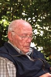
Patients who didn’t regularly wear eyeglasses had a 40% higher rate of developing AKs on periocular skin. This is consistent with reports in the ophthalmologic literature that eyeglasses can reduce the flux of UV radiation filtered to the periocular area, according to Dr. Kachiu C. Lee, a dermatology resident at Brown University in Providence, R.I.
Her hypothesis was that eyeglasses wearers would also have a lower rate of periocular nonmelanoma skin cancer. However, this wasn’t borne out, possibly because too few of the malignancies occurred to be able to demonstrate a protective effect, she noted. Although all suspected skin cancers were biopsied for histologic diagnosis, only 19 periocular squamous cell carcinomas and 71 periocular basal cell carcinomas occurred during follow-up.
A significant study limitation was that patients weren’t surveyed as to their use of sunglasses. "That’s a huge potential confounding variable," Dr. Lee said.
The study was sponsored by the U.S. Department of Veterans Affairs. Dr. Lee reported having no financial conflicts.
RALEIGH, N.C. – Wearing glasses was associated with a significantly lower rate of developing actinic keratoses on periocular skin, according to the results of a large multicenter clinical trial.
During an average 4-year prospective follow-up conducted at 6-month intervals in the Veterans Affairs Topical Tretinoin Chemoprevention Trial, the 1,131 elderly participants, most of whom were male, developed 3,291 periocular actinic keratoses.
Patients who didn’t regularly wear eyeglasses had a 40% higher rate of developing AKs on periocular skin. This is consistent with reports in the ophthalmologic literature that eyeglasses can reduce the flux of UV radiation filtered to the periocular area, according to Dr. Kachiu C. Lee, a dermatology resident at Brown University in Providence, R.I.
Her hypothesis was that eyeglasses wearers would also have a lower rate of periocular nonmelanoma skin cancer. However, this wasn’t borne out, possibly because too few of the malignancies occurred to be able to demonstrate a protective effect, she noted. Although all suspected skin cancers were biopsied for histologic diagnosis, only 19 periocular squamous cell carcinomas and 71 periocular basal cell carcinomas occurred during follow-up.
A significant study limitation was that patients weren’t surveyed as to their use of sunglasses. "That’s a huge potential confounding variable," Dr. Lee said.
The study was sponsored by the U.S. Department of Veterans Affairs. Dr. Lee reported having no financial conflicts.
AT THE ANNUAL MEETING OF THE SOCIETY FOR INVESTIGATIVE DERMATOLOGY
Major Finding: Patients who didn’t regularly wear eyeglasses had a 40% higher rate of developing AKs on periocular skin.
Data Source: A 4-year prospective study of 1,131 elderly participants, most of whom were male, who developed 3,291 periocular actinic keratoses.
Disclosures: The study was sponsored by the U.S. Department of Veterans Affairs. Dr. Lee reported having no financial conflicts.
Treatment of Actinic Keratoses

What Is the Role of Field-Directed Therapy in the Treatment of Actinic Keratosis? Part 2: Commonly Used Field-Directed and Lesion-Directed Therapies
BRAF-Plus-MEK Inhibition Slows Melanoma
Combining the BRAF inhibitor dabrafenib with the MEK inhibitor trametinib dramatically delays metastatic melanoma progression without the skin toxicities associated with vemurafenib therapy.
Median progression-free survival reached 10.8 months in the subset of 24 patients given the recommended dose of the two oral experimental agents in the dose-escalation portion of a phase I/II trial involving 77 patients without prior therapy targeting the BRAF kinase gene. The median for the entire cohort was 7.4 months, which was said to be comparable to results from past trials of single-agent vemurafenib.
Moreover, there were fewer dermatologic side effects than with any BRAF inhibitor alone seen to date, Dr. Jeffrey S. Weber said during a press briefing in advance of the upcoming annual meeting of the American Society of Clinical Oncology.
"Obviously, we have to be cautious. It’s only a cohort of 24 patients, but it looks extremely encouraging," he said.
Overall, cutaneous squamous cell carcinoma occurred in 3% of patients, which compares favorably with a 15%-20% incidence with dabrafenib and other BRAF inhibitors, said Dr. Weber, director of the Donald A. Adam Comprehensive Melanoma Research Center at the Moffitt Cancer Center in Tampa.
Similarly, actinic keratosis occurred in 5% of patients and skin papilloma in 2%, compared with a 20%-40% incidence seen with BRAF-targeted monotherapy. Skin rashes occurred in 22%, but the acneform rash often seen with MEK (MAP/ERK kinase) inhibitors was essentially absent in these patients, he said.
Notably, grade 3 or worse squamous cell carcinoma was reported in 12% of patients given the oral BRAF inhibitor vemurafenib (Zelboraf) in the pivotal BRIM-3 (BRAF Inhibitor in Melanoma-3) trial. Vemurafenib was approved last August for the first-line treatment of both metastatic and unresectable melanomas with V600E mutations in the BRAF gene, a mutation that occurs in roughly half of melanomas.
(Data to be presented at ASCO will show that median overall survival reached 13.2 months with vemurafenib vs 9.6 months with dacarbazine chemotherapy, according to Emmy Wang, senior manager, corporate relations at Genentech. Overall, up to 24% of patients in clinical trials experienced squamous cell carcinoma, which was easily treated, she noted.)*
The dramatic reduction in dermatologic toxicity observed in the current trial was offset, however, by a corresponding increase in pyrexia. Grade 3 or 4 pyrexia, which is relatively uncommon with a BRAF inhibitor alone, was observed in 8% of patients and led to dose reductions or delays in 23% of those patients, Dr. Weber acknowledged.
Other grade 3/4 events included nausea in 34% of patients, fatigue in 37%, and chills in 38% of patients, leading to dose reductions in 10%.
The investigators were initially surprised that combining BRAF and MEK inhibition reduced skin toxicity. But as evidence began to accumulate on BRAF inhibition in normal cells, Dr. Weber said they and other researchers realized there is a paradoxical activation of the MAP (mitogen-activated protein) kinase pathway through promotion of c-Raf signaling, which then leads down that pathway. If activation can be blocked with a MEK inhibitor, however, that would lead to a decrease in the off-target effects on normal cells that occur with a BRAF inhibitor, he explained.
ASCO president-elect Dr. Sandra M. Swain, who comoderated the press conference, said the findings show that researchers are finding more creative ways to effectively treat one of the most challenging cancers.
"We know cancers are smart," said Dr. Swain, medical director of the Cancer Institute at Washington Hospital Center in Washington, D.C. "They find mechanisms to escape or work around pathways, and in this case we are seeing a very innovative approach that ostensibly blocks off some of these side pathways. This is very exciting research."
The current analysis focused on 77 of 125 patients with V600 BRAF-mutant solid tumors enrolled in the phase I/II trial who were treated with four escalating doses of dabrafenib and trametinib, a MEK 1/2 inhibitor. They all had measurable disease according to RECIST criteria, 91% had V600E-mutant tumors, and 26% had prior brain metastases. Their mean age was 52 years.
The subset of 24 patients with the longest progression-free survival received the recommended dose of twice-daily dabrafenib 150 mg and daily trametinib 2 mg, which will be tested in a phase III trial, Dr. Weber said.
Among all 77 patients, responses were observed in 44 (57%), including 6 complete and 38 partial responses. Among the 24 patients on the recommended dose, the response rate reached 63%, including 2 complete responses and 13 partial responses, with the remainder all having stable disease.
Survival data on the cohort will be reported at a future date, he said. Results are also anticipated from the trial’s expansion cohort that includes patients with prior BRAF inhibition therapy as well as patients with BRAF-mutant colorectal cancer.
The abstract can be viewed at www.abstract.asco.org, and will be formally presented at ASCO at 3:30 p.m. June 4.
The trial was funded by GlaxoSmithKline. Dr. Weber reports consulting for and receiving honoraria and research funding from GSK. His coauthors report similar relationships, as well as employment/leadership positions and stock ownership with GSK.
* Updated: This paragraph was added on May 18, 2012.
FROM THE ANNUAL MEETING OF THE AMERICAN SOCIETY OF CLINICAL ONCOLOGY
Photodamage Effectively Treated With Quasi-Ablative Approach
KISSIMMEE, FLA. – The nonablative 1540-nm fractional laser can be safely converted to a quasi-ablative device for treating facial actinic keratoses and photodamage, according to Dr. Moshe Lapidoth.
By slightly modifying technique, a 50%-75% improvement in actinic keratoses and photodamage was noted in 17 patients who underwent two to three treatments at 4-week intervals, Dr. Lapidoth reported at the annual meeting of the American Society for Laser Medicine and Surgery. The laser was used in noncontact mode hovering 5-10 mm above the skin rather than in contact mode.
The patients had actinic keratoses and photodamage requiring ablative treatment of the epidermis. Treatment was applied using a fluence of 75 J/cm2, a 15-ms pulse duration, and a 10-mm spot size. Two blinded assessors and the participants evaluated clinical improvement at 3 months after the final treatment using a quartile grading scale (Lasers Med. Sci. 2012 April 27 [doi: 10.1007/s10103-012-1103-6])
A score of 0 was associated with no improvement; a score of 1 with 1%-25% improvement, a score of 2 with 26%-50% improvement, a score of 3 with 51%-75% improvement, and a score of 4 with 76%-100% improvement. The mean score for actinic keratoses was 3.4, and for skin appearance was 3.2, said Dr. Lapidoth, head of the laser department at Rabin Medical Center in Israel.
Side effects after each treatment included erythema, mild edema, erosion in two patients, and mild desquamation, but no scarring or postinflammatory pigmentary changes occurred, he said.
Although more current fractional laser devices are designed to be either ablative or nonablative, the 1540-nm fractional laser was designed to be nonablative.
"The question is, ‘Can you take a 1540-nm laser, which is totally nonablative, and turn it to be ablative or quasi-ablative?’ and the answer is, ‘yes,’ Dr. Lapidoth said.
This treatment approach can be used in conjunction with contact mode, he added, explaining that the nonablative contact mode can be used first to target the dermis for treatment of fine wrinkles and scars, followed by the ablative noncontact mode to treat actinic keratoses and resistant lentigines and other lesions.
Dr. Lapidoth reported having no disclosures.
FROM THE ANNUAL MEETING OF THE AMERICAN SOCIETY FOR LASER MEDICINE AND SURGERY
Major Finding: A quasi-ablative technique using a nonablative 1540-nm fractional laser resulted in a 50%-75% improvement in actinic keratoses and skin appearance in 17 patients.
Data Source: This was a prospective study of 17 patients treated.
Disclosures: Dr. Lapidoth reported having no disclosures.