User login
Better Atopy Outcome Measures Demanded
RALEIGH, N.C. – Atopic dermatitis is a disease that until now has gone underappreciated by regulatory authorities, the pharmaceutical industry, and the general public, according to thought leaders determined to turn the situation around.
"The first and probably foremost reason atopic dermatitis has been somewhat ignored is that it is perceived as a childhood disease, and that makes development of drugs very, very difficult. First, you have to go through studies in adults and then you have to go through children," Dr. Lisa A. Beck noted during a special plenary session devoted to atopic dermatitis at the conference.

Another stumbling block is that researchers haven’t adequately documented the negative effects atopic dermatitis can have on daily life for patients and caregivers. Clinicians who care for atopy patients are aware of it. The outside world is not, observed Dr. Beck, professor of dermatology and medicine at the University of Rochester (N.Y.). Also, better diagnostic criteria for atopic dermatitis are needed. The diagnosis isn’t always as straightforward as in psoriasis, which is "the great comparator in our range of diseases," she said.
A damaging perception that’s particularly entrenched within the regulatory agencies is that atopic dermatitis is not high priority because it lacks serious comorbidities, Dr. Beck continued.
Research Barriers
"That’s something we come up against in atopic dermatitis all the time: the idea that it’s a disease of misery rather than a disease of mortality," agreed Dr. Neil Graham of Regeneron Pharmaceuticals in Tarrytown, N.Y. "However, our view is that human misery is something we should be trying to treat therapeutically, and that this has value both economically and to society. I think we’ve seen this accomplished successfully with psoriasis, which is the template we can use in atopic dermatitis."
Biopharmaceutical companies are interested in applying the psoriasis drug development template to atopic dermatitis. Regeneron is developing a biologic agent that simultaneously blocks interleukins-4 and -13 for the treatment of atopic dermatitis and other diseases whose predominant mechanism involves Th2-driven eosinophilic inflammation, including eosinophilic asthma, chronic sinusitis with nasal polyps, and conjunctival allergic disease. The company wants an agent that can be subcutaneously injected every week or two, noted Dr. Graham.
"We hope eventually to intervene in children and potentially interrupt the atopic march. It will probably take us many years to get there," he said.
Regulatory agencies will almost certainly require that clinical trials involving any biologic agent under development for atopic dermatitis initially be restricted to patients with moderate to severe disease.
That’s a problem, according to Dr. Graham, because the Food and Drug Administration defines the clinical severity of atopic dermatitis using the Investigator’s Global Assessment (IGA) scale: a limited tool that’s not up to the task of capturing the full impact of the disease. Better means of defining the moderate to severely affected patient subset are essential. Also, a standardized, clinically relevant measure of disease severity reduction in response to treatment is needed – something akin to the Psoriasis Area Severity Index (PASI) 50 or -75 as used in psoriasis.
Dr. Eric Simpson agreed that regulatory authorities haven’t devoted sufficient attention to study design requirements or definitions of therapeutic success. For instance, the FDA requires evidence of improvement in the IGA as the bar that must be cleared in order to obtain approval of drugs for atopic dermatitis. Yet the IGA has never been adequately validated, nor has the metric’s definition been standardized. Indeed, the definition changes over time, from one phase-III clinical trial to the next.

"The inter-relater reliability of the IGA is unknown. I think it could be a major issue," said Dr. Simpson of the Oregon Health and Science University, Portland.
He proposed that participants in the SID special session on atopic dermatitis create a position paper recommending better outcome measures to the FDA. "We need to work with the agency to do what’s best for good clinical research," he said.
Identifying Biomarkers
According to Dr. Graham, atopic dermatitis is on the radar of major biotech companies that have biologic agents for psoriasis. They are working to develop biologics for atopic dermatitis, targeting a variety of pathways including interleukins-5, -4, and -13, as well as IgE.
Reliable biomarkers are needed to make drug development in atopic dermatitis go quickly and efficiently. After all, biomarker data are now routinely incorporated into early-phase development of new drugs for psoriasis, and these data are included in support of new drug applications to the FDA. But objective biomarkers have been notably lacking in atopic dermatitis, he noted.
Dr. Emma Guttman-Yassky announced help is at hand. While efforts to identify blood biomarkers have proved deeply disappointing, skin biomarkers are another story. She and her coinvestigators have identified a set of reliable skin biomarkers of therapeutic response in atopic dermatitis patients.
"We believe that these biomarkers represent the molecular fingerprints of the disease and might be key to future development of targeted treatments," said Dr. Guttman-Yassky, director of occupational and contact dermatitis at Mount Sinai Medical Center, New York.
She and her coinvestigators took lesional and nonlesional skin biopsies from 12 atopic dermatitis patients before and after undergoing narrow-band UVB phototherapy three times weekly for 12 weeks, a regimen the patients responded to with a mean 81% reduction in SCORAD index scores. Reversal of epidermal hyperplasia and abnormal keratinocyte differentiation – the histologic hallmarks of atopic dermatitis – was associated with elimination of inflammatory leukocytes and Th2-associated cytokines and chemokines (J. Allergy Clin. Immunol. 2011;128:583-93).
Among the skin biomarkers of atopic dermatitis that reversed with effective therapy were inflammatory dendritic epidermal cells, myeloid and plasmacytoid dendritic cells, CD3+ T cells, and interleukins-4 and -13.
This study, as well as another by Dr. Guttman-Yassky and her colleagues (in press), demonstrated a major role for interleukin-22 in atopic dermatitis. The cytokine was markedly overexpressed in lesional skin, with resultant upregulation of the antimicrobial protein’s S100A7, S100A8, and S100A9. After narrow-band UVB therapy, expression of IL-22 expression was downregulated, as were the IL-22-induced chemokines S100A7-9.
Expression of the three antimicrobial proteins in lesional skin also appears to be useful as a biomarker defining acute disease. Their levels jump within 3 days after onset of acute atopic dermatitis, according to Dr. Guttman-Yassky.
"Are we there yet in terms of biomarkers in atopic dermatitis? That’s the million-dollar question. Regarding biomarker availability, I think yes. We now have biomarkers that are highly informative on the reversal of disease pathology with treatment," she said. "I believe that the next step is clinical trials with specific immune antagonists that incorporate biomarkers of response. I think biomarkers are key if we want to know if new therapies really work. This will enhance early decisions and reduce the cost of developing new treatments."
An important implication of the narrow-band UVB/biomarker study in terms of atopic dermatitis pathogenesis is that the data argue against a fixed genetic phenotype, since the atopic dermatitis epidermal phenotype was shown to be reversed with broad-based immune-targeted phototherapy. Instead, the study results argue in favor of what Dr. Guttman-Yassky called "the inside-out hypothesis" of atopic dermatitis, which postulates that the epidermal abnormalities that define the disease are caused by underlying immune activation.
Her studies were supported by the National Institutes of Health.
HOME Project Seeks Standardization
During the same session, Dr. Hywel C. Williams provided an update on a major international project called Harmonising Outcome Measures for Eczema (HOME). The goal of the HOME project is to develop a consensus on a core set of outcome measures to be used in all atopic dermatitis clinical research. This should make future studies easier to compare, contrast, and synthesize in meta-analyses.
A minimum set of core outcome measures in atopic dermatitis is needed because the situation can be chaotic, noted Dr. Williams, professor of dermatology and director of the Center for Evidence-Based Dermatology at the University of Nottingham (U.K.).
"There are more than 20 named scales for atopic dermatitis, including SCORAD, POEM, SASSAD, ADASI, ADAM, EASI, and the FSSS. Some have been only partly tested. Many have not been tested at all. At international meetings I see people shouting at each other, trying to communicate what the results of their studies mean. Some people are in the SCORAD camp, some are in the SASSAD camp, others are in the EASI camp. How can we possibly communicate? This is a shameful situation which we really have to put right if we are to progress," he said.
The HOME project is modeled after Outcome Measures in Rheumatology, or OMERACT, an international consensus group that meets every 2 years with the goal of raising the quality of rheumatologic research. HOME comprises atopic dermatitis researchers, clinical experts, journal editors, patient advocates, and representatives from the European regulatory agency. The FDA was invited but didn’t attend last year’s HOME meeting in Amsterdam, where working groups were formed to identify the best evidence-based instruments for assessing atopic disease signs, symptoms, flares, quality of life, and treatment efficacy and safety. The HOME group will meet next year in San Diego on April 6-7.
Aside from Dr. Graham, a Regeneron employee, the other speakers in the special session reported receiving research grants from various sources but declared having no conflicts of interest with regard to their presentations.
RALEIGH, N.C. – Atopic dermatitis is a disease that until now has gone underappreciated by regulatory authorities, the pharmaceutical industry, and the general public, according to thought leaders determined to turn the situation around.
"The first and probably foremost reason atopic dermatitis has been somewhat ignored is that it is perceived as a childhood disease, and that makes development of drugs very, very difficult. First, you have to go through studies in adults and then you have to go through children," Dr. Lisa A. Beck noted during a special plenary session devoted to atopic dermatitis at the conference.
Another stumbling block is that researchers haven’t adequately documented the negative effects atopic dermatitis can have on daily life for patients and caregivers. Clinicians who care for atopy patients are aware of it. The outside world is not, observed Dr. Beck, professor of dermatology and medicine at the University of Rochester (N.Y.). Also, better diagnostic criteria for atopic dermatitis are needed. The diagnosis isn’t always as straightforward as in psoriasis, which is "the great comparator in our range of diseases," she said.
A damaging perception that’s particularly entrenched within the regulatory agencies is that atopic dermatitis is not high priority because it lacks serious comorbidities, Dr. Beck continued.
Research Barriers
"That’s something we come up against in atopic dermatitis all the time: the idea that it’s a disease of misery rather than a disease of mortality," agreed Dr. Neil Graham of Regeneron Pharmaceuticals in Tarrytown, N.Y. "However, our view is that human misery is something we should be trying to treat therapeutically, and that this has value both economically and to society. I think we’ve seen this accomplished successfully with psoriasis, which is the template we can use in atopic dermatitis."
Biopharmaceutical companies are interested in applying the psoriasis drug development template to atopic dermatitis. Regeneron is developing a biologic agent that simultaneously blocks interleukins-4 and -13 for the treatment of atopic dermatitis and other diseases whose predominant mechanism involves Th2-driven eosinophilic inflammation, including eosinophilic asthma, chronic sinusitis with nasal polyps, and conjunctival allergic disease. The company wants an agent that can be subcutaneously injected every week or two, noted Dr. Graham.
"We hope eventually to intervene in children and potentially interrupt the atopic march. It will probably take us many years to get there," he said.
Regulatory agencies will almost certainly require that clinical trials involving any biologic agent under development for atopic dermatitis initially be restricted to patients with moderate to severe disease.
That’s a problem, according to Dr. Graham, because the Food and Drug Administration defines the clinical severity of atopic dermatitis using the Investigator’s Global Assessment (IGA) scale: a limited tool that’s not up to the task of capturing the full impact of the disease. Better means of defining the moderate to severely affected patient subset are essential. Also, a standardized, clinically relevant measure of disease severity reduction in response to treatment is needed – something akin to the Psoriasis Area Severity Index (PASI) 50 or -75 as used in psoriasis.
Dr. Eric Simpson agreed that regulatory authorities haven’t devoted sufficient attention to study design requirements or definitions of therapeutic success. For instance, the FDA requires evidence of improvement in the IGA as the bar that must be cleared in order to obtain approval of drugs for atopic dermatitis. Yet the IGA has never been adequately validated, nor has the metric’s definition been standardized. Indeed, the definition changes over time, from one phase-III clinical trial to the next.
"The inter-relater reliability of the IGA is unknown. I think it could be a major issue," said Dr. Simpson of the Oregon Health and Science University, Portland.
He proposed that participants in the SID special session on atopic dermatitis create a position paper recommending better outcome measures to the FDA. "We need to work with the agency to do what’s best for good clinical research," he said.
Identifying Biomarkers
According to Dr. Graham, atopic dermatitis is on the radar of major biotech companies that have biologic agents for psoriasis. They are working to develop biologics for atopic dermatitis, targeting a variety of pathways including interleukins-5, -4, and -13, as well as IgE.
Reliable biomarkers are needed to make drug development in atopic dermatitis go quickly and efficiently. After all, biomarker data are now routinely incorporated into early-phase development of new drugs for psoriasis, and these data are included in support of new drug applications to the FDA. But objective biomarkers have been notably lacking in atopic dermatitis, he noted.
Dr. Emma Guttman-Yassky announced help is at hand. While efforts to identify blood biomarkers have proved deeply disappointing, skin biomarkers are another story. She and her coinvestigators have identified a set of reliable skin biomarkers of therapeutic response in atopic dermatitis patients.
"We believe that these biomarkers represent the molecular fingerprints of the disease and might be key to future development of targeted treatments," said Dr. Guttman-Yassky, director of occupational and contact dermatitis at Mount Sinai Medical Center, New York.
She and her coinvestigators took lesional and nonlesional skin biopsies from 12 atopic dermatitis patients before and after undergoing narrow-band UVB phototherapy three times weekly for 12 weeks, a regimen the patients responded to with a mean 81% reduction in SCORAD index scores. Reversal of epidermal hyperplasia and abnormal keratinocyte differentiation – the histologic hallmarks of atopic dermatitis – was associated with elimination of inflammatory leukocytes and Th2-associated cytokines and chemokines (J. Allergy Clin. Immunol. 2011;128:583-93).
Among the skin biomarkers of atopic dermatitis that reversed with effective therapy were inflammatory dendritic epidermal cells, myeloid and plasmacytoid dendritic cells, CD3+ T cells, and interleukins-4 and -13.
This study, as well as another by Dr. Guttman-Yassky and her colleagues (in press), demonstrated a major role for interleukin-22 in atopic dermatitis. The cytokine was markedly overexpressed in lesional skin, with resultant upregulation of the antimicrobial protein’s S100A7, S100A8, and S100A9. After narrow-band UVB therapy, expression of IL-22 expression was downregulated, as were the IL-22-induced chemokines S100A7-9.
Expression of the three antimicrobial proteins in lesional skin also appears to be useful as a biomarker defining acute disease. Their levels jump within 3 days after onset of acute atopic dermatitis, according to Dr. Guttman-Yassky.
"Are we there yet in terms of biomarkers in atopic dermatitis? That’s the million-dollar question. Regarding biomarker availability, I think yes. We now have biomarkers that are highly informative on the reversal of disease pathology with treatment," she said. "I believe that the next step is clinical trials with specific immune antagonists that incorporate biomarkers of response. I think biomarkers are key if we want to know if new therapies really work. This will enhance early decisions and reduce the cost of developing new treatments."
An important implication of the narrow-band UVB/biomarker study in terms of atopic dermatitis pathogenesis is that the data argue against a fixed genetic phenotype, since the atopic dermatitis epidermal phenotype was shown to be reversed with broad-based immune-targeted phototherapy. Instead, the study results argue in favor of what Dr. Guttman-Yassky called "the inside-out hypothesis" of atopic dermatitis, which postulates that the epidermal abnormalities that define the disease are caused by underlying immune activation.
Her studies were supported by the National Institutes of Health.
HOME Project Seeks Standardization
During the same session, Dr. Hywel C. Williams provided an update on a major international project called Harmonising Outcome Measures for Eczema (HOME). The goal of the HOME project is to develop a consensus on a core set of outcome measures to be used in all atopic dermatitis clinical research. This should make future studies easier to compare, contrast, and synthesize in meta-analyses.
A minimum set of core outcome measures in atopic dermatitis is needed because the situation can be chaotic, noted Dr. Williams, professor of dermatology and director of the Center for Evidence-Based Dermatology at the University of Nottingham (U.K.).
"There are more than 20 named scales for atopic dermatitis, including SCORAD, POEM, SASSAD, ADASI, ADAM, EASI, and the FSSS. Some have been only partly tested. Many have not been tested at all. At international meetings I see people shouting at each other, trying to communicate what the results of their studies mean. Some people are in the SCORAD camp, some are in the SASSAD camp, others are in the EASI camp. How can we possibly communicate? This is a shameful situation which we really have to put right if we are to progress," he said.
The HOME project is modeled after Outcome Measures in Rheumatology, or OMERACT, an international consensus group that meets every 2 years with the goal of raising the quality of rheumatologic research. HOME comprises atopic dermatitis researchers, clinical experts, journal editors, patient advocates, and representatives from the European regulatory agency. The FDA was invited but didn’t attend last year’s HOME meeting in Amsterdam, where working groups were formed to identify the best evidence-based instruments for assessing atopic disease signs, symptoms, flares, quality of life, and treatment efficacy and safety. The HOME group will meet next year in San Diego on April 6-7.
Aside from Dr. Graham, a Regeneron employee, the other speakers in the special session reported receiving research grants from various sources but declared having no conflicts of interest with regard to their presentations.
EXPERT ANALYSIS FROM THE ANNUAL MEETING OF THE SOCIETY FOR INVESTIGATIVE DERMATOLOGY

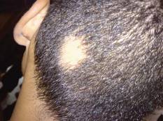
New Era in Alopecia Areata Therapy Beckons
RALEIGH, N.C. – Recent genetic insights into the roots of alopecia areata have fostered the development of new therapeutic targets, with clinical trials offering promising results.
The investigational drugs are, for the first time, addressing the underlying disease mechanism, Angela M. Christiano, Ph.D., said at the annual meeting of the Society for Investigative Dermatology.

Alopecia areata affects roughly 5.3 million Americans of all ages and ethnic groups. The dramatic hair loss exacts an underappreciated, often devastating toll in terms of patient self-esteem and quality of life. To date, treatment has been unsatisfactory, with intralesional injections of glucocorticoids being the mainstay. Wigs are big.
But all of that may be about to change. "Our translational studies offer us the beginning of a therapeutic arsenal," said Dr. Christiano, professor of dermatology and genetics at Columbia University in New York.
She and her colleagues have identified interleukin-15 as a novel and highly promising therapeutic target in alopecia areata.
Interleukin-15 is required for the growth and sustenance of the natural killer-type CD8+ cells that surround the growing end of the hair follicle during active disease. This dense infiltrate of activated T cells, known as the "swarm of bees," is the pathognomonic finding in alopecia areata.
Interleukin-15 is an attractive therapeutic target because it can be addressed along the IL-15 signaling pathway by using Janus kinase inhibitors. Dr. Christiano and her coworkers have found evidence that both the oral and compounded topical formulations of tofacitinib and ruxolitinib are effective for the prevention and treatment of alopecia areata in a mouse model of the disease.
Tofacitinib inhibits the Janus kinase 3 (JAK-3) enzyme located along the IL-15 signaling pathway. The investigational oral agent recently received a thumbs-up recommendation for the treatment of rheumatoid arthritis from a Food and Drug Administration advisory panel.
In Dr. Christiano’s mouse model of alopecia areata, when tofacitinib was administered when mice received diseased grafts, 100% of the animals retained their hair. The same phenomenon occurred when the mice were treated prophylactically with oral ruxolitinib (Jakafi), a JAK1/2 inhibitor marketed for the treatment of myelofibrosis and under investigation for the treatment of other diseases.
Dr. Christiano and her coworkers also formulated the two JAK inhibitors into a 0.5% cream for topical application. The topical agents prevented the development of alopecia areata in mice, and they triggered hair regrowth when applied to mice with established disease. At the molecular level, these drugs resulted in reversal of a pathologic interferon-gamma gene expression signature.
"The follicular expression of molecules changes back to an immune-privileged phenotype. We’re very encouraged by what we’ve seen so far with this," she said.
Alopecia has long been recognized as having a strong genetic component. The relative risk of the disease in first-degree relatives of affected individuals is increased 10-fold. Twin concordance studies have demonstrated that when one monozygotic twin has alopecia areata, there is more than a 50% chance that the other twin will be affected, whereas there is zero concordance in dizygotic twin pairs.
It’s also well recognized in the research community that the normal hair follicle is one of the few tissues in the human body that enjoys a state of immune privilege protecting it from autoimmune attack. Normal hair follicles are cloaked from immune recognition. The collapse of this immune privilege is what allows the "swarm of bees" leading to alopecia areata.
"Notably, the stem cell compartment is spared from the disease, which gives patients the ability, even after many years of longstanding disease, to experience spontaneous regrowth for reasons we really don’t understand," Dr. Christiano explained.
While the JAK inhibitors haven’t made it to clinical studies, another novel therapy in alopecia areata will be the focus of a small clinical trial slated to begin in July. Abatacept (Orencia) is a biologic agent approved for the treatment of rheumatoid arthritis. It is also in ongoing clinical trials for other autoimmune diseases, including type 1 diabetes. Abatacept is a soluble fusion protein that serves as a selective costimulation modulator and T-cell activation suppressant. Last summer, the intravenously administered formulation of abatacept was joined by a subcutaneous version permitting patient self-treatment.
"That makes it more appealing for the treatment of alopecia areata," she noted.
Dr. Christiano’s research is funded by the National Institutes of Health, American Skin Association, and National Alopecia Areata Foundation. She reported having no financial conflicts.
RALEIGH, N.C. – Recent genetic insights into the roots of alopecia areata have fostered the development of new therapeutic targets, with clinical trials offering promising results.
The investigational drugs are, for the first time, addressing the underlying disease mechanism, Angela M. Christiano, Ph.D., said at the annual meeting of the Society for Investigative Dermatology.
Alopecia areata affects roughly 5.3 million Americans of all ages and ethnic groups. The dramatic hair loss exacts an underappreciated, often devastating toll in terms of patient self-esteem and quality of life. To date, treatment has been unsatisfactory, with intralesional injections of glucocorticoids being the mainstay. Wigs are big.
But all of that may be about to change. "Our translational studies offer us the beginning of a therapeutic arsenal," said Dr. Christiano, professor of dermatology and genetics at Columbia University in New York.
She and her colleagues have identified interleukin-15 as a novel and highly promising therapeutic target in alopecia areata.
Interleukin-15 is required for the growth and sustenance of the natural killer-type CD8+ cells that surround the growing end of the hair follicle during active disease. This dense infiltrate of activated T cells, known as the "swarm of bees," is the pathognomonic finding in alopecia areata.
Interleukin-15 is an attractive therapeutic target because it can be addressed along the IL-15 signaling pathway by using Janus kinase inhibitors. Dr. Christiano and her coworkers have found evidence that both the oral and compounded topical formulations of tofacitinib and ruxolitinib are effective for the prevention and treatment of alopecia areata in a mouse model of the disease.
Tofacitinib inhibits the Janus kinase 3 (JAK-3) enzyme located along the IL-15 signaling pathway. The investigational oral agent recently received a thumbs-up recommendation for the treatment of rheumatoid arthritis from a Food and Drug Administration advisory panel.
In Dr. Christiano’s mouse model of alopecia areata, when tofacitinib was administered when mice received diseased grafts, 100% of the animals retained their hair. The same phenomenon occurred when the mice were treated prophylactically with oral ruxolitinib (Jakafi), a JAK1/2 inhibitor marketed for the treatment of myelofibrosis and under investigation for the treatment of other diseases.
Dr. Christiano and her coworkers also formulated the two JAK inhibitors into a 0.5% cream for topical application. The topical agents prevented the development of alopecia areata in mice, and they triggered hair regrowth when applied to mice with established disease. At the molecular level, these drugs resulted in reversal of a pathologic interferon-gamma gene expression signature.
"The follicular expression of molecules changes back to an immune-privileged phenotype. We’re very encouraged by what we’ve seen so far with this," she said.
Alopecia has long been recognized as having a strong genetic component. The relative risk of the disease in first-degree relatives of affected individuals is increased 10-fold. Twin concordance studies have demonstrated that when one monozygotic twin has alopecia areata, there is more than a 50% chance that the other twin will be affected, whereas there is zero concordance in dizygotic twin pairs.
It’s also well recognized in the research community that the normal hair follicle is one of the few tissues in the human body that enjoys a state of immune privilege protecting it from autoimmune attack. Normal hair follicles are cloaked from immune recognition. The collapse of this immune privilege is what allows the "swarm of bees" leading to alopecia areata.
"Notably, the stem cell compartment is spared from the disease, which gives patients the ability, even after many years of longstanding disease, to experience spontaneous regrowth for reasons we really don’t understand," Dr. Christiano explained.
While the JAK inhibitors haven’t made it to clinical studies, another novel therapy in alopecia areata will be the focus of a small clinical trial slated to begin in July. Abatacept (Orencia) is a biologic agent approved for the treatment of rheumatoid arthritis. It is also in ongoing clinical trials for other autoimmune diseases, including type 1 diabetes. Abatacept is a soluble fusion protein that serves as a selective costimulation modulator and T-cell activation suppressant. Last summer, the intravenously administered formulation of abatacept was joined by a subcutaneous version permitting patient self-treatment.
"That makes it more appealing for the treatment of alopecia areata," she noted.
Dr. Christiano’s research is funded by the National Institutes of Health, American Skin Association, and National Alopecia Areata Foundation. She reported having no financial conflicts.
EXPERT ANALYSIS FROM THE ANNUAL MEETING OF THE SOCIETY FOR INVESTIGATIVE DERMATOLOGY

Drug Samples Found to Sway Acne Prescribing
RALEIGH, N.C. – Offering free drug samples to newly diagnosed acne patients was found to increase the likelihood of prescribing more expensive medications, according to a new study.
"The benefits of free sample distribution in dermatology clinics must be weighed against the significant negative impact that free samples have on prescribing and prescription costs. Clinical trials comparing the efficacy of new branded generic drugs with existing alternatives should increasingly be used to justify their increased retail cost," said study investigator Michael Hurley, a Stanford (Calif.) University medical student.
The investigators analyzed all prescriptions written for newly diagnosed acne patients in 2010 at two dermatology clinics, one in an academic medical center that does not allow samples, and the second at an affiliated neighborhood clinic that does.
At the no-samples clinic, 17% of prescriptions written by office-based dermatologists for acne patients at their initial office visit were for branded or branded-generic drugs, compared with a 74% rate at the neighborhood clinic allowing free samples.
The average prescription costs were also higher at the clinic that uses free samples. The average retail cost of the top 20 prescribed acne medications at each site (which collectively accounted for roughly 70% of all acne prescriptions) was $204 at the neighborhood clinic, compared with $70.49 at the clinic with no free samples. After the average number of prescriptions written per visit was taken into account, this amounted to a cost difference of about $260 per office visit, reported Mr. Hurley.
In a multivariate regression analysis accounting for patient characteristics and other potential confounding factors, dermatologists who provided free samples of acne drugs were 3.4-fold more likely to prescribe a branded or branded generic drug than a less expensive generic.
After identifying the marked difference in dermatologists’ acne drug prescribing patterns at the two clinics in Northern California, Mr. Hurley and his coinvestigators analyzed national data to establish trends.
Data from the National Disease and Therapeutic Index – a national survey of physician self-reported office visits, diagnoses, and treatments conducted by IMS Health – were assessed. The data showed that in 2010, 79% of all prescriptions written by office-based dermatologists for acne patients at their initial visit were for branded or branded-generic drugs.
The data also found that the proportion of acne prescriptions written with a free sample increased from 38% to 51% in the last decade. Prescriptions for branded generic medications rose similarly, most likely because of the increased use of free samples of those drugs, said Mr. Hurley. Meanwhile the percentage of acne prescriptions for generic drugs has remained flat, and in absolute numbers has actually decreased, he said.
Upon close scrutiny of dermatologists’ acne prescribing nationally in 2010, 2005, and 2001, Mr. Hurley concluded that although dermatologists’ drug preferences do change over time as new medications enter the market, their prescribing consistently remains closely related to what’s available as a free sample.
For example, during 2010 the top five acne medications prescribed by dermatologists were (in descending order) Epiduo, doxycycline hyclate, Metrogel, Solodyn, and Differin. The top five prescribed with a free sample were Epiduo, Metrogel, Solodyn, Ziana, and Oracea.
The top five agents prescribed overall in 2005 were Differin, Benzaclin, Duac, Retin-A-Micro, and doxycycline hyclate, whereas the top five prescribed with a free sample during that year were Differin, Duac, Benzaclin, Retin-A-Micro, and Metrogel.
Mr. Hurley reported having no financial conflicts.
AT THE ANNUAL MEETING OF THE SOCIETY FOR INVESTIGATIVE DERMATOLOGY
Major Finding: At the no-samples clinic, 17% of prescriptions written by office-based dermatologists for acne patients at their initial office visit were for branded or branded-generic drugs, compared with a 74% rate at the neighborhood clinic allowing free samples.
Data Source: All prescriptions written for newly diagnosed acne patients in 2010 at two dermatology clinics were analyzed, one in an academic medical center that does not allow samples, and the second at an affiliated neighborhood clinic that does.
Disclosures: Mr. Hurley reported having no financial conflicts.
Invasive SCC Rates Doubled in Last 20 Years
RALEIGH, N.C. – The incidence of invasive squamous cell carcinoma has more than doubled among U.S. nonphysician health professionals in the last 20 years, with marked sex differences evident in body-site distribution.
Dr. Khang Nguyen said his meta-analysis of data drawn from the Nurses' Health Study, the Nurses' Health Study II, and the Health Professionals Follow-Up Study was spurred by the fact that the National Cancer Institute’s SEER (Surveillance, Epidemiology and End Results) database and other national databases that record cancer statistics don’t track squamous cell carcinoma (SCC).
"Much of what we know about the epidemiology of squamous cell carcinoma comes from studies from the 1990s and earlier," noted Dr. Nguyen of Brigham and Women’s Hospital, Boston.
To help rectify this situation, he and his coinvestigators analyzed cases of pathologist-confirmed invasive SCC in the roughly 121,000 female participants in the Nurses’ Health Study, the 117,000 in the Nurses’ Health Study II, and 51,000 men in the Health Professionals Follow-Up Study. There were 1,580 cases in the Nurses’ Health Study, 468 in the Nurses’ Health Study II, and 1,194 in the Health Professionals Follow-Up Study.
Among participants in the Nurses’ Health Study, the incidence of invasive SCC climbed from 40 per 100,000 person-years in 1987 to a peak of 120 per 100,000 person-years in 2005 before declining to 80 per 100,000 in 2007. Rates were lower in the Nurses’ Health Study II, a younger cohort. Among men in the Health Professionals Follow-Up Study, the incidence was 80 per 100,000 person-years in 1987, peaking at more than 160 per 100,000 person-years in 2002, and then falling back slightly to 140 per 100,000 person-years in 2007.
Invasive SCC occurred more often in the head and neck region among men, and in the thigh, buttock, legs, and feet region in women.
The significant risk factors for the malignancy, which emerged from a multivariate logistic regression analysis, included a history of painful or blistering sunburns (with an associated 2.2-fold increased risk) and a history of six or more sunburns in childhood. Other independent risk factors for invasive SCC were a family history of melanoma and lighter hair color.
The Nurses’ Health Study, the Nurses’ Health Study II, and the Health Professionals Follow-Up Study were funded by the National Institutes of Health. Dr. Nguyen’s analysis was funded in part by a grant from the Doris Duke Charitable Foundation.
AT THE ANNUAL MEETING OF SOCIETY FOR INVESTIGATIVE DERMATOLOGY
Major Finding: Rates of invasive squamous cell carcinoma jumped two- to threefold between 1987 and 2007 in American nonphysician health professionals.
Data Source: This finding came from a meta-analysis of three large, prospective epidemiologic studies: the Nurses’ Health Study, the Nurses’ Health Study II, and the Health Professionals Follow-Up Study.
Disclosures: Dr. Nguyen’s analysis was funded in part by a grant from the Doris Duke Charitable Foundation.
Melanoma In Situ Diagnoses Ramping Up
RALEIGH, N.C. – The ratio of melanoma in situ to invasive melanoma jumped 12-fold among American health professionals during a recent 3-decade period.
Just how much of this steep rise in melanoma in situ diagnoses represents a true increase in the development of in situ lesions as opposed to the product of improved screening techniques or overdiagnosis due to medicolegal pressure remains a matter for conjecture. The new analysis of melanoma trends in the Nurses' Health Study and the Health Professionals Follow-Up Study provides evidence suggesting that all three factors may be involved, according to Erin X. Wei of Massachusetts General Hospital, Boston.
Her analysis included roughly 121,000 female nurses followed prospectively in the Nurses’ Health Study since 1976 and 51,000 men followed in the Health Professionals Follow-Up Study since 1986. During the follow-up period, new cases of invasive melanoma increased dramatically, but they were far outpaced by the rise in cases of melanoma in situ, she said at the annual meeting of the Society for Investigative Dermatology.
For example, while the ratio of in situ to invasive melanoma among women was 0.09:1 in 1977, by 2002 it had reached 1:1.
Support for the notion that the rise in melanoma in situ is due in part to overdiagnosis in response to medicolegal concerns comes from the finding that the bulk of the increase involves lesions reported in this study as having Not-Otherwise-Specified pathology. Lentigo maligna cases have remained relatively flat over time, she noted.
Also, in this large patient population, as well as in studies from elsewhere around the world, the increase in melanoma in situ has not been accompanied by a shift in the pattern of invasive melanoma thickness or by a reduction in melanoma mortality, as would be expected in response to a major increase in detection of invasive melanoma precursor lesions.
But perhaps in situ melanomas are often not precursors to invasive melanoma. If they were precursors, one would expect them to be diagnosed at a younger average age than invasive melanoma. Such was not the case in this analysis. Indeed, while roughly 40% of invasive melanomas were diagnosed prior to age 60, that was true for only 20% of melanoma in situ. Nurses diagnosed with melanoma in situ were an average of 6 years older than those diagnosed with invasive melanoma; among men, the difference was 2 years.
Melanoma in situ and invasive melanomas tended to differ in their anatomic distribution. Invasive melanomas were far less likely than in situ lesions to occur on the head and neck, and much more likely to occur on the lower extremities. This suggests some in situ melanomas are indolent and do not become malignant over time, Ms. Wei continued.
In support of the existence of a true biologic increase in the incidence of melanoma in situ, Ms. Wei and her coworkers found that older individuals were two- to threefold more likely to develop their lesions on chronically sun-exposed areas than on intermittently sun-exposed areas.
An intriguing gender difference in the anatomic distribution of melanoma in situ was evident. Among men, more than 98% of all melanoma in situ were found on the upper half of the body. In women, the lesions were more evenly distributed anatomically, with 27% of in situ lesions being found on the lower extremities.
The Nurses’ Health Study and Health Professionals Follow-up Study are funded by the National Institutes of Health. Ms. Wei reported having no financial conflicts.
FROM THE ANNUAL MEETING OF THE SOCIETY FOR INVESTIGATIVE DERMATOLOGY
Major Finding: Roughly 40% of invasive melanomas in a large prospectively followed population were diagnosed prior to age 60, compared with 20% of in situ melanomas. This casts doubt on the notion that melanoma in situ is necessarily a precursor lesion for invasive melanoma.
Data Source: This was an analysis of data from the prospective 121,000-subject Nurses’ Health Study and 51,000-man Health Professionals Follow-Up Study.
Disclosures: The Nurses’ Health Study and Health Professionals Follow-Up Study are funded by the National Institutes of Health. Ms. Wei reported having no financial conflicts.
Firmocidin Puts Big Hurt on MRSA
RALEIGH, N.C. – A novel small-molecule antibiotic produced by Staphylococcus epidermidis and dubbed "firmocidin" shows early promise as a potential treatment for methicillin-resistant S. aureus and other major skin pathogens, Dr. Teruaki Nakatsuji reported at the annual meeting of the Society for Investigative Dermatology.
S. epidermidis is a commensal microorganism that appears to act as "an antimicrobial shield" in healthy human skin, protecting against some of the major cutaneous bacterial pathogens. It accomplishes this by producing firmocidin, explained Dr. Nakatsuji of the University of California, San Diego.
He and his coworkers in the laboratory of Dr. Richard L. Gallo, professor of medicine and pediatrics and chief of the division of dermatology at the university, discovered the antibiotic by screening clinical isolates of S. epidermidis and identifying strains possessing pronounced antimicrobial activity. Next, they zeroed in on the antibiotic component. Using mass spectrometry and nuclear magnetic resonance spectrometry, they determined that the structure of the purified product didn’t match that of any known molecule. They named it firmocidin.
In in vitro studies, firmocidin effectively killed S. aureus, both group A and B streptococcus, and methicillin-resistant S. aureus. In these cell culture assays, firmocidin’s antimicrobial activity was stronger than that of benzoyl peroxide.
Importantly, however, firmocidin’s antibiotic activity was selective. It didn’t affect the growth of S. epidermidis or other commensal bacteria, nor did it affect human keratinocytes or sebocytes. Thus, it could potentially maintain a normal microbiome while inhibiting bacterial pathogens. Its mechanism of action involves inhibition of DNA synthesis by interfering with A-T base pair matching, he continued.
The University of California has applied for patent protection for firmocidin and has announced it is seeking business partners for its commercialization. Possibilities include its development as an antiseptic or a topical or systemic antibiotic for treatment of skin infections that does not disrupt the healthy skin microflora.
Dr. Nakatsuji reported having no financial conflicts.
AT THE ANNUAL MEETING OF THE SOCIETY FOR INVESTIGATIVE DERMATOLOGY
Major Finding: Firmocidin, a novel antibiotic produced by Staphylococcus epidermidis, showed roughly 99% killing rates against S. aureus, group A and group B streptococcus, and methicillin-resistant S. aureus, in in vitro studies.
Data Source: These were studies aimed at advancing future clinical applications for firmocidin in the treatment of bacterial skin infections.
Disclosures: Dr. Nakatsuji reported having no financial conflicts.
Ear Lesion Marks Atopic Disease Severity
RALEIGH, N.C. – The infra-auricular fissure present in the overwhelming majority of atopic dermatitis patients provides a simple and convenient guide to disease severity, according to the results of a prospective study.
Infra-auricular fissures are highly prevalent in atopic dermatitis patients of all ages and racial/ethnic groups and in both genders. And the form the lesion takes shows a good correlation with disease severity as measured with the Eczema Area and Severity Index (EASI) and other metrics, Dr. Gil Yosipovitch said at the annual meeting of the Society for Investigative Dermatology.
"I’m not saying this is an ideal test, but I think that it’s a very easy bedside marker that we could use in our clinics. We can’t use the EASI all the time in busy fast-track clinics," observed Dr. Yosipovitch, professor of dermatology at Wake Forest University in Winston-Salem, N.C.
He and his coinvestigators assigned an infra-auricular fissure score of 1 to atopic dermatitis patients with an infra-auricular line and a score of 2 in those with an infra-auricular line accompanied by erythema and a small fissure. Those patients with a deeper infra-auricular fissure with significant erosion got a 3.
An infra-auricular fissure was present in every one of 81 consecutive children and adults with clinician-diagnosed atopic dermatitis. Fifty-eight percent had an infra-auricular fissure score of 1, 27% had a 2, and 15% scored 3. The infra-auricular fissure score showed a strong correlation with results on the validated EASI, with a correlation coefficient of 0.6.
Moreover, among the 29 patients seen at follow-up 1-2 months later, reductions in visual analog scale itch intensity, EASI score, and Investigator Global Assessment scores in response to treatment showed a significant correlation with improvement in the infra-auricular fissure score.
Dr. Yosipovitch reported having no financial conflicts.
AT THE ANNUAL MEETING OF THE SOCIETY FOR INVESTIGATIVE DERMATOLOGY
How to Boost Skin Self-Exam Rates
RALEIGH, N.C. – Interactive computer-assisted patient education combined with a hands-on, dermatologist-led tutorial and monthly reminders proved successful in increasing the performance of skin self-examinations in a randomized controlled trial.
The intervention also resulted in significantly increased patient confidence in the ability to detect melanoma, Savina Aneja reported at the annual meeting of the Society for Investigative Dermatology.
The fact that the intervention was triple-pronged was probably the key to its success, added Ms. Aneja, a medical student at Case Western Reserve University in Cleveland.
"We know that patients exhibit different learning styles. ... Because we used a multimodal approach we appealed to a large number of different learning styles," she said.
The study included 210 adult patients in a university dermatology clinic. Their mean age was 53 years. Those randomized to the intervention arm completed an interactive computer-based program called Skinsafe on the day of enrollment. Developed in the United Kingdom, Skinsafe is designed to increase patient awareness of melanoma risk factors and symptoms, the importance of sun-protective behaviors, and skin self-examination. Patients spent 15-40 minutes to complete the Skinsafe program in a dermatologist’s office.
The intervention group also took part in a hands-on tutorial on skin self-examination, and they signed up to receive monthly reminders to perform them. Half of the subjects opted to be reminded via e-mail, 18% via the mail, 17% by phone call, and 15% by text message.
Patients in the both groups received a brochure on melanoma detection.
The primary study end point was self-reported change in skin self-examination rates over the course of 3 months of follow-up. At baseline, 43% of subjects performed skin self-exams at home. Three months later, 61% of controls and 79% of patients in the intervention group reported regular self-exams, Ms. Aneja said.
The subgroup that opted to receive monthly text-message reminders saw the greatest improvement. "We hypothesize that this is due to a greater degree of personalization. They could select the date and time of their monthly reminders," she said.
The text messages also were more likely to reach their intended recipient than either phone calls or letters, she noted.
A key secondary end point was the number of subjects at the study’s end who felt "very or somewhat confident" in their ability to detect melanoma. This figure rose by an absolute 10% over baseline in controls, compared with an absolute 40% increase, to 76%, in the intervention group.
Ms. Aneja reported having no financial conflicts.
AT THE ANNUAL MEETING OF THE SOCIETY FOR INVESTIGATIVE DERMATOLOGY
Major Finding: Performance of skin self-examinations increased by an absolute 34% in subjects who participated in a multimodal intervention aimed at that goal, compared with a 19% increase in controls.
Data Source: The randomized controlled 3-month-long trial involved 210 adults.
Disclosures: The presenter reported having no financial disclosures.
Clinical Trials May Overestimate Benefits of Psoriasis Drugs
RALEIGH, N.C. – Absolute differences in response rates for treatments of moderate to severe plaque psoriasis as commonly used in everyday clinical practice "are small and may not be clinically significant," Dr. Joy Wan said at the annual meeting of the Society for Investigative Dermatology.
The results of a multicenter, comparative effectiveness study provide a picture of how psoriasis treatments are performing in real-world practice as opposed to the setting of clinical trials, which typically feature a nonrepresentative patient population and physician investigators having expertise in the treatment under study, according to Dr. Wan.
The results suggest that the go-to treatments for moderate to severe plaque psoriasis aren’t as effective in routine practice as they were in randomized trials (Arch. Dermatol. 2012;148:487-94). For example, only 48% of patients on adalimumab (Humira) were clear or almost clear based on Physician Global Assessment (PGA) ratings, compared with a 73% rate in an earlier clinical trial, noted Dr. Wan of the University of Pennsylvania, Philadelphia.
The comparative effectiveness study was cross sectional, and involved 713 consecutive patients with moderate to severe plaque psoriasis seen for routine follow-up care in 10 practices participating in the Dermatology Clinical Effectiveness Research Network (DCERN), a nationwide network of private and academic dermatology practices established in 2010.
The study participants (mean age, 49 years; 50% men) had a median 19-year duration of psoriasis; 23% also had psoriatic arthritis. All patients in the study were on monotherapy with methotrexate, etanercept, adalimumab, ustekinumab, or narrow-band UVB phototherapy (NBUVB). In all, 27% of the patients were on etanercept, 24% on methotrexate, 21% on adalimumab, 17% on NBUVB, and the rest on ustekinumab. Patients had been on methotrexate, adalimumab, or etanercept for a median of 12 months; ustekinumab for 4 months; and NBUVB for 2 months. On average, participants had been on only one systemic therapy or phototherapy prior to their current treatment. Patients were evaluated using PGA ratings and the Dermatology Life Quality Index.
One striking finding, noted Dr. Wan, was that recommended dosing often was not optimal. Notably, 36% of patients on etanercept were on 50 mg twice weekly and 12% on adalimumab were on 40 mg once weekly; those are double the recommended maintenance doses based on clinical trial data. And only 11% of patients on NBUVB received the optimal dosing frequency of at least 12 sessions within the previous 4 weeks.
The primary study end point was the proportion of patients rated clear or almost clear on PGA. The rates were 24% with methotrexate, 48% with adalimumab, 34% with etanercept, 36% for ustekinumab, and 28% with NBUVB.
Using methotrexate as the reference standard in analyses adjusted for more than 20 factors, including patient age, gender, and treatment duration, patients on adalimumab were 2.15-fold more likely to have clear or almost clear skin. Patients on ustekinumab were 1.57-fold more likely and those on etanercept 1.45-fold more likely to have clear or almost clear skin than those on methotrexate. The response rate to NBUVB wasn’t significantly different from that for methotrexate.
The results suggest that the biologic agents are outperforming methotrexate. That being said, the median PGA scores for all of the therapies hovered in the range of 1.3-1.7 on a scale of 0 to 5. This indicates that the absolute differences in treatment effectiveness are "quite small," Dr. Wan said. Moreover, there were no significant differences between treatments in terms of quality of life.
"The proportion of patients who said psoriasis had no or only a small effect on their quality of life was generally high across the board, ranging from 68% with narrow-band UVB to 78% with adalimumab," she said.
The number of patients who would need to be treated with a given agent in order to achieve one additional "clear" or "almost clear" response beyond what would be expected if patients were treated with methotrexate – was 4 for adalimumab, 8 for ustekinumab, 10 for etanercept, and 12 for NBUVB.
This cross-sectional study provides a useful snapshot of how psoriasis therapies are performing in current practice. The next step will be to perform a longitudinal comparative effectiveness study, according to Dr. Wan.
The DCERN network was formed in response to the fact that comparative effectiveness research was becoming a hot research trend, she said. The Institute of Medicine has identified comparative effectiveness research as a top priority, with psoriasis and acne as the main diseases of interest within dermatology.
"Congress has spent billions of dollars to jump-start comparative effectiveness research efforts," Dr. Wan noted.

Dr. Alexa B. Kimball rose from the audience to comment on the interpretation of the study data.
"There’s a relatively consistent pattern in clinical practice: Many patients are started on methotrexate; then if need be they are moved to etanercept or adalimumab, then to ustekinumab. So in a sense, since you’re finding that the clinical improvement is not so different across these therapies, doesn’t that suggest that patients get to a certain level of improvement and then are no longer switched? In fact, it may be that what you’re really showing is the level of improvement patients tend to be satisfied at," said Dr. Kimball of Harvard Medical School, Boston.
The study was sponsored by the National Institutes of Health. Dr. Wan reported having no financial conflicts. Dr. Kimball is a clinical trial investigator of biologic agents.
RALEIGH, N.C. – Absolute differences in response rates for treatments of moderate to severe plaque psoriasis as commonly used in everyday clinical practice "are small and may not be clinically significant," Dr. Joy Wan said at the annual meeting of the Society for Investigative Dermatology.
The results of a multicenter, comparative effectiveness study provide a picture of how psoriasis treatments are performing in real-world practice as opposed to the setting of clinical trials, which typically feature a nonrepresentative patient population and physician investigators having expertise in the treatment under study, according to Dr. Wan.
The results suggest that the go-to treatments for moderate to severe plaque psoriasis aren’t as effective in routine practice as they were in randomized trials (Arch. Dermatol. 2012;148:487-94). For example, only 48% of patients on adalimumab (Humira) were clear or almost clear based on Physician Global Assessment (PGA) ratings, compared with a 73% rate in an earlier clinical trial, noted Dr. Wan of the University of Pennsylvania, Philadelphia.
The comparative effectiveness study was cross sectional, and involved 713 consecutive patients with moderate to severe plaque psoriasis seen for routine follow-up care in 10 practices participating in the Dermatology Clinical Effectiveness Research Network (DCERN), a nationwide network of private and academic dermatology practices established in 2010.
The study participants (mean age, 49 years; 50% men) had a median 19-year duration of psoriasis; 23% also had psoriatic arthritis. All patients in the study were on monotherapy with methotrexate, etanercept, adalimumab, ustekinumab, or narrow-band UVB phototherapy (NBUVB). In all, 27% of the patients were on etanercept, 24% on methotrexate, 21% on adalimumab, 17% on NBUVB, and the rest on ustekinumab. Patients had been on methotrexate, adalimumab, or etanercept for a median of 12 months; ustekinumab for 4 months; and NBUVB for 2 months. On average, participants had been on only one systemic therapy or phototherapy prior to their current treatment. Patients were evaluated using PGA ratings and the Dermatology Life Quality Index.
One striking finding, noted Dr. Wan, was that recommended dosing often was not optimal. Notably, 36% of patients on etanercept were on 50 mg twice weekly and 12% on adalimumab were on 40 mg once weekly; those are double the recommended maintenance doses based on clinical trial data. And only 11% of patients on NBUVB received the optimal dosing frequency of at least 12 sessions within the previous 4 weeks.
The primary study end point was the proportion of patients rated clear or almost clear on PGA. The rates were 24% with methotrexate, 48% with adalimumab, 34% with etanercept, 36% for ustekinumab, and 28% with NBUVB.
Using methotrexate as the reference standard in analyses adjusted for more than 20 factors, including patient age, gender, and treatment duration, patients on adalimumab were 2.15-fold more likely to have clear or almost clear skin. Patients on ustekinumab were 1.57-fold more likely and those on etanercept 1.45-fold more likely to have clear or almost clear skin than those on methotrexate. The response rate to NBUVB wasn’t significantly different from that for methotrexate.
The results suggest that the biologic agents are outperforming methotrexate. That being said, the median PGA scores for all of the therapies hovered in the range of 1.3-1.7 on a scale of 0 to 5. This indicates that the absolute differences in treatment effectiveness are "quite small," Dr. Wan said. Moreover, there were no significant differences between treatments in terms of quality of life.
"The proportion of patients who said psoriasis had no or only a small effect on their quality of life was generally high across the board, ranging from 68% with narrow-band UVB to 78% with adalimumab," she said.
The number of patients who would need to be treated with a given agent in order to achieve one additional "clear" or "almost clear" response beyond what would be expected if patients were treated with methotrexate – was 4 for adalimumab, 8 for ustekinumab, 10 for etanercept, and 12 for NBUVB.
This cross-sectional study provides a useful snapshot of how psoriasis therapies are performing in current practice. The next step will be to perform a longitudinal comparative effectiveness study, according to Dr. Wan.
The DCERN network was formed in response to the fact that comparative effectiveness research was becoming a hot research trend, she said. The Institute of Medicine has identified comparative effectiveness research as a top priority, with psoriasis and acne as the main diseases of interest within dermatology.
"Congress has spent billions of dollars to jump-start comparative effectiveness research efforts," Dr. Wan noted.
Dr. Alexa B. Kimball rose from the audience to comment on the interpretation of the study data.
"There’s a relatively consistent pattern in clinical practice: Many patients are started on methotrexate; then if need be they are moved to etanercept or adalimumab, then to ustekinumab. So in a sense, since you’re finding that the clinical improvement is not so different across these therapies, doesn’t that suggest that patients get to a certain level of improvement and then are no longer switched? In fact, it may be that what you’re really showing is the level of improvement patients tend to be satisfied at," said Dr. Kimball of Harvard Medical School, Boston.
The study was sponsored by the National Institutes of Health. Dr. Wan reported having no financial conflicts. Dr. Kimball is a clinical trial investigator of biologic agents.
AT THE ANNUAL MEETING OF THE SOCIETY FOR INVESTIGATIVE DERMATOLOGY
Cool, Dry Weather Boosts Eczema Risk
RALEIGH, N.C. – Climate and weather can be added to the short list of factors known to influence the prevalence of atopic dermatitis, according to Dr. Jonathan I. Silverberg.
He presented a first-of-its-kind analysis in which he merged data from the Department of Health and Human Services’ 2007 National Survey of Children’s Health with state-by-state data from the National Oceanic and Atmospheric Administration’s National Climatic Data Center and the National Weather Service.
The National Survey of Children’s Health involved in-depth telephone interviews with parents in 91,642 households having one or more children under age 18 years.
Among the key findings: The prevalence of eczema was significantly lower in areas of the country with high relative humidity during the previous 2 years, especially during the months of November through April. The prevalence of eczema was also lower in areas with a high-to-extreme UV index, and with a higher-than-average outdoor air temperature, noted Dr. Silverberg of St. Luke’s–Roosevelt Hospital Center, New York.
In contrast, eczema prevalence was increased in regions with a high heating degree day index, which is a statewide, population-based measure of the energy demand needed to heat indoor structures by 1° F for 1 day using a baseline temperature of 65° F.
Children living in areas in the top tertile nationally in terms of mean relative humidity had a 20% lower risk of having eczema than did those residing in the lowest tertile. Children living in the top tertile for mean annual outdoor air temperature had a 23% lower prevalence of eczema than did those in the lowest tertile. Similarly, children living in areas with a high-to-extreme UV index, a measure which incorporates clear-sky days, had a 24% lower eczema prevalence than did children living under the condition of a low-to-moderate UV index.
Children residing in regions in the top tertile in terms of heating degree days had a 30% higher prevalence of eczema than did those living in the lowest tertile.
The most likely explanation for the effects climactic factors exert upon eczema prevalence involves the environmental impact upon skin barrier function. However, this study can’t pinpoint causality. Other possible mechanisms that might account for the observed association include vitamin D status, immune responses, or allergen exposures, according to Dr. Silverberg.
Previously established risk factors for eczema include family history, race/ethnicity, urban living, and socioeconomic status.
This study was funded in part by a grant from the National Institute of Arthritis and Musculoskeletal and Skin Diseases. Dr. Silverberg reported having no financial conflicts.
AT THE ANNUAL MEETING OF THE SOCIETY FOR INVESTIGATIVE DERMATOLOGY
Major Finding: Children living in areas of the country in the top tertiles for mean annual outdoor temperature and highest relative humidity were at 23% and 20% lower risks, respectively, for prevalent eczema than were those in the lowest tertiles.
Data Source: Department of Health and Human Services’ 2007 National Survey of Children’s Health data (91,642 households) were merged with state-by-state data from the National Oceanic and Atmospheric Administration’s National Climatic Data Center and the National Weather Service.
Disclosures: The study was funded in part by a grant by the National Institute of Arthritis and Musculoskeletal and Skin Diseases. Dr. Silverberg reported having no financial disclosures.