User login
Restrict Mohs surgery or risk drop in reimbursement
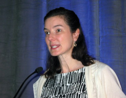
SAN FRANCISCO – Dermatologists and dermatologic surgeons risk a decreased reimbursement value for Mohs surgery if they don’t review the published criteria on its appropriate use, according to Dr. Sumaira Aasi.
Although it can be difficult, it is worth the effort to convince some patients that electrodessication, a curettage procedure, or some other management strategy is appropriate for their skin lesions, she said at the annual meeting of the Pacific Dermatologic Association.
"The reason is, at the end of the day, we might be killing the goose that laid the golden egg ourselves" if too many Mohs surgeries are done for inappropriate reasons, said Dr. Aasi of Stanford (Calif.) University.
SDr. Aasi urged the audience at the meeting to study criteria published in 2012 for the appropriate use of Mohs surgery, and to pay particular attention to scenarios deemed inappropriate for the procedure (J. Am. Acad. Dermatol. 2012;67:531-50).
There are an estimated 4 million nonmelanoma skin cancers in the United States each year, and the use of Mohs surgery increased by 400% from 1995 to 2009, Dr. Aasi said (Dermatol. Clin. 2012;30:167-75).
Some experts argue that the increase is a response to the increase in skin cancer cases, and others say the increasing number of Mohs surgeons is driving the rising rate of the procedure. "Regardless, it’s a problem," Dr. Aasi said.
Federal reimbursement programs don’t care if there is an epidemic of skin cancer, she said. If a surgery is being "overused," it is considered overvalued, and reimbursement rates are subject to change, she explained.
The top two CPT codes flagged by the federal government as potentially misvalued in the July 19, 2013, Federal Register were codes 17311 for Mohs surgery on the face, head, and neck and 17313 for Mohs surgery on the trunk and extremities, Dr. Aasi noted.
The 2012 appropriate-use criteria were created to help maintain the value of Mohs surgery in Medicare reimbursements, Dr. Aasi said. The American Academy of Dermatology, the American Society of Dermatologic Surgery, the American College of Mohs Surgery, and the American Society of Mohs Surgery convened a consensus panel of eight Mohs surgeons and nine non-Mohs surgeons. The panel considered 270 clinical scenarios and rated 74% appropriate for Mohs surgery, 17% inappropriate, and 9% uncertain, meaning there were conflicting data in the literature or not enough information to make a determination of the appropriateness of Mohs surgery in that scenario.
"I still think that Mohs surgeons in this panel were a little highly represented," Dr. Aasi said. Overall, the indications the panel deemed appropriate for Mohs surgery were "pretty generous," she added.
The consensus panel was asked to implicitly consider cost in deciding on the appropriateness of Mohs surgery for each scenario. "It’s an implicit consideration but a very important consideration," said Dr. Aasi.
Invasive melanoma was not included in the scenarios because if its complexity, but some scenarios did include melanoma in situ or lentigo maligna melanoma. The scenarios included different body areas, aggressive or nonaggressive skin cancers, tumor size, and the patient’s immune status. Agreement by at least 12 panel members was considered a consensus on the use of Mohs surgery.
Examples of scenarios that were considered inappropriate for Mohs surgery included nonaggressive basal cell or squamous cell carcinomas smaller than 2 cm on the trunk or extremities. Other lesions deemed inappropriate for Mohs included primary actinic keratosis (AK) with focal squamous cell cancer in situ, bowenoid AK, or squamous cell carcinoma in situ AK-type, labels that Dr. Aasi found "worrisome" because their definitions are unclear.
"As a Mohs surgeon, I found it a little surprising that they had to consider scenarios where actinic keratoses were being considered a possibility for Mohs surgery," she said.
Physicians can use the appropriate-use criteria to talk with other health care providers, third-party payers, and patients, said Dr. Aasi. Talking with patients can be most difficult, because if they’ve heard of Mohs surgery, they often want "the technique where I [the patient] know right away that I’m going to be clear," she said.
When Mohs surgery is inappropriate, "I try to convince them that, no, you don’t need it. It’s like an atomic bomb being used to kill a flea," she said.
The appropriate-use criteria do not compare different treatment modalities with Mohs surgery, Dr. Aasi emphasized, but they aim to state simply whether or not Mohs surgery would be appropriate in selected scenarios.
"I urge everyone to read" the criteria, she said.
Dr. Aasi reported having no financial disclosures.
On Twitter @sherryboschert
EXPERT ANALYSIS FROM THE PDA ANNUAL MEETING
Fewer than 1% of doctors mention sunscreen to patients
Physicians miss 99.9% of their opportunities to counsel patients about use of sunscreen, and dermatologists pass up 98% of those teaching moments, based on data from approximately 18 billion patient visits recorded between January 1989 and December 2010. The findings were published online on Sept. 4 in JAMA Dermatology.
Despite the rising incidence of skin cancer and recommendations from medical organizations that clinicians counsel patients about sun-protective behaviors, "sun-protection counseling ranks among the lowest topics of primary prevention discussed between physicians and patients," said Dr. Kristie Akamine of Wake Forest University in Winston-Salem, N.C., and her colleagues.
To identify trends in sunscreen recommendations by different specialties, the researchers reviewed data from the National Ambulatory Medical Care Survey (NAMCS), an ongoing survey conducted by the National Center for Health Statistics (JAMA Dermatol. 2013 Sept. 4 [doi:10.1001/jamadermatol.2013.4741]).

Overall, sunscreen was recommended at only 12.9 million of 18.3 billion patient visits (0.07%). Most of the appointments at which sunscreen was recommended were visits to a dermatologist (86%), followed by visits to family physicians or general practitioners (10%), pediatricians (1.4%), other specialists (1.4%), and internists (1.1%).
Although dermatologists were the most frequent recommenders of sunscreen, the mention of sunscreen was recorded at only 1.6% of all dermatology visits and 11% of visits associated with a diagnosis of skin cancer, the researchers said. Of note, dermatologists recommended use of sunscreen to skin cancer patients less frequently than did general/family physicians (11% vs. 56%), they added.
Overall, sunscreen was least often recommended for children younger than 10 years; by contrast, patients in their 70s were most likely to receive a recommendation for sunscreen use.
In addition, white patients were nine times more likely than black patients to receive a recommendation for sunscreen use.
Across all specialties, patients with a diagnosis of actinic keratosis accounted for 21% of the visits at which sunscreen was recommended.
The study was limited by several factors, including the cross-sectional nature of the data, which included both new and follow-up visits, and the lack of information about whether sunscreen was discussed at an earlier visit or not documented by the physician on the survey report, Dr. Akamine and her associates noted.
The results, however, suggest that sunscreen use recommendation is less frequent than advised by multiple medical organizations. "The high incidence and morbidity of skin cancer can be greatly reduced with the implementation of sun-protection behaviors, which patients should be counseled about at outpatient visits," the researchers said.
Lead author Dr. Akamine had no financial conflicts to disclose. Corresponding author Dr. Steven Feldman disclosed financial relationships with multiple pharmaceutical companies, but this study was not sponsored by a pharmaceutical company.
On Twitter @hsplete
Physicians miss 99.9% of their opportunities to counsel patients about use of sunscreen, and dermatologists pass up 98% of those teaching moments, based on data from approximately 18 billion patient visits recorded between January 1989 and December 2010. The findings were published online on Sept. 4 in JAMA Dermatology.
Despite the rising incidence of skin cancer and recommendations from medical organizations that clinicians counsel patients about sun-protective behaviors, "sun-protection counseling ranks among the lowest topics of primary prevention discussed between physicians and patients," said Dr. Kristie Akamine of Wake Forest University in Winston-Salem, N.C., and her colleagues.
To identify trends in sunscreen recommendations by different specialties, the researchers reviewed data from the National Ambulatory Medical Care Survey (NAMCS), an ongoing survey conducted by the National Center for Health Statistics (JAMA Dermatol. 2013 Sept. 4 [doi:10.1001/jamadermatol.2013.4741]).
Overall, sunscreen was recommended at only 12.9 million of 18.3 billion patient visits (0.07%). Most of the appointments at which sunscreen was recommended were visits to a dermatologist (86%), followed by visits to family physicians or general practitioners (10%), pediatricians (1.4%), other specialists (1.4%), and internists (1.1%).
Although dermatologists were the most frequent recommenders of sunscreen, the mention of sunscreen was recorded at only 1.6% of all dermatology visits and 11% of visits associated with a diagnosis of skin cancer, the researchers said. Of note, dermatologists recommended use of sunscreen to skin cancer patients less frequently than did general/family physicians (11% vs. 56%), they added.
Overall, sunscreen was least often recommended for children younger than 10 years; by contrast, patients in their 70s were most likely to receive a recommendation for sunscreen use.
In addition, white patients were nine times more likely than black patients to receive a recommendation for sunscreen use.
Across all specialties, patients with a diagnosis of actinic keratosis accounted for 21% of the visits at which sunscreen was recommended.
The study was limited by several factors, including the cross-sectional nature of the data, which included both new and follow-up visits, and the lack of information about whether sunscreen was discussed at an earlier visit or not documented by the physician on the survey report, Dr. Akamine and her associates noted.
The results, however, suggest that sunscreen use recommendation is less frequent than advised by multiple medical organizations. "The high incidence and morbidity of skin cancer can be greatly reduced with the implementation of sun-protection behaviors, which patients should be counseled about at outpatient visits," the researchers said.
Lead author Dr. Akamine had no financial conflicts to disclose. Corresponding author Dr. Steven Feldman disclosed financial relationships with multiple pharmaceutical companies, but this study was not sponsored by a pharmaceutical company.
On Twitter @hsplete
FROM JAMA DERMATOLOGY
Major Finding: Overall, physicians recommended sunscreen at 12.9 million of 18.3 billion patient visits (0.07%).
Data Source: The National Ambulatory Medical Care Survey, an ongoing survey conducted by the National Center for Health Statistics.
Disclosures: Lead author Dr. Akamine had no financial conflicts to disclose. Corresponding author Dr. Steven Feldman disclosed financial relationships with multiple pharmaceutical companies, but this study was not sponsored by a pharmaceutical company.

Fraxel for Actinic Keratosis: A New Therapeutic Option

Weiss and colleagues (J Am Acad Dermatol. 2013;68:98-102) conducted a 6-month safety, tolerance, and efficacy trial of nonablative 1927-nm fractional resurfacing of facial actinic keratosis (AK) with the Fraxel Dual (Solta Medical) laser. Twenty-four patients (5 male; 19 female) underwent up to 4 facial treatments with the 1927-nm laser with a 6-month follow-up period. The average patient age was 60 years, and Fitzpatrick skin types I and II were most common. Skin biopsy was performed in 7 patients prior to the initial procedure and at the 6-month follow-up.
Overall, there was an 86.6% reduction in absolute number of AK lesions at the 6-month follow-up visit. Cosmetic improvement was assessed on a 4-point scale. At the end of the 6-month study, the patients graded their improvement as 3.04 and investigators graded the improvement as 3.54. All 7 patient biopsies confirmed AK prior to treatment. At 6-month follow-up, 6 specimens (85.7%) showed histologic evidence of AK clearance.
What’s the issue?
The fractionated 1927-nm nonablative thulium laser is approved by the US Food and Drug Administration for the treatment of AK. Fraxel works by creating thermal zones, and the thermal damage targets AK lesions in the superficial skin. Actinic keratosis, the second most common condition treated by dermatologists, has many therapeutic options, including cryosurgery, photodynamic therapy, and multiple topical agents.
According to this study, 1927-nm fractional resurfacing appears to be a promising option for facial AK treatment. The added cosmetic benefit is a huge plus for many patients. More studies with long-term follow-up are needed.
How do you use Fraxel Dual to treat AK?

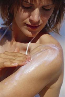
Daily sunscreen slowed skin aging in middle-aged adults
Daily sunscreen users were significantly less likely than discretionary sunscreen users to show signs of skin aging after 4.5 years, according to a study of young and middle-aged adults.
However, beta-carotene supplements appeared to have no effect on skin aging.
The findings from the randomized controlled trial were published online June 3 in Annals of Internal Medicine (2013;158:781-90).
"No known randomized studies in humans have evaluated the effect of sunscreen on surface changes associated with skin aging," wrote Maria Celia B. Hughes, MMedSci., of the Queensland Institute of Medical Research, Australia, and her colleagues.
The investigators used data from the Nambour (Australia) Skin Cancer Prevention Trial, in which 1,621 adults were studied from 1992 to 1996 to test the effect of sunscreen use or dietary supplements on skin cancer risk, photoaging, and actinic keratosis development.
To determine whether consistent, daily sunscreen use could prevent progression of skin aging, researchers randomized Nambour study participants under age 55 into four groups: daily use of broad-spectrum sunscreen plus 30 mg of beta-carotene; daily sunscreen use plus a placebo supplement; discretionary sunscreen use plus 30 mg of beta-carotene; and discretionary sunscreen use plus a placebo supplement.
The investigators focused on 903 adults younger than 55 to exclude the potential effects of growing old on participants’ skin aging.
Skin aging was assessed by comparing skin microtopography based on impressions taken of the backs of participants’ hands at baseline in 1992 and 4.5 years later in 1996. Assessors were blinded to the treatment groups.
"Most of the study participants were fair skinned, and more than 90% burned on acute sun exposure," the researchers noted. The groups were similar in terms of phenotype, sun exposure, and pretrial sunscreen use. All groups reported similar amounts of sun exposure during the study period; 78% of daily sunscreen users and 76% of discretionary sunscreen users reported being outdoors for less than 50% of their weekend time. In addition, the use of other sun protection measures, including seeking shade and wearing a hat, was similar among the groups.
By the end of the study, 77% of the daily sunscreen users applied sunscreen at least 3-4 days per week, vs. 33% of the discretionary users.
Overall, 58% of the participants in the current study met criteria for moderate photoaging at baseline in 1992, and 49% met those criteria in 1996. However, at the end of the 4.5-year period, daily sunscreen users were 24% less likely to show signs of skin aging than were discretionary users, a statistically significant difference.
When the odds of having a higher microtopography grade in 1996 than in 1992 were adjusted for sunburns and photoaging of the neck, the researchers noted, "only the daily sunscreen intervention group showed no detectable increase in microtopography grade."
No significant differences in skin aging were seen in participants randomized to beta-carotene vs. placebo.
The study was limited by several factors, including limited outcome data, which reduced the power to detect moderate treatment effects. In addition, the data were insufficient to rule out an effect of beta carotene on skin aging, the researchers noted.
However, "these results have important clinical implications," the researchers said. "A unit increase in microtopography significantly correlates with risk for actinic keratoses and skin cancer." Thus, the cosmetic benefits of reducing skin changes in middle age also may reduce cancer risk.
The National Health and Medical Research Council of Australia funded the study.
FROM ANNALS OF INTERNAL MEDICINE
Major finding: After 4.5 years, daily sunscreen users were 24% less likely to show signs of skin aging than were discretionary sunscreen users.
Data source: A randomized, controlled, community-based trial of 903 adults younger than 55 years.
Disclosures: The National Health and Medical Research Council of Australia funded the study.

Laser-assisted PDT cleared majority of AKs in immunocompromised patients
BOSTON – Fractional laser-assisted photodynamic therapy can effectively treat actinic keratoses in organ transplant recipients, but at a cost of more intense postoperative skin reactions than with laser therapy alone, based on data from a randomized clinical trial.
Intensified ablative fractional laser-assisted photodynamic therapy (AFXL-PDT) was significantly more effective at clearing actinic keratoses (AKs) and warty lesions on the dorsal aspect of the hands of organ transplant recipients than AFXL alone, reported Dr. Merete Haedersdal of the University of Copenhagen and a visiting scientist at the Wellman Center for Photomedicine at Massachusetts General Hospital, Boston.
The treatment was painful, and 3 of 10 patients reported intense inflammation a week afterward, Dr. Haedersdal noted.

However, "this new treatment modality also works well for immunosuppressed patients, and I do think it works very well for immunosuppressed patients with multiple AKs," she said at the annual meeting of the American Society for Laser Medicine and Surgery.
High-risk population
Organ transplant recipients are at a 100-fold or greater risk for developing squamous cell carcinomas, which can arise from AKs, because of their chronic immunosuppressive treatment regimens. Cancers that develop in these patients tend to be quite aggressive, with a high rate of metastases and an attributable morality rate of 6%-8%, said Dr. Haedersdal.
"Therefore, there is a huge motivation to come up with intensified treatment regimens for these patients," she said.
PDT is a well-established therapy in transplant recipients, but it is less effective in these patients than in patients with intact immune responses. It is also less effective for thick lesions or lesions on the extremities.
Dr. Haedersdal and her colleagues previously demonstrated the efficacy of AFXL-PDT for treating AKs with a carbon dioxide laser in immunocompetent patients. In the current study, they compared the therapy with ablative fractional CO2 laser alone in 10 organ transplant recipients with a total of 680 AKs and 409 wartlike lesions on the dorsal hands, and a collective history of 21 previous squamous cell carcinomas.
In a randomized intrapatient trial, the participants first underwent targeted ablation of localized keratotic lesions, and then were randomly assigned to receive one field treatment with AFXL-PDT on one hand, and AFXL alone on the other.
For AFXL, energy was delivered with the laser set to 30 W, a 0.12-mm spot size, a 1.32- to 2.06-millisecond pulse duration, and 40-60 mJ with the target of 4.3%-5.2% coverage; settings were based on the severity of skin atrophy.
After laser exposure, lesions randomized to receive PDT were treated with methyl aminolevulinate applied under occlusion for 3 hours, and were then exposed to red diode light at 37 J/cm2.
‘Really impressive’ cure rate
The combined modality completely cleared 73% of all AKs, compared with 31% cleared by AFXL alone (P = .002), and 37% of all wartlike lesions, compared with 14% for AFXL (P = .02) at 4 months’ follow-up. The patients were clinically assessed by raters blinded to treatment type.
"Normally, when we deliver PDT for these patients, we have – from just a single treatment – cure rates on the acral lesions of about 30%-40%, so this was really impressive to get a complete cure rate for AKs at a level of 73%," Dr. Haedersdal said.
Overall, AKs treated with AFXL-PDT were rated as improved by a median of 83%, 15% as unchanged, and 3% as worsened, compared with AFXL-only rates of 52%, 47%, and 4%, respectively.
Thinner AKs responded better to treatment than thick lesions, with an odds ratio (OR) for grade 2 vs. grade 1 lesions of 0.34 (P = .001) and an OR for grade 3 vs. grade 1 of 0.21 (P = .001).
Safety data showed that AFXL treatment was generally not painful, with a mean visual analog scale (VAS) pain score of 1, and the PDT was more painful, with a mean VAS of 4.5 during LED illumination. Seven of 10 patients requested anesthesia during the procedure, Dr. Haedersdal noted.
In addition, at 1 week post treatment, 3 of 10 patients reported intense inflammation of the treated sites, 3 had pigment changes with AFXL-PDT, and 1 had a pigment change with AFXL alone.
The take-home message, Dr. Haedersdal said, is: "Do not deliver laser-assisted PDT to large areas; you have to deliver it to refined areas."
However, eight patients gave a favorable overall assessment to the combined treatments, and the remaining two rated the combined therapy and laser-only therapy as being equally effective.
The study was supported by a research grant to Dr. Haedersdal from Galderma. She also disclosed serving on a Galderma advisory board.
BOSTON – Fractional laser-assisted photodynamic therapy can effectively treat actinic keratoses in organ transplant recipients, but at a cost of more intense postoperative skin reactions than with laser therapy alone, based on data from a randomized clinical trial.
Intensified ablative fractional laser-assisted photodynamic therapy (AFXL-PDT) was significantly more effective at clearing actinic keratoses (AKs) and warty lesions on the dorsal aspect of the hands of organ transplant recipients than AFXL alone, reported Dr. Merete Haedersdal of the University of Copenhagen and a visiting scientist at the Wellman Center for Photomedicine at Massachusetts General Hospital, Boston.
The treatment was painful, and 3 of 10 patients reported intense inflammation a week afterward, Dr. Haedersdal noted.
However, "this new treatment modality also works well for immunosuppressed patients, and I do think it works very well for immunosuppressed patients with multiple AKs," she said at the annual meeting of the American Society for Laser Medicine and Surgery.
High-risk population
Organ transplant recipients are at a 100-fold or greater risk for developing squamous cell carcinomas, which can arise from AKs, because of their chronic immunosuppressive treatment regimens. Cancers that develop in these patients tend to be quite aggressive, with a high rate of metastases and an attributable morality rate of 6%-8%, said Dr. Haedersdal.
"Therefore, there is a huge motivation to come up with intensified treatment regimens for these patients," she said.
PDT is a well-established therapy in transplant recipients, but it is less effective in these patients than in patients with intact immune responses. It is also less effective for thick lesions or lesions on the extremities.
Dr. Haedersdal and her colleagues previously demonstrated the efficacy of AFXL-PDT for treating AKs with a carbon dioxide laser in immunocompetent patients. In the current study, they compared the therapy with ablative fractional CO2 laser alone in 10 organ transplant recipients with a total of 680 AKs and 409 wartlike lesions on the dorsal hands, and a collective history of 21 previous squamous cell carcinomas.
In a randomized intrapatient trial, the participants first underwent targeted ablation of localized keratotic lesions, and then were randomly assigned to receive one field treatment with AFXL-PDT on one hand, and AFXL alone on the other.
For AFXL, energy was delivered with the laser set to 30 W, a 0.12-mm spot size, a 1.32- to 2.06-millisecond pulse duration, and 40-60 mJ with the target of 4.3%-5.2% coverage; settings were based on the severity of skin atrophy.
After laser exposure, lesions randomized to receive PDT were treated with methyl aminolevulinate applied under occlusion for 3 hours, and were then exposed to red diode light at 37 J/cm2.
‘Really impressive’ cure rate
The combined modality completely cleared 73% of all AKs, compared with 31% cleared by AFXL alone (P = .002), and 37% of all wartlike lesions, compared with 14% for AFXL (P = .02) at 4 months’ follow-up. The patients were clinically assessed by raters blinded to treatment type.
"Normally, when we deliver PDT for these patients, we have – from just a single treatment – cure rates on the acral lesions of about 30%-40%, so this was really impressive to get a complete cure rate for AKs at a level of 73%," Dr. Haedersdal said.
Overall, AKs treated with AFXL-PDT were rated as improved by a median of 83%, 15% as unchanged, and 3% as worsened, compared with AFXL-only rates of 52%, 47%, and 4%, respectively.
Thinner AKs responded better to treatment than thick lesions, with an odds ratio (OR) for grade 2 vs. grade 1 lesions of 0.34 (P = .001) and an OR for grade 3 vs. grade 1 of 0.21 (P = .001).
Safety data showed that AFXL treatment was generally not painful, with a mean visual analog scale (VAS) pain score of 1, and the PDT was more painful, with a mean VAS of 4.5 during LED illumination. Seven of 10 patients requested anesthesia during the procedure, Dr. Haedersdal noted.
In addition, at 1 week post treatment, 3 of 10 patients reported intense inflammation of the treated sites, 3 had pigment changes with AFXL-PDT, and 1 had a pigment change with AFXL alone.
The take-home message, Dr. Haedersdal said, is: "Do not deliver laser-assisted PDT to large areas; you have to deliver it to refined areas."
However, eight patients gave a favorable overall assessment to the combined treatments, and the remaining two rated the combined therapy and laser-only therapy as being equally effective.
The study was supported by a research grant to Dr. Haedersdal from Galderma. She also disclosed serving on a Galderma advisory board.
AT LASER 2013
Major finding: Laser-assisted photodynamic therapy completely cleared 73% of actinic keratoses on the hands of organ transplant recipients.
Data source: A randomized clinical trial comparing laser-assisted PDT with laser treatment alone in 10 patients.
Disclosures: The study was supported by a research grant to Dr. Haedersdal from Galderma. She also disclosed serving on a Galderma advisory board.
'Slow Mohs' advised for lentigo maligna
MAUI, HAWAII – "Slow Mohs" has gained near-universal acceptance among skin cancer specialists as a definitive surgical technique for complete removal of lentigo maligna melanoma while simultaneously sparing normal tissue, according to Dr. Ellen Marmur of Mount Sinai School of Medicine, New York.
The big advantage that slow Mohs has over standard wide local excision with 0.5- to 1-cm margins is a 5-year cure rate approaching 100%. In contrast, standard excision has a recurrence rate of up to 20%, she said at the Hawaii Dermatology Seminar, sponsored by Global Academy for Medical Education/Skin Disease Education Foundation.

Slow Mohs is a modified form of Mohs micrographic surgery. The surgery compares with conventional Mohs: It is staged, margin-controlled excision. But in slow Mohs, rush permanent sections are sent off to the pathologist rather than the frozen sections integral to conventional Mohs.
Dr. Marmur relies upon slow Mohs, with "bread-loafing" of the central tumor by the pathologist, because the permanent sections better preserve the tumor’s microscopic features. Interpreting atypical melanocytes in frozen sections can be quite a challenge. However, she added, some Mohs surgeons have found that using rapid immunostains also markedly improves the sensitivity and specificity of frozen sections in lentigo maligna surgery.
Slow Mohs takes place over the course of days, she said. "Basically, you do your Wood’s lamp to define the lesion diameter, you measure out your margins, you excise the tumor, pack the area with a bandage, and send the patient home. You rush your pathology, and you don’t do any reconstruction until you get the margins clear."
A pathology report that comes back stating narrow margins are present is "a heart stopper," she added.
"You have the option of observing the area if the margin is clear but the tumor was close to the margin. That’s a good approach for an elderly patient or when the lentigo maligna was in a cosmetically important area."
Lentigo maligna melanoma accounts for 4% of all cases of melanoma. It typically arises on sun-damaged skin in individuals in their 70s or older. Common sites include the malar area, forehead, nose, and temple. The differential diagnosis includes seborrheic keratosis, pigmented actinic keratosis, and pigmented nevus.
Lentigo maligna becomes lentigo maligna melanoma when malignant melanoma cells invade the dermis and deeper appendages. Roughly 5% of lentigo malignas eventually progress to invasive melanoma, according to Dr. Marmur. Typically, a lentigo maligna undergoes extended gradual horizontal growth before beginning a vertical growth phase.
"It spreads like an oil slick for many years," Dr. Marmur said at the seminar.
Established treatment modalities for patients who aren’t surgical candidates include cryotherapy, radiotherapy, and topical imiquimod 5%. All have disadvantages, including high 5-year recurrence rates.
Dr. Marmur noted that a newer nonsurgical therapy drawing considerable interest involves off-label use of topical combination therapy with imiquimod and tazarotene gel. The concept is to use the topical retinoid to disrupt the stratum corneum in order to enhance imiquimod penetration, thereby achieving a greater inflammatory response than possible with imiquimod alone.
Initial data have been published by researchers at the University of Utah, Salt Lake City. They randomized 90 patients with 91 lentigo malignas to imiquimod 5% cream applied 5 days per week for 3 months or to the imiquimod regimen plus tazarotene 0.1% gel on the other 2 days per week. After 3 months of topical therapy, patients underwent conservative staged excision with frozen section analysis with Melan A immunostaining to confirm negative margins.
Of those treated with dual topical therapy for 3 months, 29 of 37 lesions (78%) had complete responses with no residual lentigo maligna at the time of staged excision. So did 27 of 42 (64%) treated with imiquimod alone (Arch. Dermatol. 2012;148:592-6).
The modest difference in outcome was not significant (P = .17). Nevertheless, the Utah investigators wrote that topical pretreatment appears to reduce surgical defect sizes, an important consideration in lentigo maligna because the lesions are often large and located on cosmetically sensitive facial sites. At the patient’s first visit, the researchers saucerize the entire tumor to remove all visible evidence of lentigo maligna and nip in the bud any invasive element that might be present. One month later, after the wound has healed by secondary intention, the patient begins topical imiquimod therapy 5 days per week. If no inflammation is observed, tazarotene gel is added on the other 2 days per week. After 3 months of topical therapy, the patient goes off treatment for 2 months so the inflammatory response can subside. Then a staged excision is performed with 2-mm margins around the perimeter of the original tumor outline.
"This is an interesting thought, but it’s still not considered standard of care," Dr. Marmur commented in describing the Utah research.
Session chair Dr. Allan C. Halpern, chief of the dermatology service at Memorial Sloan-Kettering Cancer Center in New York, said he and his colleagues, like the Utah group, are using a lot of topical imiquimod as adjunctive therapy for lentigo maligna.
"Because we’re a referral center, we see all those patients whose melanomas unfortunately do recur at the edge of large surgeries. We see a lot of patients whose local surgeons have chased around their faces for a certain amount of time. So I think the general concept of the cure rates of surgery is a bit overstated. I’m not trying to sell an off-label use of a drug, but I think sometimes imiquimod can be very helpful," Dr. Halpern said.
He offered a clinical pearl in diagnosing lentigo maligna: Consider a broad shave biopsy of a suspicious lesion rather than a deeper, smaller-diameter biopsy.
"Sampling errors are a real problem with lentigo maligna. We learned early on that if you take a large lentigo maligna and do multiple biopsies of the same lesion and send them to what we think are our very expert pathologists, on average, we get two or three back that were melanoma and two or three back that were pigmented actinic keratoses. The sampling error tends to be more in the horizontal than in the depth for these very early lesions," according to Dr. Halpern.
Dr. Marmur and Dr. Halpern reported having no relevant financial interests. SDEF and this news organization are owned by the same parent company.
MAUI, HAWAII – "Slow Mohs" has gained near-universal acceptance among skin cancer specialists as a definitive surgical technique for complete removal of lentigo maligna melanoma while simultaneously sparing normal tissue, according to Dr. Ellen Marmur of Mount Sinai School of Medicine, New York.
The big advantage that slow Mohs has over standard wide local excision with 0.5- to 1-cm margins is a 5-year cure rate approaching 100%. In contrast, standard excision has a recurrence rate of up to 20%, she said at the Hawaii Dermatology Seminar, sponsored by Global Academy for Medical Education/Skin Disease Education Foundation.
Slow Mohs is a modified form of Mohs micrographic surgery. The surgery compares with conventional Mohs: It is staged, margin-controlled excision. But in slow Mohs, rush permanent sections are sent off to the pathologist rather than the frozen sections integral to conventional Mohs.
Dr. Marmur relies upon slow Mohs, with "bread-loafing" of the central tumor by the pathologist, because the permanent sections better preserve the tumor’s microscopic features. Interpreting atypical melanocytes in frozen sections can be quite a challenge. However, she added, some Mohs surgeons have found that using rapid immunostains also markedly improves the sensitivity and specificity of frozen sections in lentigo maligna surgery.
Slow Mohs takes place over the course of days, she said. "Basically, you do your Wood’s lamp to define the lesion diameter, you measure out your margins, you excise the tumor, pack the area with a bandage, and send the patient home. You rush your pathology, and you don’t do any reconstruction until you get the margins clear."
A pathology report that comes back stating narrow margins are present is "a heart stopper," she added.
"You have the option of observing the area if the margin is clear but the tumor was close to the margin. That’s a good approach for an elderly patient or when the lentigo maligna was in a cosmetically important area."
Lentigo maligna melanoma accounts for 4% of all cases of melanoma. It typically arises on sun-damaged skin in individuals in their 70s or older. Common sites include the malar area, forehead, nose, and temple. The differential diagnosis includes seborrheic keratosis, pigmented actinic keratosis, and pigmented nevus.
Lentigo maligna becomes lentigo maligna melanoma when malignant melanoma cells invade the dermis and deeper appendages. Roughly 5% of lentigo malignas eventually progress to invasive melanoma, according to Dr. Marmur. Typically, a lentigo maligna undergoes extended gradual horizontal growth before beginning a vertical growth phase.
"It spreads like an oil slick for many years," Dr. Marmur said at the seminar.
Established treatment modalities for patients who aren’t surgical candidates include cryotherapy, radiotherapy, and topical imiquimod 5%. All have disadvantages, including high 5-year recurrence rates.
Dr. Marmur noted that a newer nonsurgical therapy drawing considerable interest involves off-label use of topical combination therapy with imiquimod and tazarotene gel. The concept is to use the topical retinoid to disrupt the stratum corneum in order to enhance imiquimod penetration, thereby achieving a greater inflammatory response than possible with imiquimod alone.
Initial data have been published by researchers at the University of Utah, Salt Lake City. They randomized 90 patients with 91 lentigo malignas to imiquimod 5% cream applied 5 days per week for 3 months or to the imiquimod regimen plus tazarotene 0.1% gel on the other 2 days per week. After 3 months of topical therapy, patients underwent conservative staged excision with frozen section analysis with Melan A immunostaining to confirm negative margins.
Of those treated with dual topical therapy for 3 months, 29 of 37 lesions (78%) had complete responses with no residual lentigo maligna at the time of staged excision. So did 27 of 42 (64%) treated with imiquimod alone (Arch. Dermatol. 2012;148:592-6).
The modest difference in outcome was not significant (P = .17). Nevertheless, the Utah investigators wrote that topical pretreatment appears to reduce surgical defect sizes, an important consideration in lentigo maligna because the lesions are often large and located on cosmetically sensitive facial sites. At the patient’s first visit, the researchers saucerize the entire tumor to remove all visible evidence of lentigo maligna and nip in the bud any invasive element that might be present. One month later, after the wound has healed by secondary intention, the patient begins topical imiquimod therapy 5 days per week. If no inflammation is observed, tazarotene gel is added on the other 2 days per week. After 3 months of topical therapy, the patient goes off treatment for 2 months so the inflammatory response can subside. Then a staged excision is performed with 2-mm margins around the perimeter of the original tumor outline.
"This is an interesting thought, but it’s still not considered standard of care," Dr. Marmur commented in describing the Utah research.
Session chair Dr. Allan C. Halpern, chief of the dermatology service at Memorial Sloan-Kettering Cancer Center in New York, said he and his colleagues, like the Utah group, are using a lot of topical imiquimod as adjunctive therapy for lentigo maligna.
"Because we’re a referral center, we see all those patients whose melanomas unfortunately do recur at the edge of large surgeries. We see a lot of patients whose local surgeons have chased around their faces for a certain amount of time. So I think the general concept of the cure rates of surgery is a bit overstated. I’m not trying to sell an off-label use of a drug, but I think sometimes imiquimod can be very helpful," Dr. Halpern said.
He offered a clinical pearl in diagnosing lentigo maligna: Consider a broad shave biopsy of a suspicious lesion rather than a deeper, smaller-diameter biopsy.
"Sampling errors are a real problem with lentigo maligna. We learned early on that if you take a large lentigo maligna and do multiple biopsies of the same lesion and send them to what we think are our very expert pathologists, on average, we get two or three back that were melanoma and two or three back that were pigmented actinic keratoses. The sampling error tends to be more in the horizontal than in the depth for these very early lesions," according to Dr. Halpern.
Dr. Marmur and Dr. Halpern reported having no relevant financial interests. SDEF and this news organization are owned by the same parent company.
EXPERT ANALYSIS FROM SDEF HAWAII DERMATOLOGY SEMINAR