User login
CVD Risk Rises With Higher NSAID Doses in Ankylosing Spondylitis
TOPLINE:
Higher doses of nonsteroidal anti-inflammatory drugs (NSAIDs) increase the risk for cardiovascular diseases (CVDs) such as ischemic heart disease, stroke, and congestive heart failure in patients with ankylosing spondylitis (AS) compared with lower doses.
METHODOLOGY:
- NSAIDs can suppress inflammation and relieve pain in patients with AS, but long-term treatment with NSAIDs poses concerns regarding gastrointestinal and renal toxicities and increased CVD risk.
- This nationwide cohort study used data from the Korean National Health Insurance database to investigate the risk for CVD associated with an increasing NSAID dosage in a real-world AS cohort.
- Investigators recruited 19,775 patients (mean age, 36.1 years; 75% men) with newly diagnosed AS and without any prior CVD between January 2010 and December 2018, among whom 99.7% received NSAID treatment and 30.2% received tumor necrosis factor inhibitor treatment.
- A time-varying approach was used to assess the NSAID exposure, wherein periods of NSAID use were defined as “NSAID-exposed” and periods longer than 1 month without NSAID use were defined as “NSAID-unexposed.”
- The primary outcome was the composite outcome of ischemic heart disease, stroke, or congestive heart failure.
TAKEAWAY:
- During the follow-up period of 98,290 person-years, 1663 cases of CVD were identified, which included 1157 cases of ischemic heart disease, 301 cases of stroke, and 613 cases of congestive heart failure.
- After adjusting for confounders, each defined daily dose increase in NSAIDs raised the risk for incident CVD by 10% (adjusted hazard ratio [aHR], 1.10; 95% CI, 1.08-1.13).
- Similarly, increasing the dose of NSAIDs was associated with an increased risk for ischemic heart disease (aHR, 1.08; 95% CI, 1.05-1.11), stroke (aHR, 1.09; 95% CI, 1.04-1.15), and congestive heart failure (aHR, 1.12; 95% CI, 1.08-1.16).
- The association between increasing NSAID dose and increased CVD risk was consistent across various subgroups, with NSAIDs posing a greater threat to cardiovascular health in women than in men.
IN PRACTICE:
The authors wrote, “Taken together, these results suggest that increasing the dose of NSAIDs is associated with a higher cardiovascular risk in AS, but that the increased risk might be lower than that in the general population.”
SOURCE:
First author Ji-Won Kim, MD, PhD, of the Division of Rheumatology, Department of Internal Medicine, Daegu Catholic University School of Medicine, Daegu, the Republic of Korea, and colleagues had their work published online on April 9 in Annals of the Rheumatic Diseases.
LIMITATIONS:
The study was of retrospective nature. The levels of acute phase reactants and AS disease activity could not be determined owing to a lack of data in the National Health Insurance database. The accuracy of the diagnosis of cardiovascular outcomes on the basis of the International Classification of Disease codes was also questionable.
DISCLOSURES:
The study was supported by the National Research Foundation of Korea. The authors declared no conflicts of interest.
A version of this article appeared on Medscape.com.
Traffic Noise Negatively Impacts Health
New research by Thomas Münzel, MD, senior professor of cardiology at Johannes Gutenberg University Mainz in Mainz, Germany, and colleagues again emphasized the harmful effects of noise on the heart and blood vessels. An analysis of current epidemiologic data provided strong indications that transportation noise is closely related to cardiovascular and cerebrovascular diseases, according to a statement on the data analysis. The results were published in Circulation Research.
Morbidity and Mortality
Epidemiologic studies have shown that road, rail, or air traffic noise increases the risk for cardiovascular morbidity and mortality, with strong evidence for ischemic heart disease, heart failure, and stroke, according to the scientists. These factors could favor vascular (endothelial) dysfunction, inflammation, and hypertension, thereby increasing cardiovascular risk.
Consequences and Pathomechanisms
In the current publication, the authors provided an overview of epidemiologic research on the effects of transportation noise on cardiovascular risk factors and diseases, discussed mechanistic insights from the latest clinical and experimental studies, and proposed new risk markers to address noise-induced cardiovascular effects in the general population. An integrated analysis in the article demonstrated that for every 10 dB(A) increase, the risk for cardiovascular diseases such as heart attack, stroke, and heart failure significantly increases by 3.2%.
The authors also explained the possible effects of noise on changes in gene networks, epigenetic pathways, circadian rhythms, signal transmission along the neuronal-cardiovascular axis, oxidative stress, inflammation, and metabolism. Finally, current and future noise protection strategies are described, and the existing evidence on noise as a cardiovascular risk factor is discussed.
Confirmed Cardiovascular Risk Factor
“As an increasing proportion of the population is exposed to harmful traffic noise, efforts to reduce noise and laws for noise reduction are of great importance for future public health,” said Dr. Münzel. “It is also important for us that due to the strong evidence, traffic noise is finally recognized as a risk factor for cardiovascular diseases.”
Heart Attack Outcomes
Dr. Münzel and other researchers from Mainz have been studying the cardiovascular consequences of air pollution and traffic noise for several years. For example, they found that heart attacks in people and animals exposed to high noise levels earlier in life healed poorly. These results were published last year in Cardiovascular Research. According to the authors, the findings suggest that traffic noise may play a significant role in the development and course of coronary heart disease, such as after a heart attack.
The scientists initially found in animal experiments that exposure to aircraft noise for 4 days led to increased inflammation in the vessels. Compared with mice not exposed to aircraft noise, the noise-exposed animals showed an increase in free radicals; these animals exhibited a significant inflammatory response and had impaired vessel function.
The researchers explained that the experimental data showed aircraft noise alone triggers a proinflammatory transcription program that promotes the infiltration of immune cells into cardiovascular tissue in animals with acute myocardial infarction. They noted an increased infiltration of CD45+ cells into the vessels and heart, dominated by neutrophils in vessel tissue and Ly6Chigh monocytes in heart tissue. This infiltration creates a proinflammatory milieu that adversely affects the outcome after myocardial infarction by predisposing the heart tissue to greater ischemic damage and functional impairment. Exposure of animals to aircraft noise before induction of myocardial infarction by left anterior descending (LAD) coronary artery ligation impaired left ventricular function and increased infarct size after cardiac ischemia. In addition, noise exposure exacerbated infarct-induced endothelial dysfunction of peripheral vessels as early as 24 hours after LAD ligation.
Clinical Confirmation
These experimental results were confirmed by observations in the population-based Gutenberg Health Study. The researchers analyzed data from 100 patients with heart attack. The lead and senior authors of the study Michael Molitor, MD, and Philip Wenzel, MD, of the University of Mainz, explained, “From our studies, we have learned that exposure to aircraft noise before a heart attack significantly amplifies subsequent cardiovascular inflammation and exacerbates ischemic heart failure, which is favored by inflammation-promoting vascular conditioning. Our translational results show that people who have been exposed to noise in the past have a worse course if they experience a heart attack later in life.”
Study participants who had experienced a heart attack in their medical history had elevated levels of C-reactive protein if they had been exposed to aircraft noise in the past and subsequently developed noise annoyance reactions (0.305 vs 1.5; P = .0094). In addition, left ventricular ejection fraction in these patients after a heart attack was worse than that in patients with infarction without noise exposure in their medical history (62.5 vs 65.6; P = .0053).
The results suggest that measures to reduce environmental noise could help improve the clinical outcomes of heart attack patients, according to the authors.
Mental Health Effects
Traffic noise also may be associated with an increased risk for depression and anxiety disorders, as reported 2 years ago by the German Society for Psychosomatic Medicine and Medical Psychotherapy. Evolution has programmed the human organism to perceive noises as indicators of potential sources of danger — even during sleep. “Noise puts the body on alert,” explained Manfred E. Beutel, MD, director of the Clinic for Psychosomatic Medicine and Psychotherapy at the University of Mainz. As a result, the autonomic nervous system activates stress hormones such as adrenaline and cortisol, leading to an increase in heart rate and blood pressure. If noise becomes chronic, chronic diseases can develop. “Indeed, observational and experimental studies have shown that persistent noise annoyance promotes incident hypertension, cardiovascular diseases, and type 2 diabetes,” said Dr. Beutel.
Depression Risk Doubled
Among the negative effects of noise annoyance are also mental illnesses, as has become increasingly clear. “Noise annoyance disrupts daily activities and interferes with feelings and thoughts, sleep, and recovery,” said Dr. Beutel. The interruptions trigger negative emotional reactions such as anger, distress, exhaustion, flight impulses, and stress symptoms. “Such conditions promote the development of depression over time,” said Dr. Beutel. This observation was confirmed by the large-scale Gutenberg Health Study using the example of the Mainz population, which suffers to a large extent from noise annoyance because of the nearby Frankfurt Airport. “With increasing noise annoyance, the rates of depression and anxiety disorders steadily increased, until the risks eventually doubled with extreme annoyance,” said Dr. Beutel. Other studies point in the same direction. For example, a meta-analysis found a 12% increase in the risk for depression per 10-dB increase in noise. Another study found an association between nocturnal noise annoyance and the use of antidepressants.
Fine Particulate Matter
According to an evaluation of the Gutenberg Study, people perceive noise annoyance from aircraft noise as the most pronounced, followed by road, neighborhood, industrial, and railway noise. Noise occurs most frequently in urban areas that also produce air pollution such as fine particulate matter. “Fine particulate matter is also suspected of promoting anxiety and depression,” said Dr. Beutel, “because the small particles of fine particulate matter can enter the bloodstream and trigger inflammatory processes there, which in turn are closely related to depression.”
This story was translated from Univadis Germany, which is part of the Medscape professional network, using several editorial tools, including AI, as part of the process. Human editors reviewed this content before publication. A version of this article appeared on Medscape.com.
Hereditary Amyloidosis: 5 Things to Know
Amyloidosis is a condition marked by the accumulation of insoluble beta-sheet fibrillar protein aggregates in tissues that can be acquired or hereditary. Hereditary amyloidogenic transthyretin (hATTR) amyloidosis is an autosomal-dominant disease caused by pathogenic variants in the TTR gene. The TTR protein is essential for transporting thyroxine and retinol-binding protein and is primarily synthesized in the liver, becoming unstable as a result of the pathogenic mutations. Inherited pathogenic variants lead to the protein’s misfolding, aggregation, and deposition as amyloid fibrils in different organs, resulting in progressive multisystem dysfunction. hATTR amyloidosis is a heterogenous disease, characterized by a wide range of clinical manifestations affecting the peripheral (both somatic and autonomic) nervous system, heart, kidneys, and central nervous system (CNS); however, the heart and peripheral nerves appear to be the main targets of the TTR-related pathologic process. Without treatment, the prognosis is poor, with an average life expectancy of 7-11 years; however, in recent years, the development of new therapeutics has brought new hope to patients.
Here are five things to know about hereditary amyloidosis.
1. Diagnosis of hereditary amyloidosis requires a high level of suspicion.
The diagnosis of hATTR amyloidosis presents a significant challenge, particularly in nonendemic regions where a lack of family history and heterogeneity of clinical presentation can delay diagnosis by 4-5 years. A timely diagnosis requires clinicians to maintain a high index of suspicion, especially when evaluating patients with neuropathic symptoms. Early diagnosis is crucial to begin patients on recently available disease-modifying therapies that can slow the disease course. Failure to recognize is the major barrier to improved patient outcomes.
Confirming the diagnosis involves detecting amyloid deposits in tissue biopsy specimens from various possible sites, including the skin, nerves, myocardium, and others. However, the diagnosis can be challenging owing to the uneven distribution of amyloid fibrils, sometimes requiring multiple biopsies or alternative diagnostic approaches, such as TTR gene sequencing, to confirm the presence of an amyloidogenic pathogenic variant. Biopsy for hATTR amyloidosis is not required if imaging of the clinical phenotype and genetic testing are consistent.
Once diagnosed, the assessment of organ involvement is essential, using nerve conduction studies, cardiac investigations (eg, echocardiography, ECG, scintigraphy), ophthalmologic assessments, and complete renal function evaluations to fully understand the extent of disease impact.
2. Hereditary amyloidosis diseases are classified into two primary categories.
Hereditary amyloidosis represents a group of diseases caused by inherited gene mutations and is classified into two main types: ATTR (transthyretin-related) and non-TTR. Most cases of hereditary amyloidosis are associated with the TTR gene. Mutations in this protein lead to different forms of ATTR amyloidosis, categorized on the basis of the specific mutation involved, such as hATTR50M (genotype Val50Met), which is the most prevalent form.
ATTR mutations result in a variety of health issues, manifesting in three primary forms:
- Neuropathic ATTR (genotype Val50Met): Early symptoms include sensorimotor polyneuropathy of the legs, carpal tunnel syndrome, autonomic dysfunction, constipation/diarrhea, and impotence; late symptoms include cardiomyopathy, vitreous opacities, glaucoma, nephropathy, and CNS symptoms.
- Cardiac ATTR (genotype Val142Ile): This type is characterized by cardiomegaly, conduction block, arrhythmia, anginal pain, congestive heart failure, and sudden death.
- Leptomeningeal ATTR (genotype Asp38Gly): This is characterized by transient focal neurologic episodes, intracerebral and/or subarachnoid hemorrhages, dementia, ataxia, and psychosis.
Non-TTR amyloidoses are rarer than are ATTR variations and involve mutations in different genes that also have significant health impacts. These include proteins such as apolipoprotein AI, fibrinogen A alpha, lysozyme, apolipoprotein AII, gelsolin, and cystatin C. Each type contributes to a range of symptoms and requires individualized management approaches.
3. Heightened disease awareness has increased the recognized prevalence of hereditary amyloidosis.
hATTR amyloidosis has historically been recognized as a rare disease, with significant clusters in Portugal, Brazil, Sweden, and Japan and alongside smaller foci in regions such as Cyprus and Majorca. This disease›s variable incidence across Europe is now perceived to be on the rise. It is attributed to heightened disease awareness among healthcare providers and the broader availability of genetic testing, extending its recognized impact to at least 29 countries globally. The genetic landscape of hATTR amyloidosis is diverse, with over 140 mutations identified in the TTR gene. Among these, the Val50Met mutation is particularly notable for its association with large patient clusters in the endemic regions.
Morbidity and mortality associated with hATTR amyloidosis are significant, with an average lifespan of 7-11 years post diagnosis; however, survival rates can vary widely depending on the specific genetic variant and organ involvement. Early diagnosis can substantially improve outcomes; yet, for many, the prognosis remains poor, especially in cases dominated by cardiomyopathy. Genetics play a central role in the disease›s transmission, with autosomal-dominant inheritance patterns and high penetrance among carriers of pathogenic mutations. Research continues to uncover the broad spectrum of genetic variations contributing to hATTR amyloidosis, with ongoing studies poised to expand our understanding of its molecular underpinnings and potential treatment options.
4. The effect on quality of life is significant both in patients living with hATTR amyloidosis and their caregivers.
hATTR amyloidosis imposes a multifaceted burden on patients and their caregivers as the disease progresses. Symptoms range from sensorimotor impairment and gastrointestinal or autonomic dysfunction to heart failure, leading to significant health-related quality-of-life deficits. The systemic nature of hATTR amyloidosis significantly affects patients› lifestyles, daily activities, and general well-being, especially because it typically manifests in adulthood — a crucial time for occupational changes. The progression of hATTR amyloidosis exacerbates the challenges in maintaining employment and managing household chores, with symptomatic patients often unable to work and experiencing difficulties with absenteeism and presenteeism when they are able to work.
hATTR amyloidosis leads to physical, mental, occupational, and social limitations for patients, and it also places a considerable strain on their families and caregivers, who report poor mental health, work impairment, and a high time commitment (mean, 45.9 h/wk) to providing care.
5. There have been significant advancements in therapeutic options for early-stage hATTR amyloidosis.
After diagnosis, prompt initiation of treatment is recommended to delay the progression of hATTR amyloidosis; a multidisciplinary approach is essential, incorporating anti-amyloid therapy to inhibit further production and/or deposition of amyloid aggregates. Treatment strategies also include addressing symptomatic therapy and managing cardiac, renal, and ocular involvement. Although many therapies have been developed, especially for the early stages of hATTR amyloidosis, therapeutic benefits for patients with advanced disease remain limited.
Recent advancements in the treatment of hATTR amyloidosis have introduced RNA-targeted therapies including patisiran, vutrisiran, and eplontersen, which have shown efficacy in reducing hepatic TTR synthesis and the aggregation of misfolded monomers into amyloid deposits. These therapies, ranging from small interfering RNA formulations to antisense oligonucleotides, offer benefits in managing both cardiomyopathy and neuropathy associated with hATTR amyloidosis , administered through various methods, including intravenous infusions and subcutaneous injections. In addition, the stabilization of TTR tetramers with the use of drugs such as tafamidis and diflunisal has effectively prevented the formation of amyloidogenic monomers. Moreover, other investigational agents, including TTR stabilizers like acoramidis and tolcapone, as well as novel compounds that inhibit amyloid formation and disrupt fibrils, are expanding the therapeutic landscape for hATTR amyloidosis , providing hope for improved management of this complex condition.
Dr. Gertz is a professor and consultant in the Department of Hematology, Mayo Clinic, Rochester, Minnesota. He has disclosed the following relevant financial relationships: Received income in an amount equal to or greater than $250 from AstraZeneca, Ionis, and Alnylym.
A version of this article appeared on Medscape.com.
Heart Failure the Most Common Complication of Atrial Fibrillation, Not Stroke
FROM BMJ
The lifetime risk of atrial fibrillation (AF) increased from 2000 to 2022 from one in four to one in three, a Danish population-based study of temporal trends found.
Heart failure was the most frequent complication linked to this arrhythmia, with a lifetime risk of two in five, twice that of stroke, according to investigators led by Nicklas Vinter, MD, PhD, a postdoctoral researcher at the Danish Center for Health Service Research in the Department of Clinical Medicine at Aalborg University, Denmark.
Published in BMJ, the study found the lifetime risks of post-AF stroke, ischemic stroke, and myocardial infarction improved only modestly over time and remained high, with virtually no improvement in the lifetime risk of heart failure.

“Our work provides novel lifetime risk estimates that are instrumental in facilitating effective risk communication between patients and their physicians,” Dr. Vinter said in an interview. “The knowledge of risks from a lifelong perspective may serve as a motivator for patients to commence or intensify preventive efforts.” AF patients could, for example, adopt healthier lifestyles or adhere to prescribed medications, Dr. Vinter explained.
“The substantial lifetime risk of heart failure following atrial fibrillation necessitates heightened attention to its prevention and early detection,” Dr. Vinter said. “Furthermore, the high lifetime risk of stroke remains a critical complication, which highlights the importance of continuous attention to the initiation and maintenance of oral anticoagulation therapy.”
The Study
The cohort consisted of 3.5 million individuals (51.7% women) who did not have AF as of age 45 or older. These individuals were followed until incident AF, migration, death, or end of follow-up, whichever came first.
All 362,721 individuals with incident AF (53.6% men) but no prevalent complication were further followed over two time periods (2000-2010 and 2011-2020) until incident heart failure, stroke, or myocardial infarction.
Among the findings:
- Lifetime AF risk increased from 24.2% in 2000-2010 to 30.9% in 2011-2022, for a difference of 6.7% (95% confidence interval [CI], 6.5%-6.8%).
- Lifetime AF risk rose across all subgroups over time, with a larger increase in men and individuals with heart failure, myocardial infarction, stroke, diabetes, and chronic kidney disease.
- Lifetime risk of heart failure was 42.9% in 2000-2010 and 42.1% in 2011-2022, for a difference of −0.8% (95% CI, −3.8% to 2.2%).
- The lifetime risks of post-AF stroke and of myocardial infarction decreased slightly between the two periods, from 22.4% to 19.9% for stroke (difference −2.5%, 95% CI, −4.2% to −0.7%) and from 13.7% to 9.8% for myocardial infarction (−3.9%, 95% CI, −5.3% to −2.4%). No differential decrease between men and women emerged.
“Our novel quantification of the long-term downstream consequences of atrial fibrillation highlights the critical need for treatments to further decrease stroke risk as well as for heart failure prevention strategies among patients with atrial fibrillation,” the Danish researchers wrote.
Offering an outsider’s perspective, John P. Higgins, MD, MBA, MPhil, a sports cardiologist at McGovern Medical School at The University of Texas Health Science Center at Houston, said, “Think of atrial fibrillation as a barometer of underlying stress on the heart. When blood pressure is high, or a patient has underlying asymptomatic coronary artery disease or heart failure, they are more likely to have episodes of atrial fibrillation.”
According to Dr. Higgins, risk factors for AF are underappreciated in the United States and elsewhere, and primary care doctors need to be aware of them. “We should try to identify these risk factors and do primary prevention to improve risk factors to reduce the progression to heart failure and myocardial infarction and stroke. But lifelong prevention is even better, he added. “Doing things to prevent actually getting risk factors in the first place. So a healthy lifestyle including exercise, diet, hydration, sleep, relaxation, social contact, and a little sunlight might be the long-term keys and starting them at a young age, too.”
In an accompanying editorial, Jianhua Wu, PhD, a professor of biostatistics and health data science with the Wolfson Institute of Population Health at Queen Mary University of London, and a colleague, cited the study’s robust observational research and called the analysis noteworthy for its quantification of the long-term risks of post-AF sequelae. They cautioned, however, that its grouping into two 10-year periods (2000-2010 and 2011-2020) came at the cost of losing temporal resolution. They also called out the lack of reporting on the ethnic composition of the study population, a factor that influences lifetime AF risk, and the absence of subgroup analysis by socioeconomic status, which affects incidence and outcomes.

The editorialists noted that while interventions to prevent stroke dominated AF research and guidelines during the study time period, no evidence suggests these interventions can prevent incident heart failure. “Alignment of both randomised clinical trials and guidelines to better reflect the needs of the real-world population with atrial fibrillation is necessary because further improvements to patient prognosis are likely to require a broader perspective on atrial fibrillation management beyond prevention of stroke,” they wrote.
In the meantime this study “challenges research priorities and guideline design, and raises critical questions for the research and clinical communities about how the growing burden of atrial fibrillation can be stopped,” they wrote.
This work was supported by the Danish Cardiovascular Academy, which is funded by the Novo Nordisk Foundation, and The Danish Heart Foundation. Dr. Vinter has been an advisory board member and consultant for AstraZeneca and has an institutional research grant from BMS/Pfizer unrelated to the current study. He reported personal consulting fees from BMS and Pfizer. Other coauthors disclosed research support from and/or consulting work for private industry, as well as grants from not-for-profit research-funding organizations. Dr. Higgins had no competing interest to declare. The editorial writers had no relevant financial interests to declare. Dr. Wu is supported by Barts Charity.
FROM BMJ
The lifetime risk of atrial fibrillation (AF) increased from 2000 to 2022 from one in four to one in three, a Danish population-based study of temporal trends found.
Heart failure was the most frequent complication linked to this arrhythmia, with a lifetime risk of two in five, twice that of stroke, according to investigators led by Nicklas Vinter, MD, PhD, a postdoctoral researcher at the Danish Center for Health Service Research in the Department of Clinical Medicine at Aalborg University, Denmark.
Published in BMJ, the study found the lifetime risks of post-AF stroke, ischemic stroke, and myocardial infarction improved only modestly over time and remained high, with virtually no improvement in the lifetime risk of heart failure.
“Our work provides novel lifetime risk estimates that are instrumental in facilitating effective risk communication between patients and their physicians,” Dr. Vinter said in an interview. “The knowledge of risks from a lifelong perspective may serve as a motivator for patients to commence or intensify preventive efforts.” AF patients could, for example, adopt healthier lifestyles or adhere to prescribed medications, Dr. Vinter explained.
“The substantial lifetime risk of heart failure following atrial fibrillation necessitates heightened attention to its prevention and early detection,” Dr. Vinter said. “Furthermore, the high lifetime risk of stroke remains a critical complication, which highlights the importance of continuous attention to the initiation and maintenance of oral anticoagulation therapy.”
The Study
The cohort consisted of 3.5 million individuals (51.7% women) who did not have AF as of age 45 or older. These individuals were followed until incident AF, migration, death, or end of follow-up, whichever came first.
All 362,721 individuals with incident AF (53.6% men) but no prevalent complication were further followed over two time periods (2000-2010 and 2011-2020) until incident heart failure, stroke, or myocardial infarction.
Among the findings:
- Lifetime AF risk increased from 24.2% in 2000-2010 to 30.9% in 2011-2022, for a difference of 6.7% (95% confidence interval [CI], 6.5%-6.8%).
- Lifetime AF risk rose across all subgroups over time, with a larger increase in men and individuals with heart failure, myocardial infarction, stroke, diabetes, and chronic kidney disease.
- Lifetime risk of heart failure was 42.9% in 2000-2010 and 42.1% in 2011-2022, for a difference of −0.8% (95% CI, −3.8% to 2.2%).
- The lifetime risks of post-AF stroke and of myocardial infarction decreased slightly between the two periods, from 22.4% to 19.9% for stroke (difference −2.5%, 95% CI, −4.2% to −0.7%) and from 13.7% to 9.8% for myocardial infarction (−3.9%, 95% CI, −5.3% to −2.4%). No differential decrease between men and women emerged.
“Our novel quantification of the long-term downstream consequences of atrial fibrillation highlights the critical need for treatments to further decrease stroke risk as well as for heart failure prevention strategies among patients with atrial fibrillation,” the Danish researchers wrote.
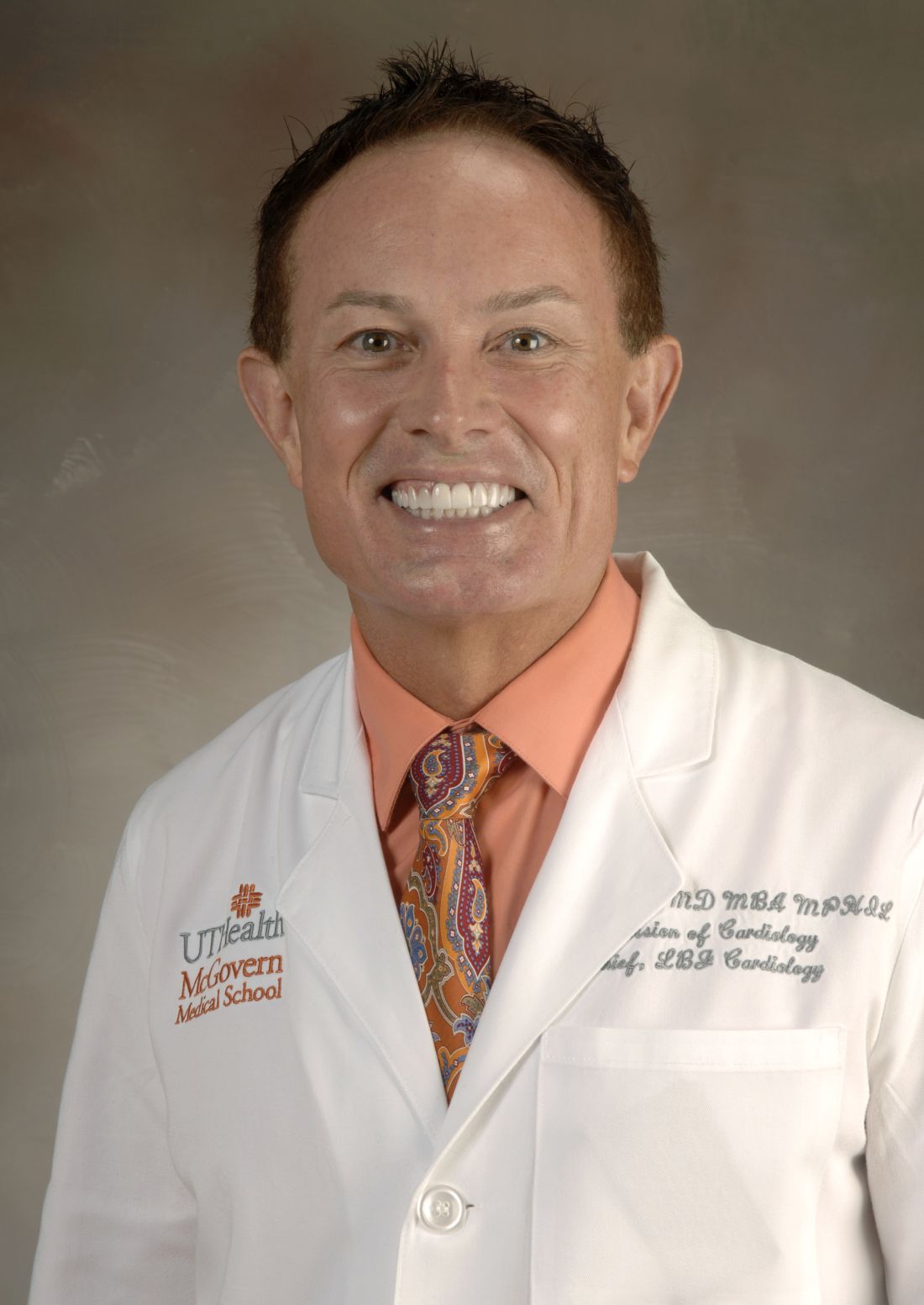
Offering an outsider’s perspective, John P. Higgins, MD, MBA, MPhil, a sports cardiologist at McGovern Medical School at The University of Texas Health Science Center at Houston, said, “Think of atrial fibrillation as a barometer of underlying stress on the heart. When blood pressure is high, or a patient has underlying asymptomatic coronary artery disease or heart failure, they are more likely to have episodes of atrial fibrillation.”
According to Dr. Higgins, risk factors for AF are underappreciated in the United States and elsewhere, and primary care doctors need to be aware of them. “We should try to identify these risk factors and do primary prevention to improve risk factors to reduce the progression to heart failure and myocardial infarction and stroke. But lifelong prevention is even better, he added. “Doing things to prevent actually getting risk factors in the first place. So a healthy lifestyle including exercise, diet, hydration, sleep, relaxation, social contact, and a little sunlight might be the long-term keys and starting them at a young age, too.”
In an accompanying editorial, Jianhua Wu, PhD, a professor of biostatistics and health data science with the Wolfson Institute of Population Health at Queen Mary University of London, and a colleague, cited the study’s robust observational research and called the analysis noteworthy for its quantification of the long-term risks of post-AF sequelae. They cautioned, however, that its grouping into two 10-year periods (2000-2010 and 2011-2020) came at the cost of losing temporal resolution. They also called out the lack of reporting on the ethnic composition of the study population, a factor that influences lifetime AF risk, and the absence of subgroup analysis by socioeconomic status, which affects incidence and outcomes.
The editorialists noted that while interventions to prevent stroke dominated AF research and guidelines during the study time period, no evidence suggests these interventions can prevent incident heart failure. “Alignment of both randomised clinical trials and guidelines to better reflect the needs of the real-world population with atrial fibrillation is necessary because further improvements to patient prognosis are likely to require a broader perspective on atrial fibrillation management beyond prevention of stroke,” they wrote.
In the meantime this study “challenges research priorities and guideline design, and raises critical questions for the research and clinical communities about how the growing burden of atrial fibrillation can be stopped,” they wrote.
This work was supported by the Danish Cardiovascular Academy, which is funded by the Novo Nordisk Foundation, and The Danish Heart Foundation. Dr. Vinter has been an advisory board member and consultant for AstraZeneca and has an institutional research grant from BMS/Pfizer unrelated to the current study. He reported personal consulting fees from BMS and Pfizer. Other coauthors disclosed research support from and/or consulting work for private industry, as well as grants from not-for-profit research-funding organizations. Dr. Higgins had no competing interest to declare. The editorial writers had no relevant financial interests to declare. Dr. Wu is supported by Barts Charity.
FROM BMJ
The lifetime risk of atrial fibrillation (AF) increased from 2000 to 2022 from one in four to one in three, a Danish population-based study of temporal trends found.
Heart failure was the most frequent complication linked to this arrhythmia, with a lifetime risk of two in five, twice that of stroke, according to investigators led by Nicklas Vinter, MD, PhD, a postdoctoral researcher at the Danish Center for Health Service Research in the Department of Clinical Medicine at Aalborg University, Denmark.
Published in BMJ, the study found the lifetime risks of post-AF stroke, ischemic stroke, and myocardial infarction improved only modestly over time and remained high, with virtually no improvement in the lifetime risk of heart failure.
“Our work provides novel lifetime risk estimates that are instrumental in facilitating effective risk communication between patients and their physicians,” Dr. Vinter said in an interview. “The knowledge of risks from a lifelong perspective may serve as a motivator for patients to commence or intensify preventive efforts.” AF patients could, for example, adopt healthier lifestyles or adhere to prescribed medications, Dr. Vinter explained.
“The substantial lifetime risk of heart failure following atrial fibrillation necessitates heightened attention to its prevention and early detection,” Dr. Vinter said. “Furthermore, the high lifetime risk of stroke remains a critical complication, which highlights the importance of continuous attention to the initiation and maintenance of oral anticoagulation therapy.”
The Study
The cohort consisted of 3.5 million individuals (51.7% women) who did not have AF as of age 45 or older. These individuals were followed until incident AF, migration, death, or end of follow-up, whichever came first.
All 362,721 individuals with incident AF (53.6% men) but no prevalent complication were further followed over two time periods (2000-2010 and 2011-2020) until incident heart failure, stroke, or myocardial infarction.
Among the findings:
- Lifetime AF risk increased from 24.2% in 2000-2010 to 30.9% in 2011-2022, for a difference of 6.7% (95% confidence interval [CI], 6.5%-6.8%).
- Lifetime AF risk rose across all subgroups over time, with a larger increase in men and individuals with heart failure, myocardial infarction, stroke, diabetes, and chronic kidney disease.
- Lifetime risk of heart failure was 42.9% in 2000-2010 and 42.1% in 2011-2022, for a difference of −0.8% (95% CI, −3.8% to 2.2%).
- The lifetime risks of post-AF stroke and of myocardial infarction decreased slightly between the two periods, from 22.4% to 19.9% for stroke (difference −2.5%, 95% CI, −4.2% to −0.7%) and from 13.7% to 9.8% for myocardial infarction (−3.9%, 95% CI, −5.3% to −2.4%). No differential decrease between men and women emerged.
“Our novel quantification of the long-term downstream consequences of atrial fibrillation highlights the critical need for treatments to further decrease stroke risk as well as for heart failure prevention strategies among patients with atrial fibrillation,” the Danish researchers wrote.
Offering an outsider’s perspective, John P. Higgins, MD, MBA, MPhil, a sports cardiologist at McGovern Medical School at The University of Texas Health Science Center at Houston, said, “Think of atrial fibrillation as a barometer of underlying stress on the heart. When blood pressure is high, or a patient has underlying asymptomatic coronary artery disease or heart failure, they are more likely to have episodes of atrial fibrillation.”
According to Dr. Higgins, risk factors for AF are underappreciated in the United States and elsewhere, and primary care doctors need to be aware of them. “We should try to identify these risk factors and do primary prevention to improve risk factors to reduce the progression to heart failure and myocardial infarction and stroke. But lifelong prevention is even better, he added. “Doing things to prevent actually getting risk factors in the first place. So a healthy lifestyle including exercise, diet, hydration, sleep, relaxation, social contact, and a little sunlight might be the long-term keys and starting them at a young age, too.”
In an accompanying editorial, Jianhua Wu, PhD, a professor of biostatistics and health data science with the Wolfson Institute of Population Health at Queen Mary University of London, and a colleague, cited the study’s robust observational research and called the analysis noteworthy for its quantification of the long-term risks of post-AF sequelae. They cautioned, however, that its grouping into two 10-year periods (2000-2010 and 2011-2020) came at the cost of losing temporal resolution. They also called out the lack of reporting on the ethnic composition of the study population, a factor that influences lifetime AF risk, and the absence of subgroup analysis by socioeconomic status, which affects incidence and outcomes.
The editorialists noted that while interventions to prevent stroke dominated AF research and guidelines during the study time period, no evidence suggests these interventions can prevent incident heart failure. “Alignment of both randomised clinical trials and guidelines to better reflect the needs of the real-world population with atrial fibrillation is necessary because further improvements to patient prognosis are likely to require a broader perspective on atrial fibrillation management beyond prevention of stroke,” they wrote.
In the meantime this study “challenges research priorities and guideline design, and raises critical questions for the research and clinical communities about how the growing burden of atrial fibrillation can be stopped,” they wrote.
This work was supported by the Danish Cardiovascular Academy, which is funded by the Novo Nordisk Foundation, and The Danish Heart Foundation. Dr. Vinter has been an advisory board member and consultant for AstraZeneca and has an institutional research grant from BMS/Pfizer unrelated to the current study. He reported personal consulting fees from BMS and Pfizer. Other coauthors disclosed research support from and/or consulting work for private industry, as well as grants from not-for-profit research-funding organizations. Dr. Higgins had no competing interest to declare. The editorial writers had no relevant financial interests to declare. Dr. Wu is supported by Barts Charity.
Consider Skin Cancer, Infection Risks in Solid Organ Transplant Recipients
SAN DIEGO — because of their suppressed immune systems.
“There are over 450,000 people with a solid organ transplant living in the United States. If you do the math, that works out to about 40 organ transplant recipients for every dermatologist, so there’s a lot of them out there for us to take care of,” Sean Christensen, MD, PhD, associate professor of dermatology, Yale University, New Haven, Connecticut, said at the annual meeting of the American Academy of Dermatology (AAD). “If we expand that umbrella to include all types of immunosuppression, that’s over 4 million adults in the US.”
Dr. Christensen encouraged dermatologists to be aware of the varying risks for immunosuppressive drugs and best screening practices for these patients, and to take advantage of a validated skin cancer risk assessment tool for transplant patients.
During his presentation, he highlighted five classes of immunosuppressive drugs and their associated skin cancer risks:
- Calcineurin inhibitors (tacrolimus or cyclosporine), which cause severe immune suppression and pose a severe skin cancer risk. They may also cause gingival hyperplasia and sebaceous hyperplasia.
- Antimetabolites (mycophenolate mofetil or azathioprine), which cause moderate to severe immune suppression and pose a severe skin cancer risk.
- Mammalian target of rapamycin inhibitors (sirolimus or everolimus), which cause severe immune suppression and pose a moderate skin cancer risk. They also impair wound healing.
- Corticosteroids (prednisone), which cause mild to severe immune suppression and pose a minimal skin cancer risk.
- A decoy receptor protein (belatacept), which causes severe immune suppression and poses a mild skin cancer risk.
“Most of our solid-organ transplant recipients will be on both a calcineurin inhibitor and an antimetabolite,” Dr. Christensen said. “In addition to the skin cancer risk associated with immunosuppression, there is an additive risk” that is a direct effect of these medications on the skin. “That means our transplant recipients have a severely and disproportionate increase in skin cancer,” he noted.
Up to half of solid-organ transplant recipients will develop skin cancer, Dr. Christensen said. These patients have a sixfold to 10-fold increased risk for basal cell carcinoma (BCC), a 35- to 65-fold increased risk for squamous cell carcinoma (SCC), a twofold to sevenfold increased risk for melanoma, and a 16- to 100-fold increased risk for Merkel cell carcinoma.
Transplant recipients with SCC, he said, have a twofold to threefold higher risk for metastasis (4%-8% nodal metastasis) and twofold to fivefold higher risk for death (2%-7% mortality) from SCC.
As for other kinds of immunosuppression, HIV positivity, treatment with 6-mercaptopurine or azathioprine (for inflammatory bowel disease and rheumatoid arthritis), and antitumor necrosis factor agents (for psoriasis, inflammatory bowel disease, and rheumatoid arthritis) have been linked in studies to a higher risk for nonmelanoma skin cancer.
Dr. Christensen also highlighted graft-versus-host disease (GVHD). “It does look like there is a disproportionate and increased risk of SCC of the oropharynx and of the skin in patients who have chronic GVHD. This is probably due to a combination of both the immunosuppressive medications that are required but also from chronic and ongoing inflammation in the skin.”
Chronic GVHD has been linked to a 5.3-fold increase in the risk for SCC and a twofold increase in the risk for BCC, he added.
Moreover, new medications for treating GVHD have been linked to an increased risk for SCC, including a 3.2-fold increased risk for SCC associated with ruxolitinib, a Janus kinase (JAK) 1 and JAK2 inhibitor, in a study of patients with polycythemia vera and myelofibrosis; and a case report of SCC in a patient treated with belumosudil, a rho-associated coiled-coil-containing protein kinase-2 kinase inhibitor, for chronic GVHD. Risk for SCC appears to increase based on duration of use with voriconazole, an antifungal, which, he said, is a potent photosynthesizer.
Dr. Christensen also noted the higher risk for infections in immunocompromised patients and added that these patients can develop inflammatory disease despite immunosuppression:
Staphylococcus, Streptococcus, and Dermatophytes are the most common skin pathogens in these patients. There’s a significantly increased risk for reactivation of herpes simplex, varicella-zoster viruses, and cytomegalovirus. Opportunistic and disseminated fungal infections, such as mycobacteria, Candida, histoplasma, cryptococcus, aspergillus, and mucormycosis, can also appear.
More than 80% of transplant recipients develop molluscum and verruca vulgaris/human papillomavirus infection. They may also develop noninfectious inflammatory dermatoses.
Risk Calculator
What can dermatologists do to help transplant patients? Dr. Christensen highlighted the Skin and UV Neoplasia Transplant Risk Assessment Calculator, which predicts skin cancer risk based on points given for race, gender, skin cancer history, age at transplant, and site of transplant.
The tool, validated in a 2023 study of transplant recipients in Europe, is available online and as an app. It makes recommendations to users about when patients should have initial skin screening exams. Those with the most risk — 45% at 5 years — should be screened within 6 months. “We can use [the tool] to triage these cases when we first meet them and get them plugged into the appropriate care,” Dr. Christensen said.
He recommended seeing high-risk patients at least annually. Patients with a prior SCC and a heavy burden of actinic keratosis should be followed more frequently, he said.
In regard to SCC, he highlighted a 2024 study of solid organ transplant recipients that found the risk for a second SCC after a first SCC was 74%, the risk for a third SCC after a second SCC was 83%, and the risk for another SCC after five SCCs was 92%.
Dr. Christensen disclosed relationships with Canfield Scientific Inc. (consulting), Inhibitor Therapeutics (advisory board), and Sol-Gel Technologies Ltd. (grants/research funding).
A version of this article first appeared on Medscape.com.
FROM AAD 2024
ACC Consensus Guidance on What’s New in HFrEF Treatment
The American College of Cardiology has published a new update to its consensus decision pathway for the treatment of heart failure with reduced ejection fraction (HFrEF).
Chair of the consensus document Writing Committee Thomas M. Maddox, MD, explained to this news organization that this new Decision Pathway provides a practical, streamlined update to frontline clinicians treating patients with heart failure and incorporates evidence from the 2022 AHA/ACC/HFSA Guideline for the Management of Heart Failure.
“While the AHA/ACC/HFSA Guidelines are wonderful in that they collate all the latest scientific evidence, they don’t speak as much to the practicalities of delivering the care. This is what this Decision Pathway document comes in — it is designed to help frontline clinicians with the practical reality of managing these patients,” Dr. Maddox, who is director of the Healthcare Innovation Lab at BJC HealthCare and the Washington University School of Medicine in St Louis, Missouri, commented.
The document, “Expert Consensus Decision Pathway for Optimization of Heart Failure Treatment: Answers to 10 Pivotal Issues About Heart Failure With Reduced Ejection Fraction,” was published online on March 8 in the Journal of the American College of Cardiology.
The authors provided guidance on introducing the numerous evidence-based therapies now available for HFrEF, improving adherence, overcoming treatment barriers, acknowledging contraindications and situations for which few data exist, affording expensive therapies, treating special cohorts, and making the transition to palliative care.
Rather than focusing on extensive text, the document provided practical tips, tables, and figures to make clear the steps, tools, and provisos needed to treat patients with heart failure successfully and expeditiously, they added.
Dr. Maddox reported that there are three main updated areas of advice on the treatment of heart failure in the new document.
Valsartan/Sacubitril First Line
One of the major changes involves an elevation for the status of the angiotensin receptor-neprilysin inhibitor (ARNI), Entresto (valsartan/sacubitril).
“It is now clear that this agent is superior to ACE-inhibitors or angiotensin receptor blockers in terms of reducing heart failure hospitalization and death, whereas previously it was seen as somewhat equivalent,” Dr. Maddox said. “So, barring a contraindication or another problem with getting the medication, this agent should be one of the first line medicines for all patients with heart failure and a reduced ejection fraction.”
Dual Sodium-Glucose Cotransporter 1/2 (SGLT1/2) Inhibitor
A second update involves the addition of sotagliflozin (a dual inhibitor of both SGLT1 and SGLT2) to the SGLT2 inhibitors as another first-line medication for patients with heart failure and reduced ejection fraction.
“We now have evidence that both SGLT2 and SGLT1 inhibitors are beneficial in reducing heart failure hospitalization and death. Previously we only had evidence on SGLT2 inhibitors — dapagliflozin and empagliflozin. Sotagliflozin is a newer agent, which inhibits both SGLT1 and SGLT2, and it turns out that inhibiting both are beneficial in heart failure. So, this gives us a third med in this category,” Dr. Maddox noted.
Rapid Initiation of the Four Pillars of Therapy
The document stated that more data have emerged recently to support early and rapid initiation and titration of the “four pillars” of medical therapy in heart failure to maximize the benefits of patient-reported outcomes and reduction in hospitalizations and mortality.
The four pillars of therapy are ARNI, a beta-blocker, a mineralocorticoid antagonist, and an SGLT inhibitor.
As an example, four-class medication initiation reduced the hazard of cardiovascular death or hospital admission for heart failure significantly (hazard ratio, 0.38) compared with therapy with just an angiotensin-converting enzyme inhibitor/angiotensin receptor blocker plus a beta-blocker, the document reported.
“What we realize now is that the more quickly we can get patients on all four of these drug classes and escalate to target doses or maximally tolerated doses ideally within 3 months, the better the outcome,” Dr. Maddox said.
“Unfortunately, right now there is very incomplete realization and recognition of that in clinical practice. So, we are trying to highlight the importance of this to encourage clinicians to be more aggressive in making this happen.”
“In all patients with heart failure and reduced ejection fraction, getting them on all four of these medicines as quickly as possible will give the best outcome. We’ve seen evidence in support of this from several broad population trials,” he added. “There are times when they can’t take all four but we should do our best to get there.”
Practical Considerations
Dr. Maddox pointed out that the Consensus Document is also trying to account for practical realities and barriers to heart failure treatment.
“When we think about these recommendations — and evidence that getting patients on all these medicines is valuable, we also focus on the fact that there are three major barriers that can get in the way of this and how to think about overcoming those barriers,” he said.
The barriers are comorbidities/side effects of medications, costs of the medicines, and systems of care that are needed to ensure patients can be treated with multiple medications in a timely fashion.
In terms of comorbidities/side effects, Dr. Maddox explained that patients with heart failure are generally older and are likely to have other comorbidities. “The more medicines we give, the more likely we are to run into side effects. So, we have produced some guidance on how to monitor for adverse effects and ways to mitigate these effects so the guideline recommended therapies can be continued without creating new harms.”
He gave the example of mineralocorticoid antagonists, which can sometimes elevate potassium levels, particularly if there is some underlying kidney disease, so clinicians are advised to recommend a low-potassium diet for these patients or the use of potassium binding agents that will also lower the amount of potassium in the blood stream; in this way, patients are able to continue the mineralocorticoid antagonist.
On costs, Dr. Maddox noted that the valsartan/sacubitril combination drug and SGLT inhibitors are new medicines and are expensive.
“They can be prohibitively expensive for patients who have suboptimal pharmacy benefits or who are uninsured.”
The Consensus Document therefore provided some guidance on ways to identify rebate programs, access insurance, and find different pathways to obtaining those drugs at a more reasonable price. It also advocated for policy changes to allow these medicines to be more accessible to more people.
More Use of Digital Tools
On the issue of systems of care, Dr. Maddox noted that the preexisting model of delivering care, which almost always involves the patient coming into the doctor’s office, invokes a high burden on both the system and most especially, the patient.
“Patients do not want to come back and forth to the doctor’s office multiple times in a few weeks. This is often a nonstarter, particularly for patients with busy lives,” he commented.
The Consensus Document advised more use of digital tools to provide remote care and contact with patients including sensors that can measure variables such as heart rate and blood pressure and video appointments.
“We are still working out what are the right models of care and how they can be performed safely and how they can be funded. But I think at the end of the day, this will give us more practical ways of getting people on multiple heart failure medicines and monitoring them safely without causing an undue burden for them logistically,” Dr. Maddox said.
He pointed out that there are a record number of medicines now available to treat heart failure, and while this is welcome, many of these patients are also on multiple other medications for other comorbidities as well.
“If you start giving patients seven, eight, or nine different medicines that they have to take every day, sometimes multiple times a day — that’s complicated medically, logistically, and financially. The potential for interaction and complications increases with every additional medication.”
Dr. Maddox also noted that patients have limits on how many medications they will accept. “It really helps if we have an engaged patient who has a good relationship with the care team to try to develop the right treatment plan that is going to meet their needs and give them the best possible health outcomes.”
It can take many visits to get the patient on all these medications and then up-titrate to target doses.
“We try and do a couple of things in each appointment. Often, we tend to start one or maybe two drugs at a time at a relatively low dose to avoid side effects, so we can be talking about 12-16 different encounters in total,” he said.
He recommended making a plan and the use of new technologies to manage each incremental step.
A Team Approach
Another issue that is discussed in the document is the use of a healthcare team to manage all the necessary appointments.
“It is no longer practical that one person can be the engineer for all this. It should be a team effort,” Dr. Maddox stated.
Responsibilities can be allocated across physicians, nurses, pharmacists, and even case managers, so that the team can take more of a population approach and develop a system to get patients on the multiple medications as quickly as possible.
“While this can still be quite a big burden for the patient, we need to figure out a system to make this as palatable as possible for them. Practices need to tailor this themselves according to what resources they have,” he added.
While most new patients will be routed to cardiologists to start their treatment plans, once on their initial medications and these have been up titrated to target levels, they should be able to be managed by primary care doctors, who will have the most holistic view of the patient and their other comorbidities, Dr. Maddox advised.
“Following this guidance should lead to more patients receiving evidence-based care which leads to better health outcomes, but delivered in a practical way that fits with their life reality and logistical needs,” he concluded.
A version of this article appeared on Medscape.com.
Many Older Adults Don’t Receive Palliative Care Before Death
A prognostic tool may facilitate the early identification of older adults in the community who would benefit from palliative care in their final years, new research from Canada suggested.
The analysis of data from close to a quarter million community-dwelling older adults in Ontario with at least one interRAI (Resident Assessment Instrument) home care assessment showed that only half of those with an estimated survival of fewer than 3 months received at least one palliative home care visit before death.
“One of the challenges and a barrier to accessing palliative home care is the difficulty of predicting survival,” Amy Hsu, PhD, an investigator at the Bruyère Research Institute in Ottawa, Ontario, Canada, told this news organization. “Clinicians are good at prognosticating when a patient might be entering their last 3-6 weeks of life, but they have a harder time predicting if someone will survive 6 months or longer.”
The team developed the Risk Evaluation for Support: Predictions for Elder-life in their Communities Tool (RESPECT) to see whether access to predicted survival data could inform conversations about a patient’s status and palliative care needs.
The study was published online in the Canadian Medical Association Journal.
Setting Care Goals
Researchers analyzed population health administrative data from Ontario involving home care clients who received at least one interRAI Home Care assessment between April 2018 and September 2019. The cohort included 247,377 adults (62% women) with a mean age of 80.1 years at the time of assessment. Comorbidities, including congestive heart failure, coronary artery disease, cancer, and chronic obstructive pulmonary disease, as well as symptoms of health instability, were more prevalent among those at higher risk of dying.
The team used an updated, validated version of RESPECT to predict survival.
Only 2.6% of home care clients had received a clinician diagnosis of an end-stage disease, which was more prevalent among those at highest mortality risk (77.9%). Most clients (74.5%) required extensive assistance in performing instrumental activities of daily living (ADLs, score ≤ 4), and half (50.3%) were less able to perform ADLs in the last 3 months of life.
Within the cohort, 75% of patients with a predicted median survival of fewer than 3 months, 55.4% of those with a predicted median survival between 3 and 6 months, and 40.7% of those with a predicted median survival between 6 and 12 months died within 6 months of the home care assessment.
Among decedents, 50.6% of those with a RESPECT-estimated median survival of fewer than 3 months received at least one nonphysician palliative home care visit before death. Less than a third (27.8%) received at least one palliative home care visit from a physician.
The proportion of those who received at least one nonphysician visit fell to 38.7% among those with a median survival of between 3 and 6 months and to 29.5% among those with a median survival of between 6 and 12 months.
Patients who received at least one palliative home care visit (from either physicians or nonphysician home care providers) within 6 months of an assessment had clinical characteristics similar to those who did not receive a visit. However, those who did not receive palliative home care were more likely to not have been identified by a clinician as being in their past 6 months of life.
“These results reinforce the role of clinicians in identifying older adults who may be in their last 6 months of life as an important component for the receipt of palliative home care and highlight the value of RESPECT in supplementing clinicians’ assessments of prognosis,” the authors wrote.
“Our goal is to use data and tools like RESPECT to help individuals living with a life-limiting illness have conversations about what their end-of-life care goals and wishes may be and discuss whether a referral to palliative care is appropriate or needed,” Dr. Hsu added. “Data about life expectancy could be helpful for framing these conversations.”
The researchers are working with partners in home, community care, and long-term care to implement RESPECT in their settings.
‘Valuable Tool’
Guohua Li, MD, DrPH, professor of epidemiology and anesthesiology at Columbia University Mailman School of Public Health and Vagelos College of Physicians and Surgeons in New York City, commented on the findings for this news organization. He noted that the study is “rigorously designed and meticulously analyzed. The findings are of high validity and population health significance.”
The findings are comparable with what is seen in the United States and Canada, he said, where about 50% of terminally ill patients die at home or in hospice. However, palliative care outside of North America “varies greatly, and in many developing countries, [it] is still in its infancy.”
As a mortality risk prediction algorithm, “RESPECT seems to perform reasonably well,” he said. “If incorporated into the electronic health record, it could be a valuable tool for clinicians to identify patients with less than 6 months of life expectancy and deliver palliative care to these patients. RESPECT appears to be particularly beneficial for home care patients without a clinically diagnosed terminal disease.”
That said, he added, “RESPECT should be viewed as a clinical decision support tool. It is no substitute for clinicians or clinical judgment. Based on the data presented in the paper, the algorithm tends to overestimate the short-term mortality risk for home care patients, therefore resulting in many false alarms.”
The study was supported by the Canadian Institutes of Health Research and the Associated Medical Services. Dr. Hsu is an executive lead on the steering committee of the Ontario Centres for Learning, Research, and Innovation in Long-Term Care. Funding for the centers comes from the Ontario Ministry of Health and Ministry of Long-Term Care and is partially administered by the Bruyère Research Institute. Dr. Li reported no relevant financial interests.
A version of this article appeared on Medscape.com.
FROM THE CANADIAN MEDICAL ASSOCIATION JOURNAL
FDA Clears Medical Grade Over-the-Counter Pulse Oximeter
The MightySat Medical, an over-the-counter medical fingertip pulse oximeter, has received clearance from the US Food and Drug Administration (FDA) for use without a prescription, according to a press release from manufacturer Masimo.
The device is the first medical fingertip pulse oximeter available directly to consumers without a prescription that includes the same technology used by many hospitals, according to the company.
According to the FDA, home pulse oximeters are currently generally of two classes: hospital-grade prescription devices which have been vetted for accuracy through clinical trials, and over-the-counter devices which are sold direct to consumers but often estimate oxygen saturation. FDA communication on pulse oximeter accuracy states "OTC oximeters that are sold as either general wellness or sporting/aviation products are not intended for medical purposes, so they do not undergo FDA review."
Pulse oximeter use is important for patients diagnosed with breathing problems or lung diseases such as asthma, chronic obstructive pulmonary disease, pulmonary fibrosis, lung cancer, flu, pneumonia, or COVID-19 to collect accurate data on arterial blood oxygen saturation that they can share with their healthcare providers, according to the company. Patients with cardiac conditions, including pulmonary hypertension and heart failure may also benefit from pulse oximeter monitoring.
However, challenges of pulse oximeter use include measuring accuracy when patients are moving, measuring patients with poor circulation, and measuring patients with cool, thick, or darker skin. The MightySat Medical is designed to provide reliable measures of oxygen saturation and pulse rate across all patient groups, the manufacturers wrote in the press release.
Asked for additional comment, Diego J. Maselli, MD, FCCP, Professor and Chief in the division of Pulmonary Diseases and Critical Care at UT Health at San Antonio, noted, "Over the past decades, there has been an increased interest in home monitoring of medical conditions, particulrly with the development of more portable and accessible technology."
"This was heightended by the COVID-19 pandemic where telemedicine was frequently required as a means of delivering care," Dr. Maselli continued. "One of the important characteristics to monitor was the oxgen saturation in patients that had an active COVID-19 infection as it would dictate management and was part of the protocol for monitoring the clinical course of infection. Because of this need, many companies developed portable pulse oximeters for home use. This resulted in widespread use of pulse oximeters at home and other places outside clinic or hospital."
Other over-the-counter pulse oximeters that are not cleared by the FDA may create confusion among patients about the accuracy of their measurements, according to the company.
Dr. Maselli also commented that pulse oximeters' value can vary. "Unfortunately, these devices vary in quality and reliability and patients may not be fully aware of this. Most recently, the FDA approved a hospital-grade pulse oximeter that requires no prescription. This device may provide a more accurate reading in a wide range of clinical situations outside the healthcare setting. Patients should be aware that there are different grades of pulse oximeter before selecting one for home use. In addition, patients should work closely with their providers to better select the monitoring modaility that best fits their clinical situation," he said.
MightySat Medical is indicated for individuals aged 18 years and older who are well or poorly perfused under no motion conditions and is not intended as a diagnostic or screening tool for lung disease, according to the release. Treatment decisions based on data from the device should be made only in consultation with a healthcare provider, the company said. Dr. Maselli serves as a member of the CHEST Physician editorial board.
The FDA’s website offers further guidance related to at-home pulse oximeter use, with recommendations and limitations, as well as information on initiatives to ensure accurate and equitable pulse oximetry for all patients.
A version of this article appeared on Medscape.com.
Just 2000 Steps a Day Linked to Reduced Heart Failure Risk
TOPLINE:
Accelerometer-measured physical activity (PA), whether light, moderate, or vigorous, is associated with lower risk for heart failure (HF) in older women while more sedentary time is associated with higher HF risk in these women, results of a new study suggest.
METHODOLOGY:
- The analysis included 5951 women aged 63-99 years (mean age, 78.6 years), including 33.7% Black, 17.2% Hispanic, and 49.2% White individuals without HF from the Objective Physical Activity and Cardiovascular Health (OPACH) study, an ancillary to the Women’s Health Initiative Long-Life Study.
- Participants wore an accelerometer on their hip 24 hours a day for up to 7 consecutive days except when in water, kept nightly sleep logs, completed questionnaires to provide information on medical history and sociodemographic and lifestyle factors, and self-rated their general health status.
- Researchers recorded their use of assistive walking devices; determined body mass index as well as blood pressure; obtained fasting serum glucose, total and high-density lipoprotein cholesterol, triglyceride, and high-sensitivity C-reactive protein concentrations; and scored participants’ multimorbidity.
- They determined intensity-specific PA using vector magnitude acceleration cut points (light PA, 19-518 counts/15 s; moderate to vigorous PA [MVPA], > 518) and steps per day using dedicated software, and they quantified sedentary time (total and mean bout duration).
- The primary outcome was overall self-reported HF later adjudicated by physicians using medical record reviews; secondary endpoints were heart failure with reduced ejection fraction (HFrEF) and preserved EF (HFpEF), classified by an EF of < 45% or 45% or > 45%, respectively, after cardiac imaging.
TAKEAWAY:
- A total of 407 HF cases (including 257 HFpEF and 110 HFrEF) were identified during a mean of 7.5 years of follow-up.
- HFrEF was not associated with PA measures in the fully adjusted model (which controlled extensively for health and physical functioning status), but overall HF and HFpEF were inversely associated with total PA (per 1-standard deviation [SD] increment: hazard ratio [HR] 0.85; 95% CI, 0.75-0.95 and HR, 0.78; 95% CI, 0.67-0.91, respectively), light PA (HR, 0.88; 95% CI, 0.78-0.98 and HR, 0.80; 95% CI, 0.70-0.93, respectively) and MVPA (HR, 0.84; 95% CI, 0.73-0.97 and HR, 0.85; 95% CI, 0.72-1.01, respectively).
- With regard to daily steps, each 1-SD increment was associated with a significant 26% lower risk for overall HF (HR 0.74; 95% CI, 0.63-0.88) and 29% lower risk for HFpEF (HR, 0.71; 95% CI, 0.57-0.88), with these inverse risks becoming significant at about 2000 steps/d, “far less than the often touted 10,000 steps/d for promoting health benefits,” noted the authors.
- but not HFrEF; mean sedentary bout duration was significantly inversely associated with HFrEF (per 1 SD: HR, 0.76; 95% CI, 0.61-0.97), although the relatively small number of cases at the extremes of bout duration may contribute to this unexpected inverse association, said the authors.
IN PRACTICE:
The implications of promoting PA, regardless of intensity, for primary HFpEF prevention in later life, “has profound public health and clinical relevance,” the authors concluded. They noted that HFpEF is a “burgeoning epidemic” that disproportionately affects women and minorities with limited available therapies.
STUDY DETAILS:
The study, led by Michael J. LaMonte, PhD, MPH, University at Buffalo-SUNY, Buffalo, New York, was published online on February 21, 2024, in JAMA Cardiology.
LIMITATIONS:
There was only a single accelerometer assessment of PA and sedentary exposures and relatively small numbers of HFrEF cases, which restricted analytic precision. Although researchers controlled for several established vascular biomarkers, they did not have HF-specific measures such as cardiac troponin or N-terminal pro–brain natriuretic peptide. It’s unknown if the findings can be generalized to men and populations dissimilar to women in OPACH.
DISCLOSURES:
The study was funded in part by the National Heart, Lung, and Blood Institute, National Institutes of Health, and US Department of Health and Human Services. LaMonte reported receiving grants from the National Institutes of Health during the conduct of the study; see paper for disclosures of the other authors.
A version of this article appeared on Medscape.com.
Spinal Cord Injury Tied to Greater Risk for Heart Disease
TOPLINE:
Spinal cord injury (SCI) is associated with a significantly greater risk for heart disease than that of the general non-SCI population, especially among those with severe disability, new observational data suggest.
METHODOLOGY:
- Researchers analyzed data from Korea’s National Health Insurance Service on 5083 patients with cervical, thoracic, or lumbar SCI (mean age, 58; 75% men) and 1:3 age- and sex-matched non-SCI controls.
- The study endpoint was new-onset myocardial infarction (MI), heart failure (HF), or atrial fibrillation (AF) during a mean follow-up of 4.3 years.
- Covariates included low income, living in an urban or rural area, alcohol consumption, smoking status, physical activity engagement, body mass index, and blood pressure; comorbidities included hypertension, type 2 diabetes, and dyslipidemia.
TAKEAWAY:
- A total of 169 MI events (7.3 per 1000 person-years), 426 HF events (18.8 per 1000 person-years), and 158 AF events (6.8 per 1000 person-years) occurred among SCI survivors.
- After adjustment, SCI survivors had a higher risk for MI (adjusted hazard ratio [aHR], 2.41), HF (aHR, 2.24), and AF (aHR, 1.84) than that of controls.
- Cervical and lumbar SCI survivors had an increased risk for heart disease compared with controls regardless of disability, and the risk was slightly higher for those with a disability; for cervical SCI survivors with a disability, aHRs for MI, HF, and AF, respectively, were 2.30, 2.05, and 1.73; for lumbar SCI survivors with a disability, aHRs were 2.79, 2.35, and 2.47.
- Thoracic SCI survivors with disability had a higher risk for MI (aHR, 5.62) and HF (aHR, 3.31) than controls.
IN PRACTICE:
“[T]he recognition and treatment of modifiable cardiovascular risk factors must be reinforced in the SCI population, [and] proper rehabilitation and education should be considered to prevent autonomic dysreflexia or orthostatic hypotension,” the authors wrote.
In an accompanying editorial, Christopher R. West, PhD, and Jacquelyn J. Cragg, PhD, both of the University of British Columbia, Vancouver, Canada, noted that clinical guidelines for cardiovascular and cardiometabolic disease after SCI don’t include approaches to help mitigate the risk for cardiac events such as those reported in the study; therefore, they wrote, the findings “should act as ‘call-to-arms’ to researchers and clinicians to shift gears from tradition and begin studying the clinical efficacy of neuraxial therapies that could help restore autonomic balance [in SCI], such as targeted neuromodulation.”
SOURCE:
The study was led by Jung Eun Yoo, MD, PhD of Seoul National University College of Medicine, Seoul, South Korea, and published online on February 12 in the Journal of the American College of Cardiology.
LIMITATIONS:
The database was not designed for the SCI population, so data are incomplete. The incidence of thoracic SCI was particularly low. Because SCI survivors may have impaired perception of chest pain in ischemic heart disease, those with asymptomatic or silent heart disease may not have been captured during follow-up. All study participants were Korean, so the findings may not be generalizable to other ethnicities.
DISCLOSURES:
This research was partially supported by a grant from the Korea Health Technology R&D Project through the Korea Health Industry Development Institute, funded by the Ministry of Health and Welfare, South Korea. The study authors and the editorialists had no relevant relationships to disclose.
A version of this article appeared on Medscape.com.