User login
Fitness Curbs Mortality in Men With Diabetes
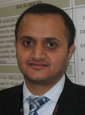
HOUSTON – Physical fitness nearly halved the risk of death in men with type 2 diabetes, regardless of whether they had left ventricular hypertrophy, a longitudinal study of 866 patients found.
During a follow-up period as long as 24 years (with a median 9-year follow-up), 236 men who had left ventricular hypertrophy and were within the lower 50th percentile of physical fitness were 20% more likely to die, compared with 225 men in a reference group who did not have left ventricular hypertrophy and also had a low level of fitness. The difference in mortality risk between these two groups did not reach statistical significance.
In contrast, compared with the reference group, the risk of death was 41% lower in 218 men who were physically fit (in the upper 50th percentile) and did not have left ventricular hypertrophy and 43% lower in 187 men who were fit and did have left ventricular hypertrophy, Dr. Khaled Alswat and his associates reported in a poster presentation at the annual meeting of the Endocrine Society.
The risk reductions in the two fit groups were statistically significant.
"We, as doctors, do a lot of testing, and one of those tests should be an exercise test" for patients with diabetes who have an increased risk for heart disease, said Dr. Alswat, an endocrinology fellow at Veterans Affairs Medical Center and George Washington University, Washington. An exercise stress test provides objective measures that physicians can use to work with patients on improving their physical fitness, he said in an interview.
The patients underwent a standardized exercise stress test and echocardiographic evaluation at the VA Medical Center during 1986-2011. Those who had a peak exercise capacity of at least six metabolic equivalence tasks (METs) were considered fit, and the rest were defined as having a low level of fitness (within the lower 50th percentile).
Left ventricular hypertrophy was defined by a left ventricular mass index, calculated by dividing left ventricular mass by height in meters to the power of 2.7. A left ventricular mass index greater than 48 g/m2.7 indicated left ventricular hypertrophy.
During follow-up, 346 patients died, for an annual death rate of 4%.
Smoking significantly increased mortality risk by 54%, a multivariate Cox proportional hazards analysis showed. The study controlled for the potential influences of age, body mass index, hypertension, smoking, and medications.
The adjusted mortality risk declined by 17% for every one-MET increase in fitness, Dr. Alswat reported.
Previous studies had shown that higher exercise capacity was associated with lower mortality risk in people with diabetes, but it had not been clear whether this applies to people with diabetes and left ventricular hypertrophy, he said.
Dr. Alswat reported having no relevant financial disclosures.
AT THE ANNUAL MEETING OF THE ENDOCRINE SOCIETY
Major Finding: Among men with diabetes, physical fitness decreased mortality risk by 43% in those with (and by 41% in those without) left ventricular hypertrophy compared with men without left ventricular hypertrophy who had a low level of fitness.
Data Source: Longitudinal study of 866 men who underwent exercise stress tests and echocardiographic evaluations at one institution from 1986 to 2011 and were followed for a median of 9 years.
Disclosures: Dr. Alswat reported having no relevant financial disclosures.
2-D Echo Is Inadequate Cardiomyopathy Screen in Childhood Cancer Survivors
Transthoracic two-dimensional echocardiography appears to be inadequate for identifying cardiomyopathy in adults who survive childhood cancer, according to a cross-sectional study published online July 16 in the Journal of Clinical Oncology.
Compared with cardiac magnetic resonance imaging (CMRI), which is considered the reference standard to which other cardiac imaging techniques are compared, 2-D echocardiography had a sensitivity of only 25% and a false-negative rate of 75% in identifying cardiomyopathy in a study of 134 adult survivors of childhood cancer, said Dr. Gregory T. Armstrong of the department of epidemiology and cancer control at St. Jude Children’s Research Hospital, Memphis, and his associates.
In these relatively young and apparently healthy study subjects who had never been diagnosed as having any cardiac abnormality, nearly half (48%) were found to have the reduced cardiac mass indicative of cancer therapy–related injury. And fully 11% of subjects who were judged to have a normal ejection fraction (EF) on 2-D echocardiography were actually proved to have an EF of less than 50% on CMRI, the researchers noted.
That number easily could have been higher, but there happened to be a low absolute number of patients (16) with this degree of EF impairment in the small cohort, they pointed out.
Adults who survive childhood cancer are at risk for cardiomyopathy because of their exposure to chemotherapy and radiotherapy. Current guidelines recommend screening such adults by transthoracic 2-D echocardiography because it is noninvasive, widely available, and less expensive than other techniques.
However, the quality of the acoustic windows obtained on 2-D echo varies widely, and the method depends on geometric assumptions that may not be valid in patients who have dilated or remodeled ventricles. Three-dimensional echocardiography yields somewhat more accurate results but is not as widely available. CMRI is the most accurate noninvasive imaging technique, but is more expensive and is even less widely available, Dr. Armstrong and his colleagues explained.
They assessed the accuracy of 2-D and 3-D echocardiography against CMRI as a screen for cardiomyopathy in a longitudinal cohort of 134 adults who had been treated at St. Jude’s for childhood cancer 18-38 years earlier. All had received chest-directed radiotherapy and/or anthracycline chemotherapy, both of which are known to impair cardiac function during treatment and to raise the risk of reduced left ventricular function later in life.
The most common pediatric malignancies were acute lymphoblastic leukemia (44 subjects) and Hodgkin’s lymphoma (37 subjects).
The median age at echocardiographic screening in adulthood was 39 years (range, 22-53 years).
Of the study subjects, 20 were unable to complete CMRI for a variety of reasons. Future studies that compare imaging techniques should take into consideration this relatively high noncompletion rate (15%) for CMRI, especially in cost-benefit analyses, Dr. Armstrong and his colleagues said (J. Clin. Oncol. 2012 July 16 [doi:10.1200/JCO.2011.40.3584]).
In the remaining 114 subjects, 2-D echocardiography consistently overestimated left ventricular ejection fraction (LVEF) and underestimated both end-systolic and end-diastolic ventricular volumes.
In all, 16 subjects were identified as having markedly decreased LVEF (50% or more) by CMRI, but only 4 of them were so identified by 2-D echocardiography and only 11 of them by 3-D echocardiography.
Compared with CMRI, the sensitivity of 2-D echocardiography was only 25%; that of 3-D echo was better but still inadequate, at only 53%. And false-negative rates were high with both 2-D echocardiography (75%) and 3-D echocardiography (47%).
Of particular concern was the finding that on CMRI, 32% of the study subjects had an LVEF that was well below normal. The rate in the subgroup of patients who had received both chest irradiation and anthracycline during childhood cancer treatment was even higher, at 42%.
A total of 48% of the study subjects had a cardiac mass that was at least 2 standard deviations below normal for their age and sex, a clear sign of cardiotoxicity from their childhood cancer treatment. "Notably, even patients who received less than 150 mg/m2 of anthracyclines had a high prevalence of reduced EF (27%), stroke volume (29%), or cardiac mass (56%)," the investigators said.
Estimates derived from Medicare data suggest that at roughly $449 each, CMRI examinations cost about $217 more than does echocardiography ($232 each). Given the high rate of cardiomyopathy discovered in this cohort, and the poor sensitivity of echocardiography as a screening tool, this cost difference may be small enough to warrant a switch in the current screening recommendations from echocardiography to CMRI.
The additional cost of a CMRI-only screening strategy per case of cardiotoxicity correctly identified would be only $1,973, they noted.
The study findings suggest that in this high-risk patient population that was exposed to cardiotoxic therapy during childhood, "consideration should be given to referring survivors with an EF of 50%-59% on [2-D echocardiography] for comprehensive cardiology assessment that includes cardiac history, symptom index, and examination; biomarker assessment; consideration of [CMRI]; functional assessment by treadmill testing; and possibly medical therapy to prevent progression of disease," Dr. Armstrong and his associates said.
This study was supported by the American Society of Clinical Oncology and the American Lebanese-Syrian Associated Charities. Dr. Armstrong’s associates reported ties to General Electric and Philips Healthcare.
FROM THE JOURNAL OF CLINICAL ONCOLOGY
Major Finding: Compared with cardiac MRI, 2-D echocardiography had only a 25% sensitivity at identifying cardiomyopathy and a 75% false-negative rate, whereas 3-D echo had only a 53% sensitivity and a 47% false-negative rate.
Data Source: A cross-sectional study of simultaneous assessment of cardiac structure and function using 2-D echo, 3-D echo, and CMRI in 134 adult survivors of childhood cancer who had no apparent cardiotoxicity from their cancer treatment.
Disclosures: This study was supported by the American Society of Clinical Oncology and the American Lebanese-Syrian Associated Charities. Dr. Armstrong’s associates reported ties to General Electric and Philips Healthcare.
Studies Clash on Cardiac Effects of TKIs in Kidney Cancer
CHICAGO – Take your pick: The tyrosine kinase inhibitors sunitinib and sorafenib do/do not appear to have significant cardiac toxicity when used in adjuvant therapy for renal cell carcinoma.
Conflicting studies presented at the annual meeting of the American Society of Clinical Oncology suggest that – for now at least – it’s a toss-up.

A cardiac substudy of the phase III ECOG (Eastern Cooperative Oncology Group) E2805 ASSURE (Adjuvant Sunitinib or Sorafenib for Unfavorable Renal Carcinoma) trial, comparing either sunitinib (Sutent) or sorafenib (Nexavar) with placebo in patients with resected renal cell carcinoma (RCC), showed that neither TKI was associated with significant declines in left ventricular ejection fraction (LVEF) or other cardiac adverse events when compared with placebo, said Dr. Naomi B. Haas of the University of Pennsylvania, Philadelphia.
Left ventricular dysfunction that did occur with the TKIs was reversible, and ischemic events were uncommon and not clearly linked to therapy, she added.
"The implications for patients: Further prospective study on the effects of these agents is needed in patients who have preexisting cardiac dysfunction. This was a well population we were looking at," said Dr. Haas.
However, a retrospective study by Dr. Phillip S. Hall and colleagues at Stanford (Calif.) University found evidence of significant cardiac toxicity in patients with metastatic renal cell carcinoma that was treated with both agents and with other targeted therapies at their institution.
"Cardiovascular toxicity is an important adverse event related to targeted-therapy administration. Close monitoring for the development of CV toxicity with the use of these agents should become standard of care, as early detection of asymptomatic patients could preempt symptomatic toxicity and reduce treatment-related morbidity and mortality," they wrote in a poster presentation.
TKIs on Trial
Previous studies, most of them retrospective, have reported cardiac dysfunction with TKI use ranging from 1% to 28%. The proposed mechanism of action is through the metabolic dysfunction of cardiac myocytes, Dr. Haas said.
She and her coinvestigators in the ECOG E2805 ASSURE trial looked at data from a cardiac substudy, and asked whether either sorafenib or sunitinib was associated with a decline in LVEF, clinically significant heart failure (HF) or other effects, using multigated acquisition scans (MUGA) at baseline and at 3, 6, and 12 months (study end) or at the end of treatment.
There were nine cases of the primary end point (a decline in LVEF of 16% or greater from baseline) among 397 patients on sunitinib, seven among 394 patients on sorafenib, and five among 502 patients on placebo. The respective event rates were 2.3%, 1.8%, and 1.0%; these differences were not clinically significant.
The numbers for other cardiac events – including LVEF decline of 16% or more below the institutional level of normal occurring after 6 months, or a grade 2 or 3 left ventricular systolic or diastolic dysfunction – were also similar among the groups, occurring in 12, 11, and 11 patients, respectively.
"Looking at the data as they stand, it on the face of it is very reassuring, with the primary end point being met in a very small proportion of patients," commented the invited discussant Dr. Tim Eisen, professor of oncology at the University of Cambridge (England).
He pointed out, however, that new cardiac events were seen in the study past 6 months of therapy, which indicated that investigators should continue to monitor patients for cardiotoxicities throughout the course of therapy and in follow-up.
TKIs in Practice
The Stanford investigators looked at the incidence of hypertension, left ventricular dysfunction, changes in serum markers of cardiovascular toxicity, and heart failure in 159 patients with metastatic RCC who were treated from 2004 through 2011. They found that 116 of 159 patients (73%) developed cardiovascular toxicities.
"Sunitinib was the most frequently used and most common offending agent, with 66 of 101 sunitinib-treated patients (65%) developing a form of CV toxicity, or 32 of 101 (32%) excluding hypertension. However, it was notable that CV toxicity was observed in 68%, 66%, and 51% of patients treated with bevacizumab, sorafenib, and pazopanib as well," the investigators wrote.
They noted that there were fewer toxicities with mTOR (mammalian target of rapamycin) inhibitors than with TKIs, but the sample sizes were small.
The ECOG E2805 trial was supported by the National Cancer Institute. The Stanford study was internally funded. Dr. Haas reported having a consulting or advisory role to Boehringer Ingelheim, Dendreon, Novartis, and Pfizer, and receiving research funding from GlaxoSmithKline. Dr. Hall reported having no relevant disclosures. Dr. Eisen has received honoraria and serves in a consulting or advisory role to Astellas and AVEO.
Cardiovascular toxicity, kidney cancer
CHICAGO – Take your pick: The tyrosine kinase inhibitors sunitinib and sorafenib do/do not appear to have significant cardiac toxicity when used in adjuvant therapy for renal cell carcinoma.
Conflicting studies presented at the annual meeting of the American Society of Clinical Oncology suggest that – for now at least – it’s a toss-up.
A cardiac substudy of the phase III ECOG (Eastern Cooperative Oncology Group) E2805 ASSURE (Adjuvant Sunitinib or Sorafenib for Unfavorable Renal Carcinoma) trial, comparing either sunitinib (Sutent) or sorafenib (Nexavar) with placebo in patients with resected renal cell carcinoma (RCC), showed that neither TKI was associated with significant declines in left ventricular ejection fraction (LVEF) or other cardiac adverse events when compared with placebo, said Dr. Naomi B. Haas of the University of Pennsylvania, Philadelphia.
Left ventricular dysfunction that did occur with the TKIs was reversible, and ischemic events were uncommon and not clearly linked to therapy, she added.
"The implications for patients: Further prospective study on the effects of these agents is needed in patients who have preexisting cardiac dysfunction. This was a well population we were looking at," said Dr. Haas.
However, a retrospective study by Dr. Phillip S. Hall and colleagues at Stanford (Calif.) University found evidence of significant cardiac toxicity in patients with metastatic renal cell carcinoma that was treated with both agents and with other targeted therapies at their institution.
"Cardiovascular toxicity is an important adverse event related to targeted-therapy administration. Close monitoring for the development of CV toxicity with the use of these agents should become standard of care, as early detection of asymptomatic patients could preempt symptomatic toxicity and reduce treatment-related morbidity and mortality," they wrote in a poster presentation.
TKIs on Trial
Previous studies, most of them retrospective, have reported cardiac dysfunction with TKI use ranging from 1% to 28%. The proposed mechanism of action is through the metabolic dysfunction of cardiac myocytes, Dr. Haas said.
She and her coinvestigators in the ECOG E2805 ASSURE trial looked at data from a cardiac substudy, and asked whether either sorafenib or sunitinib was associated with a decline in LVEF, clinically significant heart failure (HF) or other effects, using multigated acquisition scans (MUGA) at baseline and at 3, 6, and 12 months (study end) or at the end of treatment.
There were nine cases of the primary end point (a decline in LVEF of 16% or greater from baseline) among 397 patients on sunitinib, seven among 394 patients on sorafenib, and five among 502 patients on placebo. The respective event rates were 2.3%, 1.8%, and 1.0%; these differences were not clinically significant.
The numbers for other cardiac events – including LVEF decline of 16% or more below the institutional level of normal occurring after 6 months, or a grade 2 or 3 left ventricular systolic or diastolic dysfunction – were also similar among the groups, occurring in 12, 11, and 11 patients, respectively.
"Looking at the data as they stand, it on the face of it is very reassuring, with the primary end point being met in a very small proportion of patients," commented the invited discussant Dr. Tim Eisen, professor of oncology at the University of Cambridge (England).
He pointed out, however, that new cardiac events were seen in the study past 6 months of therapy, which indicated that investigators should continue to monitor patients for cardiotoxicities throughout the course of therapy and in follow-up.
TKIs in Practice
The Stanford investigators looked at the incidence of hypertension, left ventricular dysfunction, changes in serum markers of cardiovascular toxicity, and heart failure in 159 patients with metastatic RCC who were treated from 2004 through 2011. They found that 116 of 159 patients (73%) developed cardiovascular toxicities.
"Sunitinib was the most frequently used and most common offending agent, with 66 of 101 sunitinib-treated patients (65%) developing a form of CV toxicity, or 32 of 101 (32%) excluding hypertension. However, it was notable that CV toxicity was observed in 68%, 66%, and 51% of patients treated with bevacizumab, sorafenib, and pazopanib as well," the investigators wrote.
They noted that there were fewer toxicities with mTOR (mammalian target of rapamycin) inhibitors than with TKIs, but the sample sizes were small.
The ECOG E2805 trial was supported by the National Cancer Institute. The Stanford study was internally funded. Dr. Haas reported having a consulting or advisory role to Boehringer Ingelheim, Dendreon, Novartis, and Pfizer, and receiving research funding from GlaxoSmithKline. Dr. Hall reported having no relevant disclosures. Dr. Eisen has received honoraria and serves in a consulting or advisory role to Astellas and AVEO.
Cardiovascular toxicity, kidney cancer
Cardiovascular toxicity, kidney cancer
AT THE ANNUAL MEETING OF THE AMERICAN SOCIETY OF CLINICAL ONCOLOGY
Major Finding: Whereas LVEF declines of 16% or greater from baseline were seen in 1.8%-2.3% of kidney cancer patients treated with sunitinib or sorafenib in a randomized trial, most patients on targeted therapies, including sunitinib and sorafenib, developed cardiovascular toxicities, including hypertension, in a single-center study.
Data Source: Investigators from the ECOG E2805 trial and Stanford University presented prospective and retrospective findings, respectively.
Disclosures: The ECOG E2805 trial was supported by the National Cancer Institute. Dr. Haas reported having a consulting or advisory role to Boehringer Ingelheim, Dendreon, Novartis, and Pfizer, and receiving research funding from GlaxoSmithKline. Dr. Hall reported having no relevant disclosures. Dr. Eisen has received honoraria and serves in a consulting or advisory role to Astellas and AVEO.
FDA Announces Recall of Cardiac Diagnostic Tests
Certain lots of Alere Triage cardiac diagnostic tests have been recalled because their use could result in an increase in false-positive or false-negative results, the Food and Drug Administration announced on July 11.
The recall may affect laboratory supplies: The statement says that there may not be enough of these products unaffected by the recall to meet the demand in all laboratories.
The recalled products – used to aid in the diagnosis of heart failure, myocardial infarction, and other conditions – are the Triage CardioProfiler Panel PN 97100CP, Triage Cardiac Panel PN 97000HS, Triage Profiler SOB Panel PN 97300, Triage BNP PN 98000XR, and Triage D-dimer PN 98100, according to a letter issued by the manufacturer, Alere San Diego.
As many as 98,100 test kits may be defective, the FDA statement said.
"There have been reports of patients receiving inappropriate clinical management which may have been due to such erroneous results," and the product "may cause serious adverse health consequences, including death," according to the FDA statement. Quality control tests may not detect false-positive and false-negative results within lots, which are unpredictable, the statement said. For example, some lots affected by the recall provide a troponin I result that is greater than 0.05 ng/mL, which additional testing determines is lower than 0.05 ng/mL.
The manufacturer is advising that the affected product be discarded, and that unaffected lots or other methods of performing these analyses be used instead.
The lot numbers of the recalled products are available at www.alere.com/assets/articles/TriageProductRecallLotsMay22.pdf.
The recall was initiated in May.
The recall notice is available at www.fda.gov/Safety/MedWatch/SafetyInformation/SafetyAlertsforHumanMedicalProducts/ucm311405.htm. The manufacturer can be contacted at 877-308-8287. Adverse events associated with any of these products should be reported to the FDA’s MedWatch program at 800-332-1088 or www.fda.gov/medwatch/.
What's the Dose?
Physicians struggle every day to pick the right drug dosage for the treatment and prevention of disease. For the acute illnesses, efficacy is evident within hours or days. For the prevention of chronic disease, however, the outcome is uncertain at best. Therefore, we rely on randomized clinical trials to provide evidence that a specific drug and dosage are safe and effective.
Unfortunately, because of the limited average follow-up of 3-5 years, randomized clinical trials (RCTs) do not provide efficacy and safety information for lifetime therapy that is often advocated for the prevention of chronic disease.
For both the patient and physician, the side effects become the deciding factor. The physician usually chooses the smallest dose in order to avoid toxicity and presumably to achieve some benefit. The patient takes the drug irregularly at best.
As an example, consider the appropriate dosage for statin therapy for the prevention of atherosclerotic cardiovascular disease. Although numerous RCTs have defined the effective dose of a number of statins, recent trends in therapeutics have advocated that rather than using the dose that was used in RCTs, clinicians should increase the dose in order to reach a specific LDL cholesterol blood level.
Choosing the dosage of a drug in an RCT is a less-than-perfect exercise. Here’s how it usually goes:
Phase I trials – often based on pharmacokinetic data derived from animal studies – examine the physiological characteristics of the drug in healthy human volunteers in order to determine an effective and safe dosage prior to a phase II trial.
Phase II trials are larger; they usually examine the effect of several different dosages on a target population, and are focused not on physiological effects but on clinical outcomes and safety, in order to choose the best dosage for a phase III study. Because of their small size, these phase II studies are underpowered and prone to providing misleading dose choices.
Nevertheless, one or two doses are chosen to be used in the definitive phase III RCT, which includes enough patients to provide proof of benefit and safety of the drug based solely on its effect on mortality and morbidity.
Information is often collected in regard to the physiological effects of the drug on, for example, LDL cholesterol (in the case of statins) or heart rate (in the case of beta-blocking drugs). The proof of benefit, however, is determined by clinical outcomes, not on the physiological or "surrogate" measurements.
In the process of designing an RCT, we often make presumptions about mechanisms and will identify certain parameters that theoretically provide insight into the presumed benefit. However, many of the drugs we use have physiological effects that extend beyond the specific therapeutic target. We often remain ignorant about the mechanism by which drugs express their benefit long after their proof of benefit is demonstrated.
Statins, for instance, have a variety of pleiotropic effects. One of the most interesting is their ability to modulate inflammation, a process that is thought to be central to the progression of atherosclerotic disease. Although we presume that their effect is on LDL cholesterol, that presumption may be incorrect. Similarly, beta-blockers have well-known effects on heart rate and blood pressure, but their effect on modulating the up-regulated sympathetic nervous system in heart failure has presumed importance well beyond their effect on heart rate and blood pressure.
It is tempting to make presumptions about the effect of a drug intervention on the basis of surrogate measures like heart rate or LDL cholesterol effects, but their mechanisms of action on mortality and morbidity of disease may be unrelated to that measure.
RCTs have come a long way from relying on "surrogate" end points as the basis for making therapeutic decisions. More than 20 years ago, the CAST (Cardiac Arrhythmia Suppression Trial) was the watershed RCT that excluded the surrogate as a measure of therapeutic efficacy (J. Am. Coll. Cardiol. 1991;18:14-9). At a time when ventricular premature contraction (VPC) suppression was the "mantra" to prevent sudden death, CAST examined the pharmacologic suppression of VPCs in post–MI patients and found that, as the drugs decreased ventricular ectopy, mortality increased.
The use of the seemingly appropriate and obvious "surrogate" of LDL cholesterol lowering as a measure of therapeutic efficacy may be just as illusory. As enticing as surrogates are, the contemporary drive to lower LDL cholesterol may be as misdirected as the target to decrease the frequency of VPCs to prevent sudden death.
Like many things in life and science, things may not be what they seem.
Dr. Goldstein, the medical editor of Cardiology News, is a professor of medicine at Wayne State University and division head emeritus of cardiovascular medicine at Henry Ford Hospital, both in Detroit. He is on data safety monitoring committees for the National Institutes of Health and several pharmaceutical companies.
ICDs' Mortality Benefit Persists Up to 12 Years
BOSTON – More than a decade’s worth of follow-up of participants in the SCD-HeFT trial confirms that implantable cardioverter defibrillators in patients with moderate heart failure and reduced left ventricular systolic function can significantly reduce mortality, Dr. Jeanne Poole reported at the annual meeting of the Heart Rhythm Society.
ICD therapy was most beneficial in patients with New York Heart Association (NYHA) class II disease and ischemic heart failure, reported Dr. Poole, professor of medicine and director of the arrhythmia service and electrophysiology laboratory at the University of Washington in Seattle.
But as the original analysis of SCD-HeFT (Sudden Cardiac Death in Heart Failure Trial) showed, ICDs did not appear to benefit patients with NYHA class III disease, for whom cardiac resynchronization therapy (CRT) was not available at the time of enrollment (N. Engl. J. Med. 2005;352:225-37). Additionally, ICDs benefited patients with ischemic, but not nonischemic, heart failure, Dr. Poole noted.
Despite the significant reduction in mortality seen in some patients, "the mortality we observed at median follow-up of 11 years is substantial and reflects the reality of patients diagnosed at least a decade ago with heart failure," she said at a late-breaking abstracts session.
The SCD-HeFT trial was designed to see whether amiodarone (Cordarone, Pacerone) or a single-lead ICD, programmed conservatively to shock only, could reduce all-cause mortality compared with placebo in patients with ischemic or nonischemic NYHA class II-III heart failure with ejection fraction 35% or less.
In all, 829 patients were assigned to receive ICDs, 845 to amiodarone, and 847 to placebo during 1997-2001. The trial ended in October 2003.
An intention-to-treat analysis at 5 years (median follow-up 45.5 months) showed that although amiodarone was no better than placebo at preventing deaths, ICD treatment was associated with a 7.2% absolute risk reduction (hazard ratio, 0.77; P = .007).
The current analysis carried follow-up out an additional 5 or more years. The investigators contacted the 148 original trial enrollment sites asking for data on the patients. Two of the sites reported that all of the patients enrolled there had died, 110 others provided mortality data (89 included clinical or arrhythmia data), and 36 sites did not respond or chose not to participate.
Mortality data were available for 2,294 of the original 2,521 participants (91%).
The 12-year all-cause mortality for patients randomized to ICD treatment was 59%, compared with 64% for patients randomized to placebo (HR, 0.87; P = .028), translating into an absolute risk reduction of 5%.
Among patients with NYHA class II heart failure at enrollment, the all-cause mortality rate was significantly lower than among patients originally assigned to placebo (HR, 0.76; P = .001). However, patients with class III disease at enrollment did no better than did controls (HR, 1.06).
Similarly, patients with an ischemic heart failure etiology did better than did placebo patients (HR, 0.81; P = .001), but those with nonischemic origin did not.
Consistent with the observations in the original trial, amiodarone did not confer a survival benefit compared with placebo.
Study limitations include vital status determination on only 91% of the original participants, limited data on new ICD implants during follow-up, and limited data on long-term use of amiodarone. Additionally, "long-term mortality for patients in the original randomized treatment groups may have been confounded by multiple clinical and advanced heart failure therapies after SCD-HeFT was completed," Dr. Poole noted.
Dr. Christine M. Albert, director of the center for arrhythmia prevention at Brigham and Women’s Hospital in Boston, said in an interview that the long-term data show that clinicians need better tools than just ejection fraction for determining which patients with heart failure are most at risk and could benefit from more aggressive interventions.
"SCD-HeFT showed a 5% absolute difference. It would be nice to find a group of indicators that would tell you who is really going to be at risk for arrhythmic death but live 10 years with their heart failure. Unfortunately, because they don’t have the updated information about therapy, it’s difficult to make a lot of interpretation of their results," she said.
Dr. Albert comoderated the session in which the data were presented, but was not involved in the study.
The study was funded by the National Heart, Lung, and Blood Institute with a subsidiary grant for St. Jude Medical Corp., maker of the ICD used in the study. Dr. Poole disclosed being on the speakers bureau for St. Jude Medical, Medtronic Inc., and Boston Scientific Corp. Dr. Albert disclosed receiving research support from St. Jude Medical.
FROM THE ANNUAL MEETING OF THE HEART RHYTHM SOCIETY
Major Finding: The 12-year all-cause mortality rate for patients randomized to ICD in the SCD-HeFT trial was 59%, compared with 64% for patients randomized to placebo (HR, 0.87; P = .028), translating into an absolute risk reduction of 5%.
Data Source: Study was a follow-up of mortality data on patients originally enrolled in a prospective randomized trial.
Disclosures: The study was funded by the NHLBI with a subsidiary grant for St. Jude Medical Corp, maker of the ICD used in the study. Dr. Poole disclosed being on the speakers bureaus for St. Jude Medical, Medtronic, and Boston Scientific. Dr. Albert disclosed receiving research support from St. Jude Medical.
Shift From Atrial Overdrive Pacing for AF Prevention Urged
BOSTON – Continuous atrial overdrive pacing does not prevent the development of new atrial fibrillation and is not a useful feature of pacemakers for patients with no history of AF, a researcher said at the annual meeting of the Heart Rhythm Society.
A secondary analysis of data from the Asymptomatic Atrial Fibrillation and Stroke Evaluation in Pacemaker Patients and the Atrial Fibrillation Reduction Atrial Pacing Trial (ASSERT) of atrial pacing in older patients with no history of AF showed that continuous atrial overdrive pacing (CAOP) had "no discernible effect" on the incidence of new atrial tachyarrhythmia, AF longer than 6 minutes, or AF burden, reported Dr. Stefan Hohnloser, director of clinical electrophysiology at J.W. Goethe University in Frankfurt, Germany.
Atrial preventive pacing "was associated with a high rate of crossover to alternate pacing mode, an increase in AF burden in patients with minimal ventricular pacing, more false-positive detection of atrial fibrillation by the pacemaker, and more frequent pacemaker generator replacement," Dr. Hohnloser said.
There have been 24 small or moderately sized studies of atrial preventive pacing in more than 10,000 patients, yet the results of those studies have been muddied by relatively short follow-up, the use of many different devices from different manufacturers, different pacing algorithms, and variations in atrial lead placements, hence the rationale for the secondary goal of ASSERT, he said.
ASSERT was a randomized study of 2,343 patients aged 65 and older with a history of hypertension but no history of AF or prior use of a vitamin K antagonist. The primary hypothesis that subclinical AF detected by pacemakers or implantable cardioverter defibrillators (ICDs) could predict increased risk of stroke or systemic embolism was borne out by the results.
The same could not be said, however, for the secondary hypothesis that CAOP could prevent development of AF and clinical end points, Dr. Hohnloser said.
After a 3-month run-in period to determine the presence or absence of subclinical AF, patients were randomized in a single-blind fashion to have the CAOP feature of their devices switched on or kept off. Patients were followed every 3 months. Independent adjudicators read all device-stored electrograms longer than 6 minutes.
Over 2.5 years of follow-up, there were no significant differences between the CAOP-on or -off groups in time to atrial tachyarrhythmia or to a composite clinical end point of stroke, myocardial infarction, cardiovascular death, systemic embolism, or heart-failure hospitalization, he said.
A subgroup analysis showed that atrial lead position, atrioventricular node disease with or without sinus node disease, sinus node disease alone, or history of heart failure were not significant predictors of treatment effect by randomization. However, patients who spent less than the median time in ventricular pacing at 6 months (59%) had significantly more of the primary outcome events, compared with patients who spent more than 59% of the time in ventricular pacing, he reported.
In all, 11.4% of patients assigned to CAOP on at study entry were crossed over to CAOP off, compared with only 1.0% of patients in the off group who were crossed over to continuous atrial overdrive pacing, a significant difference.
One or more false-positive AF detections occurred in 23% of patients with continuous pacing, compared with 7.7% of those with it turned off, for a significant relative risk of 2.99.
Pacemaker generator replacement was required in 4.4% of patients with CAOP on, compared with 2.5% of those with it off (relative risk, 1.70; P = .02). This result was expected, Dr. Hohnloser said, because of the extra workload on the pacemakers in continuous overdrive.
The results confirm that patients not already in atrial fibrillation do not appear to benefit from CAOP, but the study did not address whether the strategy benefits patients who already have AF, commented Dr. Richard I. Fogel from the St. Vincent Medical Group, Indianapolis, in an interview.
"The question didn’t address whether it decreases the atrial fibrillation burden. But I think it’s very clear that if you don’t have atrial fibrillation and you have had it, you shouldn’t use this algorithm. Although you have to wonder whether there aren’t some subsets of patients who might benefit," he said. Dr. Fogel moderated the late-breaking abstracts session but was not involved in the study.
The ASSERT trial was funded by St. Jude Medical. Dr. Hohnloser disclosed serving as a consultant, member of the steering committee, and speakers bureau member for St. Jude Medical and other companies. Dr. Fogel disclosed that he has received grants for clinical research and grants for educational activities from St. Jude Medical, Medtronic, and Guidant and owns stock in Medtronic and Guidant.
FROM THE ANNUAL MEETING OF THE HEART RHYTHM SOCIETY
Major Finding: Over 2.5 years of follow-up, there were no significant differences in time to atrial tachyarrhythmia or to a composite clinical end point of stroke, MI, cardiovascular death, systemic embolism, or heart failure hospitalization between patients with no history of atrial fibrillation who had pacemakers/ICDs set to continuous atrial overdrive pacing on or off.
Data Source: The substudy was a secondary analysis of data from the randomized, prospective ASSERT trial.
Disclosures: ASSERT was funded by St. Jude Medical. Dr. Hohnloser disclosed serving as a consultant, member of the steering committee, and speakers bureau member for St. Jude Medical and other companies. Dr. Fogel disclosed that he has received grants for clinical research and grants for educational activities from St. Jude Medical, Medtronic, and Guidant and owns stock in Medtronic and Guidant.
Longer ICD Detection Window Reduces Inappropriate Shocks
BOSTON – Tweaking implantable cardioverter defibrillator settings to lengthen the detection window is safe and significantly reduces inappropriate antitachycardia pacing and shocks, an investigator said at the annual meeting of the Heart Rhythm Society.
Patients with ICDs programmed with a number of intervals to detect (NID) of 30/40 beats had a 37% reduction in ventricular therapies (antitachycardia pacing and shocks), compared with patients with ICDs programmed with an NID of 18/24 beats, with no significant differences in syncope or deaths between the groups, reported Dr. Maurizio Gasparini of Istituto Clinico Humanitas IRCCS, Milan.
"This strategy is demonstrated to be safe and effective in reducing unnecessary ICD therapy, and increasing consequently the quality of life of these patients," Dr. Gasparini said on behalf of coinvestigators in the randomized ADVANCE III (Avoid Delivering Therapies for Nonsustained Arrhythmias in ICD Patients III) trial.
In a previous trial, Dr. Gasparini and colleagues showed that 66% of ventricular fibrillation (VF) episodes, and 91% of fast ventricular tachycardia (FVT) episodes terminated spontaneously within 30 beats (Eur. Heart. J. 2009;30:2758-67), yet two major ICD manufacturers still have nominal (in-the-box) settings of only 2-3 seconds for a VF detection window, potentially leading to unpleasant and unnecessary shocks, he said.
The ADVANCE III investigators enrolled 1,902 patients from 94 centers with single-chamber, dual-chamber, or cardiac resynchronization therapy-defibrillator (CRT-D) ICDs. In all, 891 of those assigned to NID 18/24 programming with antitachycardia pacing (ATP) during charging and 876 patients assigned to NID 30/40 with ATP had available clinical data for the primary end point: a 20% or greater reduction in ATP and shocks for spontaneous arrhythmia with a cycle length of 320 ms or less.
The patients were predominantly male (84% in each arm) with a mean age of 65. Nearly half of patients in each group had New York Heart Association class III or IV heart failure, and 60% had coronary artery disease. The mean left ventricular ejection fraction in each group was 30%.
The devices were implanted for primary prevention in about 75% of patients in each arm. About 40% had CRT-Ds, 31% had dual-chamber devices, and 29% had single-chamber ICDs.
At a median follow-up of 12 months in an intention-to-treat analysis, 97 patients assigned to NID 30/40 had experienced 346 therapies (ATP or shock deliveries), compared with 149 patients and 557 therapies in those assigned to NID 18/24 (incidence rate ratio [IRR] 0.63, P less than .001), meeting the primary end point.
A Kaplan-Meier analysis also showed that the longer detection window was significantly better at keeping patients therapy free over 12 months.
There were no significant differences in syncopal events, which occurred in 1.5% of patients in the 30/40 group, compared with 0.8% in the 18/24 group, or in deaths, which occurred in 5.1% of patients randomized to the 30/40 strategy, and 5.9% of those assigned to 18/24.
The results suggest that "in many cases the nominal ICD settings are probably too conservative," Dr. Gasparini said.
Session comoderator Dr. Christine M. Albert, director of the center for arrhythmia prevention at Brigham and Women’s Hospital in Boston, challenged the safety findings, noting that the incidence of syncope in both treatment arms was extremely low.
Dr. Gasparini agreed, but noted that in each arm of the study population, about 20% of participants had experienced one or more syncopal episodes prior to device implantation.
"This was a population that theoretically may have a high incidence of syncope; nonetheless, we did not observe very high incidence of it," he said.
The study was supported by Medtronic. Dr. Gasparini reported having no conflicts of interest. Two of the study coauthors are Medtronic employees. Dr. Albert has previously received research support from St. Jude Medical and was a consultant to Novartis.
FROM THE ANNUAL MEETING OF THE HEART RHYTHM SOCIETY
Major Finding: The incidence rate ratio of ICD therapy (antitachycardia pacing or shocks) was 37% lower among patients with implantable cardioverter defibrillators programmed to a longer arrhythmia detection interval (30/40 beats), compared with patients assigned to ICDs programmed to an 18/24-beat detection interval.
Data Source: This was a randomized prospective multicenter trial.
Disclosures: The study was supported by Medtronic. Dr. Gasparini reported having no conflicts of interest. Two of the study coauthors are Medtronic employees. Dr. Albert has previously received research support from St. Jude Medical and was a consultant to Novartis.
Mechanical Circulatory Support for Select Patients Nears Transplant Survival Rates
SAN FRANCISCO – Data from the Interagency Registry for Mechanically Assisted Circulatory Support has identified significant risk factors for death following implantation of left ventricular assist devices as destination therapy. The findings will allow better targeting of patients who can expect 1- and 2-year survival rates on left ventricular assist devices (LVADs) that approach those seen with heart transplantation, according to Dr. James K. Kirklin.
"If we focus on the subset of stable patients without renal or severe right ventricular failure and without previous cardiac surgery, the 2-year survival with a continuous flow device is 80%," said Dr. Kirklin, professor of medicine and director of the division of cardiothoracic surgery at the University of Alabama at Birmingham.
LVAD destination therapy is not appropriate for patients with rapid hemodynamic deterioration, cardiogenic shock, or right ventricular failure, Dr. Kirklin said at the annual meeting of the American Association for Thoracic Surgery. But there is a significant subset of patients who do benefit, including many patients who have advanced heart failure and are not candidates for transplantation.
"Of particular importance is the INTERMACS [Interagency Registry for Mechanically Assisted Circulatory Support] level, which describes subsets of NYHA Class IV patients," Dr. Kirklin said. For example, level 1 describes patients in cardiogenic shock at the time of LVAD implant and was "the most important risk factor for early mortality." Of 112 patients with level 1 designation, 32 died during the study.
Also, right ventricular failure severe enough to require right ventricular assist device support in the same operation was the strongest predictor of mortality in the constant phase (hazard ratio, 3.2).
LVADs are increasingly being used as destination therapy, with LVADs accounting for nearly one-third of all mechanical circulatory support activity in the U.S.
Dr. Kirklin and his colleagues studied 5,407 patients who received a durable ventricular assist device or total artificial heart between June 2006 and December 2011 in the National Heart, Lung, and Blood Institute’s INTERMACS. The 1,287 patients who were not transplant-eligible and received a LVAD for destination therapy comprised the study group.
Significant risk factors for death included older age, larger body mass index, diabetes, a history of coronary artery bypass graft surgery, cardiogenic shock, low sodium, increased bilirubin, and use of a pulsatile flow LVAD, the results of a multivariate analysis indicated.
This is a "timely and important presentation," said invited study discussant Dr. Soon J. Park. "[The researchers] report that a significant percentage of destination therapy patients are achieving a survival rate that is comparable to those who undergo heart transplantation. Such a finding is especially astonishing in that these patients, by definition, were those deemed inappropriate [for] donor hearts. For a significant fraction of patients who were destined to die because heart transplant was not an option for them ... a vast majority will now be able to enjoy life on VAD support." Dr. Park is a cardiovascular surgeon at the Mayo Clinic Transplant Center in Rochester, Minn.
Representative survival after cardiac transplantation from the International Society for Heart and Lung Transplant Registry is approximately 80% at 2 years in a population with generally less comorbidities than the current destination therapy study group, Dr. Kirklin said. Of note, the INTERMACS destination therapy patients were older, a mean of 67 years, compared with 55 years in the transplant reference group. This survival rate of 80% was figured into the multivariate mortality risk factor analysis.
"Heart transplant had been the only option to prolong lives in patients with end-stage heart failure. With recent developments in VAD therapy, heart transplant seems to be the only viable option no longer," Dr. Park said. "VAD therapy can be rendered immediately and abundantly and it is going to change the natural history of end-stage heart failure dramatically, whether [the patient is] transplant eligible or not."
During a discussion that followed presentation of the study results, Dr. John Conte, director of mechanical circulatory support and a professor of surgery at Johns Hopkins Medicine, asked Dr. Kirklin if he is now ready to refer a subset of patients to mechanical support instead of heart transplantation.
"Yes," Dr. Kirklin replied. "I must say the actual identification of patients from the transplant list who could be triaged to mechanical support will depend on their comorbidity. "Remember that the low-risk, 40-year-old patient undergoing cardiac transplantation without other important comorbidities is not the patient one would want to triage to mechanical support. It will only be those patients on the transplant list with multiple, adverse comorbidities that we will initially select for triage."
Dr. Park asked Dr. Kirklin if it would ever make sense to consider VAD as primary therapy and reserve heart transplant as a secondary therapy.
"Of course the goal in the future will be to have an array of therapies which maximize long-term survival for patients, so whether that means initial VAD therapy followed by transplant or initial transplant followed by total artificial heart of course depends upon the rigorous analyses we will need to do in the future," Dr. Kirklin replied.
Although permanent treatment is the intention of destination therapy, the clinical situation may evolve over time and some patients may be considered for cardiac transplantation or device explant, Dr. Kirklin said. "These outcomes can be tracked. In the current era, less than 5% of DT [destination therapy] patients underwent transplant or explant within 2 years."
Dr. Kirklin is the principal investigator of the INTERMACS NHLBI contract.
FROM THE ANNUAL MEETING OF THE AMERICAN ASSOCIATION FOR THORACIC SURGERY
Major Finding: An 80% 2-year survival rate was reported for patients placed on continuous flow mechanical circulatory support as destination therapy if they were stable, had no renal or severe right ventricular failure, and no history of previous cardiac surgery.
Data Source: A study was done of 1,287 patients who received a left ventricular assist device as destination therapy in the Interagency Registry for Mechanically Assisted Circulatory Support between June 2006 and December 2011.
Disclosures: Dr. Kirklin said he is the principal investigator of the INTERMACS NHLBI contract.
ACC Urges Shift to Patient-Centered Care
It’s time for cardiologists to pay more attention to patients’ needs and to advocate for patient-centered care with insurers and policy makers, according to a new health policy statement from the American College of Cardiology.
The statement outlines a new treatment paradigm of shared decision making with patients, and a systemic approach to care that, for the first time, urges creation of patient-centered medical homes led by cardiovascular specialists.

"In true patient-centered care, the focus is on the patient, not on the disease," Dr. Mary Norine Walsh, chair of the ACC’s patient-centered care committee, said in an interview. "Beyond knowing the technical aspects of the disease, we need to do a better job of understanding patients’ perception of their disease and their goals and life experience, so we can together chart a course for how we are going to manage the disease," said Dr. Walsh, medical director of the heart failure and cardiac transplantation programs at St. Vincent Indianapolis Hospital.
The bottom line is, physicians need to communicate better with patients, at a level that takes into account their health literacy, and patients need to be more actively engaged in their own care, according to the policy statement, which was published in the Journal of the American College of Cardiology (2012, May 14:doi:10.1016/j.jacc.2012.03.016).
"As clinicians, we have been taught for many years to give patients orders and expect things to happen," said Dr. Alfred A. Bove, professor emeritus, Temple University, Philadelphia, and vice chair of the patient-centered care committee, in a statement. "But when it comes to the day-to-day management of chronic conditions like heart disease, we have to empower patients to be actively involved in their own care. We won’t be effective unless we move toward a patient-centered approach. This initiative is intended to help us get there."
The statement is not designed to tell physicians how to manage their practices, said Dr. Walsh. It’s a call to payers and policy makers to help give physicians the tools they need to move to patient-centered care, she said. "The onus is not on the doctors here," said Dr. Walsh.
However, it does urge physicians to start thinking about educating patients in a variety of ways, including pamphlets, online programs, community events, and group education sessions. The statement estimates that 89 million American adults – one-third the population – are not literate enough, health-wise, to follow through on recommendations for tests and treatments and self-monitoring. Educational content should also be tailored to patients’ individual health situations and needs.
The next step should be to create easy-to-use decision aids for patients – so they are no longer passive recipients of recommendations for care – and self-management programs, including web portals, so that patients also engage in monitoring chronic conditions. Such portals are currently rare but will likely be required for physicians who want to qualify under stage 2 of the meaningful use criteria for Medicare’s electronic health record incentive program.
Dr. Walsh admitted that, even in her practice, she relies on outdated technology – giving patients a printed-out grid for monitoring their blood pressure – that is not ideal for engaging them in their own care. But for starters, she says, the ACC has a portal of sorts, the CardioSmart website, which offers patients some decision aids and information about various conditions and tests and treatments in lay-friendly language.
The ACC is also hoping to make inroads with the idea that cardiovascular specialists can lead medical homes, for certain patients. "We know that not every cardiologist will be a home for every patient," said Dr. Walsh. But for transplant patients, those with ventricular assist devices or congenital heart disease, the primary physician is a cardiologist or a surgeon, she said.
The specialist-led medical home will not operate to the exclusion of the traditional medical home; it will be one of many models, said Dr. Walsh.
The policy statement envisions a care team "to manage patients with advanced cardiac disease across the continuum of care from the stable outpatient environment to the level of intensive in-hospital care without changing care teams." Such a team would be directed by cardiologists with advanced training in cardiovascular disease management and include nurse practitioners, pharmacists, educators, and technologists with expertise in echocardiography, myocardial perfusion imaging, and other advanced imaging.
Overall, the policy statement "clearly puts a stake in the ground as far as what we as a professional society feel is important," said Dr. Walsh, who added that she expected that the statement will be used to educate physicians and payers and be taken to all of its discussions on Capitol Hill.
Dr. Walsh reported no relevant conflicts. Dr. Bove disclosed that he is a consultant for Health Station Networks. Most other authors and reviewers reported no relevant conflicts. Three who had potential conflicts were not permitted to draft text or vote on text or recommendations.
It’s time for cardiologists to pay more attention to patients’ needs and to advocate for patient-centered care with insurers and policy makers, according to a new health policy statement from the American College of Cardiology.
The statement outlines a new treatment paradigm of shared decision making with patients, and a systemic approach to care that, for the first time, urges creation of patient-centered medical homes led by cardiovascular specialists.
"In true patient-centered care, the focus is on the patient, not on the disease," Dr. Mary Norine Walsh, chair of the ACC’s patient-centered care committee, said in an interview. "Beyond knowing the technical aspects of the disease, we need to do a better job of understanding patients’ perception of their disease and their goals and life experience, so we can together chart a course for how we are going to manage the disease," said Dr. Walsh, medical director of the heart failure and cardiac transplantation programs at St. Vincent Indianapolis Hospital.
The bottom line is, physicians need to communicate better with patients, at a level that takes into account their health literacy, and patients need to be more actively engaged in their own care, according to the policy statement, which was published in the Journal of the American College of Cardiology (2012, May 14:doi:10.1016/j.jacc.2012.03.016).
"As clinicians, we have been taught for many years to give patients orders and expect things to happen," said Dr. Alfred A. Bove, professor emeritus, Temple University, Philadelphia, and vice chair of the patient-centered care committee, in a statement. "But when it comes to the day-to-day management of chronic conditions like heart disease, we have to empower patients to be actively involved in their own care. We won’t be effective unless we move toward a patient-centered approach. This initiative is intended to help us get there."
The statement is not designed to tell physicians how to manage their practices, said Dr. Walsh. It’s a call to payers and policy makers to help give physicians the tools they need to move to patient-centered care, she said. "The onus is not on the doctors here," said Dr. Walsh.
However, it does urge physicians to start thinking about educating patients in a variety of ways, including pamphlets, online programs, community events, and group education sessions. The statement estimates that 89 million American adults – one-third the population – are not literate enough, health-wise, to follow through on recommendations for tests and treatments and self-monitoring. Educational content should also be tailored to patients’ individual health situations and needs.
The next step should be to create easy-to-use decision aids for patients – so they are no longer passive recipients of recommendations for care – and self-management programs, including web portals, so that patients also engage in monitoring chronic conditions. Such portals are currently rare but will likely be required for physicians who want to qualify under stage 2 of the meaningful use criteria for Medicare’s electronic health record incentive program.
Dr. Walsh admitted that, even in her practice, she relies on outdated technology – giving patients a printed-out grid for monitoring their blood pressure – that is not ideal for engaging them in their own care. But for starters, she says, the ACC has a portal of sorts, the CardioSmart website, which offers patients some decision aids and information about various conditions and tests and treatments in lay-friendly language.
The ACC is also hoping to make inroads with the idea that cardiovascular specialists can lead medical homes, for certain patients. "We know that not every cardiologist will be a home for every patient," said Dr. Walsh. But for transplant patients, those with ventricular assist devices or congenital heart disease, the primary physician is a cardiologist or a surgeon, she said.
The specialist-led medical home will not operate to the exclusion of the traditional medical home; it will be one of many models, said Dr. Walsh.
The policy statement envisions a care team "to manage patients with advanced cardiac disease across the continuum of care from the stable outpatient environment to the level of intensive in-hospital care without changing care teams." Such a team would be directed by cardiologists with advanced training in cardiovascular disease management and include nurse practitioners, pharmacists, educators, and technologists with expertise in echocardiography, myocardial perfusion imaging, and other advanced imaging.
Overall, the policy statement "clearly puts a stake in the ground as far as what we as a professional society feel is important," said Dr. Walsh, who added that she expected that the statement will be used to educate physicians and payers and be taken to all of its discussions on Capitol Hill.
Dr. Walsh reported no relevant conflicts. Dr. Bove disclosed that he is a consultant for Health Station Networks. Most other authors and reviewers reported no relevant conflicts. Three who had potential conflicts were not permitted to draft text or vote on text or recommendations.