User login
Sacubitril/valsartan enhances glycemic control in diabetic patients with heart failure
Washington – Patients with heart failure with reduced ejection fraction and comorbid diabetes whose heart failure was treated with sacubitril/valsartan experienced significantly enhanced glycemic control in addition to reduced morbidity and mortality due to heart failure in the landmark PARADIGM-HF trial, Jelena P. Seferovic, MD, reported at the annual meeting of the American College of Cardiology.
PARADIGM-HF was a randomized, double-blind clinical trial of oral sacubitril/valsartan, an angiotensin receptor–neprilysin inhibitor, or ARNI, at 97 mg/103 mg twice daily versus enalapril at 10 mg twice daily in 8,442 patients with heart failure with reduced ejection fraction (HFrEF). The primary results, which demonstrated significant reductions in heart failure morbidity and mortality in the sacubitril/valsartan group, have been published (N Engl J Med. 2014 Sep 11;371[11]:993-1004). The findings led to Food and Drug Administration approval of the drug, marketed as Entresto, for HFrEF, as well as a top level Class I recommendation for the drug’s use in the 2016 ACC/AHA heart failure management guidelines.
Dr. Seferovic presented a new post hoc secondary analysis of PARADIGM-HF focused on the 3,778 participants with HFrEF and diabetes. During the first year of study follow-up, hemoglobin A1c decreased by 0.26% from a baseline of 7.44% in the ARNI group, significantly greater than the 0.16% reduction in enalapril-treated controls. This benefit persisted during years 2 and 3.
Moreover, this effect was accompanied by a 29% reduction in new use of insulin in the ARNI group, 7% of whom began insulin therapy during follow-up compared with 10% of controls. Similarly, a 23% reduction in new use of oral antihyperglycemic drugs was seen in the ARNI group, reported Dr. Seferovic of Brigham and Women’s Hospital, Boston.
“Our post hoc study findings should be considered hypothesis-generating, but they suggest that sacubitril/valsartan, which has already been proven to reduce morbidity and mortality in heart failure, might provide additional metabolic benefits in patients with diabetes. Also, our findings suggest that diabetic patients who are treated with sacubitril/valsartan for their heart failure may require dose adjustment of their antihyperglycemic therapy,” she said.
Dr. Seferovic added that this need for dose adjustment is not just a theoretical concern. It actually occurred during the PARADIGM-HF trial.
The impetus for the post hoc analysis was driven in part by animal studies demonstrating that neprilysin inhibition improves insulin sensitivity. Although the mechanism of metabolic benefit for sacubitril/valsartan is not fully understood, it’s known that neprilysin is expressed in adipocytes, cardiac myocytes, smooth muscle cells, and endothelial cells. Neprilysin increases postprandial lipid oxidation, promotes adiponectin release, and boosts the oxidative capacity of muscle. Moreover, neprilysin is responsible for breakdown of natriuretic peptides as well as glucagon-like peptide 1.
“We hypothesize that the beneficial antihyperglycemic effect of sacubitril/valsartan revealed in this analysis is most probably due to the neprilysin inhibition and modulation of its circulating substrates,” said Dr. Seferovic.
One audience member asked Dr. Seferovic if she deemed the 0.14% greater absolute reduction in HbA1c achieved with the ARNI over the course of 3 years to be clinically meaningful. She responded with an emphatic yes.
“We believe any decrease in HbA1c is clinically important,” she declared.
Comorbid diabetes is present in up to 40% of patients with heart failure and has been shown to be a strong independent risk factor for heart failure progression.
Simultaneously with her presentation of the new PARADIGM-HF analysis at ACC 17, the study results were published online in The Lancet Diabetes & Endocrinology.
In an accompanying editorial, Gregory Giamouzis, MD, and Javed Butler, MD, cited a handful of reasons why the new findings are important and exciting. For example, polypharmacy has been linked to poor treatment adherence in heart failure, so a drug that can simultaneously improve diabetes and heart failure is attractive. Also, beta blockers and diuretics – cornerstones of heart failure therapy – have been implicated in worsening hyperglycemia.
“Thus, any heart failure therapy that is protective against incident diabetes or worsening glycemic control is a welcome addition,” according to Dr. Giamouzis of the University of Thessaly, Greece, and Dr. Butler of Stony Brook (N.Y.) University.
They added that important unresolved questions include whether sacubitril/valsartan will provide meaningful metabolic benefits in patients with heart failure and comorbid metabolic syndrome, as well as the effects of the ARNI in diabetic patients with heart failure with preserved ejection fraction. The latter issue is the focus of the ongoing major multicenter PARAGON-HF trial.
The PARADIGM-HF trial was sponsored by Novartis. Dr. Seferovic reported having no financial conflicts.
At ACC 17
Key clinical point:
Major finding: Sacubitril/valsartan reduced the need for new use of insulin by 29% in diabetic patients with heart failure with reduced ejection fraction.
Data source: A secondary post hoc analysis of the 3,778 participants in the randomized pivotal PARADIGM-HF trial who had heart failure with reduced ejection fraction and comorbid diabetes.
Disclosures: The PARADIGM-HF trial was sponsored by Novartis. The study presenter reported having no financial conflicts.
Digoxin definitively dissed for AF
WASHINGTON – In what could prove to be the final word in the clinical controversy over the safety of prescribing digoxin in patients with atrial fibrillation, a secondary analysis of the roughly 18,000-patient ARISTOTLE trial has come down emphatically on the side of avoiding the venerable drug.
“The clinical implications of our analysis are that in the absence of randomized trial data showing its safety and efficacy, digoxin should generally not be prescribed for patients with atrial fibrillation, particularly if symptoms can be alleviated with other treatments. And in patients with atrial fibrillation already taking digoxin, monitoring its serum concentration may be important, targeting blood levels below 1.2 ng/mL,” Renato D. Lopes, MD, PhD, said at the annual meeting of the American College of Cardiology.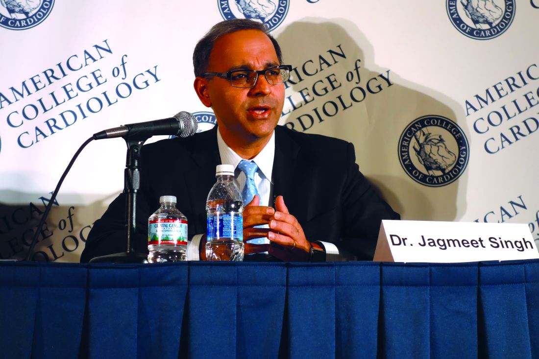
A randomized clinical trial of digoxin in AF is extremely unlikely, added Dr. Lopes, professor of medicine at Duke University in Durham, N.C.
ARISTOTLE was a randomized trial of apixaban (Eliquis) versus warfarin for stroke prevention in AF. The results of this landmark study, previously reported (N Engl J Med. 2011 Sep 15;365[11]:981-92), demonstrated that apixaban was the superior oral anticoagulant in preventing stroke or systemic embolism, caused less bleeding, and resulted in lower mortality.
ARISTOTLE had some unique features that rendered the study database an exceptional resource for use in a large observational study of digoxin’s safety in patients with AF. It included a detailed serial assessment of concomitant medications as well as measurements of serum digoxin levels, left ventricular ejection fraction, creatinine clearance, and biomarkers including vasoactive intestinal peptide, troponins T and I, N-terminal pro–brain-type natriuretic peptide, and growth differentiation factor 15. These were among the 48 clinical variables included in multivariate adjusted analyses of mortality risk.
One-third of ARISTOTLE participants were on digoxin at study entry, a prevalence typical of what’s seen in clinical practice. Among the 5,824 subjects with AF already on digoxin at the start of the trial, the risk of death during follow-up proved independently related to baseline serum digoxin concentration. Patients with a level from 0.9 ng/mL to less than 1.2 ng/mL had a 16% increased risk of death during study follow-up, compared with digoxin nonusers, a trend that didn’t reach statistical significance. However, the 11% of AF patients with a serum concentration of 1.2 ng/mL or above were at a significant 56% increased risk for death.
When serum digoxin concentration is looked at as a continuous, rather than dichotomous variable, for each 0.5-ng/mL increase in drug concentration, the adjusted risk of all-cause mortality at 1 year of study follow-up climbed by 19%.
Moreover, among 781 AF patients who initiated digoxin during the study, the risk of death was increased by 78%, compared with that of 2,343 extensively matched controls. The most common cause of this excess mortality was sudden death, and in a closer look at that endpoint, the investigators found that the risk of sudden death was increased fourfold in new users of digoxin. This increased risk occurred early: Most sudden deaths occurred within the first 6 months after going on the drug, suggesting a causal relationship, although not providing definitive proof, Dr. Lopes noted.
Forty-three percent of ARISTOTLE participants had heart failure at enrollment. Interestingly, the increased risk of death associated with on-study initiation of digoxin was of similar magnitude, regardless of whether comorbid heart failure was present. The mortality risk was 58% greater in new users with heart failure, compared with matched nonusers with heart failure, and twofold greater in new users without heart failure than in their matched controls.
The benefits of apixaban over warfarin were consistent regardless of whether or not patients were on digoxin.
Discussant Kristen K. Patton, MD, was effusive in her response to the new ARISTOTLE findings.
“This was a really, truly, beautiful observational analysis,” declared Dr. Patton, an electrophysiologist at the University of Washington, Seattle.
“I think in cardiology, where our hearts have been broken before due to flawed observational studies, it’s really important for people to understand that observational data, when analyzed well, with appropriate propensity matching, with new-user analysis and close attention to clinical variables that are important, can really change practice in a good way. I think that’s what we see here,” she said.
A beaming Dr. Lopes responded that it’s likely that some of the past conflicting studies were marred by survival bias – that is, an inability to account for the fact that patients already on digoxin at the outset of a study have already declared themselves to be more tolerant of the drug. Past studies also didn’t adjust for biomarker levels.
“We could adjust for things we know today are associated with death in atrial fibrillation,” he observed.
Dr. Patton added that the most surprising study finding to her involved the new users of digoxin. She suspects that the reported figure of a 78% increased risk of all-cause mortality during study follow-up actually markedly underestimates the true size of that risk during the initial months on the drug. Dr. Lopes agreed.
She also said she found worrisome and disappointing the increased mortality risk reported with initiation of digoxin in AF patients with heart failure. That hasn’t been seen in other studies.
Dr. Lopes said the investigators utilized multiple means of identifying patients with heart failure and are certain they captured the full population of affected patients.
“We feel very confident that, when you have atrial fibrillation together with heart failure, it might be a different story than without atrial fibrillation,” the cardiologist said.
Discussant Jagmeet P. Singh, MD, associate chief of cardiology at Massachusetts General Hospital and professor of medicine at Harvard Medical School, Boston, said the ARISTOTLE analysis carries an eye-opening take-home message: “If you have to initiate digoxin, you have to follow the serum levels more closely than we ever have before. How frequently, I don’t know – maybe monthly instead of at the 6-monthly intervals that we often do. And I think maybe arrhythmia monitoring in the initial stages of putting patients on digoxin will be key to see if there are any additional proarrhythmic effects.”
The original ARISTOTLE trial was sponsored by Bristol-Myers Squibb and Pfizer. However, the ARISTOTLE digoxin analysis was sponsored by the Duke Clinical Research Institute. Dr. Lopes reported serving as a consultant to and/or receiving research grants from Bristol-Myers Squibb, Pfizer, Bayer, Boehringer Ingleheim, Daiichi Sankyo, GlaxoSmithKline, Medtronic, Merck, and Portola.
AT ACC 17
Key clinical point:
Major finding: Patients with atrial fibrillation who initiated digoxin therapy had a fourfold increased risk of sudden death, compared with extensively matched controls.
Data source: This was a secondary observational analysis drawn from the roughly 18,000-patient, randomized, multicenter ARISTOTLE trial.
Disclosures: This ARISTOTLE digoxin analysis was sponsored by the Duke Clinical Research Institute. The study presenter reported serving as a consultant to and/or recipient of research grants from numerous companies, including Bristol-Myers Squibb and Pfizer, which cosponsored the original ARISTOTLE trial.
Moderate exercise benefits hypertrophic cardiomyopathy patients
WASHINGTON – A regimen of moderately intense exercise for 16 weeks produced no adverse effects and led to a clinically meaningful improvement in exercise capacity in a multicenter, randomized trial involving 136 patients with hypertrophic cardiomyopathy.
While finding that regular exercise produced a statistically significant improvement, compared with control patients fulfilled the study’s primary endpoint, it was perhaps as important that patients with hypertrophic cardiomyopathy (HCM) randomized to the exercise arm experienced no ill effects, a finding that bucked conventional wisdom that, once diagnosed, HCM patients must adhere to a largely inactive lifestyle.
“I see patients with HCM who tell me that, when they were first diagnosed, they were told by their physician not to do anything at all,” said Dr. Saberi, a cardiologist at the University of Michigan in Arbor. Although she cautioned that the findings from this modestly sized trial cannot prove that exercise is safe for HMC patients – something that would require randomizing more than 2,000 patients and following them for about 3 years – the findings provide some level of reassurance that regular, moderately intense exercise as simple as walking can benefit patients and likely not cause any problems. “We saw no signal of harm,” she stressed in an interview. “I would recommend moderate-intensity, regular exercise. Physicians can feel comfortable using the exercise prescription” used in the study, she advised.
Concurrently with Dr. Saberi’s report at the meeting, the results also appeared in an article published online (JAMA. 2017 Mar 17. doi: 10.1001/jama.2017.2503).
The Study of Exercise Training in Hypertrophic Cardiomyopathy (RESET-HCM) ran during 2010-2015 at the University of Michigan and Stanford University, and enrolled 136 patients 18-80 years old diagnosed with HCM but without more severe manifestations such as a history of exercise-induced syncope or ventricular arrhythmia, a left ventricular ejection fraction less than 55%, recent clinical decompensation, or a history of a severe hypotensive response to exercise. Enrolled patients averaged about 50 years old, their baseline peak oxygen consumption, VO2, was about 22 mL/kg per min, and their maximal ventricular wall thickness was about 21 mm.
Following a comprehensive baseline heart function and imaging assessment, including a cardiopulmonary exercise test, the 67 patients in the intervention arm received an individualized exercise prescription based on their heart rate reserve. At a minimum, they started with three exercise sessions per week lasting at least 20 minutes on their own, a regimen designed to bring them to 60% of their heart rate reserve. Their prescription gradually increased to four to seven exercise periods weekly lasting up to 60 minutes and designed to bring them to 70% of their heart rate reserve. Although most patients accomplished this through walking, some engaged in activities that included cycling, swimming, jogging, or elliptical training. The researchers told the 69 control patients to continue doing their usual activities. Among the patients who entered the study, 57 in the exercise group and 56 controls remained for the full 16 weeks and had a complete final assessment.
After 16 weeks, the average change in peak VO2, was 1.35 mL/kg per min in the intervention group and 0.08 mL/kg per min in the control patients, a statistically significant difference for the primary endpoint and a 6% increase in exercise capacity, compared with baseline for the intervention group. Dr. Saberi noted that, in a prior study of patients with chronic heart failure, a 6% increase in peak VO2 linked with an 8% decrease in cardiovascular mortality of heart failure hospitalization.
This result is “a glimmer of hope for using exercise in HCM patients,” she said. “There are no other disease-modifying treatments for HCM, so this is the first ‘treatment’ to have any impact on the disease.”
The patients who exercised had no identified major adverse effects or changes in cardiac morphology after 16 weeks. Assessment of physical function quality of life using the SF-36v2 showed an average 8-point improvement in the patients who exercised, compared with the controls. In addition, “a lot of patients in the exercise group told us that they felt better,” Dr. Saberi said.
Because patients with HCM are often advised to limit their activity, the consequence is “we increasingly see HCM patients who are obese and have complications such as sleep apnea and atrial fibrillation. Patients are even fearful of going up and down stairs.” She stressed the simplicity and ease of the intervention she and her colleagues tested, which can involve nothing more than regular, daily walks of 20 minutes or more. Dr. Saberi advised tailoring the intensity and frequency of the exercise intervention to each patient and avoiding having the patient push beyond what they find comfortable.
A multicenter U.S. registry, the Lifestyle and Exercise in Hypertrophic Cardiomyopathy Study (LIVE-HCM), is now enrolling HCM patients that should eventually follow enough patients for a long enough period of time to definitely prove whether moderate exercise is safe for HCM patients, but the results will not be available for several more years, Dr. Saberi said. A recent analysis estimated that one in every 200 adults has HCM (J Am Coll Cardiol. 2015 Mar 31;65[12]:1249-54), which means the disease likely affects more than one million Americans.
mzoler@frontlinemedcom.com
On Twitter @mitchelzoler
AT ACC 17
Key clinical point:
Major finding: Average exercise capacity, peak VO2, increased by 1.27 mL/kg per min in exercising patients compared with nonexercising controls.
Data source: RESET-HCM, a multicenter, randomized trial with 136 hypertrophic cardiomyopathy patients.
Disclosures: Dr. Saberi had no disclosures.
Cannabis associated with increased risk of heart failure and stroke
Cannabis use was associated with an increased risk of cerebrovascular accidents and heart failure in a retrospective analysis of the Nationwide Inpatient Sample (NIS).
Aditi Kalla, MD, a cardiology fellow at Einstein Medical Center in Philadelphia, and her colleagues analyzed data from nearly 21 million adult patients aged 18-55 years from the NIS 2009-2010 database. Approximately 1.5% (316,397) were diagnosed as cannabis users.
Cannabis users also were more likely to report cardiac risk factors such as hypertension (19.9% vs. 15.7% of nonusers), tobacco use (47.2% vs. 11.4%), alcohol use (28.1% vs 3.8%), and obesity (7% vs. 6.5%). They were older, on average, with a mean age of 33 years, compared with 26 years, and were likely to be male (60%), Dr. Kalla noted during a press briefing held in advance of the annual meeting of the American College of Cardiology.
Using multivariate regression analysis to adjust for these traditional cardiovascular risk factors, the investigators found cannabis remained an independent predictor for heart failure, with an odds ratio of 1.1 (P less than .01) and cerebrovascular accident, with an OR of 1.24 (P less than .001).
“Even when we corrected for known risks, we still found a higher rate of both stroke and heart failure in these patients,” Dr. Kalla said. “That leads us to believe that there is something else going on besides just obesity or diet-related cardiovascular side effects.”
Dr. Kalla noted that an expert analysis published by the ACC in September 2016 linked cannabinoid receptor type 1 with atherogenesis.
Further research is needed on the topic of cannabis and cardiovascular effects, especially as the legalization of medical and recreational cannabis spreads across the country, Dr. Kalla said. “Decriminalization of cannabis has passed in several states, bringing the total count now up to 28 states, plus the District of Columbia. We now need to be more knowledgeable of the risks and benefits of cannabis, as patients in these states may inquire into the use of it, or even ask us for prescriptions for it.”
While the NIS provided a large and strong data set for this analysis, the number of cannabis users likely was underreported because cannabis was legal in just 14 states at the time, Dr. Kalla noted. The study also was limited by a lack of specific information regarding cannabis intake, method of intake (ingestion or smoking), quantity and frequency of use, and whether use was medical or recreational.
The information collected also excluded whether patients used marijuana for medical or recreational purpose and how it was taken, by smoking or ingestion.
ezimmerman@frontlinemedcom.com
On Twitter @EAZTweets
FROM ACC 17
Key clinical point:
Major finding: Cannabis users showed 26% increased risk (OR, 1.24) of stroke and 10% increased risk (OR, 1.1) of heart failure.
Data source: Retrospective study of over 20 million patients’ records aged 18-55 years gathered from the Nationwide Inpatient Sample 2009-2010 database.
Disclosures: Researchers reported no relevant conflicts of interest.
Prediction: LVADs will rule end-stage heart failure
SNOWMASS, COLO. – Multifaceted progress in mechanical circulatory support as long-term therapy in end-stage heart failure is happening at a brisk pace, Y. Joseph C. Woo, MD, reported at the Annual Cardiovascular Conference at Snowmass.
declared Dr. Woo, professor and chair of the department of cardiothoracic surgery at Stanford (Calif.) University.
That’s quite a prediction, especially considering the source: Stanford is where the late Dr. Norman Shumway – widely considered “the father of heart transplantation” – performed the first adult heart transplant in the United States in 1968.
Dr. Woo was coauthor of an American Heart Association policy statement on the future of cardiovascular disease in the United States, which forecast a 25% increase in heart failure between 2010 and 2030 (Circulation. 2011 Mar 1;123[8]:933-44). There is simply no way that heart transplantation can begin to meet the projected growing need for effective therapy in patients with end-stage disease.
Here’s what Dr. Woo sees as the future of MCS:
Minimally invasive implantation
At Stanford, LVAD implantations are now routinely done off-pump on a beating heart.
“We clamp only when there is a sound reason, like the presence of left ventricular thrombus, where you run the risk of embolization without the cross clamp,” the surgeon said.
Concomitant valvular surgery
At Stanford and other centers of excellence, surgeons perform additional procedures as warranted while they implant an LVAD, including atrial fibrillation ablation, revascularization of the right heart coronaries, patent foramen ovale closure, and repair of the tricuspid, pulmonic, or aortic valves.
Enhanced right ventricular management
Survival is greatly impaired if a patient with an LVAD later requires the addition of a right ventricular assist device. This realization has led to the development of multiple preoperative risk scoring systems by the Stanford group (Ann Thorac Surg. 2013 Sep;96[3]:857-63) and others, including investigators at the Deutsche Herzzentrum Berlin, the world’s busiest heart transplant center. The purpose is to identify upfront those patients who are likely to later develop right heart failure so they can receive biventricular MCS from the start.
Adjunctive biologic therapies
Intramyocardial injection of 25 million allogeneic mesenchymal precursor cells during LVAD implantation appeared to be safe and showed a promising efficacy signal in a 30-patient, multicenter, double-blind, placebo-controlled, National Institutes of Health–sponsored proof of concept study in which Dr. Woo was a coinvestigator (Circulation. 2014 Jun 3;129[22]:2287-96).
The goal of this research effort is to provide a cell therapy assist to the LVAD as a bridge to recovery of left ventricular function such that the device might eventually no longer be needed, he explained.
These cells are immune privileged. They can be transplanted into recipients without need for immunosuppressive therapy or HLA matching, basically as an off the shelf product. Rather than transforming into cardiomyocytes, it appears that the mechanism by which the donor cells enhance cardiac performance in heart failure is via secretion of a shower of growth and angiogenic factors.
Based upon the encouraging results of the initial study, a 90-patient, phase II, double-blind clinical trial is underway. In order to better evaluate efficacy, this time the patients will receive 150 million mesenchymal precursor cells rather than 25 million.
New technologies
The developmental pipeline is chock full of MCS devices. The trend is to go smaller and simpler. HeartWare is developing a miniaturized version of its approved continuous flow centrifugal force LVAD. The ReliantHeart aVAD, an intraventricular device less than 2.5 cm in diameter, is approved in Europe and under study in the U.S. The Thoratec HeartMate III is a smaller version of the HeartMate II, which is FDA-approved as destination therapy. And the Circulite Synergy micropump, designed to provide partial circulatory support to patients who don’t require a full-force LVAD, is the size of a AA battery.
Dr. Woo reported having no financial conflicts.
bjancin@frontlinemedcom.com
EXPERT ANALYSIS FROM THE CARDIOVASCULAR CONFERENCE AT SNOWMASS
Big changes ahead in heart failure management
SNOWMASS, COLO. – Advances in remote telemedical management show enormous promise as a means of preventing costly heart failure hospitalizations, William T. Abraham, MD, said at the Annual Cardiovascular Conference at Snowmass.
He characterized the decades-old conventional approach to heart failure management as a reactive strategy focused on making medication adjustments based upon weight changes and worsening signs and symptoms. But these physiologic markers of acute decompensation aren’t actionable; that is, they don’t occur until it’s too late to successfully intervene to prevent hospitalization.
It’s now clear that objective, measurable changes in intracardiac and pulmonary artery pressures as well as increases in lung fluid volume precede symptomatic decompensation episodes by several weeks. These early harbingers are reliably detectable by telemedical monitoring via small implantable pressure sensors or, noninvasively, using wearable sensors embedded in a vest.
“By moving upstream, we hope to develop a more proactive preventive approach to managing heart failure patients so that we can implement a medical intervention during this presymptomatic phase of worsening heart failure and avert a heart failure hospitalization,” the cardiologist explained.
That has already been demonstrated in several studies in which remote physicians checked the home monitoring data daily and promptly increased the dose of diuretics when pressure readings or lung fluid volume climbed above normal: The elevated readings quickly retreated and heart failure hospitalizations occurred much less frequently than with conventional management.
“Well-structured outpatient care could reduce the need for hospital admission, facilitate early intervention, prevent crisis management, and avoid complications or disease management in these patients,” Dr. Abraham observed.
Finding best telemedicine options
But this high-tech patient management strategy is not quite ready for prime time use in daily clinical practice.
“We’re still sorting through this field and trying to figure out the best telemedicine options,” according to Dr. Abraham.
He cited several recent large, well-conducted randomized trials that have persuasively shown that there’s no point in applying home telemedicine in order to quickly respond to changes in a heart failure patient’s blood pressure, weight, and symptom status.
“The horse is already out of the barn at that point in time,” according to the cardiologist.
For example, the BEAT-HF (Better Effectiveness After Transition–Heart Failure) trial included 1,437 patients hospitalized for heart failure at eight California academic medical centers who were randomized to usual care or an intervention that combined health-coaching telephone calls and protocols for physician or nurse response to daily telemetric data on patient blood pressure, symptoms, heart rate, weight, and symptoms. The intervention turned out to have absolutely no effect on the 180-day rate of readmissions, which was roughly 50% in both groups (JAMA Intern Med. 2016 Mar;176[3]:310-8).
The PCDM (Patient-Centered Disease Management) trial was a multicenter Veterans Affairs study that randomized heart failure patients to usual care or a comprehensive intervention involving collaborative care by a cardiologist, psychiatrist, primary care physician, and nurse coordinator; screening and treatment for depression; and home telemonitoring of heart failure decompensation symptoms. The multifaceted intervention had no effect on the 1-year readmission rate (JAMA Intern Med. 2015 May;175[5]:725-32).
“To date, I would challenge you to find any adequately powered randomized, controlled trial in heart failure disease management that demonstrates that the way we’ve been doing things really keeps heart failure patients out of the hospital. So, it is time for a paradigm shift and some new technologies in our armamentarium,” Dr. Abraham said.
CardioMEMS system
Several remote telemedical management systems for heart failure, which measure early preclinical harbingers of acute decompensation, have received Food and Drug Administration approval. Many more are in development. Dr. Abraham highlighted two approved systems for which he was a coinvestigator in clinical trials.
The CardioMEMS system uses a pressure sensor the size of a small paper clip that is placed in a branch of the pulmonary artery, where it readily becomes endothelialized. Device implantation can be carried out by any cardiologist who can perform a right heart catheterization, be it an electrophysiologist, interventionalist, heart failure specialist, or general cardiologist. It takes only about an additional 7 minutes to deploy the sensor following a standard diagnostic right heart catheterization. The system doesn’t use a battery and has no moving parts.
“Now, with more than 10 years experience, we have yet to see a sensor failure. It’s a highly reliable system,” according to the cardiologist.
Once a day the patient lies down, pushes a button, and the sensor is simultaneously powered up while the data on pulmonary artery pressure waveforms and systolic and diastolic pressures is extracted using radiofrequency energy.
In the randomized, multicenter, controlled, single-blind CHAMPION (CardioMEMS Heart Sensor Allows Monitoring of Pressure to Improve Outcomes in NYHA Class III Heart Failure Patients) trial, Dr. Abraham and coinvestigators demonstrated that pulmonary artery pressure-guided heart failure management reduced the rate of heart failure hospitalizations in the CardioMEMS group by 33% during the first 18 months of the trial and by 48% in the subsequent 13 months (Lancet. 2016 Jan 30;387[10017]:453-61).
“It’s important to note that the results were positive in diastolic as well as systolic heart failure. Patients with a baseline left ventricular ejection fraction of 40% or more saw a 50% reduction in the risk of heart failure hospitalizations, and those with an LVEF of 50% or more saw a 70% reduction,” Dr. Abraham said.
Other implantable hemodynamic monitors
Numerous other implantable pressure sensors are in development for management of heart failure, including other pulmonary artery pressure sensors as well as left atrial pressure monitors.
“You’re going to hear a lot more about this field of implantable hemodynamic monitors in the future,” the cardiologist predicted.
Dr. Abraham was also a coinvestigator in an observational study of a wearable sensor based upon radar technology developed for the military and subsequently applied to rescue searches through rubble for earthquake survivors. This remote dielectric sensing (ReDS) technology has been miniaturized, with the sensors embedded in an FDA-approved vest. The heart failure patient dons the SensiVest for 90 seconds once per day for measurement of the absolute amount of fluid in the lungs. The data is automatically transmitted to a secured site in the cloud, where the physician can review the results and adjust medications in response to early evidence of fluid buildup.
“The normal lung is composed of 20%-35% fluid. So when that fluid content is elevated, patients with heart failure have wet lungs and they’re decompensating. You increase their diuretics to bring it back down into normal range,” he explained.
In the observational study, conducted in Israel, hospital readmission rates were reduced by 87% through the use of ReDS-guided patient management using the system marketed by Sensible Medical Innovations. Dr. Abraham and his coinvestigators are now seeking to confirm those results in the prospective, multicenter, randomized, controlled U.S. SMILE study.
Dr. Abraham reported serving as a consultant to Abbott Vascular, Medtronic, Novartis, and St. Jude Medical.
EXPERT ANALYSIS FROM THE CARDIOVASCULAR CONFERENCE AT SNOWMASS
Nonthoracic MRI safe in patients with cardiac devices
Nonthoracic MRI was safe in patients who had implanted non–MRI-conditional pacemakers or implantable cardioverter defibrillators, as long as they followed a specific safety protocol before and after the imaging procedure, according to a report published online Feb. 23 in the New England Journal of Medicine.
Patients with implanted cardiac devices have long been advised to avoid MRI because of the potential for the magnetic field to induce heating of the cardiac leads, which could in turn produce thermal injury to the myocardium and adversely affect the device’s function. Certain cardiac devices that have been proved to pose no such hazards have been designated by the Food and Drug Administration as “MRI conditional.” However, an estimated 2 million patients in the United States and another 6 million worldwide have devices that are not MRI conditional, and at least half of these patients are predicted to require an MRI during their lifetimes, said Robert J. Russo, MD, PhD, of Scripps Research Institute and the La Jolla (Calif.) Cardiovascular Research Institute, and his associates.
The MagnaSafe Registry was established to monitor device-related clinical events and device alterations among adults undergoing nonthoracic MRIs at 1.5 T. Dr. Russo and his associates analyzed data in this registry from 19 medical centers during a 5-year period. They assessed 1,000 MRIs in 818 patients with pacemakers and 500 MRIs in 428 patients with implantable cardioverter defibrillators who were followed for 6 months after the imaging procedures. Most of these scans involved the brain or spine, and the median duration of exposure to the magnetic field was 44 minutes.
According to the safety protocol, all devices were interrogated immediately before the MRI and, depending on those results, were programmed to no pacing or asynchronous pacing during the scan with all tachycardia and bradycardia therapies inactivated. Immediately after the scan, all devices were reprogrammed to baseline settings, a full device interrogation was repeated, and, if necessary, further reprogramming was performed to maintain adequate pacing and sensing. A physician, nurse practitioner, or physician’s assistant with cardiac expertise attended each scan.
There were no deaths, lead failures requiring immediate replacement, losses of capture, or full electrical resets associated with any of the 1,500 MRI scans.
Four patients developed atrial fibrillation, and two developed atrial flutter, during or after the MRI; three returned to sinus rhythm while still in the scanning room, and the other three did so within 49 hours. There were six cases requiring partial generator electrical resets. “Changes in device settings were common, but relatively few exceeded our prespecified threshold criteria for a clinically important change,” Dr. Russo and his associates wrote (N Engl J Med. 2017;376[8]:755-64).
Four patients reported feeling discomfort at the implant site during MRI, including one who felt a heating sensation and was removed from the scanner before completing the procedure. None of them had any further problems.
Some experts have suggested that to allow patients with cardiac devices to undergo MRI, the generators and leads could be removed before the procedure and replaced afterward. The findings of this study show that undergoing a nonthoracic MRI using this protocol would likely be a safer alternative, the investigators added.
This work was supported by St. Jude Medical, Biotronik, Boston Scientific, the Hewitt Foundation for Medical Research, and several philanthropic gifts. Dr. Russo reported ties to St. Jude Medical, Biotronik, Boston Scientific, and the Hewitt Foundation, and his associates reported ties to numerous industry sources.
FROM THE NEW ENGLAND JOURNAL OF MEDICINE
Key clinical point: Nonthoracic MRI was safe in patients who had implanted non–MRI-conditional pacemakers or ICDs, as long as they followed a specific safety protocol before and after the imaging procedure.
Key numerical finding: No deaths, lead failures requiring immediate replacement, losses of capture, or full electrical resets were tied to any of the 1,500 MRI scans.
Data source: A U.S. registry–based cohort study of 1,000 MRIs involving patients with pacemakers and 500 MRIs involving patients with ICDs, performed during a 5-year period.
Disclosures: This work was supported by St. Jude Medical, Biotronik, Boston Scientific, the Hewitt Foundation for Medical Research, and several philanthropic gifts. Dr. Russo reported ties to St. Jude Medical, Biotronik, Boston Scientific, and the Hewitt Foundation, and his associates reported ties to numerous industry sources.
Consider ultraslow thrombolysis for mechanical valve thrombosis
SNOWMASS, COLO. – Ultraslow infusion of a very-low-dose thrombolytic agent for treatment of mechanical prosthetic valve thrombosis appears to be as effective as surgery – the former first-line therapy – and sports a far lower stroke risk, Rick A. Nishimura, MD, said at the Annual Cardiovascular Conference at Snowmass.
“I’m not saying you have to use this, but I think it’s reasonable to consider it, especially if the patient is at high risk for surgery and low risk for thrombolysis,” according to Dr. Nishimura, professor of cardiovascular diseases and hypertension at the Mayo Clinic in Rochester, Minn. 
He added that he and his Mayo colleagues have begun using the novel therapy and are favorably impressed with the resultant complete normalization of valve gradients and low complication rate.
Dr. Nishimura was cochair of the writing committee for the current American College of Cardiology/American Heart Association guidelines for management of valvular heart disease (Circulation. 2014 Jun 10;129[23]:e521-643). Those guidelines state that emergency surgery is the treatment of choice for thrombosis of a left-sided mechanical heart valve. That strong recommendation was based on a dozen nonrandomized studies reported prior to 2013 which showed a 95% success rate with surgery compared with 75% with conventional large-bolus thrombolytic therapy, a high 10%-12% mortality with either form of therapy, and a stroke risk of 12%-14% with thrombolytic therapy, substantially higher than for surgery.
Since release of the ACC/AHA guidelines, however, there’s been an important new development: Three groups outside of the United States have pioneered ultraslow thrombolytic therapy for mechanical prosthetic valve thrombosis. The supporting evidence comes from cohort studies, with no randomized trials done to date. But the collective reported experience from these three research teams shows a 90%-95% success rate – comparable to surgery – along with stroke and mortality rates in the low single digits.
The Turkish group waits until the patient’s international normalized ratio (INR) is below 2.5, then administers 25 mg of tissue plasminogen activator guided by transesophageal echocardiography (TEE) over 25 hours.
“We traditionally give 90 mg over 1 hour, so this is very, very slow therapy,” Dr. Nishimura observed.
After the 24-hour infusion, TEE is repeated. If imaging shows the clot is not resolved, another 25 mg of tissue plasminogen activator is given over 24 hours. This process is repeated for up to 8 days as needed (Am Heart J. 2015 Aug;170[2]:409-18).
Dr. Nishimura advised reserving ultraslow thrombolytic therapy for patients who are hemodynamically stable; this treatment takes a while to work, so patients in severe heart failure should be sent straight away to surgery. The novel therapy is best suited for patients with recent-onset mechanical valve thrombosis, a low INR, TEE evidence that the clot isn’t huge, and/or when surgical expertise isn’t readily available.
Before you can treat a prosthetic mechanical valve thrombosis, however, you have to make the diagnosis. Here’s what Dr. Nishimura recommends: First, suspect the condition on the basis of clinical symptoms of heart failure and dull, muffled S1 and S2 sounds on auscultation, especially in a patient who presents with a low INR.
Next, prove that obstruction is present via Doppler echocardiographic evidence of an abnormal gradient across the mechanical valve.
Finally, determine if the mechanical valve shows abnormal disc motion with sticking leaflets. TEE is excellent for visualizing a mechanical mitral valve but isn’t helpful if it’s a mechanical aortic valve.
“Old-fashioned fluoroscopy is the best approach for looking at leaflet motion in the atrial valve and mitral valve. We’ve got 3-D cine now that provides beautiful images, but if you can get the same information with a quick fluoroscopy, go with the fluoroscopy,” the cardiologist suggested.
He reported having no financial conflicts of interest.
SNOWMASS, COLO. – Ultraslow infusion of a very-low-dose thrombolytic agent for treatment of mechanical prosthetic valve thrombosis appears to be as effective as surgery – the former first-line therapy – and sports a far lower stroke risk, Rick A. Nishimura, MD, said at the Annual Cardiovascular Conference at Snowmass.
“I’m not saying you have to use this, but I think it’s reasonable to consider it, especially if the patient is at high risk for surgery and low risk for thrombolysis,” according to Dr. Nishimura, professor of cardiovascular diseases and hypertension at the Mayo Clinic in Rochester, Minn.
He added that he and his Mayo colleagues have begun using the novel therapy and are favorably impressed with the resultant complete normalization of valve gradients and low complication rate.
Dr. Nishimura was cochair of the writing committee for the current American College of Cardiology/American Heart Association guidelines for management of valvular heart disease (Circulation. 2014 Jun 10;129[23]:e521-643). Those guidelines state that emergency surgery is the treatment of choice for thrombosis of a left-sided mechanical heart valve. That strong recommendation was based on a dozen nonrandomized studies reported prior to 2013 which showed a 95% success rate with surgery compared with 75% with conventional large-bolus thrombolytic therapy, a high 10%-12% mortality with either form of therapy, and a stroke risk of 12%-14% with thrombolytic therapy, substantially higher than for surgery.
Since release of the ACC/AHA guidelines, however, there’s been an important new development: Three groups outside of the United States have pioneered ultraslow thrombolytic therapy for mechanical prosthetic valve thrombosis. The supporting evidence comes from cohort studies, with no randomized trials done to date. But the collective reported experience from these three research teams shows a 90%-95% success rate – comparable to surgery – along with stroke and mortality rates in the low single digits.
The Turkish group waits until the patient’s international normalized ratio (INR) is below 2.5, then administers 25 mg of tissue plasminogen activator guided by transesophageal echocardiography (TEE) over 25 hours.
“We traditionally give 90 mg over 1 hour, so this is very, very slow therapy,” Dr. Nishimura observed.
After the 24-hour infusion, TEE is repeated. If imaging shows the clot is not resolved, another 25 mg of tissue plasminogen activator is given over 24 hours. This process is repeated for up to 8 days as needed (Am Heart J. 2015 Aug;170[2]:409-18).
Dr. Nishimura advised reserving ultraslow thrombolytic therapy for patients who are hemodynamically stable; this treatment takes a while to work, so patients in severe heart failure should be sent straight away to surgery. The novel therapy is best suited for patients with recent-onset mechanical valve thrombosis, a low INR, TEE evidence that the clot isn’t huge, and/or when surgical expertise isn’t readily available.
Before you can treat a prosthetic mechanical valve thrombosis, however, you have to make the diagnosis. Here’s what Dr. Nishimura recommends: First, suspect the condition on the basis of clinical symptoms of heart failure and dull, muffled S1 and S2 sounds on auscultation, especially in a patient who presents with a low INR.
Next, prove that obstruction is present via Doppler echocardiographic evidence of an abnormal gradient across the mechanical valve.
Finally, determine if the mechanical valve shows abnormal disc motion with sticking leaflets. TEE is excellent for visualizing a mechanical mitral valve but isn’t helpful if it’s a mechanical aortic valve.
“Old-fashioned fluoroscopy is the best approach for looking at leaflet motion in the atrial valve and mitral valve. We’ve got 3-D cine now that provides beautiful images, but if you can get the same information with a quick fluoroscopy, go with the fluoroscopy,” the cardiologist suggested.
He reported having no financial conflicts of interest.
EXPERT ANALYSIS FROM THE CARDIOVASCULAR CONFERENCE AT SNOWMASS
The two faces of mitral regurgitation
SNOWMASS, COLO. – Primary mitral regurgitation and secondary mitral regurgitation may sound a lot alike, but they are in fact starkly different diseases, Blase A. Carabello, MD, said at the Annual Cardiovascular Conference at Snowmass.
“They are almost entirely different in their etiologies, in their pathophysiologies, and in their therapies,” according to Dr. Carabello, professor of medicine and chief of cardiology at East Carolina University in Greenville, N.C.
“On the other hand, if you fix the valve, you fix the disease,” he said.
In contrast, in secondary MR, the dysfunctional valve is a result of the ventricular problem, not its cause.
“In secondary MR, it’s the ventricle that made the valve sick. It’s the regional wall motion abnormalities, the displacement of the papillary muscles, and the dilation of the mitral annulus that pulls the mitral valve apart and prevents it from coapting,” he explained.
Dr. Carabello, who was on the writing committee for the current American College of Cardiology/American Heart Association guidelines on management of valvular heart disease (Circulation. 2014 Jun 10;129[23]:e521-643), highlighted other key distinctions between the two diseases.
Primary MR
“Primary MR, unlike aortic regurgitation, is not well tolerated. Early repair is key,” the cardiologist stressed.
The ACC/AHA guidelines emphasize the importance of early referral for surgery for primary MR because of surgery’s proven survival benefit. The triggers for surgery, as described in the guidelines, are easy to remember, namely, “symptoms/60/50/40.” That is, it’s time to move on to surgery when any of the following occurs: The patient becomes even mildly symptomatic, the left ventricular ejection fraction (LVEF) drops to 60%, the pulmonary artery pressure climbs to 50 mm Hg, or the LV end-systolic dimension reaches 40 mm.
Dr. Carabello said he believes those surgical thresholds are conservative, and it’s best to make the surgical referral when a patient approaches one or more of those triggers, but before actually reaching them.
“That’s the way I practice: Don’t wait for any of those things. Just get it done. One advantage to early repair is the patient can’t get lost to follow-up. They’re repaired, and they can’t take it out,” he said.
It’s possible that the next iteration of the guidelines will utilize stricter thresholds for surgery. French investigators have shown that surgery for primary MR achieves a significantly higher rate of normal LV function if the operation occurs when patients have an LVEF of 64% or more and an LV end-systolic dimension of less than 37 mm (Eur J Echocardiogr. 2011 Sep;12[9]:702-10). So far, though, there hasn’t been a confirmatory study.
“I think the normal EF in primary MR is about 70%. By the time the EF in a patient with primary MR gets down into the 50s, you’re looking at an extraordinarily sick ventricle,” according to Dr. Carabello.
The impetus for including the “symptoms/60/50/40” surgical triggers in the guidelines is to encourage physicians to make the surgical referral earlier than has often been the case. Too frequently, the surgical referral is delayed until damage to the ventricle is irreversible, with a resultant worsened prognosis.
“The natural history is such that, from the time a patient with severe primary MR enters your office to the annual mortality risk reaching 50% is only about 5 years. So if you’re going to do watchful waiting, fine, but remember: You don’t have all that long to watch and wait before something bad happens to these folks,” the cardiologist cautioned.
He added that it’s possible that, in the future, cardiac biomarkers will be utilized to help predict the long-term mortality risk of patients under medical management of their primary MR. In a recent study of 1,331 patients with primary MR, the investigators showed that the ratio of B-type natriuretic peptide (BNP) to the upper limit of normal BNP, adjusted for age and sex, was a powerful independent predictor of this risk (J Am Coll Cardiol. 2016 Sep 20;68[12]:1297-307).
The guidelines state that mitral valve repair is preferable to replacement as long as the heart team determines there’s at least a 95% chance of a durable repair. That’s because the operative mortality associated with replacement is significantly higher than with repair.
Secondary MR
Unlike in primary MR, it’s unclear whether surgery prolongs life for patients with secondary MR, or if mitral repair is superior to replacement. Thus, the current guidelines recommend surgery only for patients who are still severely symptomatic despite maximal guideline-directed medical therapy for heart failure as well as cardiac resynchronization therapy, provided they have a conduction system abnormality and qualify for the device therapy.
“If you’ve done all that and they’re still sick, I think that surgery or the MitraClip may benefit them very much,” Dr. Carabello said.
In the United States, the MitraClip transcatheter device is approved only for the treatment of primary MR in inoperable patients. But in the rest of the world, roughly three-quarters of these devices are used for treatment of secondary MR. That potential indication is currently under study in the United States in the phase III COAPT trial (Cardiovascular Outcomes Assessment of the MitraClip Percutaneous Therapy for Heart Failure Patients With Functional Mitral Regurgitation).
“In the acute setting, the changes in hemodynamic variables and left ventricular performance that occur with the MitraClip are quite dramatic, with a marked decrease in wedge pressure and a markedly increased stroke volume, which is what it’s supposed to do. So I’m encouraged. We’ll wait for the results of the COAPT trial, which I believe is likely to demonstrate a reduction in hospitalizations and certainly an improvement in patient symptoms. But I doubt very much that we’ll ever see a change in lifespan because the ventricle is still sick, and we in 2017 have not found a way yet to revive a sick ventricle. It’s unlikely that fixing the MR will do so,” according to Dr. Carabello.
He reported serving on a data safety monitoring board for Edwards Lifesciences.
EXPERT ANALYSIS FROM THE CARDIOVASCULAR CONFERENCE AT SNOWMASS

Trials refine device therapy options for heart failure
SNOWMASS, COLO. – The indication for primary prophylactic implantable cardioverter-defibrillator therapy in patients with nonischemic heart failure is likely to be downgraded in the next iteration of the ACC/AHA heart failure guidelines as a consequence of the negative results of the DANISH trial, William T. Abraham, MD, predicted at the Annual Cardiovascular Conference at Snowmass.
In addition to outlining where the guideline recommendations for implantable cardioverter-defibrillator (ICD) therapy stand today, and how they’re likely to change in response to the DANISH findings, he highlighted the latest patient selection criteria for cardiac resynchronization therapy (CRT), which have grown considerably more complicated over time.
“Following the success of neurohormonal inhibitors and antagonists, our only other breakthroughs for the management of heart failure have been CRT and ICDs,” he noted.
The two device therapies are complementary, and indeed are often employed in combination.
“CRT makes patients feel better and saves lives, while ICDs prolong survival without an effect on improving heart failure per se,” the cardiologist explained.
To put the quality of life benefits of CRT into perspective, studies show that the device therapy results in a placebo-subtracted improvement on the Minnesota Living With Heart Failure Questionnaire of 9-10 points.
“This is a large and clinically meaningful improvement in quality of life. Our best drugs for heart failure – beta blockers and ACE inhibitors – improve this same measure by 4 or 5 points,” Dr. Abraham said.
Current American College of Cardiology/American Heart Association heart failure guidelines give a class I, level of evidence: A, recommendation for prophylactic ICD therapy in patients with an left ventricular ejection fraction (LVEF) of 35% or less and New York Heart Association functional class II or III symptoms despite optimal medical therapy, regardless of whether their heart failure is attributable to ischemic heart disease or nonischemic dilated cardiomyopathy.
The DANISH trial investigators looked at the evidence base for primary prevention ICDs in nonischemic heart failure and concluded it needed shoring up. The recommendation relied mainly on subgroup analyses of larger landmark trials done about 15 years ago, before major improvements in medical therapy had occurred. These reservations were the impetus for the DANISH trial, in which more than 1,100 patients with symptomatic systolic heart failure were randomized to an ICD or usual care.
The primary outcome in the DANISH trial – all-cause mortality – occurred in 21.6% of patients in the ICD group and 23.4% of controls during a median follow-up of 68 months, a nonsignificant difference (N Engl J Med. 2016 Sep 29;375[13]:1221-30).
Turning to the CRT guidelines, Dr. Abraham noted that the simple, broad, class I recommendation for this form of device therapy in patients with cardiac dyssynchrony as defined by a QRS duration greater than 120 msec contained in the 2005 ACC/AHA heart failure guidelines has been whittled down over time as new evidence has unfolded. The only class I recommendation in the current guidelines is in patients with an LVEF of 35% or less, sinus rhythm, left bundle branch block with a QRS duration of 150 msec or longer, and NYHA class II, III, or ambulatory IV symptoms while on guideline-directed medical therapy (Circulation. 2012 Oct 1;126:1784-800). “That’s the money group right there. That’s the group for whom we have the greatest confidence of producing the greatest benefit with the application of cardiac resynchronization therapy,” he explained.
Studies examining the use of CRT in heart failure patients with a non–left bundle branch morphology and a QRS duration of less than 150 msec have yielded negative findings. So have attempts to utilize echocardiographic evidence of mechanical dyssynchrony rather than ECG measurement of QRS duration to guide patient selection for CRT.
“In our practice, any patient with a left bundle branch block gets a CRT device. Our confidence in its efficacy is greater in patients with a QRS of at least 150 msec, but the studies demonstrate clear benefit for patients with left bundle branch block and a QRS of 120-149 msec as well,” according to the cardiologist.
Studies also show that patients who are dependent upon ventricular pacing benefit from CRT.
“If you have a patient who requires at least 40% or more ventricular pacing and also has reduced ejection fraction heart failure, that patient should have a CRT device rather than a dual chamber ICD or standard right-sided right ventricular pacemaker,” he said.
All of this presupposes that first and foremost the patient is already on optimized guideline-directed medical therapy.
“With optimal medical therapy, some of these patients may improve their left ventricular ejection fraction above 35%, or they may become asymptomatic and no longer have an indication for CRT,” Dr. Abraham added.
The rationale for utilizing CRT in combination with an ICD is a bit shaky, resting on a single older landmark study, the COMPANION trial (N Engl J Med. 2004; 350:2140-50).
“That study wasn’t powered to answer the question of whether CRT-D [a combined CRT/ICD device] is better than CRT. Really, this remains somewhat of an unanswered question. So where are we today? Essentially, if a patient has an indication for CRT and an indication for an ICD, we implant a combined device,” he said.
Dr. Abraham reported serving as a consultant to Abbott Vascular, Medtronic, Novartis, and St. Jude Medical.
EXPERT ANALYSIS FROM THE CARDIOVASCULAR CONFERENCE AT SNOWMASS
