User login
For MD-IQ on Family Practice News, but a regular topic for Rheumatology News
Cannabidiol may interact with rheumatologic drugs
A number of medications commonly prescribed by rheumatologists may interact with cannabidiol oil, investigators at the Imperial College Healthcare NHS Trust, London, reported.
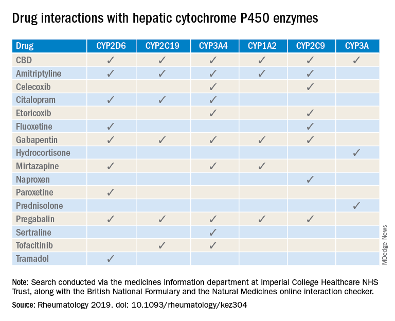
“Patients are increasingly requesting information concerning the safety of CBD oil,” Taryn Youngstein, MD, and associates said in letter to the editor in Rheumatology, but current guidelines on the use of medical cannabis do “not address the potential interactions between CBD oil and medicines frequently used in the rheumatology clinic.”
The most important potential CBD interaction, they suggested, may be with corticosteroids. Hydrocortisone and prednisolone both inhibit the cytochrome P450 enzyme CYP3A, but CBD is a potent inhibitor of CYP3A, so “concomitant use may decrease glucocorticoid clearance and increase risk of systemic [corticosteroid] side effects,” the investigators wrote.
CBD also is known to inhibit the cytochrome P450 isozymes CYP2C9, CYP2D6, CYP2C19, CYP3A4, and CYP1A2, which, alone or in combination, are involved in the metabolization of naproxen, tramadol, amitriptyline, and tofacitinib (Xeljanz), according to a literature search done via the college’s medicine information department that also used the British National Formulary and the Natural Medicines online interaction checker.
The Janus kinase inhibitor tofacitinib is included among the possible interactions, but the other Food and Drug Administration–approved JAK inhibitor, baricitinib (Olumiant), is primarily metabolized by the kidneys and should not have significant interaction with CBD, Dr. Youngstein and associates said. Most of the conventional synthetic and biologic disease-modifying antirheumatic drugs, including methotrexate, hydroxychloroquine, adalimumab (Humira), and abatacept (Orencia), also are expected to be relatively free from CBD interactions.
This first published report on interactions between CBD oil and common rheumatology medications “highlights the importance of taking comprehensive drug histories, by asking directly about drugs considered alternative medicines and food supplements,” they said.
The investigators declared no conflicts of interest, and there was no specific funding for the study.
SOURCE: Wilson-Morkeh H et al. Rheumatology. 2019 July 29. doi: 10.1093/rheumatology/kez304.
A number of medications commonly prescribed by rheumatologists may interact with cannabidiol oil, investigators at the Imperial College Healthcare NHS Trust, London, reported.
“Patients are increasingly requesting information concerning the safety of CBD oil,” Taryn Youngstein, MD, and associates said in letter to the editor in Rheumatology, but current guidelines on the use of medical cannabis do “not address the potential interactions between CBD oil and medicines frequently used in the rheumatology clinic.”
The most important potential CBD interaction, they suggested, may be with corticosteroids. Hydrocortisone and prednisolone both inhibit the cytochrome P450 enzyme CYP3A, but CBD is a potent inhibitor of CYP3A, so “concomitant use may decrease glucocorticoid clearance and increase risk of systemic [corticosteroid] side effects,” the investigators wrote.
CBD also is known to inhibit the cytochrome P450 isozymes CYP2C9, CYP2D6, CYP2C19, CYP3A4, and CYP1A2, which, alone or in combination, are involved in the metabolization of naproxen, tramadol, amitriptyline, and tofacitinib (Xeljanz), according to a literature search done via the college’s medicine information department that also used the British National Formulary and the Natural Medicines online interaction checker.
The Janus kinase inhibitor tofacitinib is included among the possible interactions, but the other Food and Drug Administration–approved JAK inhibitor, baricitinib (Olumiant), is primarily metabolized by the kidneys and should not have significant interaction with CBD, Dr. Youngstein and associates said. Most of the conventional synthetic and biologic disease-modifying antirheumatic drugs, including methotrexate, hydroxychloroquine, adalimumab (Humira), and abatacept (Orencia), also are expected to be relatively free from CBD interactions.
This first published report on interactions between CBD oil and common rheumatology medications “highlights the importance of taking comprehensive drug histories, by asking directly about drugs considered alternative medicines and food supplements,” they said.
The investigators declared no conflicts of interest, and there was no specific funding for the study.
SOURCE: Wilson-Morkeh H et al. Rheumatology. 2019 July 29. doi: 10.1093/rheumatology/kez304.
FROM RHEUMATOLOGY
Hemophilia carriers face elevated risk of joint comorbidities
Individuals who are carriers of hemophilia genes and have reduced clotting factor activity have at least a twofold higher risk of joint-related comorbidities, compared with the general population, according to new research.

In a population-based cohort study using patient registry data, Swedish researchers identified 539 potential carriers of impaired factor VIII or IX gene in the X chromosome – 213 of whom had documented factor activity – and paired them with sex‐ and birthdate‐matched controls from the general population.
They found that carriers with reduced factor activity had a 2.3-fold higher risk of a joint diagnosis, compared with the general population (95% confidence interval, 1.1-4.5). Carriers with normal factor activity did not show a statistically significant increase in joint diagnosis hazard, however carriers with unknown factor activity had a 2.4-fold higher risk of joint diagnosis, compared with controls (95% CI, 1.8-3.2). The findings were published in Haemophilia.
By the age of 60 years, around 37% of carriers with reduced or unknown factor activity had received a joint diagnosis, compared with 23% of carriers with normal factor activity.
The most common joint diagnoses across carriers and controls were knee related, including gonarthrosis and internal derangement, but these were more common among carriers. Five carriers also recorded a diagnosis of hemophilic arthropathy or systemic disorders of connective tissue in diseases classified elsewhere.
Researchers also saw a 10-fold higher risk of joint surgery (95% CI, 1.0-3.7) among carriers with reduced factor activity – although the numbers were small – and even among carriers with normal factor activity, there was a nearly twofold higher rate (95% CI, 0.9-4.6), compared with the control population.
Carriers with reduced or unknown factor activity also had a higher risk of outpatient hospitalization, compared with the general population, although no effect was seen in carriers with normal factor activity.
“Although the frequency of joint comorbidities overall was relatively low, our results clearly indicate and confirm a higher burden of joint afflictions, including an earlier age at joint diagnosis, for carriers with reduced or unknown factor activity compared with the general population, as well as more joint surgeries and related hospitalizations,” wrote Mehdi Osooli, PhD, from the Skåne University Hospital in Malmö, Sweden, and his coauthors.
The authors noted that the findings correlated with their earlier research on the incidence of arthropathy among males with mild hemophilia, who have previously been found to have a ninefold higher incidence of arthropathy‐related hospital admissions and a 16‐fold higher incidence of joint disease.
“The relatively higher incidence in the male population compared with the carriers in the current study may be explained by the lower median factor activity level, for example, levels between 5% and 40% in males with mild haemophilia compared with a median overall of 50% in carriers,” they wrote.
All authors declared that they had no conflict of interest related to the study findings. Four of the authors reported financial ties to companies including Novo Nordisk, Shire, and Bayer.
SOURCE: Osooli M et al. Haemophilia. 2019 Aug 14. doi: 10.1111/hae.13831.
Individuals who are carriers of hemophilia genes and have reduced clotting factor activity have at least a twofold higher risk of joint-related comorbidities, compared with the general population, according to new research.
In a population-based cohort study using patient registry data, Swedish researchers identified 539 potential carriers of impaired factor VIII or IX gene in the X chromosome – 213 of whom had documented factor activity – and paired them with sex‐ and birthdate‐matched controls from the general population.
They found that carriers with reduced factor activity had a 2.3-fold higher risk of a joint diagnosis, compared with the general population (95% confidence interval, 1.1-4.5). Carriers with normal factor activity did not show a statistically significant increase in joint diagnosis hazard, however carriers with unknown factor activity had a 2.4-fold higher risk of joint diagnosis, compared with controls (95% CI, 1.8-3.2). The findings were published in Haemophilia.
By the age of 60 years, around 37% of carriers with reduced or unknown factor activity had received a joint diagnosis, compared with 23% of carriers with normal factor activity.
The most common joint diagnoses across carriers and controls were knee related, including gonarthrosis and internal derangement, but these were more common among carriers. Five carriers also recorded a diagnosis of hemophilic arthropathy or systemic disorders of connective tissue in diseases classified elsewhere.
Researchers also saw a 10-fold higher risk of joint surgery (95% CI, 1.0-3.7) among carriers with reduced factor activity – although the numbers were small – and even among carriers with normal factor activity, there was a nearly twofold higher rate (95% CI, 0.9-4.6), compared with the control population.
Carriers with reduced or unknown factor activity also had a higher risk of outpatient hospitalization, compared with the general population, although no effect was seen in carriers with normal factor activity.
“Although the frequency of joint comorbidities overall was relatively low, our results clearly indicate and confirm a higher burden of joint afflictions, including an earlier age at joint diagnosis, for carriers with reduced or unknown factor activity compared with the general population, as well as more joint surgeries and related hospitalizations,” wrote Mehdi Osooli, PhD, from the Skåne University Hospital in Malmö, Sweden, and his coauthors.
The authors noted that the findings correlated with their earlier research on the incidence of arthropathy among males with mild hemophilia, who have previously been found to have a ninefold higher incidence of arthropathy‐related hospital admissions and a 16‐fold higher incidence of joint disease.
“The relatively higher incidence in the male population compared with the carriers in the current study may be explained by the lower median factor activity level, for example, levels between 5% and 40% in males with mild haemophilia compared with a median overall of 50% in carriers,” they wrote.
All authors declared that they had no conflict of interest related to the study findings. Four of the authors reported financial ties to companies including Novo Nordisk, Shire, and Bayer.
SOURCE: Osooli M et al. Haemophilia. 2019 Aug 14. doi: 10.1111/hae.13831.
FROM HAEMOPHILIA
PROMIS tools provide useful data for managing rheumatology patients
LAKE BUENA VISTA, FLA. –

The PROMIS tools – which like most patient-reported outcome (PRO) measurement tools are designed to evaluate and monitor physical, mental, and social health – can be used both for the general population and for individuals living with chronic conditions, Dr. Curtis, professor of medicine in the division of clinical immunology and rheumatology at the University of Alabama at Birmingham (UAB), said at the annual meeting of the Florida Society of Rheumatology.
The tools take a deeper dive into various symptoms and their effects; for instance, with respect to physical health, they measure fatigue, physical function, sleep disturbance, pain intensity, and pain interference – the extent to which pain “messes your patient’s life up,” explained Dr. Curtis, who also is codirector of the UAB Pharmacoepidemiology and Pharmacoeconomics Unit.
Additional physical health domains that PROs measure include dyspnea, gastrointestinal symptoms, pain behavior, pain quality, sexual function, and sleep-related impairment.
These are “things that, honestly, we don’t talk about much as a field, but absolutely affect patients with autoimmune diseases,” he said. “You know, sexual function – that doesn’t come up in my practice spontaneously very often, but there are ways you can quantify that, and for many patients that’s actually a big deal.”
The domains measured by PROMIS tools for mental health look at anxiety and depression, but also delve into alcohol use, anger, cognitive function, life satisfaction, self-efficacy for managing chronic conditions, substance use, and more. The domains for social health address ability to participate in social roles and activities, as well as companionship, satisfaction with social roles and activity, social isolation, and social support.
“You can’t go on a hike with friends [and] be far from a bathroom, because you have bad arthritis and you have Crohn’s disease. Well, that’s kind of an important thing that may or may not come up in your discussions about inflammatory arthritis associated with [inflammatory bowel disease],” he said.
Another example is a patient who is embarrassed attending social functions or wearing a swimsuit because of really bad psoriasis.
“These are the kinds of things that I’m suggesting you and I probably want to measure if we’re providing holistic care to rheumatology patients,” Dr. Curtis said.
The PROMIS tools provide a simple, user-friendly means for doing so in English, Spanish, and many other languages, he noted.
All the scales use the same 1-100 scoring range, which simplifies measurements. They are available for free by download and can be printed or used electronically for use in the office, at home, on the web, and via smartphone.
The NIH developed the PROMIS tools several years ago and validated them for multiple chronic disease populations, Dr. Curtis said, adding that the tools include multiple individual domains and overall “profiles” of varying lengths.
Most are fixed-length scales that are between 4 and 10 questions and can be completed within 30-60 seconds per scale, so several scales can be completed within 5-10 minutes.
However, some scales are longer and provide greater detail.
“The nice thing is that if you ask a few more questions you can get more precise information – there’s more of a floor and ceiling. You can detect people who do really well. You can distinguish between the marathon runners and the 5K-ers and the people who can walk 2 miles but aren’t going to run a race,” he explained.
Further, the PROMIS tools, like the 36-item Short Form Health Survey (SF-36), are benchmarked against the U.S. adult population, allowing for assessment of how a specific drug or treatment “impacts your arthritis patient on a scale that would also be relevant for somebody who doesn’t have arthritis, they have diabetes.”
The metrics and scales are the same, and that can be helpful when trying to get a payer to pay for a particular drug, he said.
“None of these are rheumatology specific; this puts PROs into a language that can help rheumatology contend for the value of the care that we provide on a scale that would be relevant for any other chronic illness, even for nonrheumatology patients,” he explained.
In addition, minimally important differences (group mean change of about 2-3 units) and minimally clinical important differences for individuals (5 units) have been established.
“So we know what the numbers mean, and this is true for all of the scales,” he said.
PROMIS tools also include computer-adaptive testing (CAT) versions, which helps to personalize the scales to provide more precise information for a given patient and eliminate irrelevant information.
Of note, PROMIS health measures are among the data that can be tracked on a smartphone using Arthritis Power, an arthritis research registry developed with the help of a recent infrastructure grant awarded to the Center for Education and Research and Therapeutics of Musculoskeletal Disorders at UAB, Dr. Curtis said.
The measures were also shown in the AWARE study to track closely with other measures, including the Clinical Disease Activity Index (CDAI), and with patient improvement on therapy.
“So these PROMIS scores are tracking with things that you and I are familiar with ... and it looks like these scores are faithfully tracking, over time, patients getting better on therapies that we would expect them to,” he said. “I think this is additional validation – not just from the National Institutes of Health and a decade of research by lots of different groups, but in our own field – that these actually correlate with disease activity ... and that when you start an effective therapy like a [tumor necrosis factor inhibitor] they’re going to improve as you would anticipate.”
Dr. Curtis reported funding from the National Institute of Arthritis and Musculoskeletal and Skin Diseases and the Patient-Centered Outcomes Research Institute. He has also consulted for or received research grants from Amgen, AbbVie, Bristol-Myers Squibb, CORRONA, Lilly, Janssen, Myriad, Novartis, Roche, Pfizer, and Sanofi/Regeneron.
LAKE BUENA VISTA, FLA. –
The PROMIS tools – which like most patient-reported outcome (PRO) measurement tools are designed to evaluate and monitor physical, mental, and social health – can be used both for the general population and for individuals living with chronic conditions, Dr. Curtis, professor of medicine in the division of clinical immunology and rheumatology at the University of Alabama at Birmingham (UAB), said at the annual meeting of the Florida Society of Rheumatology.
The tools take a deeper dive into various symptoms and their effects; for instance, with respect to physical health, they measure fatigue, physical function, sleep disturbance, pain intensity, and pain interference – the extent to which pain “messes your patient’s life up,” explained Dr. Curtis, who also is codirector of the UAB Pharmacoepidemiology and Pharmacoeconomics Unit.
Additional physical health domains that PROs measure include dyspnea, gastrointestinal symptoms, pain behavior, pain quality, sexual function, and sleep-related impairment.
These are “things that, honestly, we don’t talk about much as a field, but absolutely affect patients with autoimmune diseases,” he said. “You know, sexual function – that doesn’t come up in my practice spontaneously very often, but there are ways you can quantify that, and for many patients that’s actually a big deal.”
The domains measured by PROMIS tools for mental health look at anxiety and depression, but also delve into alcohol use, anger, cognitive function, life satisfaction, self-efficacy for managing chronic conditions, substance use, and more. The domains for social health address ability to participate in social roles and activities, as well as companionship, satisfaction with social roles and activity, social isolation, and social support.
“You can’t go on a hike with friends [and] be far from a bathroom, because you have bad arthritis and you have Crohn’s disease. Well, that’s kind of an important thing that may or may not come up in your discussions about inflammatory arthritis associated with [inflammatory bowel disease],” he said.
Another example is a patient who is embarrassed attending social functions or wearing a swimsuit because of really bad psoriasis.
“These are the kinds of things that I’m suggesting you and I probably want to measure if we’re providing holistic care to rheumatology patients,” Dr. Curtis said.
The PROMIS tools provide a simple, user-friendly means for doing so in English, Spanish, and many other languages, he noted.
All the scales use the same 1-100 scoring range, which simplifies measurements. They are available for free by download and can be printed or used electronically for use in the office, at home, on the web, and via smartphone.
The NIH developed the PROMIS tools several years ago and validated them for multiple chronic disease populations, Dr. Curtis said, adding that the tools include multiple individual domains and overall “profiles” of varying lengths.
Most are fixed-length scales that are between 4 and 10 questions and can be completed within 30-60 seconds per scale, so several scales can be completed within 5-10 minutes.
However, some scales are longer and provide greater detail.
“The nice thing is that if you ask a few more questions you can get more precise information – there’s more of a floor and ceiling. You can detect people who do really well. You can distinguish between the marathon runners and the 5K-ers and the people who can walk 2 miles but aren’t going to run a race,” he explained.
Further, the PROMIS tools, like the 36-item Short Form Health Survey (SF-36), are benchmarked against the U.S. adult population, allowing for assessment of how a specific drug or treatment “impacts your arthritis patient on a scale that would also be relevant for somebody who doesn’t have arthritis, they have diabetes.”
The metrics and scales are the same, and that can be helpful when trying to get a payer to pay for a particular drug, he said.
“None of these are rheumatology specific; this puts PROs into a language that can help rheumatology contend for the value of the care that we provide on a scale that would be relevant for any other chronic illness, even for nonrheumatology patients,” he explained.
In addition, minimally important differences (group mean change of about 2-3 units) and minimally clinical important differences for individuals (5 units) have been established.
“So we know what the numbers mean, and this is true for all of the scales,” he said.
PROMIS tools also include computer-adaptive testing (CAT) versions, which helps to personalize the scales to provide more precise information for a given patient and eliminate irrelevant information.
Of note, PROMIS health measures are among the data that can be tracked on a smartphone using Arthritis Power, an arthritis research registry developed with the help of a recent infrastructure grant awarded to the Center for Education and Research and Therapeutics of Musculoskeletal Disorders at UAB, Dr. Curtis said.
The measures were also shown in the AWARE study to track closely with other measures, including the Clinical Disease Activity Index (CDAI), and with patient improvement on therapy.
“So these PROMIS scores are tracking with things that you and I are familiar with ... and it looks like these scores are faithfully tracking, over time, patients getting better on therapies that we would expect them to,” he said. “I think this is additional validation – not just from the National Institutes of Health and a decade of research by lots of different groups, but in our own field – that these actually correlate with disease activity ... and that when you start an effective therapy like a [tumor necrosis factor inhibitor] they’re going to improve as you would anticipate.”
Dr. Curtis reported funding from the National Institute of Arthritis and Musculoskeletal and Skin Diseases and the Patient-Centered Outcomes Research Institute. He has also consulted for or received research grants from Amgen, AbbVie, Bristol-Myers Squibb, CORRONA, Lilly, Janssen, Myriad, Novartis, Roche, Pfizer, and Sanofi/Regeneron.
EXPERT ANALYSIS FROM FSR 2019
NSAIDs a significant mediator of cardiovascular risk in osteoarthritis

Writing in Arthritis & Rheumatology, researchers reported the outcomes of a longitudinal, population-based cohort study of 7,743 individuals with osteoarthritis patients and 23,229 age- and sex-matched controls without osteoarthritis.
“The prevailing hypothesis in the OA to CVD relationship has been that OA patients frequently take NSAIDs to control their pain and inflammation and that this may lead to them developing CVD,” wrote Mohammad Atiquzzaman, a PhD student at the University of British Columbia, Vancouver, and his coauthors. However they commented that no studies had so far examined this directly in patients with osteoarthritis.
Overall, people with osteoarthritis had a significant 23% higher risk of cardiovascular disease, compared with controls, after adjustment for factors such body mass index, hypertension, diabetes, hyperlipidemia, and socioeconomic status. They also had a 42% higher risk of congestive heart failure, 17% higher risk of ischemic heart disease, and 14% higher risk of stroke.
NSAID use was five times more common among people with osteoarthritis, and NSAIDs alone were associated with a greater than fourfold higher risk of cardiovascular disease, after adjusting for osteoarthritis and other potential confounders.
When the authors performed modeling to break down the effect of osteoarthritis on CVD risk into the direct effect of osteoarthritis itself and the indirect effect mediated by NSAID use, they concluded that 41% of the total effect of osteoarthritis on cardiovascular risk was mediated by NSAIDs. The effect of NSAIDs was particularly pronounced for stroke, in which cases they estimated that the drugs contributed to 64% of the increased in risk, and in ischemic heart disease, in which they contributed to 56% of the increased risk.
Subgroup analysis suggested that conventional NSAIDs were responsible for around 29% of the total increased risk of cardiovascular disease, while selective COX-2 inhibitors, or coxibs, such as celecoxib, lumiracoxib, rofecoxib, and valdecoxib mediated around 21%. For ischemic heart disease, conventional NSAIDs explained around 45% of the increased risk, while selective coxibs explained around 32% of the risk. Similarly, with congestive heart failure and stroke, the proportion of risk mediated by NSAIDs was higher for conventional NSAIDs, compared with coxibs.
The authors noted that while a number of previous studies have found osteoarthritis is an independent risk factor for cardiovascular disease, theirs was the first study to specifically examine the role that NSAIDs play in that increased risk.
However, they noted that their information on NSAID use was gleaned from prescription claims data, which did not include information on over-the-counter NSAID use. Their analysis was also unable to include information on family history of cardiovascular disease, smoking, and physical activity, which are important cardiovascular disease risk factors. They did observe that the rates of obesity were higher among the osteoarthritis group when compared with controls (29% vs. 20%), and hypertension and COPD were also more common among individuals with osteoarthritis.
There was no outside funding for the study, and the authors had no conflicts of interest to declare.
SOURCE: Atiquzzaman M et al. Arthritis Rheumatol. 2019 Aug 6. doi: 10.1002/art.41027
FROM ARTHRITIS & RHEUMATOLOGY
Preoperative tramadol fails to improve function after knee surgery
according to findings of a study based on pre- and postsurgery data.
Tramadol has become a popular choice for nonoperative knee pain relief because of its low potential for abuse and favorable safety profile, but its impact on postoperative outcomes when given before knee surgery has not been well studied, wrote Adam Driesman, MD, of the New York University Langone Orthopedic Hospital and colleagues.
In a study published in the Journal of Arthroplasty, the researchers compared patient-reported outcomes (PRO) after total knee arthroplasty among 136 patients who received no opiates, 21 who received tramadol, and 42 who received other opiates. All patients who did not have preoperative and postoperative PRO scores were excluded
All patients received the same multimodal perioperative pain protocol, and all were placed on oxycodone postoperatively for maintenance and breakthrough pain as needed, with discharge prescriptions for acetaminophen/oxycodone combination (Percocet) for breakthrough pain.
Patients preoperative assessment using the Knee Disability and Osteoarthritis Outcome Score Jr. (KOOS, JR.) were similar among the groups prior to surgery; baseline scores for the groups receiving either tramadol, no opiates, or other opiates were 49.95, 50.4, and 48.0, respectively. Demographics also were not significantly different among the groups.
At 3 months, the average KOOS, JR., score for the tramadol group (62.4) was significantly lower, compared with the other-opiate group (67.1) and treatment-naive group (70.1). In addition, patients in the tramadol group had the least change in scores on KOOS, JR., with an average of 12.5 points, compared with 19.1-point and 20.1-point improvements, respectively, in the alternate-opiate group and opiate-naive group.
The data expand on previous findings that patients given preoperative opioids had proportionally less postoperative pain relief than those not on opioids, the researchers said, but noted that they were surprised by the worse outcomes in the tramadol group given its demonstrated side-effect profile.
The study findings were limited by several factors including the retrospective design and relatively short follow-up period, as well as the inability to accurately determine outpatient medication use, not only of opioids, but of nonopioid postoperative pain medications that could have affected the results, the researchers said.
“However, given the conflicting evidence presented in this study and despite the 2013 American Academy of Orthopedic Surgeons Clinical Practice Guidelines, it is recommended providers remain very conservative in their administration of outpatient narcotics including tramadol prior to surgery,” they concluded.
chestphysiciannews@chestnet.org
SOURCE: Driesman A et al. J Arthroplasty. 2019;34(8):1662-66.
FROM THE JOURNAL OF ARTHROPLASTY
What’s hot in knee OA rehab research
TORONTO – Emerging evidence indicates that patients with knee osteoarthritis who engage in high-intensity interval training obtain significantly greater improvement in physical function than with conventionally prescribed moderate-intensity exercise, Monica R. Maly, PhD, said at the OARSI 2019 World Congress.

This was one of the key conclusions she and her coworkers drew from their analysis of the past year’s published research on diet and exercise interventions to improve outcomes in patients with OA, where obesity and physical inactivity figure prominently as modifiable lifestyle factors.
Another finding: Exercise interventions are where all the action is at present in the field of lifestyle-modification research aimed at achieving better health-related quality of life and other positive outcomes in OA. Dietary interventions are simply not a hot research topic. Indeed, her review of the past year’s literature included 38 randomized, controlled trials (RCTs) and 15 meta-analyses and systematic reviews – and all 38 RCTs addressed exercise interventions.
“It’s interesting to note that we found no new RCT data on diet to modify obesity in OA in the past year,” Dr. Maly said at the meeting sponsored by the Osteoarthritis Research Society International.
Additionally, 32 of the 38 RCTs devoted to exercise interventions for OA focused specifically on knee OA, noted Dr. Maly of the department of kinesiology at the University of Waterloo (Ont.).
Aerobic exercise dosage and intensity
Australian investigators conducted a pilot randomized trial of high-intensity interval training (HIIT) versus more conventional moderate-intensity exercise to improve health-related quality of life and physical function in 27 patients with knee OA. The exercise programs involved unsupervised home-based cycling, with participants requested to do four roughly 25-minute sessions per week for 8 weeks.
The two exercise intensity groups showed similar gains in health-related quality of life as assessed by the Western Ontario and McMaster Universities Osteoarthritis Index (WOMAC). However, the HIIT group showed significantly greater improvement in physical function as measured on the Timed Up and Go test (PeerJ. 2018 May 9;6:e4738).
Dr. Maly noted that adherence to the home-based exercise programs was a challenge: Only 17 of the 27 patients completed the 8-week Australian study, for a 37% dropout rate. However, most study withdrawals were because of family-related issues, illness, or injuries unrelated to cycling, with no signal that HIIT placed knee OA patients at higher injury risk.
Other investigators performed a systematic review of 45 studies in an effort to generate evidence-based guidance about the optimal exercise dosing in order to improve outcomes in knee OA patients. They concluded that programs comprising 24 therapeutic exercise sessions over the course of 8-12 weeks resulted in the largest improvements in measures of pain and physical function. And, importantly, one exercise session per week conferred no benefits (J Orthop Sports Phys Ther. 2018 Mar;48[3]:146-61).
“Frequency probably matters,” Dr. Maly observed.
Patients and their physicians often wonder if long-term, land-based exercise might have deleterious impacts on joint structure in patients with knee OA. Reassurance on this score was provided by a recent meta-analysis of RCTs that concluded, on the basis of moderate-strength evidence, that exercise therapy of longer than 6 months duration had no adverse effect on tibiofemoral radiographic disease severity, compared with no exercise. Nor was there evidence of a long-term-exercise–associated deterioration of tibiofemoral cartilage morphology or worsening of synovitis or effusion. Plus, the meta-analysis provided limited evidence to suggest long-term exercise had a protective effect on the composition of patellar cartilage (Semin Arthritis Rheum. 2019 Jun;48[6]:941-9).
“While there was a little bit of evidence suggesting that long-term exercise could worsen bone marrow lesions, really there was no other evidence that it could change the structure of a joint,” according to Dr. Maly.
Internet-based exercise training
Using the Internet to deliver an individually tailored exercise-training program for patients with symptomatic knee OA might sound like an efficient strategy, but in fact it proved fruitless in a large randomized trial. The 350 participants were assigned to an 8-visit, 4-month program of physical therapy, a wait-list control group, or an internet-based program that delivered tailored exercises and video demonstrations with no face-to-face contact. The bottom line is that improvement in WOMAC scores didn’t differ significantly between the three groups when evaluated at 4 and 12 months (Osteoarthritis Cartilage. 2018 Mar;26[3]:383-96).
“When we deliver exercise with the use of technology, it may require some support, including face to face,” Dr. Maly concluded from the study results.
Strength training
High-intensity resistance training such as weight lifting aimed at strengthening the quadriceps and other large muscles is often eschewed in patients with knee OA because of concern about possible damage to their already damaged joints. Intriguingly, Brazilian investigators may have found a workaround. They randomized 48 women with knee OA to 12 weeks of either supervised low-intensity resistance training performed with partial blood-flow restriction using an air cuff, to low-intensity resistance training alone, or to high-intensity resistance training. The low-intensity resistance workouts involved exercises such as leg presses and knee extensions performed at 30% of maximum effort.
The low-intensity resistance training performed with blood-flow restriction and the high-intensity strength training programs proved similarly effective in improving quadriceps muscle mass, muscle strength, and physical function to a significantly greater extent than with low-intensity resistance training alone. Moreover, low-intensity resistance training with blood-flow restriction also resulted in a significant improvement in pain scores. That finding, coupled with the fact that 4 of the 16 patients in the high-intensity resistance training group dropped out of the trial because of exercise-induced knee pain, suggests that low-intensity strength training carried out with partial blood-flow restriction may have a bright future (Med Sci Sports Exerc. 2018 May;50[5]:897-905).
Exercise plus diet-induced weight loss
How does the combination of dietary weight loss plus exercise stack up against diet alone in terms of benefits on pain and physical function in obese patients with knee OA? A systematic review and meta-analysis of nine RCTs aimed at answering that question concluded that diet-alone strategies are less effective. Both the diet-plus-exercise and diet-only interventions resulted in comparably moderate improvement in physical function. However, diet-only treatments didn’t reduce pain, whereas diet-plus-exercise interventions achieved moderate pain relief (Semin Arthritis Rheum. 2019 Apr;48[5]:765-77).
Dr. Maly reported having no financial conflicts of interest regarding her presentation.
TORONTO – Emerging evidence indicates that patients with knee osteoarthritis who engage in high-intensity interval training obtain significantly greater improvement in physical function than with conventionally prescribed moderate-intensity exercise, Monica R. Maly, PhD, said at the OARSI 2019 World Congress.
This was one of the key conclusions she and her coworkers drew from their analysis of the past year’s published research on diet and exercise interventions to improve outcomes in patients with OA, where obesity and physical inactivity figure prominently as modifiable lifestyle factors.
Another finding: Exercise interventions are where all the action is at present in the field of lifestyle-modification research aimed at achieving better health-related quality of life and other positive outcomes in OA. Dietary interventions are simply not a hot research topic. Indeed, her review of the past year’s literature included 38 randomized, controlled trials (RCTs) and 15 meta-analyses and systematic reviews – and all 38 RCTs addressed exercise interventions.
“It’s interesting to note that we found no new RCT data on diet to modify obesity in OA in the past year,” Dr. Maly said at the meeting sponsored by the Osteoarthritis Research Society International.
Additionally, 32 of the 38 RCTs devoted to exercise interventions for OA focused specifically on knee OA, noted Dr. Maly of the department of kinesiology at the University of Waterloo (Ont.).
Aerobic exercise dosage and intensity
Australian investigators conducted a pilot randomized trial of high-intensity interval training (HIIT) versus more conventional moderate-intensity exercise to improve health-related quality of life and physical function in 27 patients with knee OA. The exercise programs involved unsupervised home-based cycling, with participants requested to do four roughly 25-minute sessions per week for 8 weeks.
The two exercise intensity groups showed similar gains in health-related quality of life as assessed by the Western Ontario and McMaster Universities Osteoarthritis Index (WOMAC). However, the HIIT group showed significantly greater improvement in physical function as measured on the Timed Up and Go test (PeerJ. 2018 May 9;6:e4738).
Dr. Maly noted that adherence to the home-based exercise programs was a challenge: Only 17 of the 27 patients completed the 8-week Australian study, for a 37% dropout rate. However, most study withdrawals were because of family-related issues, illness, or injuries unrelated to cycling, with no signal that HIIT placed knee OA patients at higher injury risk.
Other investigators performed a systematic review of 45 studies in an effort to generate evidence-based guidance about the optimal exercise dosing in order to improve outcomes in knee OA patients. They concluded that programs comprising 24 therapeutic exercise sessions over the course of 8-12 weeks resulted in the largest improvements in measures of pain and physical function. And, importantly, one exercise session per week conferred no benefits (J Orthop Sports Phys Ther. 2018 Mar;48[3]:146-61).
“Frequency probably matters,” Dr. Maly observed.
Patients and their physicians often wonder if long-term, land-based exercise might have deleterious impacts on joint structure in patients with knee OA. Reassurance on this score was provided by a recent meta-analysis of RCTs that concluded, on the basis of moderate-strength evidence, that exercise therapy of longer than 6 months duration had no adverse effect on tibiofemoral radiographic disease severity, compared with no exercise. Nor was there evidence of a long-term-exercise–associated deterioration of tibiofemoral cartilage morphology or worsening of synovitis or effusion. Plus, the meta-analysis provided limited evidence to suggest long-term exercise had a protective effect on the composition of patellar cartilage (Semin Arthritis Rheum. 2019 Jun;48[6]:941-9).
“While there was a little bit of evidence suggesting that long-term exercise could worsen bone marrow lesions, really there was no other evidence that it could change the structure of a joint,” according to Dr. Maly.
Internet-based exercise training
Using the Internet to deliver an individually tailored exercise-training program for patients with symptomatic knee OA might sound like an efficient strategy, but in fact it proved fruitless in a large randomized trial. The 350 participants were assigned to an 8-visit, 4-month program of physical therapy, a wait-list control group, or an internet-based program that delivered tailored exercises and video demonstrations with no face-to-face contact. The bottom line is that improvement in WOMAC scores didn’t differ significantly between the three groups when evaluated at 4 and 12 months (Osteoarthritis Cartilage. 2018 Mar;26[3]:383-96).
“When we deliver exercise with the use of technology, it may require some support, including face to face,” Dr. Maly concluded from the study results.
Strength training
High-intensity resistance training such as weight lifting aimed at strengthening the quadriceps and other large muscles is often eschewed in patients with knee OA because of concern about possible damage to their already damaged joints. Intriguingly, Brazilian investigators may have found a workaround. They randomized 48 women with knee OA to 12 weeks of either supervised low-intensity resistance training performed with partial blood-flow restriction using an air cuff, to low-intensity resistance training alone, or to high-intensity resistance training. The low-intensity resistance workouts involved exercises such as leg presses and knee extensions performed at 30% of maximum effort.
The low-intensity resistance training performed with blood-flow restriction and the high-intensity strength training programs proved similarly effective in improving quadriceps muscle mass, muscle strength, and physical function to a significantly greater extent than with low-intensity resistance training alone. Moreover, low-intensity resistance training with blood-flow restriction also resulted in a significant improvement in pain scores. That finding, coupled with the fact that 4 of the 16 patients in the high-intensity resistance training group dropped out of the trial because of exercise-induced knee pain, suggests that low-intensity strength training carried out with partial blood-flow restriction may have a bright future (Med Sci Sports Exerc. 2018 May;50[5]:897-905).
Exercise plus diet-induced weight loss
How does the combination of dietary weight loss plus exercise stack up against diet alone in terms of benefits on pain and physical function in obese patients with knee OA? A systematic review and meta-analysis of nine RCTs aimed at answering that question concluded that diet-alone strategies are less effective. Both the diet-plus-exercise and diet-only interventions resulted in comparably moderate improvement in physical function. However, diet-only treatments didn’t reduce pain, whereas diet-plus-exercise interventions achieved moderate pain relief (Semin Arthritis Rheum. 2019 Apr;48[5]:765-77).
Dr. Maly reported having no financial conflicts of interest regarding her presentation.
EXPERT ANALYSIS FROM OARSI 2019
Occupational therapy program helps thumb OA
TORONTO – A multimodal occupational therapy intervention in patients with thumb base osteoarthritis brought clinically meaningful improvements in pain, grip strength, and function, at least short term, in a Norwegian multicenter randomized clinical trial, Anne Therese Tveter reported at the OARSI 2019 World Congress.

OA of the thumb base – that is, the carpometacarpal joint – causes more pain and dysfunction than disease involvement at many other sites because of the evolutionary importance of the opposable thumb. Current guidelines recommend conservative therapies as first line for hand OA; however, there is a dearth of high-quality evidence for multimodal occupational therapy in the special setting of thumb-base OA. This was the impetus for a randomized trial of 170 consecutive patients with thumb OA who presented to three Norwegian rheumatology departments for surgical consultation, explained Ms. Tveter, a physiotherapist at the Norwegian National Advisory Unit on Rehabilitation in Rheumatology at Diakonhjemmet Hospital in Oslo.
Participants were randomized to a 3-month, multimodal self-management intervention. It included education about OA; ergonomic principles; the importance of using separate orthoses as much as possible both day and night to stabilize the joint, improve performance, and relieve pain; and – at the heart of the program – instruction in hand exercises to enhance joint mobility, strength, and stability, as well as hand-stretching exercises. The exercises were to be done at home three times per week. Also, the active intervention group received five common assistive devices to help them in household tasks, such as opening jars. The control group received usual care, which was basically information about hand OA, she said at the meeting sponsored by the Osteoarthritis Research Society International.
Ms. Tveter presented an interim analysis focused on the 3-month outcomes. At 4 months, participants underwent surgical consultation. The study will continue for 2 years, with endpoints including the impact of the occupational therapy intervention on need for joint surgery, as well as long-term pain and function measures.
At baseline, most patients reported mild pain, with a median score of 3 on a 10-point numeric rating scale, and moderate disability. Baseline grip and pinch strength was 60%-65% of normal. The 3-month outcomes included pain at rest and during pinch- and grip-strength testing, range of motion through palmar abduction and abduction in the carpometacarpal joint, and self-reported function as measured using the validated MAP-Hand and QuickDASH physiotherapy measures. Adherence to the program was assessed by review of patient diaries.
At 3 months of follow-up, the active-intervention group showed significant improvements in all measures of pain and function except for the flexion deficit, which was minimal to begin with. In contrast, the control group showed no improvements and a trend towards deterioration in pain and function.
Specifically, the intervention group averaged a 1.4-point reduction in pain at rest on a self-reported 10-point scale, a 1.1-point improvement in pain following a grip strength test, and a 0.8-point improvement in pain following a pinch test. On the MAP-Hand self-reported test of function, the intervention group showed a 0.18-point improvement from a baseline of 2 on the 1-4 scale, coupled with an 8.1-point improvement on the QuickDASH, which is scored 0-100.
Adherence to the program was deemed acceptable: 82% of patients reported doing their hand exercises at least twice per week for at least 8 of the 12 weeks, 61% used their day orthotic devices at least 4 days per week for 8 weeks, 54% used the night orthoses at least 5 nights per week for 8 weeks, and 69% utilized at least three of the five home-assist devices. In total, 64% of patients adhered to at least three of the four program components.
Asked for the rationale in requesting that patients do their home exercises three times per week instead of daily, Ms. Tveter replied that three times per week is more realistic and is consistent with major guidelines.
“It would be nice to exercise every day. I don’t think it would be possible to get adherence to that,” she said.
She reported having no financial conflicts regarding the study, funded by scientific research grants from the Norwegian government.
TORONTO – A multimodal occupational therapy intervention in patients with thumb base osteoarthritis brought clinically meaningful improvements in pain, grip strength, and function, at least short term, in a Norwegian multicenter randomized clinical trial, Anne Therese Tveter reported at the OARSI 2019 World Congress.
OA of the thumb base – that is, the carpometacarpal joint – causes more pain and dysfunction than disease involvement at many other sites because of the evolutionary importance of the opposable thumb. Current guidelines recommend conservative therapies as first line for hand OA; however, there is a dearth of high-quality evidence for multimodal occupational therapy in the special setting of thumb-base OA. This was the impetus for a randomized trial of 170 consecutive patients with thumb OA who presented to three Norwegian rheumatology departments for surgical consultation, explained Ms. Tveter, a physiotherapist at the Norwegian National Advisory Unit on Rehabilitation in Rheumatology at Diakonhjemmet Hospital in Oslo.
Participants were randomized to a 3-month, multimodal self-management intervention. It included education about OA; ergonomic principles; the importance of using separate orthoses as much as possible both day and night to stabilize the joint, improve performance, and relieve pain; and – at the heart of the program – instruction in hand exercises to enhance joint mobility, strength, and stability, as well as hand-stretching exercises. The exercises were to be done at home three times per week. Also, the active intervention group received five common assistive devices to help them in household tasks, such as opening jars. The control group received usual care, which was basically information about hand OA, she said at the meeting sponsored by the Osteoarthritis Research Society International.
Ms. Tveter presented an interim analysis focused on the 3-month outcomes. At 4 months, participants underwent surgical consultation. The study will continue for 2 years, with endpoints including the impact of the occupational therapy intervention on need for joint surgery, as well as long-term pain and function measures.
At baseline, most patients reported mild pain, with a median score of 3 on a 10-point numeric rating scale, and moderate disability. Baseline grip and pinch strength was 60%-65% of normal. The 3-month outcomes included pain at rest and during pinch- and grip-strength testing, range of motion through palmar abduction and abduction in the carpometacarpal joint, and self-reported function as measured using the validated MAP-Hand and QuickDASH physiotherapy measures. Adherence to the program was assessed by review of patient diaries.
At 3 months of follow-up, the active-intervention group showed significant improvements in all measures of pain and function except for the flexion deficit, which was minimal to begin with. In contrast, the control group showed no improvements and a trend towards deterioration in pain and function.
Specifically, the intervention group averaged a 1.4-point reduction in pain at rest on a self-reported 10-point scale, a 1.1-point improvement in pain following a grip strength test, and a 0.8-point improvement in pain following a pinch test. On the MAP-Hand self-reported test of function, the intervention group showed a 0.18-point improvement from a baseline of 2 on the 1-4 scale, coupled with an 8.1-point improvement on the QuickDASH, which is scored 0-100.
Adherence to the program was deemed acceptable: 82% of patients reported doing their hand exercises at least twice per week for at least 8 of the 12 weeks, 61% used their day orthotic devices at least 4 days per week for 8 weeks, 54% used the night orthoses at least 5 nights per week for 8 weeks, and 69% utilized at least three of the five home-assist devices. In total, 64% of patients adhered to at least three of the four program components.
Asked for the rationale in requesting that patients do their home exercises three times per week instead of daily, Ms. Tveter replied that three times per week is more realistic and is consistent with major guidelines.
“It would be nice to exercise every day. I don’t think it would be possible to get adherence to that,” she said.
She reported having no financial conflicts regarding the study, funded by scientific research grants from the Norwegian government.
REPORTING FROM OARSI 2019
Key clinical point: A multimodal occupational therapy intervention brought significant improvements in pain and function in patients with thumb-base OA.
Major finding: The intervention resulted in a mean 1.4-point decrease in self-reported pain at rest from a baseline of 3 on a 10-point scale, while most usual care controls showed a modest trend for worsening.
Study details: This was an interim 3-month report from a 2-year, randomized, multicenter trial including 170 consecutive patients who presented for surgical consultation regarding their thumb base OA.
Disclosures: The presenter reported having no financial conflicts regarding the study, funded by Norwegian governmental scientific research grants.
Liposomal steroid brings durable pain relief in knee OA
TORONTO – A single intra-articular injection of a novel, sustained-release liposomal formulation of dexamethasone in patients with symptomatic knee osteoarthritis brought at least 6 months of pain control in a multicenter, phase 2a trial, David Hunter, MD, reported at the OARSI 2019 World Congress.

This is a product that could fill a significant unmet medical need. Current therapies for knee OA have modest efficacy, and the injectable ones provide only 2-4 weeks of benefit. The ability to obtain significant pain relief with just a couple of intra-articular injections per year would be an important therapeutic advance, observed Dr. Hunter, professor of medicine at the University of Sydney.
He presented a 24-week study of 75 patients with symptomatic knee OA randomized at 13 sites in Australia and Taiwan to a single intra-articular injection of either 12 or 18 mg of the liposomal dexamethasone or to normal saline. One knee per patient was treated.
The primary outcome was the change in the Western Ontario and McMaster Universities Osteoarthritis Index (WOMAC) pain score from baseline to week 12. The 12-mg formulation of steroid significantly outperformed placebo at that time point as well as at all others. From a mean baseline WOMAC pain score of 1.49 on the 0-4 scale, patients in the 12-mg group averaged reductions of 0.83 points at 12 weeks, 0.85 at both weeks 16 and 20, and 0.87 at week 24. A statistically significant between-group difference was seen as early as day 3 after injection.
More than half (52%) of recipients of the 12-mg dose of liposomal dexamethasone, a product known for now simply as TLC599, maintained at least 30% pain relief at all visits through the study close at 24 weeks, as did 22% of controls, the rheumatologist reported at the meeting sponsored by the Osteoarthritis Research Society International.
The 12-mg injection also proved superior to placebo for the secondary endpoint of change in WOMAC function score. From a mean baseline score of 1.53, recipients of the 12-mg dose had improvements ranging from 0.82 points at week 12 to 0.85 points at week 24.
Of note, total acetaminophen intake over the course of the trial in the 12-mg steroid group was less than one-third of that in controls.
The 18-mg dose didn’t result in significantly greater reduction in pain scores than placebo. This is because dexamethasone release in the higher-dose formulation as presently constituted turned out to be less efficient, Dr. Hunter explained.
The safety profile was closely similar in all three study arms.
In phase 3 clinical trials, TLC599 will be compared with standard intra-articular triamcinolone, according to the rheumatologist.
He reported serving as a consultant to the Taiwan Liposome Company, which sponsored the phase 2a study, as well as to a handful of other pharmaceutical companies.
TORONTO – A single intra-articular injection of a novel, sustained-release liposomal formulation of dexamethasone in patients with symptomatic knee osteoarthritis brought at least 6 months of pain control in a multicenter, phase 2a trial, David Hunter, MD, reported at the OARSI 2019 World Congress.
This is a product that could fill a significant unmet medical need. Current therapies for knee OA have modest efficacy, and the injectable ones provide only 2-4 weeks of benefit. The ability to obtain significant pain relief with just a couple of intra-articular injections per year would be an important therapeutic advance, observed Dr. Hunter, professor of medicine at the University of Sydney.
He presented a 24-week study of 75 patients with symptomatic knee OA randomized at 13 sites in Australia and Taiwan to a single intra-articular injection of either 12 or 18 mg of the liposomal dexamethasone or to normal saline. One knee per patient was treated.
The primary outcome was the change in the Western Ontario and McMaster Universities Osteoarthritis Index (WOMAC) pain score from baseline to week 12. The 12-mg formulation of steroid significantly outperformed placebo at that time point as well as at all others. From a mean baseline WOMAC pain score of 1.49 on the 0-4 scale, patients in the 12-mg group averaged reductions of 0.83 points at 12 weeks, 0.85 at both weeks 16 and 20, and 0.87 at week 24. A statistically significant between-group difference was seen as early as day 3 after injection.
More than half (52%) of recipients of the 12-mg dose of liposomal dexamethasone, a product known for now simply as TLC599, maintained at least 30% pain relief at all visits through the study close at 24 weeks, as did 22% of controls, the rheumatologist reported at the meeting sponsored by the Osteoarthritis Research Society International.
The 12-mg injection also proved superior to placebo for the secondary endpoint of change in WOMAC function score. From a mean baseline score of 1.53, recipients of the 12-mg dose had improvements ranging from 0.82 points at week 12 to 0.85 points at week 24.
Of note, total acetaminophen intake over the course of the trial in the 12-mg steroid group was less than one-third of that in controls.
The 18-mg dose didn’t result in significantly greater reduction in pain scores than placebo. This is because dexamethasone release in the higher-dose formulation as presently constituted turned out to be less efficient, Dr. Hunter explained.
The safety profile was closely similar in all three study arms.
In phase 3 clinical trials, TLC599 will be compared with standard intra-articular triamcinolone, according to the rheumatologist.
He reported serving as a consultant to the Taiwan Liposome Company, which sponsored the phase 2a study, as well as to a handful of other pharmaceutical companies.
REPORTING FROM OARSI 2019
What’s up in the osteoarthritis drug pipeline
TORONTO – Philip G. Conaghan, MBBS, PhD, observed at the OARSI 2019 World Congress.

“Not only have things not improved during my time in osteoarthritis-land, they’ve gotten worse. We’ve lost therapies,” said Dr. Conaghan, professor of musculoskeletal medicine at the University of Leeds (England) and director of the Leeds Institute of Rheumatic and Musculoskeletal Medicine.
Specifically, opioids are now shunned because of the ongoing epidemic of addiction and a belated recognition that opioids are not a good option for pain relief in OA patients who want to have active lives. And acetaminophen has fallen by the wayside in light of recent evidence of lack of effectiveness: “It’s what our patients have been telling us for a long period of time,” he noted at the meeting sponsored by the Osteoarthritis Research Society International.
But change is in the air.
“I think we’ve got some things coming that look promising. What do I think will be the fastest to get to market? The peripheral nerve modulators look to me like the ones closest to going forward,” according to the rheumatologist, who provided an overview of the OA drug development pipeline, organized by treatment target.
Nerves
“Nerves as a treatment target in OA is a hot area. We’ve seen quite a slew of products recently looking at this. I think it’s a really fascinating area to play in: looking at how we modulate peripheral nociceptive pain,” Dr. Conaghan continued.
Tanezumab, an inhibitor of nerve growth factor, demonstrated very good pain relief and improvement in physical function in a phase 3 trial, although the occurrence of rapidly progressive OA in a subset of patients has bedeviled the drug development program. The hope is that a new subcutaneous drug delivery system coupled with careful patient pretreatment screening will mitigate the problem.
Tanezumab’s efficacy has contributed to a new understanding of the nature of pain in OA.
“I know I’m going to upset some people, but if you think central sensitization is the biggest driver of pain, I’d have to argue that the tanezumab program is the biggest single argument against that, since tanezumab is a large monoclonal antibody that doesn’t cross the blood-brain barrier and yet it has some of the best pain responses that we’ve seen,” Dr. Conaghan said.
Another peripheral nerve modulator, known for now as CNTX-4975, exhibited dose-dependent improvement in knee OA pain in the 175-patient, phase 2b TRIUMPH trial (Arthritis Rheumatol. 2019 Mar 19. doi: 10.1002/art.40894). CNTX-4975, which is delivered by intra-articular injection, is a synthetic form of capsaicin specific to pain nociceptors within the joint. Other sensory fibers remain unaffected.
Cartilage
Sprifermin, a recombinant human fibroblast growth factor 18 given by intra-articular injection, stimulates chondrocyte growth and decreases type 1 collagen expression. At year 2 in the ongoing 549-patient, 5-year, phase 2 FORWARD study, a dose-dependent increase in cartilage thickness at the tibiofemoral joint became apparent in sprifermin-treated patients, compared with those on placebo. This cartilage rebuilding effect was maintained at year 3, Dr. Conaghan said.
Bone
At the OARSI meeting, Dr. Conaghan and coinvestigators presented the results of a 6-month, open-label extension of their previously reported 6-month, placebo-controlled, phase 2 study of MIV-711, a potent selective reversible inhibitor of cathepsin K. The disease-modifying effects of MIV-711 seen in the first 6 months of the study, based on MRI-based measurements of changes in three-dimensional bone shape and cartilage thickness, were maintained in the second 6 months. Notably, MIV-711 slowed the rate of increase in bone area in the medial femur region and reduced loss of cartilage thickness relative to placebo. MIV-711 has also been shown to achieve a rapid and sustained reduction in the bone turnover biomarkers CTX-1 and -2, providing a rational mechanism to explain the drug’s observed structural benefits.
“So we’ve now got two agents – sprifermin for cartilage and MIV-711 for bone – showing that it’s possible to get some structural change, but no symptomatic benefit within the period of those trials,” the rheumatologist noted.
Wnt pathway inhibition
Samumed has launched a phase 3 clinical trials program, known as STRIDES, for lorecivivint, the company’s investigational small molecule inhibitor of the Wnt pathway. In phase 2 studies, including one led by Dr. Conaghan, intra-articular injection of lorecivivint, previously known as SMO4690, improved pain and physical function as well as medial joint space width. The drug’s potential effects on multiple tissues offers the promise of providing both symptomatic improvement and modification of the course of structural disease progression.
Inflammation
Lutikizumab, an anti–interleukin-1 alpha/beta immunoglobulin, proved to be a disappointment in a recent phase 2, placebo-controlled trial carried out in 350 patients with knee OA and synovitis. The IL-1 inhibitor had no benefit on synovitis, joint space narrowing, or cartilage thickness. Nor was it significantly better than placebo for pain reduction (Arthritis Rheumatol. 2019 Jul;71[7]:1056-69).
Anti–tumor necrosis factor agents haven’t exactly set the OA world on fire, either.
“In rheumatoid arthritis we know they’re stupendously effective, but the data from a number of trials in OA show they’re not so effective on symptoms and signs,” Dr. Conaghan said.
Colchicine and hydroxychloroquine are other anti-inflammatory agents which, while in theory might be helpful, have in actuality shown no benefit for OA symptoms in controlled clinical trials and are now considered dead ends.
On the other hand, the sustained delivery of intra-articular corticosteroids through the use of microsphere technology is advancing smartly through the developmental pipeline. Dr. Conaghan was first author of a multicenter, double-blind, phase 3 trial of FX006, a sustained-release formulation of triamcinolone acetonide, which showed that a single intra-articular injection provided at least 3 months of pain relief in knee OA patients, along with reduced systemic drug exposure, compared with standard intra-articular corticosteroids (J Bone Joint Surg Am. 2018 Apr 18;100[8]:666-77).
FX006 also performed well in another phase 3 trial, this one featuring repeated dosing on a flexible schedule based upon patient response (Rheumatol Ther. 2019 Mar;6[1]:109-24).
Reassuringly, this slow-release corticosteroid doesn’t appear to worsen glycemic control in knee OA patients with type 2 diabetes (Rheumatology [Oxford]. 2018 Dec 1;57[12]:2235-41).
“This is the start of a revolution in nanotechnology and the ability to slowly deliver a variety of drugs within the joint,” Dr. Conaghan predicted.
Although he was tasked at OARSI 2019 with providing an overview of the OA pharmacologic pipeline, he stressed that in his clinical practice, as opposed to his work as a clinical trialist, he functions more like a physical therapist.
“I actually spend my whole time in the OA clinic being a physical therapist and trying to get people strong, because that does work and it has no side effects. It’s just that nobody does it. We have a real adherence problem,” he said. “My favorite thought is this: keep people strong. If a patient can’t get out of a chair easily or can’t undo a jar, then they’ve got a problem.”
Dr. Conaghan reported receiving research funding from and serving as a consultant to many of the companies developing novel drug treatments for OA.
TORONTO – Philip G. Conaghan, MBBS, PhD, observed at the OARSI 2019 World Congress.
“Not only have things not improved during my time in osteoarthritis-land, they’ve gotten worse. We’ve lost therapies,” said Dr. Conaghan, professor of musculoskeletal medicine at the University of Leeds (England) and director of the Leeds Institute of Rheumatic and Musculoskeletal Medicine.
Specifically, opioids are now shunned because of the ongoing epidemic of addiction and a belated recognition that opioids are not a good option for pain relief in OA patients who want to have active lives. And acetaminophen has fallen by the wayside in light of recent evidence of lack of effectiveness: “It’s what our patients have been telling us for a long period of time,” he noted at the meeting sponsored by the Osteoarthritis Research Society International.
But change is in the air.
“I think we’ve got some things coming that look promising. What do I think will be the fastest to get to market? The peripheral nerve modulators look to me like the ones closest to going forward,” according to the rheumatologist, who provided an overview of the OA drug development pipeline, organized by treatment target.
Nerves
“Nerves as a treatment target in OA is a hot area. We’ve seen quite a slew of products recently looking at this. I think it’s a really fascinating area to play in: looking at how we modulate peripheral nociceptive pain,” Dr. Conaghan continued.
Tanezumab, an inhibitor of nerve growth factor, demonstrated very good pain relief and improvement in physical function in a phase 3 trial, although the occurrence of rapidly progressive OA in a subset of patients has bedeviled the drug development program. The hope is that a new subcutaneous drug delivery system coupled with careful patient pretreatment screening will mitigate the problem.
Tanezumab’s efficacy has contributed to a new understanding of the nature of pain in OA.
“I know I’m going to upset some people, but if you think central sensitization is the biggest driver of pain, I’d have to argue that the tanezumab program is the biggest single argument against that, since tanezumab is a large monoclonal antibody that doesn’t cross the blood-brain barrier and yet it has some of the best pain responses that we’ve seen,” Dr. Conaghan said.
Another peripheral nerve modulator, known for now as CNTX-4975, exhibited dose-dependent improvement in knee OA pain in the 175-patient, phase 2b TRIUMPH trial (Arthritis Rheumatol. 2019 Mar 19. doi: 10.1002/art.40894). CNTX-4975, which is delivered by intra-articular injection, is a synthetic form of capsaicin specific to pain nociceptors within the joint. Other sensory fibers remain unaffected.
Cartilage
Sprifermin, a recombinant human fibroblast growth factor 18 given by intra-articular injection, stimulates chondrocyte growth and decreases type 1 collagen expression. At year 2 in the ongoing 549-patient, 5-year, phase 2 FORWARD study, a dose-dependent increase in cartilage thickness at the tibiofemoral joint became apparent in sprifermin-treated patients, compared with those on placebo. This cartilage rebuilding effect was maintained at year 3, Dr. Conaghan said.
Bone
At the OARSI meeting, Dr. Conaghan and coinvestigators presented the results of a 6-month, open-label extension of their previously reported 6-month, placebo-controlled, phase 2 study of MIV-711, a potent selective reversible inhibitor of cathepsin K. The disease-modifying effects of MIV-711 seen in the first 6 months of the study, based on MRI-based measurements of changes in three-dimensional bone shape and cartilage thickness, were maintained in the second 6 months. Notably, MIV-711 slowed the rate of increase in bone area in the medial femur region and reduced loss of cartilage thickness relative to placebo. MIV-711 has also been shown to achieve a rapid and sustained reduction in the bone turnover biomarkers CTX-1 and -2, providing a rational mechanism to explain the drug’s observed structural benefits.
“So we’ve now got two agents – sprifermin for cartilage and MIV-711 for bone – showing that it’s possible to get some structural change, but no symptomatic benefit within the period of those trials,” the rheumatologist noted.
Wnt pathway inhibition
Samumed has launched a phase 3 clinical trials program, known as STRIDES, for lorecivivint, the company’s investigational small molecule inhibitor of the Wnt pathway. In phase 2 studies, including one led by Dr. Conaghan, intra-articular injection of lorecivivint, previously known as SMO4690, improved pain and physical function as well as medial joint space width. The drug’s potential effects on multiple tissues offers the promise of providing both symptomatic improvement and modification of the course of structural disease progression.
Inflammation
Lutikizumab, an anti–interleukin-1 alpha/beta immunoglobulin, proved to be a disappointment in a recent phase 2, placebo-controlled trial carried out in 350 patients with knee OA and synovitis. The IL-1 inhibitor had no benefit on synovitis, joint space narrowing, or cartilage thickness. Nor was it significantly better than placebo for pain reduction (Arthritis Rheumatol. 2019 Jul;71[7]:1056-69).
Anti–tumor necrosis factor agents haven’t exactly set the OA world on fire, either.
“In rheumatoid arthritis we know they’re stupendously effective, but the data from a number of trials in OA show they’re not so effective on symptoms and signs,” Dr. Conaghan said.
Colchicine and hydroxychloroquine are other anti-inflammatory agents which, while in theory might be helpful, have in actuality shown no benefit for OA symptoms in controlled clinical trials and are now considered dead ends.
On the other hand, the sustained delivery of intra-articular corticosteroids through the use of microsphere technology is advancing smartly through the developmental pipeline. Dr. Conaghan was first author of a multicenter, double-blind, phase 3 trial of FX006, a sustained-release formulation of triamcinolone acetonide, which showed that a single intra-articular injection provided at least 3 months of pain relief in knee OA patients, along with reduced systemic drug exposure, compared with standard intra-articular corticosteroids (J Bone Joint Surg Am. 2018 Apr 18;100[8]:666-77).
FX006 also performed well in another phase 3 trial, this one featuring repeated dosing on a flexible schedule based upon patient response (Rheumatol Ther. 2019 Mar;6[1]:109-24).
Reassuringly, this slow-release corticosteroid doesn’t appear to worsen glycemic control in knee OA patients with type 2 diabetes (Rheumatology [Oxford]. 2018 Dec 1;57[12]:2235-41).
“This is the start of a revolution in nanotechnology and the ability to slowly deliver a variety of drugs within the joint,” Dr. Conaghan predicted.
Although he was tasked at OARSI 2019 with providing an overview of the OA pharmacologic pipeline, he stressed that in his clinical practice, as opposed to his work as a clinical trialist, he functions more like a physical therapist.
“I actually spend my whole time in the OA clinic being a physical therapist and trying to get people strong, because that does work and it has no side effects. It’s just that nobody does it. We have a real adherence problem,” he said. “My favorite thought is this: keep people strong. If a patient can’t get out of a chair easily or can’t undo a jar, then they’ve got a problem.”
Dr. Conaghan reported receiving research funding from and serving as a consultant to many of the companies developing novel drug treatments for OA.
EXPERT ANALYSIS FROM OARSI 2019
How common is accelerated knee OA?
TORONTO – Accelerated knee osteoarthritis – a particularly noxious form of the joint disease – occurred in more than one in seven women who developed knee osteoarthritis in the prospective, long-term Chingford Cohort Study, Jeffrey B. Driban, PhD, reported at the OARSI 2019 World Congress.
This finding from a unique prospective study of 1,003 middle-aged U.K. women who were followed for the development of knee osteoarthritis (OA) for 15 years is important because the participants represented a typical community-based population sample. And yet the Chingford results are consistent with and confirmatory of those found earlier in the Osteoarthritis Initiative, a U.S. cohort study of nearly 4,800 individuals, even though the Osteoarthritis Initiative featured a population enriched with established risk factors for knee OA, Dr. Driban explained at the meeting, sponsored by the Osteoarthritis Research Society International.
In Chingford, accelerated knee OA accounted for 15% of all incident cases of knee OA during follow-up, and for 17% of all newly affected knees, whereas 20% of incident knee OA in the Osteoarthritis Initiative was accelerated knee OA, noted Dr. Driban of Tufts University, Boston.
Accelerated knee OA is defined by rapidly progressive structural damage. Affected individuals streak from no radiographic evidence of knee OA to advanced-stage disease marked by a Kellgren-Lawrence score of 3 or more within 4 years, whereas the typical form of knee OA follows a more gradual course. Also, accelerated knee OA features greater pain and disability.
In the Chingford study, the cumulative incidence of accelerated knee OA was 3.9%, while typical knee OA occurred in 21.7% of women. During years 6-15 of follow-up, 21% of women with accelerated knee OA underwent total knee replacement, compared with 2% of those with typical knee OA and 0.9% of women without knee OA.
Dr. Driban reported having no financial conflicts regarding his analysis of the Chingford Cohort Study and the Osteoarthritis Initiative, supported by Arthritis Research UK and the National Institutes of Health, respectively.
SOURCE: Driban JB et al. Osteoarthritis Cartilage. 2019 Apr;27[suppl 1]:S250-S251, Abstract 352.
REPORTING FROM OARSI 2019