User login
For MD-IQ on Family Practice News, but a regular topic for Rheumatology News
VIDEO: Prescription-strength ibuprofen worsens blood pressure more than other NSAIDs
BARCELONA – Prescription-strength ibuprofen has a bigger adverse effect on blood pressure than celecoxib or naproxen, a finding that suggests a likely mechanism for the worse cardiovascular event rate documented in ibuprofen-treated arthritis patients in the PRECISION trial, Frank Ruschitzka, MD, said at the annual congress of the European Society of Cardiology.
“Prescription-strength ibuprofen is under pressure – it has a high incidence of new-onset hypertension, particularly when compared to the more selective COX-2 inhibitor celecoxib. Before we did this study, many would have said it’s the other way around,” observed Dr. Ruschitzka, professor of cardiology at the University of Zurich.
He presented the results of PRECISION-ABPM (Prospective Randomized Evaluation of Celecoxib Integrated Safety Versus Ibuprofen or Naproxen Ambulatory Blood Pressure Measurement).
“These results will have impact on your daily practice when you go home,” the cardiologist promised.
PRECISION-ABPM was a prespecified double-blind, randomized, 60-center substudy of the published PRECISION trial, which included 24,081 U.S. patients who needed daily NSAIDs for arthritis and were also at increased cardiovascular risk. They were randomized to the COX-2 inhibitor celecoxib at 100-200 mg b.i.d. or the nonselective NSAIDs ibuprofen at 600-800 mg three times a day or naproxen at 375-500 mg twice daily. Participants also received a proton pump inhibitor to protect against NSAID-related GI bleeding. In the on-treatment analysis, the ibuprofen group was significantly more likely to experience cardiovascular and all-cause mortality and renal events than were those on celecoxib (N Engl J Med. 2016 Dec 29;375[26]:2519-29).
The PRECISION-ABPM substudy included 444 arthritis patients, 92% of whom had osteoarthritis. During the 4-month study, investigators amassed roughly 60,000 automated blood pressure measurements across the three study arms.
The primary outcome was change from baseline in mean 24-hour systolic blood pressure (SBP). It increased by 3.7 mm Hg in the ibuprofen group and declined by 0.3 mm Hg in the celecoxib group, while the naproxen group occupied the middle ground with a 1.6-mm Hg increase.
The nearly 4-mm Hg increase in mean 24-hour SBP at 4 months in the ibuprofen group is of sufficient magnitude to be clinically important, Dr. Ruschitzka noted. He noted that fully 23.2% of ibuprofen-treated patients who had normal baseline blood pressure developed hypertension as defined by a mean 24-hour SBP of at least 130 and/or a diastolic blood pressure of at least 80 mm Hg. In contrast, incident hypertension occurred in only 10.3% of the celecoxib group and 19% of naproxen-treated patients. Thus, the likelihood of developing hypertension was 61% less with celecoxib than ibuprofen and 51% less with celecoxib than naproxen.
Not treating chronic arthritic pain to avoid the cardiovascular risk of NSAIDs is not a legitimate option.
“Pain is a cardiovascular risk factor,” Dr. Ruschitzka emphasized. “It’s unethical not to treat it. If you don’t treat pain, the patient’s blood pressure goes up, heart rate goes up, and you’re driving patients into inactivity.”
Although he’s convinced there’s no such thing as a safe NSAID from a cardiovascular risk standpoint, the PRECISION and PRECISION-ABPM data show celecoxib is less unsafe than ibuprofen. And as for the oft-heard statement that naproxen is the safest NSAID for the heart, Dr. Ruschitzka snorted, “What an urban legend.”
Discussant Scott Solomon, MD, opined that, while PRECISION-ABPM doesn’t support the notion that conventional NSAIDs such as naproxen or ibuprofen are any safer than celecoxib, it would be wrong to conclude from the study that celecoxib doesn’t affect blood pressure and is safer than the others from a cardiovascular standpoint. That’s because the three study drugs weren’t compared in an equipotent way. Because of safety concerns, the Food and Drug Administration required that the daily dose of celecoxib be capped at the low end of the therapeutic range, while no such constraints were placed on the two nonselective NSAIDS.
“Compared to placebo, all NSAIDs likely raise blood pressure, especially in patients prone to hypertension, those with chronic kidney disease, the elderly – and this is exactly the type of patients who require NSAIDs for arthritis. Whichever NSAID is chosen, clinicians should be aware of this effect and treat hypertension according to guidelines,” said Dr. Solomon, director of noninvasive cardiology at Brigham and Women’s Hospital, Boston, and professor of medicine at Harvard Medical School.
Dr. Solomon has been a key figure in the COX-2 inhibitor controversy of the last decade. He was lead author of a 2005 review of data from clinical trials of COX-2 inhibitors for colorectal adenoma prevention, which concluded that the drugs had a cardiovascular safety issue in that setting (N Engl J Med. 2005 Mar 17;352[11]:1071-80).
“Our analysis of celecoxib concluded that a dose-dependent increase in cardiovascular events was there, was real, but notably occurred at doses which were substantially higher than what we typically use for patients with arthritis,” he said.
That report triggered a fevered reaction.
“Amid an enormous amount of hype, hyperbole, and hysteria, the safety of these agents was thrown into question, leading to the withdrawal of all but one of them from the market and a black-box warning around the one remaining agent, celecoxib,” he recalled.
Dr. Ruschitzka discussed his findings in a video interview.
PRECISION-ABPM was sponsored by Pfizer. Dr. Ruschitzka and Dr. Solomon reported having no financial conflicts of interest regarding their presentations.
AT THE ESC CONGRESS 2017
Key clinical point:
Major finding: Incident hypertension occurred within 4 months in 23.2% of arthritis patients on ibuprofen, compared with 10.3% taking celecoxib and 19% on naproxen.
Data source: This was a randomized, double-blind, multicenter, prospective trial including 444 arthritis patients at increased cardiovascular risk who underwent 4 months of ambulatory blood pressure monitoring after being assigned to prescription-strength ibuprofen, naproxen, or celecoxib.
Disclosures: The PRECISION-ABPM trial was sponsored by Pfizer. The presenter reported having no financial conflicts of interest.

Novel knee osteoarthritis drugs target pain, joint space narrowing
MADRID – Two independently conducted, randomized, phase 2 trials of novel agents for moderate to severe knee osteoarthritis have produced promising results, as reported at the European Congress of Rheumatology.
In the 24-week, dose-ranging TRIUMPH trial involving 175 patients, treatment with the synthetic trans-capsaicin CNTX-4975 was associated with improvements in several clinical parameters, such as pain when walking, knee stiffness, and physical function, as well as being generally well tolerated.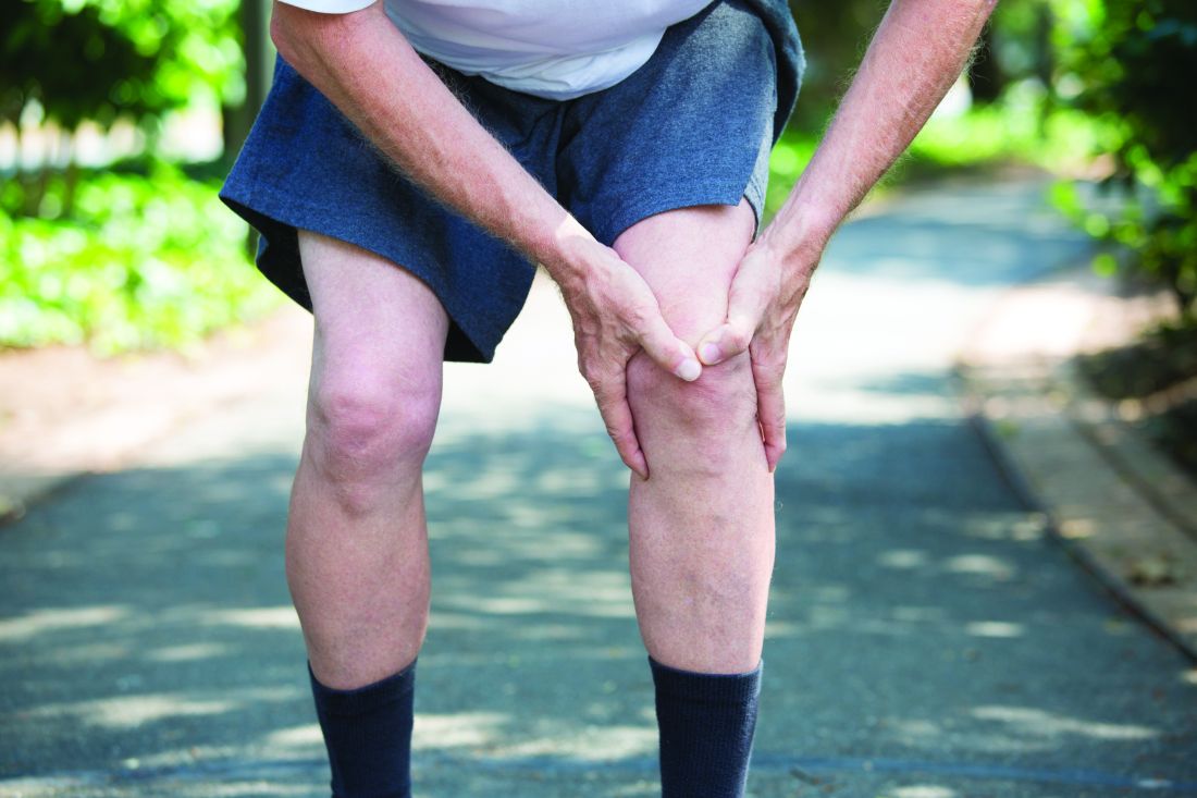
These findings suggest that both could be future alternatives to using nonsteroidal anti-inflammatory drugs (NSAIDs), injected corticosteroids, and opioid analgesics, which are currently used to help manage knee OA.
Synthetic trans-capsaicin CNTX-4975
“Few effective pharmacologic therapies are available to manage the chronic pain of OA,” said Randall Stevens, MD, chief medical officer for Centrexion Therapeutics (Boston), the company developing CNTX-4975. Both NSAIDs and corticosteroids are associated with substantial toxicities, he argued, and opioid analgesics are not ideal to use long term because of the risk of side effects and addiction.
CNTX-4975 is a nonopioid analgesic that acts directly on the pain fibers in the knee, Dr. Stevens explained. Specifically, after a single injection, it targets the capsaicin receptor (transient receptor potential vanilloid 1, TRPV1) to inactivate only the local fibers transmitting pain signals to the brain. It does not affect other sensory fibers involved in sensation to touch or pressure, he said.
The aim of the phase 2b TRIUMPH trial was to examine the efficacy and safety of two doses (0.5 mg and 1.0 mg) of the synthetic trans-capsaicin versus placebo in patients with chronic, stable, moderate to severe knee OA who had been experiencing knee pain for at least 2 months or more. For inclusion, patients could be aged between 45 and 80 years, have a body mass index of up to 45 kg/m2, and had to have failed treatment or not be able to tolerate oral or intra-articular analgesics. Patients who had undergone recent knee surgery were excluded.
The primary endpoint was the change in pain with walking from baseline to week 12 measured as the area under the curve (AUC) for daily Western Ontario and McMaster Universities Osteoarthritis Index (WOMAC) A1 score. Significant improvements were seen, with a least squares mean difference (LSMD) of –0.8 (P = .07) and –1.6 (P less than .0001) with the 0.5 mg and 1.0 mg doses of CNTX-4975, respectively, versus placebo.
The effects of CNTX-4975 were maintained to 24 weeks, Dr. Stevens reported, with significant improvements in pain with walking, comparing the 1.0-mg dose with placebo (LSMD, –1.35; P = .0002).
“These are the largest effect sizes I’ve seen with osteoarthritis knee pain,” Dr. Stevens said.
The investigators saw improvements in other efficacy endpoints, such as the weekly pain with walking (WOMAC A1) score and the change in weekly average joint stiffness (WOMAC B subscale, LSMD: −2.5; P = .0013) and physical function (WOMAC C subscale, LSMD: −18.3; P = .004) versus placebo at week 12. Numerically greater improvements occurred at week 24.
A similar percentage of patients experienced any treatment-emergent adverse events in the 1.0-mg (29.6%) CNTX-4975 treated and placebo groups (30%). Although a higher percentage of patients given the lower CNTX-4975 dose reported side effects (47.1%), most were mild to moderate and were considered unrelated to study treatment. Arthralgia was the most common side effect with CNTX-4975 1.0 mg versus placebo (7% vs. 5.7%).
“With these findings, we are moving in to phase 3 with the 1.0-mg dose,” Dr. Stevens said.
Wnt inhibitor SM04690
Promising data from the phase 2 trial of the Wnt inhibitor SM04690 also suggest that it, too, could soon be heading into phase 3 trials.
Several early clinical studies have been completed already, noted Yusuf Yazici, MD, chief medical officer of Samumed (San Diego), the California-based company that is developing SMO4690.
SM04690 is a small molecule inhibitor of the Wnt pathway, which is involved in increased bone formation and turnover, he explained. The hypothesis is that by blocking this pathway, the joint can be protected and cartilage could perhaps be regenerated.
Dr. Yazici presented radiographic outcomes at 26 weeks from a 52-week trial that “demonstrated SM04690 treatment maintained or increased medial joint space width compared to placebo.” This is important, as “joint space narrowing is indicative of OA progression and is predictive of knee surgery,” and radiographic progression remains a “gold standard” for assessing potential disease modification in OA, said Dr. Yazici, who is also clinical associate professor and director of the Seligman Center for Advanced Therapeutics at New York University.
In the phase 2 trial he presented, three doses of SM04690 (0.03 mg, 0.07 mg, and 0.23 mg), given as one 2-mL intra-articular injection into a single target knee, were tested and compared against placebo. The target knee was the knee designated as causing the patient the most pain, according to the study investigator.
Radiographs of both knees were taken at the start of treatment, at week 26, and again at week 52, with the change in medial joint space width analyzed. Clinical assessments included changes in WOMAC total, function, and pain subscale scores, and patients’ and physicians’ global assessments.
A total of 455 subjects, mean age 60 years, were randomized into the trial.
Considering all patients, the mean increase in medial joint space width from baseline to 26-week assessment versus placebo was –0.07 mm in the SM04690 0.03-mg group, –0.16 mm in the SM04690 0.07-mg group, and –0.03 mm in the SM04690 0.23-mg group.
An improvement in mean medial joint space narrowing was observed in 47.6% with SM04690 0.03 mg, 40.5% with 0.07 mg, 39.6% with 0.23 mg, and 30.5% with placebo. There was no change in a respective 7.6%, 9%, 17.8%, and 11.4%, and narrowing in the remainder (44.8%, 50.5%, 42,6%, and 58.1%) in this intent-to-treat population.
The investigators conducted prespecified subanalyses in a “unilateral symptomatic” population of 164 patients, and this showed slightly better results, with a higher probability of improved medial joint space width with SM04690 than with placebo (odds ratio, 5.2; 95% confidence interval, 2.1-12.8; P less than .001).
However, an important limitation of the study is that it was not formerly powered to look at radiographic progression, Dr. Yazici said. Nevertheless, radiographs were centrally read and high intra- and inter-observer reproducibility was observed.
Safety and clinical outcomes from the trial, which were reported separately in a poster at the meeting, showed no undue concerns and clinically meaningful improvements in both patients’ and physicians’ global assessments of pain with SM04690 versus placebo.
“Radiographic and clinical outcomes from this 26-week interim analysis of a 52-week trial suggest that SM04690 has potential as a disease-modifying osteoarthritis drug (DMOAD) for knee OA,” Dr. Yazici concluded.
The studies were supported by Samumed and by Centrexion Therapeutics. Dr. Yazici is an employee and shareholder of Samumed. Dr. Stevens is an employee and shareholder of Centrexion Therapeutics.
AT THE EULAR 2017 CONGRESS
Key clinical point:
Major finding: Radiographic joint space narrowing was no worse or improved after treatment with the Wnt pathway inhibitor SM04690. Treatment with the synthetic trans-capsaicin CNTX-4975 improved pain when walking, knee stiffness, and physical function.
Data source: Two independently conducted randomized, phase 2 trials of patients with moderate to severe knee OA: a 52-week trial with SM04690 in 455 patients and a 24-week trial of CNTX-4975 in 175 patients.
Disclosures: Samumed and Centrexion Therapeutics sponsored the two separate studies. The presenting authors were employees and shareholders of their respective companies.
Erosive hand OA evades dual IL-1 blocker’s effects
MADRID – Treatment with the dual interleukin (IL) 1 blocker ABT-981 did not improve pain scores or erosive joint damage in patients with hand osteoarthritis (OA) in a phase 2a trial reported at the European Congress of Rheumatology.
The disappointing findings suggest that IL-1 may not be an effective target in erosive hand OA, said Margreet Kloppenburg, MD, of Leiden (the Netherlands) University Medical Center, even though the investigators obtained adequate pharmacodynamic results that suggested it was effectively blocking both IL-1 alpha and IL-1 beta.
The phase 2a, multicenter, randomized, double-blind, placebo-controlled study she reported followed on from a phase 1 trial showing that ABT-981 could dose-dependently reduce neutrophil counts and markers of joint inflammation in patients with knee OA. So it was perhaps natural to see if it could potentially have an effect in patients with erosive hand OA.
The aim of the phase 2a trial was to evaluate the safety and efficacy of ABT-981 in the treatment of 131 patients with erosive hand OA. After screening and a 45-day washout period, patients with confirmed erosive hand OA were randomized to receive ABT-981 200 mg injected every 2 weeks or matching placebo.
The primary endpoint was the change in Australian/Canadian Hand Osteoarthritis Index (AUSCAN) pain from baseline to 16 weeks, but no significant difference was observed. The mean change in AUSCAN pain was –9.2 for the 64 ABT-981-treated patients and –10.7 for placebo-treated patients (P = .039).
“There were similar results with AUSCAN function and tender and swollen joint counts,” Dr. Kloppenburg said.
There were also no differences seen in radiographic or MRI endpoints, such as the number of erosive joints, Kellgren and Lawrence scores, joint-space narrowing, or the presence of osteophytes.
Nevertheless, there was evidence that ABT-981 was decreasing inflammatory markers, such as high-sensitivity C-reactive protein and neutrophils. IL-1 alpha and IL-1 beta were difficult to measure, but data suggested that IL-1 was being blocked.
Disappointing results with cytokine targeting
Hand OA affects around 11% of the OA population and erosive hand OA is a very painful phenotype that affects multiple joints, Xavier Chevalier, MD, of Henri-Mondor Hospital, Paris XII University (France), said during a separate session at the congress.
While there is a rationale for using anti-inflammatory drugs for erosive hand OA, so far there just have not been many, if any, real successes. “The effect sizes are small,” Dr. Chevalier said. “There are not a lot of drugs when compared to hip and knee OA,” he observed.
His general conclusion was that targeting cytokines in hand OA had been “disappointing” with “small effect in subgroup of clinically inflamed IP [interphalangeal] joints.”
Dr. Chevalier questioned: “Is hand OA a real OA?” There are features of the disease that imply it could be more of a ligamentous or enthesitis disease, or perhaps a subchondral bone disorder.
Is erosive hand OA even an inflammatory disease? he asked. Are TNF and IL-1 the right targets? Perhaps not, given the research presented by Dr. Kloppenburg and others, he suggested. Further research needs to look for surrogate markers and try to quantify the structural evolution of the disease, he proposed.
It is important to talk about negative studies and to not make excuses for why they might be negative, Tonia Vincent, PhD, observed during the discussion that followed Dr. Chevalier’s presentation.
“If you do proper, randomized, controlled studies in decent numbers and you don’t see effect sizes, and the only times when you see things are when they are open-label, they are not randomized, you’re looking at small numbers, it just goes to show easy it is to over interpret the results of those small studies,” said Dr. Vincent, professor of musculoskeletal biology at the Kennedy Institute of Rheumatology in Oxford, England.
“My conclusion, from my own in vivo studies and from what you’ve [Dr. Chevalier] just said, is that these are the wrong targets. Just because we are rheumatologists does not mean to say everything is about cytokines,” she said.
AbbVie funded the study. Dr. Kloppenburg acknowledged receiving research support from Pfizer and consultancy fees from AbbVie, GlaxoSmithKline, Merck, and Levicept. Dr. Chevalier disclosed working as an advisor to IBSA [Institut Biochimique SA], Sanofi, and Flexion Therapeutics; as an advisory board member for Laboratoires Genevrier and Labpharma; and receiving travel and accommodation support to attend the meeting from Nordic Pharma. Dr. Vincent did not report her disclosures.
rhnews@frontlinemedcom.com
On Twitter @rheumnews1
AT THE EULAR 2017 CONGRESS
Key clinical point:
Major finding: The mean change in AUSCAN pain from baseline to week 16 was –9.2 and –10.7 comparing ABT-981 and placebo-treated patients (P = .039).
Data source: A phase 2a, multicenter, randomized, double-blind, placebo-controlled study evaluating the safety and efficacy of ABT-981 in the treatment of 131 patients with erosive hand OA.
Disclosures: AbbVie funded the study. The study presenter acknowledged receiving research support from Pfizer and consultancy fees from AbbVie, GlaxoSmithKline, Merck, and Levicept. The other speaker disclosed working as an adviser to IBSA [Institut Biochimique SA], Sanofi, and Flexion Therapeutics; as an advisory board member for Laboratoires Genevrier and Labpharma; and receiving travel and accommodation support to attend the meeting from Nordic Pharma.
Target childhood obesity now to prevent knee OA later
LAS VEGAS – Childhood overweight was associated with increased risk of patellar cartilage defects in young adulthood independent of adult weight status in what’s believed to be the first long-term prospective study to address the issue using informative MRI imaging.
“Our data indicate the importance of intervening in childhood obesity for adult joint health,” Benny E. Antony, MD, reported at the World Congress on Osteoarthritis, sponsored by the Osteoarthritis Research Society International.
He presented the 25-year prospective follow-up from the population-based Childhood Determinants of Adult Knee Cartilage Study, a substudy of the Australian Schools Health and Fitness Survey of 1985. The analysis included 322 nationally representative participants who were 7-15 years old at enrollment and 31-41 years old at follow-up, when they underwent screening MRI knee scans in which cartilage defects in the tibial, femoral, and patellar zones were rated by a modified Outerbridge scoring system.
The increased prevalence of patellar compared with tibiofemoral cartilage defects is consistent with growing evidence that knee OA typically starts in the patellar region and then spreads through the knee over time, according to Dr. Antony of the University of Tasmania in Hobart, Australia.
Among the other key findings:
• Women had a higher prevalence of cartilage defects: 43% in the whole knee and 30% at the patella, compared with rates of 34% and 20%, respectively, in men.
• The prevalence of patellar cartilage defects in young adulthood was 24.2% in those who had a normal weight both as children and young adults compared with 40% in participants who were overweight at both time points, for an adjusted 1.77-fold increased risk in subjects who were overweight across the decades, .
• Excess childhood weight per kilogram, fat mass per kilogram, and body mass per unit were each associated with 5%-12% increased risks of patellar cartilage defects 25 years later, independent of adult body weight status, in an analysis adjusted for childhood age, sex, height, duration of follow-up, and history of pediatric or adult knee injury.
• A dose-response relationship was evident between the degree of childhood overweight and the severity of cartilage defects as young adults on a 0-4 rating scale.
Dr. Antony reported having no financial conflicts of interest regarding the study, which was supported by the National Health and Medical Research Council of Australia.
AT OARSI 2017
Key clinical point:
Major finding: The prevalence of patellar cartilage defects in young adults was 24.2% in those who were normal weight both as children and young adults, compared with 40% in subjects who were overweight at both time points.
Data source: A prospective population-based cohort study of 322 subjects followed from childhood to age 31-41 years, when they were assessed via MRI for knee cartilage defects.
Disclosures: The National Health and Medical Research Council of Australia supported the study. The presenter reported having no financial conflicts.
HIV-positive patients with metabolic syndrome have high rate of hand OA
LAS VEGAS – One of the most intriguing clues that suggest the metabolic syndrome or one of its components might cause hand osteoarthritis comes from a recent study of middle-aged HIV-positive patients, David T. Felson, MD, asserted at the World Congress on Osteoarthritis.
“This is an important study. The identification of an unusual cohort, which otherwise wasn’t supposed to get a particular disease, has really helped us to determine the cause of diseases in other circumstances,” said Dr. Felson, a rheumatologist who is professor of medicine and epidemiology and director of the clinical epidemiology research and training unit at Boston University. He cited an example that also happens to have been related to HIV: Kaposi’s sarcoma in gay men “that alerted us initially to the existence of HIV infection,” he commented. 
Dr. Felson was a coinvestigator in the cross-sectional hand osteoarthritis (HOA) study, which included 152 HIV-positive patients with metabolic syndrome matched by age and gender to 149 HIV-infected individuals without it, with individuals in the Framingham (Mass.) Osteoarthritis Study serving as controls drawn from the general population. The prevalence of hand OA was 64.5% in HIV-positive subjects with metabolic syndrome – significantly greater than the 46.3% prevalence in HIV-positive patients without metabolic syndrome, and the 38.7% prevalence in the Framingham cohort.
In addition, the radiographic severity of hand OA was greater in the HIV-positive group with metabolic syndrome. In a logistic regression analysis, the presence of metabolic syndrome was associated with a 2.23-fold increased risk of hand OA in HIV-infected subjects (Ann Rheum Dis. 2016 Dec;75[12]:2101-7).
“It’s circumstantial evidence, but I would say that the report on this cohort provides us with potentially very important clues,” Dr. Felson said at the congress sponsored by the Osteoarthritis Research Society International.
Other evidence to suggest that metabolic factors are causally related to hand OA comes from animal models, as well as from large population-based cohort studies, including the Netherlands Epidemiology of Obesity (NEO) study. NEO involved 6,673 middle-aged Dutch men and women. Metabolic syndrome was associated with increased rates of both hand OA and knee OA in analyses unadjusted for body weight. However, when the Leiden University investigators adjusted for body weight, the association between metabolic syndrome and knee OA went away, whereas the association between metabolic syndrome and hand OA remained strong (Ann Rheum Dis. 2015 Oct;74[10]:1842-7).
This has uniformly been the case in other cohort studies reporting an association between metabolic syndrome and knee OA: upon adjusting for body mass index (BMI), there is no longer a residual relationship between metabolic syndrome and knee OA.
“One of the challenges in studying metabolic syndrome and knee osteoarthritis is that all the components of the metabolic syndrome are strongly correlated with obesity, and obesity is a major risk factor for knee osteoarthritis through its effects on joint loading. So obesity – at least for knee osteoarthritis – is an enormous confounder,” Dr. Felson said.
The findings from NEO and other large cohort studies underscore a key point: “Metabolic syndrome is not a risk factor for knee osteoarthritis, despite a lot of hullabaloo to the contrary. It doesn’t emerge in cohort studies as an important factor,” according to the rheum
“I’m not suggesting that there are different ultimate causes in the biology of hand osteoarthritis and knee osteoarthritis. I’m suggesting that the epidemiologic findings are different because joint load-bearing is a critical factor in knee osteoarthritis and that factor overwhelms much else. The message with regards to hand osteoarthritis is nowhere near as clear as it is for knee osteoarthritis, and the possibility that metabolic syndrome may cause hand osteoarthritis probably needs to be pursued further,” he continued.
Also worth pursuing is the possibility that hypertension increases the risk of OA. Several studies, including Framingham, have shown a modest signal of a relationship with both knee OA and hand OA that persists after adjusting for BMI. A hypothetical mechanism for such an effect might be reduced blood flow to the joints of hypertensive patients, with resultant adverse structural effects.
“Look at all the varied consequences of high blood pressure: stroke, blindness, MI, heart failure, kidney failure. Is osteoarthritis another one we need to be thinking about? I don’t know the answer, but I think it remains an open question based on the available data,” Dr. Felson observed.
As for the possibility that diabetes is causally linked to OA, he pronounced himself a skeptic.
“The diabetes association has been heralded by some, but in multiple studies, after adjustment for BMI the association goes away. I would strongly suggest to you that diabetes is not associated with osteoarthritis,” he declared.
He stressed that the possibility that metabolic factors are involved in the pathogenesis of OA isn’t simply of academic interest.
“We’re struggling in this field to find prevention and treatment opportunities. At present, there is no treatment that’s been shown to slow progression of osteoarthritis. If a causative metabolic factor could be identified, we might hope that it could reveal effective treatments for abrogating the disease,” he said.
Dr. Felson reported having no financial conflicts of interest.
LAS VEGAS – One of the most intriguing clues that suggest the metabolic syndrome or one of its components might cause hand osteoarthritis comes from a recent study of middle-aged HIV-positive patients, David T. Felson, MD, asserted at the World Congress on Osteoarthritis.
“This is an important study. The identification of an unusual cohort, which otherwise wasn’t supposed to get a particular disease, has really helped us to determine the cause of diseases in other circumstances,” said Dr. Felson, a rheumatologist who is professor of medicine and epidemiology and director of the clinical epidemiology research and training unit at Boston University. He cited an example that also happens to have been related to HIV: Kaposi’s sarcoma in gay men “that alerted us initially to the existence of HIV infection,” he commented.
Dr. Felson was a coinvestigator in the cross-sectional hand osteoarthritis (HOA) study, which included 152 HIV-positive patients with metabolic syndrome matched by age and gender to 149 HIV-infected individuals without it, with individuals in the Framingham (Mass.) Osteoarthritis Study serving as controls drawn from the general population. The prevalence of hand OA was 64.5% in HIV-positive subjects with metabolic syndrome – significantly greater than the 46.3% prevalence in HIV-positive patients without metabolic syndrome, and the 38.7% prevalence in the Framingham cohort.
In addition, the radiographic severity of hand OA was greater in the HIV-positive group with metabolic syndrome. In a logistic regression analysis, the presence of metabolic syndrome was associated with a 2.23-fold increased risk of hand OA in HIV-infected subjects (Ann Rheum Dis. 2016 Dec;75[12]:2101-7).
“It’s circumstantial evidence, but I would say that the report on this cohort provides us with potentially very important clues,” Dr. Felson said at the congress sponsored by the Osteoarthritis Research Society International.
Other evidence to suggest that metabolic factors are causally related to hand OA comes from animal models, as well as from large population-based cohort studies, including the Netherlands Epidemiology of Obesity (NEO) study. NEO involved 6,673 middle-aged Dutch men and women. Metabolic syndrome was associated with increased rates of both hand OA and knee OA in analyses unadjusted for body weight. However, when the Leiden University investigators adjusted for body weight, the association between metabolic syndrome and knee OA went away, whereas the association between metabolic syndrome and hand OA remained strong (Ann Rheum Dis. 2015 Oct;74[10]:1842-7).
This has uniformly been the case in other cohort studies reporting an association between metabolic syndrome and knee OA: upon adjusting for body mass index (BMI), there is no longer a residual relationship between metabolic syndrome and knee OA.
“One of the challenges in studying metabolic syndrome and knee osteoarthritis is that all the components of the metabolic syndrome are strongly correlated with obesity, and obesity is a major risk factor for knee osteoarthritis through its effects on joint loading. So obesity – at least for knee osteoarthritis – is an enormous confounder,” Dr. Felson said.
The findings from NEO and other large cohort studies underscore a key point: “Metabolic syndrome is not a risk factor for knee osteoarthritis, despite a lot of hullabaloo to the contrary. It doesn’t emerge in cohort studies as an important factor,” according to the rheum
“I’m not suggesting that there are different ultimate causes in the biology of hand osteoarthritis and knee osteoarthritis. I’m suggesting that the epidemiologic findings are different because joint load-bearing is a critical factor in knee osteoarthritis and that factor overwhelms much else. The message with regards to hand osteoarthritis is nowhere near as clear as it is for knee osteoarthritis, and the possibility that metabolic syndrome may cause hand osteoarthritis probably needs to be pursued further,” he continued.
Also worth pursuing is the possibility that hypertension increases the risk of OA. Several studies, including Framingham, have shown a modest signal of a relationship with both knee OA and hand OA that persists after adjusting for BMI. A hypothetical mechanism for such an effect might be reduced blood flow to the joints of hypertensive patients, with resultant adverse structural effects.
“Look at all the varied consequences of high blood pressure: stroke, blindness, MI, heart failure, kidney failure. Is osteoarthritis another one we need to be thinking about? I don’t know the answer, but I think it remains an open question based on the available data,” Dr. Felson observed.
As for the possibility that diabetes is causally linked to OA, he pronounced himself a skeptic.
“The diabetes association has been heralded by some, but in multiple studies, after adjustment for BMI the association goes away. I would strongly suggest to you that diabetes is not associated with osteoarthritis,” he declared.
He stressed that the possibility that metabolic factors are involved in the pathogenesis of OA isn’t simply of academic interest.
“We’re struggling in this field to find prevention and treatment opportunities. At present, there is no treatment that’s been shown to slow progression of osteoarthritis. If a causative metabolic factor could be identified, we might hope that it could reveal effective treatments for abrogating the disease,” he said.
Dr. Felson reported having no financial conflicts of interest.
EXPERT ANALYSIS FROM OARSI 2017
Metabolic syndrome doesn’t cause hand osteoarthritis
LAS VEGAS – Metabolic syndrome is not causally related to hand osteoarthritis, according to data from the Framingham Offspring Study.
The new Framingham analysis, which features rigorous longitudinal follow-up, puts a serious dent in the popular hypothesis that metabolic syndrome is a risk factor for osteoarthritis through the proposed mechanism of systemic inflammation, Ida K. Haugen, MD, said at the World Congress on Osteoarthritis.
In recent years, the growing obesity epidemic and the related phenomenon of the metabolic syndrome have been posited to be the hub around which a variety of chronic diseases orbit, including type 2 diabetes, cardiovascular disease, some cancers, and osteoarthritis. Hand osteoarthritis is the phenotype of osteoarthritis best suited to investigation of whether metabolic syndrome promotes osteoarthritis by creating a systemic inflammatory state. Unlike knee, hip, or ankle osteoarthritis, the obesity that is a core feature of metabolic syndrome doesn’t cause much extra loading of the finger joints, Dr. Haugen explained at the meeting sponsored by the Osteoarthritis Research Society International.
Dr. Haugen, a rheumatologist at Diakonhjemmet Hospital in Oslo, presented an analysis of 1,089 Framingham Offspring Study participants aged 50-75 years at baseline, all free of rheumatoid arthritis and all with baseline hand radiographs. Of those patients, 41% met American Heart Association criteria for metabolic syndrome. In a cross-sectional analysis at baseline, the prevalence of metabolic syndrome in the subgroup with baseline hand osteoarthritis was no different from that in those without the disease.
The focus of the study involved the 785 patients who had both baseline hand radiographs and repeat hand x-rays at 7 years of follow-up. At baseline, 199 of these patients (25%) already had hand osteoarthritis, as defined by two or more interphalangeal joints with Kellgren-Lawrence grade 2-4 findings, and 49 patients had erosive hand osteoarthritis.
In a cross-sectional analysis at baseline, there was no association between metabolic syndrome and hand osteoarthritis. In contrast to the findings in earlier studies by other investigators, metabolic syndrome was actually associated with a significantly reduced risk of erosive hand osteoarthritis. Indeed, in a logistic regression analysis adjusted for age, sex, and body mass index, having metabolic syndrome was associated with a 58% reduction in the risk of prevalent erosive hand osteoarthritis.
During a mean follow-up of 7 years, 26% of patients who were free of hand osteoarthritis at baseline developed the condition. Of those, 8% developed incident erosive hand osteoarthritis. Metabolic syndrome was unrelated to the risk of these conditions.
Moreover, among patients with baseline hand osteoarthritis, there was no association between having metabolic syndrome and worsening Kellgren-Lawrence scores over time.
“We found no dose-response relationship between the number of metabolic syndrome components and the risk of developing hand osteoarthritis during follow-up,” according to the rheumatologist. “Those with all five components present did not have any higher risk of hand osteoarthritis, compared with those with no components.”
When she and her coinvestigators looked at the impact of the individual components of metabolic syndrome, they found a significant association between hypertension and worsening Kellgren-Lawrence scores over time. In an analysis adjusted for age, sex, and body mass index, having hypertension was associated with a 47% increased likelihood of radiographic worsening in patients with hand osteoarthritis at baseline. However, the association between hypertension and incident hand osteoarthritis was weaker and not statistically significant.
Drilling down further, the investigators found a dose-response relationship between the quartile of diastolic blood pressure and the risk of having hand osteoarthritis at baseline. This was not the case for systolic blood pressure, however. The apparent association between hypertension and hand osteoarthritis is worthy of further exploration, Dr. Haugen said.
None of the other elements of the metabolic syndrome showed any relationship with hand osteoarthritis risk.
The Framingham Offspring Study is supported by the National Heart, Lung, and Blood Institute. Dr. Haugen’s involvement was supported by Extrastiftelsen. She reported having no financial conflicts of interest.
AT OARSI 2017
Key clinical point:
Major finding: Patients with metabolic syndrome are not at increased risk of developing hand osteoarthritis, erosive hand osteoarthritis, or accelerated progression of existing hand osteoarthritis.
Data source: A prospective observational study of 785 patients with hand radiographs at baseline and 7 years’ follow-up, during which 26% developed new-onset hand osteoarthritis.
Disclosures: The Framingham Offspring Study is supported by the National Heart, Lung, and Blood Institute. Dr. Haugen’s involvement was supported by Extrastiftelsen. She reported having no financial conflicts of interest.

Knee bone density improved in osteoarthritis with load-reducing shoe
LAS VEGAS – Patients with medial compartment knee osteoarthritis who wore a patented flexible mobility shoe experienced a favorable reduction in medial tibial bone mineral density that directly correlated with their improved gait biomechanics and reduced peak knee adduction moment, Najia Shakoor, MD, reported at the World Congress on Osteoarthritis.
“Our results suggest that bone can be modified with sustained load reduction and that evaluation of tibial bone density may be an inexpensive tool for evaluating the consequences of load-reducing interventions,” said Dr. Shakoor, a rheumatologist at Rush University in Chicago.
Indeed, measuring changes in medial tibial bone density over time via serial dual x-ray absorptiometry is an attractive surrogate anatomic marker of a patient’s response to a biomechanical load-reducing intervention such as a special shoe or knee brace, Dr. Shakoor noted at the meeting sponsored by the Osteoarthritis Research Society International.
After all, she added, bone density measurement is simpler than sending a patient to a motion analysis laboratory for multicamera gait analysis using a force plate to evaluate changes in the peak external knee adduction moment (a validated marker of load distribution across the tibial plateau).
Studies suggest that bone, not cartilage, bears the bulk of the load burden across the knee joint. That’s why patients with knee osteoarthritis have increased proximal tibial bone mineral density. Dr. Shakoor presented evidence that sustained reduction in dynamic knee loading results in a proportionate reduction in medial tibial bone density over the course of 6 months.
She reported on 51 patients with mild to moderate radiographic and symptomatic medial compartment knee osteoarthritis who were randomized to wear a commercially available flexible mobility shoe or a similar-looking but nonflexible control shoe for 6 hours per day for at least 6 days per week for 6 months. At baseline and again at 6 months, the participants underwent knee bone density measurement and formal gait analysis.
Peak knee adduction moment decreased by 14% over the course of 6 months in the flexible shoe group, significantly greater than the 6% reduction in the controls. Moreover, Dr. Shakoor and her coinvestigators documented a significant reduction in medial tibial bone density in the flexible shoe group. The greater the improvement in knee adduction moment, the larger the reduction in bone density.
In contrast, medial tibial bone density didn’t change significantly in the controls.
Dr. Shakoor said that she had also expected to see a reduction in the ratio of medial to lateral tibial bone density in the flexible shoe group. However, there was no statistically significant change, although there was a trend in that direction.
Asked if reduction in knee adduction moment and/or medial tibial bone density correlated with improved knee pain scores, Dr. Shakoor replied that almost everyone in the study reported improvement in pain, suggesting a placebo effect for that endpoint. In any event, the relatively small study wasn’t powered to evaluate change in pain over time.
The Arthritis Foundation funded the study. Dr. Shakoor is coinventor of the flexible shoe used in the study. The patent, owned by Rush University, has been licensed to Dr. Comfort, which markets the shoe as the Dr. Comfort Flex-OA Mobility Shoe. A percentage of the proceeds from shoe sales is distributed to the university and the coinventors.
AT OARSI 2017
Key clinical point:
Major finding: Knee osteoarthritis patients who wore a flexible mobility shoe designed to reduce dynamic loading of the joint had a 14% reduction in peak external knee adduction moment over a 6-month period, with a parallel decrease in medial tibial bone density.
Data source: A 6-month randomized trial involving 51 patients with symptomatic radiographic medial compartment knee osteoarthritis, who were assigned to wear a shoe designed to reduce dynamic knee loading or a similar-looking control shoe.
Disclosures: The Arthritis Foundation funded the study. Dr. Shakoor is coinventor of the flexible shoe used in the study. The patent, owned by Rush University, has been licensed to Dr. Comfort, which markets the shoe as the Dr. Comfort Flex-OA Mobility Shoe. A percentage of the proceeds from shoe sales is distributed to the university and the coinventors.
CRPM may be promising predictive biomarker for knee osteoarthritis
LAS VEGAS – , Anne-Christine Bay-Jensen, PhD, said at the World Congress on Osteoarthritis.

That way, investigators maximize the likelihood of obtaining a positive outcome undiluted by giving the therapy to the wrong patients.
CRPM shows preliminary evidence of passing muster on both counts, according to Dr. Bay-Jensen, head of rheumatology at Nordic Bioscience in Herløv, Denmark.
The Danish researcher introduced herself to the Las Vegas audience by announcing, “My purpose in life is to develop biomarkers for identifying phenotypes in osteoarthritis.”
Indeed, she has been a pioneer in investigating the clinical utility of CRPM, a degradation fragment of C-reactive protein that is produced in the joint and thus reflects joint-specific tissue inflammation. Unlike C-reactive protein, which is an acute phase reactant, CRPM reflects chronic inflammation, she explained at the meeting sponsored by the Osteoarthritis Research Society International.
Her early work with CRPM explored its use as a biomarker in rheumatoid arthritis (RA) patients. She demonstrated, for example, in a secondary analysis of the phase III, double-blind, placebo-controlled LITHE trial that an 11% reduction in CRPM at week 4 of treatment with tocilizumab (Actemra) plus methotrexate was associated with a fourfold increased likelihood of a clinical response at week 16. That finding indicates that utilizing CRPM as an early predictor of tocilizumab efficacy promotes a more targeted, cost-effective, and personalized use of the biologic agent.
At OARSI 2017, Dr. Bay-Jensen presented evidence that even though the mean serum CRPM is significantly higher in patients with RA than in those with knee osteoarthritis (KOA), one-third or more of KOA patients have levels of joint tissue inflammation comparable to that seen in RA. That patient subset with a highly inflammatory KOA phenotype would be the logical focus of future clinical trials of agents having potent anti-inflammatory effects, rather than potential therapies with bone- or cartilage-modifying effects.
The data came from a biomarker study of 113 patients with early RA, as well as from two Nordic Bioscience–sponsored phase III randomized, multicenter, placebo-controlled clinical trials of oral salmon calcitonin in a total of 2,306 patients with knee osteoarthritis, both of which proved negative (Osteoarthritis Cartilage. 2015 Apr;23[4]:532-43). The mean baseline CRPM in the early RA patients was 17.1 ng/mL, compared with 8.5 ng/mL in the KOA patients. However, 31% of KOA patients in one phase III oral calcitonin trial and 41% in the other had a baseline serum CRPM greater than 9 ng/mL, a level that overlapped with 75% of the RA patients.
A related substudy of the oral calcitonin trials examined CRPM as a predictive biomarker. It included 153 knees without OA at baseline, 50 of which developed radiographic evidence of KOA, as evidenced by a Kellgren-Lawrence grade of 2 or 3 during 2 years of prospective follow-up. A serum CRPM of 9 ng/mL or more at baseline was associated with a 4.6-fold increased likelihood of incident KOA during follow-up.
Nordic Bioscience, Dr. Bay-Jensen’s employer, markets numerous proprietary biomarker assays, including one for CRPM.
LAS VEGAS – , Anne-Christine Bay-Jensen, PhD, said at the World Congress on Osteoarthritis.
That way, investigators maximize the likelihood of obtaining a positive outcome undiluted by giving the therapy to the wrong patients.
CRPM shows preliminary evidence of passing muster on both counts, according to Dr. Bay-Jensen, head of rheumatology at Nordic Bioscience in Herløv, Denmark.
The Danish researcher introduced herself to the Las Vegas audience by announcing, “My purpose in life is to develop biomarkers for identifying phenotypes in osteoarthritis.”
Indeed, she has been a pioneer in investigating the clinical utility of CRPM, a degradation fragment of C-reactive protein that is produced in the joint and thus reflects joint-specific tissue inflammation. Unlike C-reactive protein, which is an acute phase reactant, CRPM reflects chronic inflammation, she explained at the meeting sponsored by the Osteoarthritis Research Society International.
Her early work with CRPM explored its use as a biomarker in rheumatoid arthritis (RA) patients. She demonstrated, for example, in a secondary analysis of the phase III, double-blind, placebo-controlled LITHE trial that an 11% reduction in CRPM at week 4 of treatment with tocilizumab (Actemra) plus methotrexate was associated with a fourfold increased likelihood of a clinical response at week 16. That finding indicates that utilizing CRPM as an early predictor of tocilizumab efficacy promotes a more targeted, cost-effective, and personalized use of the biologic agent.
At OARSI 2017, Dr. Bay-Jensen presented evidence that even though the mean serum CRPM is significantly higher in patients with RA than in those with knee osteoarthritis (KOA), one-third or more of KOA patients have levels of joint tissue inflammation comparable to that seen in RA. That patient subset with a highly inflammatory KOA phenotype would be the logical focus of future clinical trials of agents having potent anti-inflammatory effects, rather than potential therapies with bone- or cartilage-modifying effects.
The data came from a biomarker study of 113 patients with early RA, as well as from two Nordic Bioscience–sponsored phase III randomized, multicenter, placebo-controlled clinical trials of oral salmon calcitonin in a total of 2,306 patients with knee osteoarthritis, both of which proved negative (Osteoarthritis Cartilage. 2015 Apr;23[4]:532-43). The mean baseline CRPM in the early RA patients was 17.1 ng/mL, compared with 8.5 ng/mL in the KOA patients. However, 31% of KOA patients in one phase III oral calcitonin trial and 41% in the other had a baseline serum CRPM greater than 9 ng/mL, a level that overlapped with 75% of the RA patients.
A related substudy of the oral calcitonin trials examined CRPM as a predictive biomarker. It included 153 knees without OA at baseline, 50 of which developed radiographic evidence of KOA, as evidenced by a Kellgren-Lawrence grade of 2 or 3 during 2 years of prospective follow-up. A serum CRPM of 9 ng/mL or more at baseline was associated with a 4.6-fold increased likelihood of incident KOA during follow-up.
Nordic Bioscience, Dr. Bay-Jensen’s employer, markets numerous proprietary biomarker assays, including one for CRPM.
EXPERT ANALYSIS FROM OARSI 2017
Novel ibuprofen formulation cuts GI side effects in patients with knee pain flares
LAS VEGAS – A novel lipid formulation of ibuprofen given at 1,200 mg/day proved noninferior to high-dose standard ibuprofen at 2,400 mg/day for management of episodic knee pain flares in a phase III randomized trial, and it accomplished this with significantly fewer gastrointestinal side effects, Sita M.A. Bierma-Zeinstra, MD, reported.
Episodic flares of knee pain are a disabling feature of knee osteoarthritis that can occur at all stages of the disease, including prior to clinical diagnosis. There is a need for a fast-acting analgesic designed for short-term use with minimal side effects to provide pain relief during these flares. This was the motivation for developing Flarin, a lipid formulation soft-gel capsule of ibuprofen that is effective at less-than-standard doses of the conventional NSAID, she explained at the World Congress on Osteoarthritis.
The primary outcome was change in the Western Ontario and McMaster Universities Osteoarthritis Index (WOMAC) pain subscale after 5 days of treatment. The score dropped from a mean of 5.72 at baseline out of a possible maximum of 10 to 3.05 in the lipid ibuprofen group, from 5.6 to 3.26 in patients on standard ibuprofen at 1,200 mg/day, and from 5.61 to 2.82 in subjects on standard ibuprofen at 2,400 mg/day. Those between-group differences were not statistically significant, reported Dr. Bierma-Zeinstra, professor of osteoarthritis and related disorders at Erasmus University Medical Center in Rotterdam.
In contrast, the rate of gastrointestinal side effects was significantly different across the three treatment arms: 16.2% in the lipid ibuprofen group, 22.6% with conventional ibuprofen at 1,200 mg/day, and 28.3% with ibuprofen at 2,400 mg/day, she said at the congress, which was sponsored by the Osteoarthritis Research Society International.
The group on the investigational formulation of ibuprofen also showed a consistent trend for superior results on the WOMAC swelling, pain, stiffness, and function subscales after 5 days of treatment, although only the improvement in swelling achieved statistical significance, Dr. Bierma-Zeinstra continued.
Flarin is approved and marketed in the United Kingdom. Infirst Healthcare, the novel NSAID’s developer and the sponsor of the phase III randomized trial, is seeking to gain regulatory approval throughout Europe. The U.K. company also has an agreement with McNeil Consumer Pharmaceuticals to market its products in the United States, once approved.
Dr. Bierma-Zeinstra reported having received a research grant from Infirst.
AT OARSI 2017
Key clinical point:
Major finding: The incidence of GI side effects in patients being treated for an episodic knee pain flare was 16.2% after 5 days of treatment with a novel lipid formulation of ibuprofen at 1,200 mg/day, significantly lower than in patients randomized to conventional ibuprofen soft-gel capsules at 1,200 or 2,400 mg/day.
Data source: This three-arm, multicenter, international randomized trial involved 462 patients experiencing a recent-onset episodic flare of knee pain.
Disclosures: The presenter reported having received a research grant from Infirst Healthcare, which funded the study.
How to prevent secondary posttraumatic knee osteoarthritis
LAS VEGAS – A variety of evidence-based strategies are available for preventing posttraumatic knee osteoarthritis (KOA) in patients who have already sustained an anterior cruciate ligament (ACL) injury. And they’re generally ignored, according to May Arna Risberg, PhD.
“We have a lot of knowledge. We can use secondary prevention strategies. And here I think we, as physical therapists, physicians, and orthopedic surgeons, are doing a lousy job because we are sending these ACL-injured patients back to sports before they have normalized knee function and quadriceps strength,” said Dr. Risberg, professor of sports medicine at the Norwegian School of Sport Sciences in Oslo.
With no proven disease-modifying therapy for KOA available to date, secondary prevention of posttraumatic KOA is worthy of high-priority status, she said at the World Congress on Osteoarthritis. An estimated 250,00 ACL injuries occur annually in the United States, and up to one-half of affected patients, most of whom are young, active people, will experience a second ACL rupture within the first few years after undergoing their initial reconstruction. This second ACL injury greatly increases their risk of developing posttraumatic KOA within 15-20 years, while they are still relatively young, she said.
Moreover, if the second ACL injury involves meniscus surgery, the 5-year risk of posttraumatic KOA roughly triples to up to 48%.
She highlighted a few effective strategies for preventing posttraumatic KOA in patients who already have an ACL injury.
Avoid reinjury
Dr. Risberg was senior author of a recent report from the prospective Delaware-Oslo Cohort Study involving 106 athletes who underwent ACL reconstruction following injury in what she termed level I sports. These are sports that entail lots of pivoting, jumping, and hard cutting, such as basketball, soccer, and handball.
In the first 2 years after ACL repair, 30% of patients who returned to participation in a level 1 sport experienced an ACL reinjury, compared with just 8% who opted for a lower-level sport. Athletes who returned to a level 1 sport had an adjusted 4.3 times greater ACL reinjury rate than those who didn’t, Dr. Risberg noted at the congress sponsored by the Osteoarthritis Research Society International.
The good news is that this sharply increased reinjury risk was mitigated if return to a level 1 sport was delayed for at least 9 months post surgery and if the patient had regained quadriceps strength comparable to the uninjured side. For every month that return to sport was delayed out until 9 months post ACL reconstruction, the knee reinjury rate was reduced by 51% (Br J Sports Med. 2016;50:804-8).
In a meta-analysis by other investigators of 12 studies including 5,707 participants, weakness of the knee extensor muscles was independently associated with a 1.65 times increased risk of developing KOA (Osteoarthritis Cartilage. 2015 Feb;23[2]:171-7).
Attend to BMI
A discussion of the importance of maintaining a healthy body weight is an important aspect of patient education for athletes with knee injuries. In a cohort study of 988 patients who underwent primary ACL reconstruction, being overweight or obese was associated with a significantly increased risk of subsequent meniscal tears and chondral lesions (Am J Sports Med. 2015 Dec;43[12]:2966-73).
Also, it’s well established that obesity is a risk factor for knee OA, and Canadian investigators have shown that young athletes with a sports-related intra-articular knee injury were 3.75 times more likely to be overweight or obese 3-10 years post injury, compared with matched uninjured controls (Osteoarthritis Cartilage. 2015 Jul;23[7]:1122-9).
Consider prehabilitative exercise training
Dr. Risberg and coinvestigators have reported that preoperative quadriceps muscle strength deficits are predictive of impaired knee function, as measured by the Cincinnati Knee Score 2 years post surgery. She said she believes ACL reconstruction shouldn’t be done until quadriceps muscle strength is at least 80% of that in the uninjured limb (Br J Sports Med. 2009 May;43[5]:371-6). She and her coinvestigators have published the details of a 5-week progressive exercise therapy program in which they have shown results in significantly improved early postoperative knee function (J Orthop Sports Phys Ther. 2010 Nov;40[11]:705-21). They now try to have patients complete the twice-weekly, 5-week program before final decisions are reached regarding whether to have ACL reconstruction.
Test all before okaying return to sport
It’s important to know if patients who have undergone ACL reconstruction have gotten full knee function back before determining if they’re ready for full-on sports participation. In the Delaware-Oslo Cohort Study, patients who delayed their return until at least 9 months after surgery and passed the return-to-sports test had a 5.6% reinjury rate within 2 years, while those who failed the return-to-sports criteria had a 38.2% ACL reinjury rate.
The return-to-sports testing utilized in this study entailed isokinetic quadriceps strength testing, the single hop leg test, the 14-item self-rated Knee Outcome Survey–Activities of Daily Living Scale, and a self-rated Global Rating Scale of perceived function on a 0-100 scale. To be cleared for return to sports, a patient had to demonstrate having regained at least 90% of quadriceps muscle strength and hop performance along with scoring in the normative range on both of the self-rating instruments.
Surgical vs. nonsurgical treatment of ACL rupture
The evidence on this score is conflicting, according to Dr. Risberg. While most physical therapists believe ACL reconstruction doesn’t protect against later development of KOA, as reflected in a meta-analysis of published studies (J Bone Joint Surg Am. 2014 Feb 19;96[4]:292-300), a more recent retrospective comparison of 964 patients with an isolated ACL tear and an equal number of matched controls concluded that patients treated nonoperatively were six times more likely to have been diagnosed with KOA and 16.7 times more likely to have undergone total knee replacement at a mean follow-up of 13.7 years than were those treated with ACL reconstruction (Am J Sports Med. 2016 Jul;44[7]:1699-707).
Dr. Risberg’s fellow panelist Jackie Whittaker, PhD, said that, as long as quadriceps muscle strengthening is a priority, it makes sense to strengthen the hamstring as well, particularly if the ACL reconstruction utilized the hamstring tendon.
“Also, I would add that it’s important to develop a relationship with these ACL-injured people, who are often very young. Preventing a disease that they’re going to get 20 years later isn’t a priority for them. You need to develop that relationship and build it up over time. Helping them set realistic expectations is very important. And we need to do what we can to help them find some sort of competitive outlet. A lot of these kids were very competitive, and now they’ve had an injury and can’t compete. They don’t want to go back to playing just any sport. They want to be able to be competitive, and if you don’t help them find another way to express that, they sort of give up on physical activity altogether,” according to Dr. Whittaker of the University of Alberta in Edmonton.
Dr. Risberg and Dr. Whittaker reported having no financial conflicts of interest.
EXPERT ANALYSIS FROM OARSI 2017