User login
Diagnostic puzzler: Acute eyelid edema
A 68-year-old man sought care in our emergency department for unilateral ptosis following superior and inferior right eyelid edema. The patient said that the edema had developed 3 days earlier and was getting worse each day; the ptosis was impairing his vision. The patient indicated that the edema was accompanied by mild burning in the right periocular region. His medical history included arterial hypertension, which was under control, and bilateral cataract surgery 5 years ago.
On examination, we noted superior and inferior nontender painless eyelid edema on the right eye, with no signs of acute inflammation (FIGURE). The patient also had right supraciliary folliculitis that was improving; the folliculitis had been treated 3 days earlier at a primary care facility.
We performed a complete ocular examination, including visual acuity (20/20 in both eyes) and found no other significant problems. Nor did the patient have a fever or any other systemic symptoms.
FIGURE 1
Edema of the upper and lower eyelid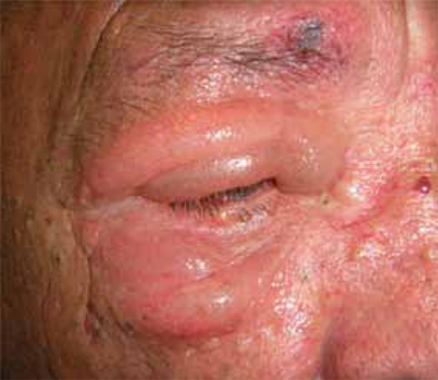
The swelling was not accompanied by inflammation or fever, but it did impair the patient’s vision.
WHAT IS YOUR DIAGNOSIS?
HOW WOULD YOU TREAT THIS PATIENT?
Diagnosis: Noninflammatory eyelid edema
The fact that the patient’s eyelid was edematous (but not tender or red) and that he’d been scratching the area following a case of folliculitis prompted us to diagnose noninflammatory eyelid edema.
Noninflammatory eyelid edema—also called noninflammatory palpebral edema—is a relatively common disorder that usually occurs after some local irritation or microtrauma. In its early phase, patients start out with nonerythematous, painless edema of the upper eyelid, which eventually affects the lower eyelid.
As was the case with our patient, this disorder is not associated with fever or any other systemic sign or symptom. Complete blood count and C-reactive protein will be negative, as no infection or systemic alteration is present.
Consider these infectious alternatives
In cases like this one, be sure to differentiate between noninflammatory palpebral edema and other common but less benign ocular conditions. Among the infectious causes of unilateral edema to rule out:
Preseptal cellulitis is an infection that is localized between the skin and the orbital septum. This disorder usually appears as a tender erythematous eyelid edema with no proptosis and no pain with eye movement.1 It is usually accompanied by an elevated white blood cell (WBC) count. Blood cultures are rarely positive, but when they are, Haemophilus influenzae is the most frequent pathogen.2 Preseptal cellulitis is treated with broad-spectrum oral antibiotics (amoxicillin/ clavulanate) for at least 7 days.
Orbital cellulitis is an infection that affects the orbital extraocular structures. It can lead to blindness, so prompt diagnosis is critical.
Orbital cellulitis usually develops when a sinus infection spreads into the orbit.3 Suspect it in a patient with proptosis, orbital pain, tenderness, conjunctival chemosis, decreased vision, elevated intraocular pressure, and pain on eye movement. Also look for an elevated WBC count. Perform blood cultures to confirm the presence of a pathogen.
If you suspect orbital cellulitis, order an orbital computed tomography scan with contrast infusion, including axial and coronal views; it may reveal an infection of the soft tissue behind the orbital septum. Treat this condition with broad-spectrum intravenous antibiotics (ceftriaxone) for 1 week, followed by 2 weeks of oral antibiotics, completing a total of 21 days of antibiotics.
Hordeolum is a localized infection that occurs when a meibomian gland becomes blocked. It usually appears as a palpable subcutaneous nodule within the eyelid and requires treatment with warm compresses and a combination of antibiotics and steroid topical ointment (tobramycin and dexamethasone ophthalmic ointment 4 times a day for a week).4
Acute dacryoadenitis is a lacrimal gland infection that usually causes unilateral, painful swelling of the outer third of the upper lid. You may also see tearing and discharge.5 Look for an elevated WBC count; however, blood cultures are rarely positive. These patients must be referred to an ophthalmologist as treatment varies from symptomatic measures in viral cases (warm compresses and a nonsteroidal anti-inflammatory drug, such as ibuprofen) to hospitalization and oral or intravenous antibiotics (eg, amoxicillin/ clavulanate for at least 7 days), depending on the severity and origin of the infection.6
Ruling out other noninfectious disorders
Noninfectious inflammatory diseases should also be part of the differential diagnosis, including:
Idiopathic orbital inflammation. Also known as orbital pseudotumor, this disorder is a nongranulomatous acute-to-subacute inflammatory disease with no systemic manifestations.7 It frequently involves the lacrimal gland and can cause abrupt pain, conjunctival edema, and lid edema. Other common signs are proptosis and ocular motility alterations, which the patient may report as diplopia and visual impairment.
This disorder can result from optic neuropathy, exudative retinal detachment, or uveitis. It is a diagnosis of exclusion and is often arrived at by seeing the patient respond to systemic corticosteroids. The specific drug and dose will vary based on disease severity.
Graves’ ophthalmopathy. This bilateral, asymmetric immunological disorder affects the conjunctiva, eyelids,8 extraocular muscles, lacrimal gland, and optic nerve. Although it is typically diagnosed clinically, thyroid hormone abnormalities (increased T4 and a drop in thyroid-stimulating hormone) help support the diagnosis. In the acute phase, it has to be treated with systemic steroids; if the condition becomes chronic, the complications may require surgery. Treatment of the underlying hyperthyroidism may, in some cases, worsen the ophthalmopathy.
One more consideration. Finally, consider a local allergic reaction in the eyelids,9 which can also cause painless palpebral edema. Expect to see erythematous edema and intense itching a few hours after exposure to the allergen. A cosmetic product is often the culprit. As you might expect, initial treatment calls for withdrawal of the offending product. Instruct the patient to apply a topical steroid ointment on the eyelid if the edema is intense.
Good news for our patient
Diagnosing noninflammatory eyelid edema requires keen observational skills and a knowledge of several alternative diagnoses. Fortunately for our patient, this benign condition did not require any local or systemic treatment. The edema resolved on its own in 6 days.
CORRESPONDENCE
Omar Rayward, MD, Calle Profesor Martín Lagos, Madrid, Spain 28040; orayward@yahoo.com
1. Bilyk JR. Periocular infection. Curr Opin Ophthalmol. 2007;18:414-423.
2. Jackson K, Baker SR. Periorbital cellulitis. Head Neck Surg. 1987;9:227-234.
3. Adam R, Gupta V, Harvey J. Question: can you identify this condition? This condition is orbital cellulitis. Can Fam Physician. 2009;55:1097.-
4. Lindsley K, Nichols JJ, Dickersin K. Interventions for acute internal hordeolum. Cochrane Database Syst Rev. 2010;(9):CD007742.-
5. Colegrove JA. Localized orbital inflammation: a case of dacryoadenitis. Optom Vis Sci. 2000;77:121-124.
6. Durand ML. Periocular infections. In: Mandell GL, Bennett JE, Dolin R, eds. Mandell, Douglas, and Bennett’s Principles and Practice of Infectious Diseases. 7th ed. Philadelphia, Pa: Elsevier Churchill Livingstone; 2009:1569–1575.
7. Cooney NL. Orbital pseudotumor. Int J Emerg Med. 2009;2:263.-
8. Imaizumi M. Recurrent upper eyelid edema as first sign of Graves’ disease. Thyroid. 2006;16:95-96.
9. Friedlaender MH. Objective measurement of allergic reactions in the eye. Curr Opin Allergy Clin Immunol. 2004;4:447-453.
A 68-year-old man sought care in our emergency department for unilateral ptosis following superior and inferior right eyelid edema. The patient said that the edema had developed 3 days earlier and was getting worse each day; the ptosis was impairing his vision. The patient indicated that the edema was accompanied by mild burning in the right periocular region. His medical history included arterial hypertension, which was under control, and bilateral cataract surgery 5 years ago.
On examination, we noted superior and inferior nontender painless eyelid edema on the right eye, with no signs of acute inflammation (FIGURE). The patient also had right supraciliary folliculitis that was improving; the folliculitis had been treated 3 days earlier at a primary care facility.
We performed a complete ocular examination, including visual acuity (20/20 in both eyes) and found no other significant problems. Nor did the patient have a fever or any other systemic symptoms.
FIGURE 1
Edema of the upper and lower eyelid
The swelling was not accompanied by inflammation or fever, but it did impair the patient’s vision.
WHAT IS YOUR DIAGNOSIS?
HOW WOULD YOU TREAT THIS PATIENT?
Diagnosis: Noninflammatory eyelid edema
The fact that the patient’s eyelid was edematous (but not tender or red) and that he’d been scratching the area following a case of folliculitis prompted us to diagnose noninflammatory eyelid edema.
Noninflammatory eyelid edema—also called noninflammatory palpebral edema—is a relatively common disorder that usually occurs after some local irritation or microtrauma. In its early phase, patients start out with nonerythematous, painless edema of the upper eyelid, which eventually affects the lower eyelid.
As was the case with our patient, this disorder is not associated with fever or any other systemic sign or symptom. Complete blood count and C-reactive protein will be negative, as no infection or systemic alteration is present.
Consider these infectious alternatives
In cases like this one, be sure to differentiate between noninflammatory palpebral edema and other common but less benign ocular conditions. Among the infectious causes of unilateral edema to rule out:
Preseptal cellulitis is an infection that is localized between the skin and the orbital septum. This disorder usually appears as a tender erythematous eyelid edema with no proptosis and no pain with eye movement.1 It is usually accompanied by an elevated white blood cell (WBC) count. Blood cultures are rarely positive, but when they are, Haemophilus influenzae is the most frequent pathogen.2 Preseptal cellulitis is treated with broad-spectrum oral antibiotics (amoxicillin/ clavulanate) for at least 7 days.
Orbital cellulitis is an infection that affects the orbital extraocular structures. It can lead to blindness, so prompt diagnosis is critical.
Orbital cellulitis usually develops when a sinus infection spreads into the orbit.3 Suspect it in a patient with proptosis, orbital pain, tenderness, conjunctival chemosis, decreased vision, elevated intraocular pressure, and pain on eye movement. Also look for an elevated WBC count. Perform blood cultures to confirm the presence of a pathogen.
If you suspect orbital cellulitis, order an orbital computed tomography scan with contrast infusion, including axial and coronal views; it may reveal an infection of the soft tissue behind the orbital septum. Treat this condition with broad-spectrum intravenous antibiotics (ceftriaxone) for 1 week, followed by 2 weeks of oral antibiotics, completing a total of 21 days of antibiotics.
Hordeolum is a localized infection that occurs when a meibomian gland becomes blocked. It usually appears as a palpable subcutaneous nodule within the eyelid and requires treatment with warm compresses and a combination of antibiotics and steroid topical ointment (tobramycin and dexamethasone ophthalmic ointment 4 times a day for a week).4
Acute dacryoadenitis is a lacrimal gland infection that usually causes unilateral, painful swelling of the outer third of the upper lid. You may also see tearing and discharge.5 Look for an elevated WBC count; however, blood cultures are rarely positive. These patients must be referred to an ophthalmologist as treatment varies from symptomatic measures in viral cases (warm compresses and a nonsteroidal anti-inflammatory drug, such as ibuprofen) to hospitalization and oral or intravenous antibiotics (eg, amoxicillin/ clavulanate for at least 7 days), depending on the severity and origin of the infection.6
Ruling out other noninfectious disorders
Noninfectious inflammatory diseases should also be part of the differential diagnosis, including:
Idiopathic orbital inflammation. Also known as orbital pseudotumor, this disorder is a nongranulomatous acute-to-subacute inflammatory disease with no systemic manifestations.7 It frequently involves the lacrimal gland and can cause abrupt pain, conjunctival edema, and lid edema. Other common signs are proptosis and ocular motility alterations, which the patient may report as diplopia and visual impairment.
This disorder can result from optic neuropathy, exudative retinal detachment, or uveitis. It is a diagnosis of exclusion and is often arrived at by seeing the patient respond to systemic corticosteroids. The specific drug and dose will vary based on disease severity.
Graves’ ophthalmopathy. This bilateral, asymmetric immunological disorder affects the conjunctiva, eyelids,8 extraocular muscles, lacrimal gland, and optic nerve. Although it is typically diagnosed clinically, thyroid hormone abnormalities (increased T4 and a drop in thyroid-stimulating hormone) help support the diagnosis. In the acute phase, it has to be treated with systemic steroids; if the condition becomes chronic, the complications may require surgery. Treatment of the underlying hyperthyroidism may, in some cases, worsen the ophthalmopathy.
One more consideration. Finally, consider a local allergic reaction in the eyelids,9 which can also cause painless palpebral edema. Expect to see erythematous edema and intense itching a few hours after exposure to the allergen. A cosmetic product is often the culprit. As you might expect, initial treatment calls for withdrawal of the offending product. Instruct the patient to apply a topical steroid ointment on the eyelid if the edema is intense.
Good news for our patient
Diagnosing noninflammatory eyelid edema requires keen observational skills and a knowledge of several alternative diagnoses. Fortunately for our patient, this benign condition did not require any local or systemic treatment. The edema resolved on its own in 6 days.
CORRESPONDENCE
Omar Rayward, MD, Calle Profesor Martín Lagos, Madrid, Spain 28040; orayward@yahoo.com
1. Bilyk JR. Periocular infection. Curr Opin Ophthalmol. 2007;18:414-423.
2. Jackson K, Baker SR. Periorbital cellulitis. Head Neck Surg. 1987;9:227-234.
3. Adam R, Gupta V, Harvey J. Question: can you identify this condition? This condition is orbital cellulitis. Can Fam Physician. 2009;55:1097.-
4. Lindsley K, Nichols JJ, Dickersin K. Interventions for acute internal hordeolum. Cochrane Database Syst Rev. 2010;(9):CD007742.-
5. Colegrove JA. Localized orbital inflammation: a case of dacryoadenitis. Optom Vis Sci. 2000;77:121-124.
6. Durand ML. Periocular infections. In: Mandell GL, Bennett JE, Dolin R, eds. Mandell, Douglas, and Bennett’s Principles and Practice of Infectious Diseases. 7th ed. Philadelphia, Pa: Elsevier Churchill Livingstone; 2009:1569–1575.
7. Cooney NL. Orbital pseudotumor. Int J Emerg Med. 2009;2:263.-
8. Imaizumi M. Recurrent upper eyelid edema as first sign of Graves’ disease. Thyroid. 2006;16:95-96.
9. Friedlaender MH. Objective measurement of allergic reactions in the eye. Curr Opin Allergy Clin Immunol. 2004;4:447-453.
Painful eye with a facial rash
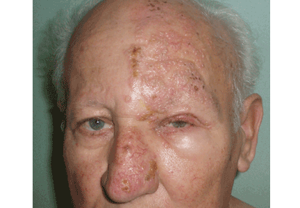
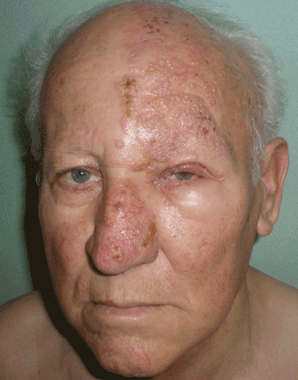
A 75-year-old man presents 4 days after painful cutaneous lesions appeared on the left side of his face, associated with severe ocular pain. Two days before the eruption, he had had an intense headache, which was diagnosed as a tension headache and was treated with oral acetaminophen (Tylenol), but with no improvement.
The remainder of his physical examination is normal. Laboratory tests, including red and white blood cell counts, hemoglobin, and basic metabolic and coagulation tests reveal no abnormalities.
Q: What is your diagnosis?
- Allergic contact dermatitis
- Herpes simplex
- Varicella
- Ramsay-Hunt syndrome
- Herpes zoster ophthalmicus and herpetic keratitis
A: Herpes zoster ophthalmicus is the correct diagnosis. It represents a reactivation of the varicella zoster virus.1
Varicella zoster virus, like others of the herpes family, has developed a complex control of virus-host interactions to ensure its survival in humans. It lies dormant in the sensory ganglia and, when reactivated, moves down the neurons and satellite cells along the sensory axons to the skin.1 The reactivation is related to diminished cell-mediated immunity, which occurs as a physiologic part of aging, which is why the elderly tend to be the most often affected. 2 The incidence of herpes zoster varies from 2.2 to 3.4 per 1,000 people per year.3 Its incidence in people over age 80 is about 10 per 1,000 people per year.3
CLINICAL PRESENTATION
Herpes zoster typically presents as a dermatome-grouped vesicular eruption over an erythematous base, accompanied or preceded by local pain. It has two main complications, postherpetic neuralgia and ocular involvement. Postherpetic neuralgia is neuropathic pain that persists or develops after the dermatomal rash has healed.4 Independent predictors of postherpetic neuralgia are older age, severe acute pain, severe rash, a shorter duration of rash before consultation, and ocular involvement.5 It occurs in 36.6% of patients over age 60, and in 47.5% over 70.6 Persistent postherpetic neuralgia has been linked to suicide in patients over 70.7
Ocular infection occurs with involvement of the ophthalmic division of the fifth cranial nerve. Before the antiviral era, it was seen in as many as 50% of patients.8 Hutchinson’s sign is skin lesions on the tip, side, or root of the nose and is an important predictor of ocular involvement.1 Lesions may include folliculopapillar conjunctivitis, episcleritis, scleritis, keratitis (dendritic, pseudodendritic, and interstitial), uveitis, and necrotizing retinitis.
DIAGNOSIS
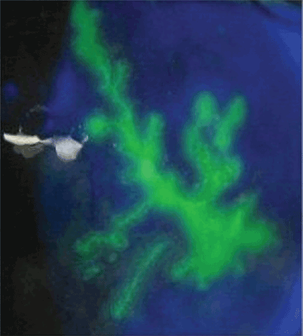
The diagnosis of herpes zoster is usually based on clinical observation of the characteristic rash, although viral culture and molecular techniques are available when definitive diagnosis is required. When ophthalmic division is affected and Hutchinson’s sign, unexplained ocular redness with pain, or complaints of visual problems are present, the patient should be referred promptly to an ophthalmologist, because serious visual impairment can occur. The fluorescein dye may show no staining or the typical dendritic keratitis (Figure 2).
TREATMENT
Oral antiviral drugs have made the treatment of zoster possible when, effectively, no treatment existed before. Ideally, an antiviral should be given within 72 hours of symptom onset. Starting treatment as early as possible—especially within 72 hours of onset—has been shown to be effective in alleviating acute pain and in preventing or limiting the duration and severity of postherpetic neuralgia.3
Acyclovir (Zovirax) 800 mg five times a day for 7 days or one of its derivatives—eg, famciclovir (Famvir), penciclovir (Denavir), or valacyclovir (Valtrex)—has been shown to be safe and effective in the treatment of active disease, as well as in preventing or shortening the duration of postherpetic neuralgia.3 It has also been shown to reduce the rate of eye involvement from 50% to 20% or 30%.9 This is why all patients with this dermatomal involvement must be treated.
Second-generation antivirals
Valacyclovir 1,000 mg three times a day and famciclovir 500 mg three times a day seem to be as effective as acyclovir in reducing zoster-associated pain, but their efficacy in reducing eye involvement has not been studied. In clinical practice, however, these second-generation antivirals may be more effective than acyclovir because patients are more likely to comply with the treatment regimen of three rather than five daily doses.
Other considerations
In patients with kidney failure, the non-nephrotoxic antiviral brivudine is preferred, but it is not available in the United States. Therefore, one must use acyclovir or one of the other drugs, carefully adjusting the dose according to the creatinine clearance and making sure the patient is well hydrated.
The efficacy of antiviral treatment that is started more than 72 hours after the onset of skin rash has never been confirmed.
Although the additional effectiveness of acyclovir eye ointment has never been established, topical acyclovir can be considered in cases of dendritic or pseudodendritic keratitis.
- Liesegang TJ. Herpes zoster ophthalmicus: natural history, risk factors, clinical presentation, and morbidity. Ophthalmology 2008; 115( suppl 2):S3–S12.
- Opstelten W, Eekhof J, Neven AK, Verheij T. Treatment of herpes zoster. Can Fam Physician 2008; 54:373–377.
- Opstelten W, Zaal MJ. Managing ophthalmic herpes zoster in primary care. BMJ 2005; 331:147–151.
- Donahue JG, Choo PW, Manson JE, Platt R. The incidence of herpes zoster. Arch Intern Med 1995; 155:1605–1609.
- Opstelten W, Zuithoff NP, van Essen GA, et al. Predicting postherpetic neuralgia in elderly primary care patients with herpes zoster: prospective prognostic study. Pain 2007; 132( suppl 1):S52–S59.
- De Morgas JM, Kierland RR. The outcome of patients with herpes zoster. AMA Arch Derm 1957; 75:193–196.
- Hess TM, Lutz LJ, Nauss LA, Lamer TJ. Treatment of acute herpetic neuralgia. A case report and review of the literature. Minn Med 1990; 73:37–40.
- Harding SP, Lipton JR, Wells JC. Natural history of herpes zoster ophthalmicus: predictors of postherpetic neuralgia and ocular involvement. Br J Ophthalmol 1987; 71:353–358.
- Cobo LM, Foulks GN, Liesegang T, et al. Oral acyclovir in the treatment of acute herpes zoster ophthalmicus. Ophthalmology 1986; 93:763–770.
A 75-year-old man presents 4 days after painful cutaneous lesions appeared on the left side of his face, associated with severe ocular pain. Two days before the eruption, he had had an intense headache, which was diagnosed as a tension headache and was treated with oral acetaminophen (Tylenol), but with no improvement.
The remainder of his physical examination is normal. Laboratory tests, including red and white blood cell counts, hemoglobin, and basic metabolic and coagulation tests reveal no abnormalities.
Q: What is your diagnosis?
- Allergic contact dermatitis
- Herpes simplex
- Varicella
- Ramsay-Hunt syndrome
- Herpes zoster ophthalmicus and herpetic keratitis
A: Herpes zoster ophthalmicus is the correct diagnosis. It represents a reactivation of the varicella zoster virus.1
Varicella zoster virus, like others of the herpes family, has developed a complex control of virus-host interactions to ensure its survival in humans. It lies dormant in the sensory ganglia and, when reactivated, moves down the neurons and satellite cells along the sensory axons to the skin.1 The reactivation is related to diminished cell-mediated immunity, which occurs as a physiologic part of aging, which is why the elderly tend to be the most often affected. 2 The incidence of herpes zoster varies from 2.2 to 3.4 per 1,000 people per year.3 Its incidence in people over age 80 is about 10 per 1,000 people per year.3
CLINICAL PRESENTATION
Herpes zoster typically presents as a dermatome-grouped vesicular eruption over an erythematous base, accompanied or preceded by local pain. It has two main complications, postherpetic neuralgia and ocular involvement. Postherpetic neuralgia is neuropathic pain that persists or develops after the dermatomal rash has healed.4 Independent predictors of postherpetic neuralgia are older age, severe acute pain, severe rash, a shorter duration of rash before consultation, and ocular involvement.5 It occurs in 36.6% of patients over age 60, and in 47.5% over 70.6 Persistent postherpetic neuralgia has been linked to suicide in patients over 70.7
Ocular infection occurs with involvement of the ophthalmic division of the fifth cranial nerve. Before the antiviral era, it was seen in as many as 50% of patients.8 Hutchinson’s sign is skin lesions on the tip, side, or root of the nose and is an important predictor of ocular involvement.1 Lesions may include folliculopapillar conjunctivitis, episcleritis, scleritis, keratitis (dendritic, pseudodendritic, and interstitial), uveitis, and necrotizing retinitis.
DIAGNOSIS
The diagnosis of herpes zoster is usually based on clinical observation of the characteristic rash, although viral culture and molecular techniques are available when definitive diagnosis is required. When ophthalmic division is affected and Hutchinson’s sign, unexplained ocular redness with pain, or complaints of visual problems are present, the patient should be referred promptly to an ophthalmologist, because serious visual impairment can occur. The fluorescein dye may show no staining or the typical dendritic keratitis (Figure 2).
TREATMENT
Oral antiviral drugs have made the treatment of zoster possible when, effectively, no treatment existed before. Ideally, an antiviral should be given within 72 hours of symptom onset. Starting treatment as early as possible—especially within 72 hours of onset—has been shown to be effective in alleviating acute pain and in preventing or limiting the duration and severity of postherpetic neuralgia.3
Acyclovir (Zovirax) 800 mg five times a day for 7 days or one of its derivatives—eg, famciclovir (Famvir), penciclovir (Denavir), or valacyclovir (Valtrex)—has been shown to be safe and effective in the treatment of active disease, as well as in preventing or shortening the duration of postherpetic neuralgia.3 It has also been shown to reduce the rate of eye involvement from 50% to 20% or 30%.9 This is why all patients with this dermatomal involvement must be treated.
Second-generation antivirals
Valacyclovir 1,000 mg three times a day and famciclovir 500 mg three times a day seem to be as effective as acyclovir in reducing zoster-associated pain, but their efficacy in reducing eye involvement has not been studied. In clinical practice, however, these second-generation antivirals may be more effective than acyclovir because patients are more likely to comply with the treatment regimen of three rather than five daily doses.
Other considerations
In patients with kidney failure, the non-nephrotoxic antiviral brivudine is preferred, but it is not available in the United States. Therefore, one must use acyclovir or one of the other drugs, carefully adjusting the dose according to the creatinine clearance and making sure the patient is well hydrated.
The efficacy of antiviral treatment that is started more than 72 hours after the onset of skin rash has never been confirmed.
Although the additional effectiveness of acyclovir eye ointment has never been established, topical acyclovir can be considered in cases of dendritic or pseudodendritic keratitis.
- Liesegang TJ. Herpes zoster ophthalmicus: natural history, risk factors, clinical presentation, and morbidity. Ophthalmology 2008; 115( suppl 2):S3–S12.
- Opstelten W, Eekhof J, Neven AK, Verheij T. Treatment of herpes zoster. Can Fam Physician 2008; 54:373–377.
- Opstelten W, Zaal MJ. Managing ophthalmic herpes zoster in primary care. BMJ 2005; 331:147–151.
- Donahue JG, Choo PW, Manson JE, Platt R. The incidence of herpes zoster. Arch Intern Med 1995; 155:1605–1609.
- Opstelten W, Zuithoff NP, van Essen GA, et al. Predicting postherpetic neuralgia in elderly primary care patients with herpes zoster: prospective prognostic study. Pain 2007; 132( suppl 1):S52–S59.
- De Morgas JM, Kierland RR. The outcome of patients with herpes zoster. AMA Arch Derm 1957; 75:193–196.
- Hess TM, Lutz LJ, Nauss LA, Lamer TJ. Treatment of acute herpetic neuralgia. A case report and review of the literature. Minn Med 1990; 73:37–40.
- Harding SP, Lipton JR, Wells JC. Natural history of herpes zoster ophthalmicus: predictors of postherpetic neuralgia and ocular involvement. Br J Ophthalmol 1987; 71:353–358.
- Cobo LM, Foulks GN, Liesegang T, et al. Oral acyclovir in the treatment of acute herpes zoster ophthalmicus. Ophthalmology 1986; 93:763–770.