User login
Noah Worcester Dermatology Society: Annual Meeting
VIDEO: Get excited about the excimer laser for dermatitis
ASHEVILLE, N.C. – Excimer lasers can be used for patients with allergic contact dermatitis for whom topical therapies are unsuccessful or undesirable, according to Dr. Alison Ehrlich of George Washington University in Washington.
In an interview at the annual meeting of the Noah Worcester Dermatological Society, Dr. Ehrlich discussed her use of the excimer laser for dermatitis patients and shared some tips for successful treatment.
The video associated with this article is no longer available on this site. Please view all of our videos on the MDedge YouTube channel
AT NOAH 57
VIDEO: Dermatologists should embrace tissue adhesive
ASHEVILLE, N.C. – Cutaneous adhesives have many advantages in dermatologic surgery, according to Dr. John West of Seaport Dermatology and Mohs Surgery in Mystic, Conn.
In a video interview at the annual meeting of the Noah Worcester Dermatological Society, Dr. West explained some of the benefits of tissue adhesives for both doctors and patients and shared several pearls for optimal use of these products.
The video associated with this article is no longer available on this site. Please view all of our videos on the MDedge YouTube channel
AT NOAH 57

VIDEO: Lasers take on the toughest scars
ASHEVILLE, N.C. – Advances in laser and light sources are making it possible to greatly improve the treatment of atrophic and hypertrophic scars, according to Dr. Michael Gold of the Gold Skin Care Center in Nashville, Tenn. In a video interview at the annual meeting of the Noah Worcester Dermatological Society, Dr. Gold explained how laser technology is being used today to manage contracture and other elements of the most challenging scars, such as those sustained by military personnel.
The video associated with this article is no longer available on this site. Please view all of our videos on the MDedge YouTube channel
AT NOAH 57

Safely Patch Test Children for Contact Dermatitis
ASHEVILLE, N.C. – “Children really do develop contact dermatitis, and they are frequently mislabeled as cases of eczema,” Dr. Bruce Brod said at the annual meeting of the Noah Worcester Dermatological Society.
When taking a history in a child with potential contact dermatitis, keep the most likely allergens in mind, especially nickel, said Dr. Brod of the University of Pennsylvania, Philadelphia. Nickel remains the most common allergen in adults and young children, and more than a quarter of patients are positive on patch testing.
Consider all possible sources of nickel. In addition to the old standbys of jewelry and buckles, ask patients and families about the use of flip-style cell phones, as well as first generation iPads (the cases contained nickel). “Old cell phones do not make great toys,” Dr. Brod emphasized.
Patch testing children with potential contact dermatitis makes sense in several situations, including cases of new-onset dermatitis; progressing or deteriorating dermatitis; involvement of specific body sites, such as the face, eyelids, or neck folds; an increase in the total body surface area affected; clinical presentation of dyshidrosis; and dermatitis that resists standard therapies and only improves with oral or extremely potent topical steroids, he said.
Children with atopic dermatitis are more prone to irritation from patch testing, so shorten the exposure time and use a lower concentration of allergens such as nickel, formaldehyde, and rubber additives, advised Dr. Brod. Shorten the exposure time for children younger than 5 years of age, whether or not they have atopy, he added. Videos or video games can work well as a distraction.
When taking a history, consider the most the common allergens in children, defined in a recent study of patch testing results (Dermatitis 2014;25:345-55), said Dr. Brod. He offered the mnemonic MAFLPP (More Allergies for Lovable Pediatric Patients) to characterize the top categories: metals (nickel and cobalt), antibiotics (neomycin and bacitracin), fragrance (fragrances and balsam of Peru), lanolin, phenylenediamine, and preservatives (including quatemium-15 and methylisothiazolinone).
After patch testing, describe the allergen to patients and their families, and explain where it is found, Dr. Brod said. The site mypatchlink.com has helpful information. Also remind patients to read product labels, and to check pharmacy websites such as drugstore.com or cvs.com.
Members of the American Contact Dermatitis Society can access the Contact Allergen Management Program (CAMP) database to help patients identify allergen-free products based on their patch test results, he said.
Dr. Brod had no relevant financial conflicts to disclose.
EXPERT ANALYSIS FROM NOAH 57
Safely patch test children for contact dermatitis
ASHEVILLE, N.C. – “Children really do develop contact dermatitis, and they are frequently mislabeled as cases of eczema,” Dr. Bruce Brod said at the annual meeting of the Noah Worcester Dermatological Society.
When taking a history in a child with potential contact dermatitis, keep the most likely allergens in mind, especially nickel, said Dr. Brod of the University of Pennsylvania, Philadelphia. Nickel remains the most common allergen in adults and young children, and more than a quarter of patients are positive on patch testing.
Consider all possible sources of nickel. In addition to the old standbys of jewelry and buckles, ask patients and families about the use of flip-style cell phones, as well as first generation iPads (the cases contained nickel). “Old cell phones do not make great toys,” Dr. Brod emphasized.
Patch testing children with potential contact dermatitis makes sense in several situations, including cases of new-onset dermatitis; progressing or deteriorating dermatitis; involvement of specific body sites, such as the face, eyelids, or neck folds; an increase in the total body surface area affected; clinical presentation of dyshidrosis; and dermatitis that resists standard therapies and only improves with oral or extremely potent topical steroids, he said.
Children with atopic dermatitis are more prone to irritation from patch testing, so shorten the exposure time and use a lower concentration of allergens such as nickel, formaldehyde, and rubber additives, advised Dr. Brod. Shorten the exposure time for children younger than 5 years of age, whether or not they have atopy, he added. Videos or video games can work well as a distraction.
When taking a history, consider the most the common allergens in children, defined in a recent study of patch testing results (Dermatitis 2014;25:345-55), said Dr. Brod. He offered the mnemonic MAFLPP (More Allergies for Lovable Pediatric Patients) to characterize the top categories: metals (nickel and cobalt), antibiotics (neomycin and bacitracin), fragrance (fragrances and balsam of Peru), lanolin, phenylenediamine, and preservatives (including quatemium-15 and methylisothiazolinone).
After patch testing, describe the allergen to patients and their families, and explain where it is found, Dr. Brod said. The site mypatchlink.com has helpful information. Also remind patients to read product labels, and to check pharmacy websites such as drugstore.com or cvs.com.
Members of the American Contact Dermatitis Society can access the Contact Allergen Management Program (CAMP) database to help patients identify allergen-free products based on their patch test results, he said.
Dr. Brod had no relevant financial conflicts to disclose.
EXPERT ANALYSIS FROM NOAH 57
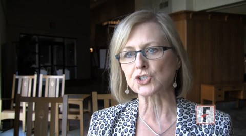
VIDEO: Collaboration Between Providers Key for Derm Patients
ASHEVILLE, N.C. – Building bridges with colleagues in other specialties, especially primary care, helps patients and improves relations among specialties, Dr. Lisa Garner said in an interview at the annual meeting of the Noah Worcester Dermatological Society.
“Communicating is key for patient care,” she said. Dr. Garner, a dermatologist in private practice in Garland, Tex., chaired the American Academy of Dermatology’s Perceptions of Dermatology task force, which assessed perceptions of dermatologists and dermatology within the house of medicine from 2012 to 2014.
Dr. Garner had no financial conflicts to disclose.
The video associated with this article is no longer available on this site. Please view all of our videos on the MDedge YouTube channel
EXPERT ANALYSIS FROM NOAH 57
VIDEO: Collaboration with primary care helps colleagues and patients
ASHEVILLE, N.C. – Building bridges with colleagues in other specialties, especially primary care, helps patients and improves relations among specialties, Dr. Lisa Garner said in an interview at the annual meeting of the Noah Worcester Dermatological Society.
“Communicating is key for patient care,” she said. Dr. Garner, a dermatologist in private practice in Garland, Tex., chaired the American Academy of Dermatology’s Perceptions of Dermatology task force, which assessed perceptions of dermatologists and dermatology within the house of medicine from 2012 to 2014.
Dr. Garner had no financial conflicts to disclose.
The video associated with this article is no longer available on this site. Please view all of our videos on the MDedge YouTube channel
EXPERT ANALYSIS FROM NOAH 57

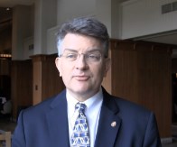
VIDEO: Updating the immune response to nonmelanoma skin cancer
ASHEVILLE, N.C. – Recent advances in basic science have shown how the local immune environment in tissue surrounding nonmelanoma skin cancer compares to adjacent normal tissue.
New Mexico Health Sciences Center’s Dr. Andrew Ondo reviewed the latest research in an interview at the annual meeting of the Noah Worcester Dermatological Society. “Each step along the way is a possible target for the treatment of squamous cell carcinoma,” said Dr. Ondo, who indicated that he had no financial conflicts to disclose.
The video associated with this article is no longer available on this site. Please view all of our videos on the MDedge YouTube channel
EXPERT ANALYSIS FROM NOAH 57
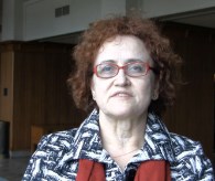
Leukocytoclastic vasculitis incidence underestimated
ASHEVILLE, N.C. – The incidence of leukocytoclastic vasculitis in the general population is 1.5 to 3 times higher than previously reported, based on data from a large, population-based study.
Based on these findings, leukocytoclastic vasculitis (LCV) is “more common than cutaneous lupus,” said Dr. David A. Wetter of the Mayo Clinic, Rochester, Minn.

Dr. Wetter and his associates reviewed data from all residents of Olmstead County, Minn., with a biopsy-proven diagnosis of LCV from January 1, 1996, through December 31, 2010.
A total of 84 patients were identified, for an overall incidence of 4.5 per 100,000 person-years. The mean age was 48 years, and approximately half were women.
The researchers divided the patients into five subtypes: cutaneous small vessel vasculitis (CSVV), IgA vasculitis, urticarial vasculitis, cryoglobulinemic vasculitis, and antineutrophil cytoplasmic antibody–associated vasculitis.
Systemic involvement occurred in 46% of the patients, with the kidneys involved in 44% of these cases. Recurrent disease was observed in 30% of the 80 patients for whom follow-up data were available.
In addition, LCV was idiopathic in 76% of CSVV patients, significantly higher than the 45% rate reported in the current textbooks, Dr. Wetter noted. Idiopathic LCV also occurred in 96% of patients with IgA vasculitis.
Overall survival was significantly decreased in the LCV patients compared to the general Minnesota population, but the reasons for this difference remain unclear and are worthy of additional study, Dr. Wetter noted. However, survival was not significantly decreased in the subset of patients with IgA vasculitis, despite the high rate of systemic involvement, he added.
Although the study was limited by its retrospective nature and relatively homogeneous white population, it is the first known population-based study of histopathologically-defined LCV, and the data support the evaluation of all patients with LCV “for extracutaneous disease and systemic etiologies for their disease,” Dr. Wetter said.
Dr. Wetter presented the study findings at the annual meeting of the Noah Worcester Dermatological Society. The data also were published in the Mayo Clinic Proceedings in 2014 (Mayo Clin. Proc. 2014;89:1515-24).
Dr. Wetter had no financial conflicts to disclose.
ASHEVILLE, N.C. – The incidence of leukocytoclastic vasculitis in the general population is 1.5 to 3 times higher than previously reported, based on data from a large, population-based study.
Based on these findings, leukocytoclastic vasculitis (LCV) is “more common than cutaneous lupus,” said Dr. David A. Wetter of the Mayo Clinic, Rochester, Minn.
Dr. Wetter and his associates reviewed data from all residents of Olmstead County, Minn., with a biopsy-proven diagnosis of LCV from January 1, 1996, through December 31, 2010.
A total of 84 patients were identified, for an overall incidence of 4.5 per 100,000 person-years. The mean age was 48 years, and approximately half were women.
The researchers divided the patients into five subtypes: cutaneous small vessel vasculitis (CSVV), IgA vasculitis, urticarial vasculitis, cryoglobulinemic vasculitis, and antineutrophil cytoplasmic antibody–associated vasculitis.
Systemic involvement occurred in 46% of the patients, with the kidneys involved in 44% of these cases. Recurrent disease was observed in 30% of the 80 patients for whom follow-up data were available.
In addition, LCV was idiopathic in 76% of CSVV patients, significantly higher than the 45% rate reported in the current textbooks, Dr. Wetter noted. Idiopathic LCV also occurred in 96% of patients with IgA vasculitis.
Overall survival was significantly decreased in the LCV patients compared to the general Minnesota population, but the reasons for this difference remain unclear and are worthy of additional study, Dr. Wetter noted. However, survival was not significantly decreased in the subset of patients with IgA vasculitis, despite the high rate of systemic involvement, he added.
Although the study was limited by its retrospective nature and relatively homogeneous white population, it is the first known population-based study of histopathologically-defined LCV, and the data support the evaluation of all patients with LCV “for extracutaneous disease and systemic etiologies for their disease,” Dr. Wetter said.
Dr. Wetter presented the study findings at the annual meeting of the Noah Worcester Dermatological Society. The data also were published in the Mayo Clinic Proceedings in 2014 (Mayo Clin. Proc. 2014;89:1515-24).
Dr. Wetter had no financial conflicts to disclose.
AT NOAH 57
Key clinical point: The data support the evaluation of all patients with LCV for extracutaneous disease and systemic etiologies.
Major finding: The incidence of leukocytoclastic vasculitis in the general population is 1.5 to 3 times higher than previously reported.
Data source: A population-based study of all residents of Olmstead County, Minn., with a biopsy-proven diagnosis of LCV from January 1, 1996, through December 31, 2010.
Disclosures: Dr. Wetter had no financial conflicts to disclose.

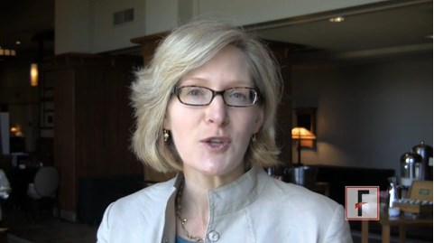
VIDEO: Larger lentigo maligna lesions increase risk
ASHEVILLE, N.C. – What are the risk factors for invasive melanoma in patients with lentigo maligna? Size, for one thing, according to Dr. Suzanne M. Olbricht.
In an interview at the annual meeting of the Noah Worcester Dermatological Society, Dr. Olbricht of the Lahey Hospital and Medical Center in Burlington, Mass., reviewed evidence suggesting that the recurrence rate is highest for large lesions. “This is important information that helps us think about the treatments we can use,” she said.
Dr. Olbricht had no financial conflicts to disclose.
The video associated with this article is no longer available on this site. Please view all of our videos on the MDedge YouTube channel
AT NOAH 57