User login
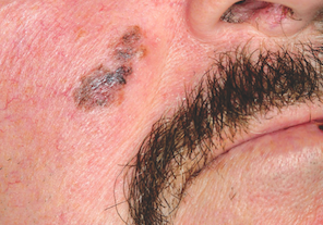
Lesion Has Doubled in Size in Two Weeks
ANSWER
The one incorrect statement is choice “a,” since the history and appearance of the lesion are quite consistent with keratoacanthoma—considered by most to be a low-grade form of squamous cell carcinoma (SCC). All the other statements are true.
DISCUSSION/TREATMENT
Keratoacanthomas (KAs) are quite common, especially in older patients with fair, sun-damaged skin. They appear on areas of the skin that have been directly exposed to the sun. The dorsal forearm is especially typical, but KAs can also appear on the face, hands, shoulders, and back.
The relatively rapid growth is in marked contrast to most other skin cancers and has been linked to the human papillomavirus DNA, which has been found in these lesions. However, by no means is this connection universally accepted as the cause, even though immune suppression, in the susceptible patient, does appear to play a role.
Microscopically, KAs bear a marked resemblance to SCCs. Indeed, they have been known to metastasize in rare instances, although most will involute (albeit with minor scarring) on their own, without treatment. The standard of treatment in this country is to remove KAs surgically and to submit the tissue for pathologic examination, which would officially show “SCC, KA type,” or “well-differentiated SCC.”
The differential diagnosis includes wart, invasive SCC, Merkel cell carcinoma, and melanoma. KAs have nothing to do with bacterial infection.
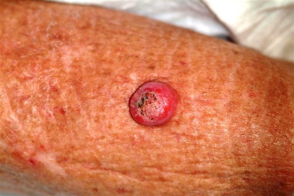
Six weeks ago, a new lesion began to appear on the forearm of this 63-year-old woman; it has grown rapidly in the ensuing time. She first went to her primary care provider, who diagnosed a staph infection and prescribed an antibiotic that hasn’t helped. The lesion, while asymptomatic, is nonetheless alarming; it has doubled in size in the past two weeks, which is why the patient now presents to the dermatology clinic. Her medical history includes well-controlled hypertension and a remote incidence of several basal cell carcinomas. On examination, the patient’s skin, in general, is severely sun-damaged, as evidenced by a weathered, leathery look to all exposed skin, as well as by multiple telangiectasias and solar lentigines. The lesion in question is a striking 1.8-cm dome-like nodule with a central keratotic core, located on the mid-dorsal forearm. Smooth and shiny, the surface of this firm nodule is also covered with tiny telangiectasias. Careful examination of the rest of the patient’s skin shows no other worrisome lesions.

Eyelids Are Irritated (But Eyes Are Fine)
ANSWER
The one incorrect answer is yeast infection (choice “c”). All the others should be included in the list of possible explanations. Given the inherent dryness and relatively low temperature of the affected area, as well as the nonresponse to imidazole cream, candida is quite unlikely.
DISCUSSION
The only way this patient could acquire a yeast infection in this area is if she were immunosuppressed. Eyelid dermatitis is seen daily in derm practices; patients are often referred by frustrated primary care providers who are unable to help.
This problem is almost never seen in male patients, which suggests the involvement of makeup, eye shadow, or skin care products, especially cleansers. But even when these are changed or eliminated, the problem often continues.
Here’s why: Women—especially those with sensitive skin in general—will often manifest that sensitivity in areas where skin is the thinnest and most accessible, and therefore the most easily damaged. Once the problem starts, it is difficult for these women to leave it alone. They scratch it, rub it, and often apply multiple products to it, all of which only serves to worsen it.
In contrast, most men use no products at all on their faces, let alone “eye creams” or special cleansers. The male version of this problem is the equally ubiquitous chronic anterior scrotal rash—which develops for many of the same reasons.
Many of these women also happen to have a history of atopic dermatitis, which not only means extraordinarily sensitive skin all over the body but also tends to involve unusually dry skin (xerosis). These women have multiple allergies to contactants, such as nickel and nail polish.
TREATMENT
As the cycle of applying different (unhelpful) products to the problem area continues, women are increasingly likely to use products with irritating ingredients. If they are to get any relief, this cycle must be broken.
I usually prescribe hydrocortisone 2.5% ointment (not the cream, which has a drying effect) or tacrolimus 0.1% ointment (either treatment to be used twice a day), plus once-a-day application of the greasiest moisturizer they can stand (eg, petroleum jelly) over the affected area.
They can use this same approach with future episodes, although there must be strict limits on the duration (two weeks) and frequency of the application. Overuse of the steroid can lead to glaucoma and permanent thinning of already thin skin.
Obviously, it would be of great utility if the patient were in fact found to be allergic to nail polish, but in my experience, most women have eliminated that as a potential cause before they get to a dermatology provider. The same goes for makeup.
The eyelid dermatitis patient certainly could be suffering from seborrhea or psoriasis; however, in such cases, the same itch-scratch-itch cycle often results, and the treatment would be identical.
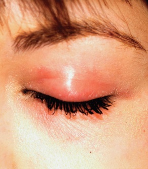
A 22-year-old woman is evaluated for a recurring itchy rash on both the upper and lower bilateral eyelids, which has been present for months. Her eyes themselves are unaffected, but the skin around them is highly irritated. The patient has already seen a number of providers who prescribed or recommended several remedies, including hydrocortisone creams, imidazole creams, and OTC moisturizers. Since none of these has proved helpful, she presents to the dermatology clinic. Her medical history includes seasonal allergies and “sensitive skin”; by the latter, she means frequent rashes or irritation brought on by various products, such as shampoos and conditioners. During this occurrence, as in several previous episodes, she has changed eye shadow and other makeup, to no avail. The patient denies any history of psoriasis, seborrhea, or other skin disease. On examination, a brisk red, dry, wrinkly rash covering the above-mentioned areas is noted. No rash is seen elsewhere, and there are no signs of psoriasis or seborrhea in any of the locations where they typically appear.

Painless Chest Lesion Suddenly Triples in Size
ANSWER
The correct answer is all of the above (choice “d”). All are reasonable diagnoses and important because they drive the decision as to how to proceed.
DISCUSSION
An incisional biopsy would also have been acceptable, but collection of the entire lesion, with modest margins where possible, is almost always the gold standard in establishing a diagnosis in cases such as this one. There are several reasons, one being that the pathologist will then have adequate tissue to examine and will have a good chance of determining whether the lesion began locally or represents spread from a distant site. The only argument against such an approach would be the potential for scarring.
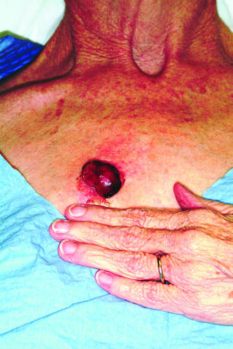
Moreover, in the majority of cases, this procedure can be curative—as in this case, in which the pathology report revealed the lesion to be a well-differentiated squamous cell carcinoma that had been traumatized, resulting in the formation of reparative inappropriate granulation tissue. It was the latter that accounted for the friability and rapid growth. Fortunately, the margins were clear, so no additional surgery or radiation was necessary.
A complete differential diagnosis would also include other types of cancer, including colon cancer and melanoma. Considering the possibilities, the patient was all too happy to have the problem taken care of in one step.
ANSWER
The correct answer is all of the above (choice “d”). All are reasonable diagnoses and important because they drive the decision as to how to proceed.
DISCUSSION
An incisional biopsy would also have been acceptable, but collection of the entire lesion, with modest margins where possible, is almost always the gold standard in establishing a diagnosis in cases such as this one. There are several reasons, one being that the pathologist will then have adequate tissue to examine and will have a good chance of determining whether the lesion began locally or represents spread from a distant site. The only argument against such an approach would be the potential for scarring.
Moreover, in the majority of cases, this procedure can be curative—as in this case, in which the pathology report revealed the lesion to be a well-differentiated squamous cell carcinoma that had been traumatized, resulting in the formation of reparative inappropriate granulation tissue. It was the latter that accounted for the friability and rapid growth. Fortunately, the margins were clear, so no additional surgery or radiation was necessary.
A complete differential diagnosis would also include other types of cancer, including colon cancer and melanoma. Considering the possibilities, the patient was all too happy to have the problem taken care of in one step.

Rapid growth and bleeding are two of the most common reasons that patients seek evaluation for lesions. Both are concerns for this 69-year-old woman who has had a chest lesion for almost two years. Recently, the lesion suddenly tripled in size and began to bleed with minor trauma. Although painless, the lesion is of particular concern since the patient was diagnosed with breast cancer about 18 years ago and underwent a right radical mastectomy at that time. The patient is a smoker with a more than 50–pack-year history. She denies any recent history of shortness of breath, cough, or weight loss. Examination reveals an impressive nodule on the right midchest, measuring 4 cm and demonstrating a smooth, vascular surface and lobular shape. The base of the lesion is pedunculated, and the lesion itself is quite mobile. After consultation with the patient, the decision to excise is quickly reached and the procedure carried out. The excised lesion is sent for pathologic exam.
Dark Lesions Prompt Skin Cancer Scare
ANSWER
The one incorrect statement above is choice “d.” Although most patients call them age spots, age is, in fact, only one factor in the development of such lesions. All the other statements are true and are discussed below.
DISCUSSION
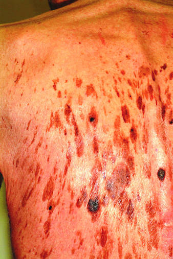
Seborrheic keratoses (SKs) are the most common benign skin lesions seen in older patients—but they’re also commonly seen on the skin of people in their 20s and 30s, so age is only one factor favoring their appearance. Heredity is the main cause in about 50% of cases. One of the most common dermatologic diagnoses, they are responsible for a large percentage of referrals to dermatology. As with this patient, SKs appear to be related to sun exposure in a significant percentage of patients, even though they have no malignant potential themselves.
Becoming thoroughly familiar with the myriad morphologic presentations of SKs is essential in learning dermatology. Their color ranges from gray to pink, tan to brown, and even black. Fortunately, they’re usually distinctly epidermal, with a “stuck-on” look that helps greatly in distinguishing them from malignant lesions. In oilier areas of the body, such as on the face, they can become much less scaly and much darker, making them difficult to distinguish from pigmented basal cell carcinoma or even melanoma in some cases; biopsy is thus necessary. Indeed, the “secret” to learning how to recognize SKs in all their varied presentations is the routine performance of punch biopsies.
SKs, when they’re as numerous as on this patient, can easily obscure a melanoma and can even coexist with the latter in the same location. This is especially true in high-risk patients (ie, those with fair, sun-damaged skin) who tan poorly. Fortunately, this patient had, by history and exam, type IV skin (with type I being the most fair, and type VI being the darkest) and was therefore at relatively low risk for skin cancer.
But on empirical grounds alone, one could correctly surmise that the mere presence of so many similar lesions, often present for years without change, effectively rules out skin cancer. The latter is far more likely to appear as a solitary lesion, as well as to be dynamic in nature (ie, to grow and/or change over time).
ANSWER
The one incorrect statement above is choice “d.” Although most patients call them age spots, age is, in fact, only one factor in the development of such lesions. All the other statements are true and are discussed below.
DISCUSSION
Seborrheic keratoses (SKs) are the most common benign skin lesions seen in older patients—but they’re also commonly seen on the skin of people in their 20s and 30s, so age is only one factor favoring their appearance. Heredity is the main cause in about 50% of cases. One of the most common dermatologic diagnoses, they are responsible for a large percentage of referrals to dermatology. As with this patient, SKs appear to be related to sun exposure in a significant percentage of patients, even though they have no malignant potential themselves.
Becoming thoroughly familiar with the myriad morphologic presentations of SKs is essential in learning dermatology. Their color ranges from gray to pink, tan to brown, and even black. Fortunately, they’re usually distinctly epidermal, with a “stuck-on” look that helps greatly in distinguishing them from malignant lesions. In oilier areas of the body, such as on the face, they can become much less scaly and much darker, making them difficult to distinguish from pigmented basal cell carcinoma or even melanoma in some cases; biopsy is thus necessary. Indeed, the “secret” to learning how to recognize SKs in all their varied presentations is the routine performance of punch biopsies.
SKs, when they’re as numerous as on this patient, can easily obscure a melanoma and can even coexist with the latter in the same location. This is especially true in high-risk patients (ie, those with fair, sun-damaged skin) who tan poorly. Fortunately, this patient had, by history and exam, type IV skin (with type I being the most fair, and type VI being the darkest) and was therefore at relatively low risk for skin cancer.
But on empirical grounds alone, one could correctly surmise that the mere presence of so many similar lesions, often present for years without change, effectively rules out skin cancer. The latter is far more likely to appear as a solitary lesion, as well as to be dynamic in nature (ie, to grow and/or change over time).

A 71-year-old man is urgently referred to dermatology by his primary care provider at the request of the patient’s children. These adult children recently noticed changes to the skin on their father’s back. On the day of the appointment, two of his children—a son and a daughter—bring the man in and already have his shirt off when you enter the room. Before you can evaluate the patient, his children express their anxiety about the lesions: “They’re so dark, and there are so many of them! We’re really concerned about this being skin cancer.” Examination reveals relatively dark skin with little evidence of sun damage, although the patient reports spending much of his life outdoors. When he is exposed to sun, he says, he tans relatively easily, holding the tan for weeks and very seldom burning. The lesions number easily in the hundreds and are tan to brown, papular, and nodular. Many coalesce into large plaques, with linear and in some cases digitate configuration. They virtually cover his back. On palpation, the surfaces of these lesions are epidermal, dry, and rough, with a warty texture. Lesions are also seen on his face, chest, and arms, though they are far fewer in number. Interestingly, both children have numerous similar lesions that can be easily seen.
Small, Painful Blisters Erupt in Patient's Groin Area
ANSWER
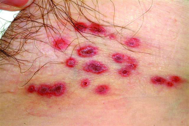
The correct answer is herpes zoster (choice “d”; see discussion). Yeast infections (choice “a”) would be quite unlikely to present with hemorrhagic discrete lesions and far more likely to present with a confluent rash. MRSA (choice “b”) can present in a wide variety of forms but is unlikely to form discrete blisters and would almost certainly involve induration in the area. Without a definitive viral culture, herpes simplex (choice “c”) could not be ruled out in this case, since it presents in a similar manner. But given the patient’s age and the morphology and location of the lesions, zoster was far more likely. See the discussion for further differentiating factors.
DISCUSSION/TREATMENT
The appearance of intralesional hemorrhage in an inflammatory lesion bespeaks a deeper, more vigorous process, since vasculature ends just below the dermoepidermal junction. This occurs infrequently enough with herpes zoster to obscure the diagnosis. Ironically, herpes simplex almost never exhibits this phenomenon, making it useful as a differentiating feature.
In this patient’s case, it seems reasonable to blame the local application of heat as a potential trigger. My experience is that stress is also a factor, though the literature does not bear out that theory.
ANSWER
The correct answer is herpes zoster (choice “d”; see discussion). Yeast infections (choice “a”) would be quite unlikely to present with hemorrhagic discrete lesions and far more likely to present with a confluent rash. MRSA (choice “b”) can present in a wide variety of forms but is unlikely to form discrete blisters and would almost certainly involve induration in the area. Without a definitive viral culture, herpes simplex (choice “c”) could not be ruled out in this case, since it presents in a similar manner. But given the patient’s age and the morphology and location of the lesions, zoster was far more likely. See the discussion for further differentiating factors.
DISCUSSION/TREATMENT
The appearance of intralesional hemorrhage in an inflammatory lesion bespeaks a deeper, more vigorous process, since vasculature ends just below the dermoepidermal junction. This occurs infrequently enough with herpes zoster to obscure the diagnosis. Ironically, herpes simplex almost never exhibits this phenomenon, making it useful as a differentiating feature.
In this patient’s case, it seems reasonable to blame the local application of heat as a potential trigger. My experience is that stress is also a factor, though the literature does not bear out that theory.

A 60-year-old woman presents with a symptomatic eruption in the left groin that began a week ago, when she first noticed a slight tingling sensation in that area. Within 48 hours, small blisters appeared, clustered in the crural fold. These became slightly painful, with a curious lancinating type of pain. The patient also began to experience fatigue and myalgia. She sought care when her lesions became hemorrhagic and was subsequently referred to dermatology. Additional history reveals that in the past month she has experienced low back pain on the left side, radiating into the groin. In response, she started to use moist hot packs on the area. The onset of her symptoms has coincided with another stressor, her husband’s job loss. She denies the use of any immunosuppressive medications and is well in other respects. Examination shows a grouped collection of targetoid hemorrhagic flaccid low blisters, from 1 to 2 cm in diameter, confined to the left crural fold. No underlying induration is felt, but there is palpable adenopathy confined to that area.
Dark Blue Facial Lesion Worries Mother of Young Boy
ANSWER
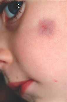
The correct answer is pilomatricoma (choice “a”), also know as calcifying epithelioma of Malherbe. See discussion.
Dermatofibromas (choice “b”) can be dark, especially in darker-skinned individuals, but are rare in children and even more so on the face.
Sebaceous cysts (choice “c”) are quite common on the face and can develop in children but are more likely to have a fluctuant feel rather than a firm one. They are also unlikely to be hyperpigmented and will usually involve an overlying punctum (comedone) on the surface.
Spitz tumors (choice “d”) can range from red to black and papular to macular. They tend to be intradermal in terms of vertical placement and are quite unlikely to be subcutaneous and indurated. They belong in the differential diagnosis because of their color and the fact that they are usually seen on children.
DISCUSSION
Pilomatricomas are uncommon but not at all rare. They usually manifest on the head, neck, or upper extremities of children as bluish subcutaneous masses devoid of overlying skin changes (eg, atrophy, comedones, or breaks in the skin). Though not seen in this case, the deep lesion often “tents” the overlying skin in one or more foci, a phenomenon termed the tent sign.
Although the exact reason they appear is not known, it is clear that they derive histologically from hair matrix cells. Quite rarely, they can undergo malignant transformation. They can also be quite a bit larger than this one and can present in multiples, with color ranging from deep blue (as in this case) to pinkish brown.
TREATMENT
For the purposes of positive identification and cosmesis, pilomatricomas are usually excised. At that time, identifying features such as the lack of an organized cyst wall and the granular, calcified contents are typically seen.
ANSWER
The correct answer is pilomatricoma (choice “a”), also know as calcifying epithelioma of Malherbe. See discussion.
Dermatofibromas (choice “b”) can be dark, especially in darker-skinned individuals, but are rare in children and even more so on the face.
Sebaceous cysts (choice “c”) are quite common on the face and can develop in children but are more likely to have a fluctuant feel rather than a firm one. They are also unlikely to be hyperpigmented and will usually involve an overlying punctum (comedone) on the surface.
Spitz tumors (choice “d”) can range from red to black and papular to macular. They tend to be intradermal in terms of vertical placement and are quite unlikely to be subcutaneous and indurated. They belong in the differential diagnosis because of their color and the fact that they are usually seen on children.
DISCUSSION
Pilomatricomas are uncommon but not at all rare. They usually manifest on the head, neck, or upper extremities of children as bluish subcutaneous masses devoid of overlying skin changes (eg, atrophy, comedones, or breaks in the skin). Though not seen in this case, the deep lesion often “tents” the overlying skin in one or more foci, a phenomenon termed the tent sign.
Although the exact reason they appear is not known, it is clear that they derive histologically from hair matrix cells. Quite rarely, they can undergo malignant transformation. They can also be quite a bit larger than this one and can present in multiples, with color ranging from deep blue (as in this case) to pinkish brown.
TREATMENT
For the purposes of positive identification and cosmesis, pilomatricomas are usually excised. At that time, identifying features such as the lack of an organized cyst wall and the granular, calcified contents are typically seen.

A 6-year-old boy is brought in by his mother for evaluation of a lesion that has been present on his face, with little change, for about two years. Though asymptomatic, the lesion is nonetheless worrisome, since the boy’s pediatrician isn’t sure what it is. The boy is in good health otherwise. His mother’s expectation is that the lesion will be removed at this initial visit to dermatology, since this is the reason she consulted the pediatrician in the first place. The 2-cm left facial lesion can be seen from across the room; it is quite large, round, and dark blue. While no changes are seen on the surface of the skin, palpation reveals definite, deep induration. No punctum can be seen on the overlying surface, and there is no palpable adenopathy in the area.
What Is This Long-Standing Loss of Pigment?
ANSWER
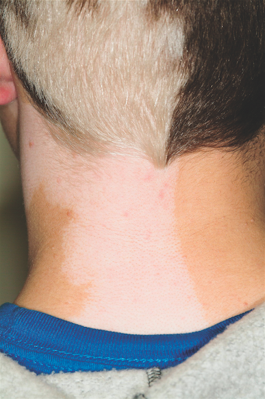
The correct answer is poliosis (choice “d”), which has been described as a reliable indicator of tuberous sclerosis when seen early in life.1 Poliosis is the specific term for focal loss of color in scalp hair. Technically, this process is a form of hypomelanosis (choice “b”), although the latter is a more generic term used to refer to loss of pigment in any area. Graying (choice “a”) usually refers to uniform loss of hair color all over the scalp—albeit with focal accentuation over the temples, as opposed to the sharply demarcated areas of complete color loss seen in poliosis. Poliosis can be seen as a feature of vitiligo (choice “c”), but the latter usually also involves pigment loss elsewhere.
DISCUSSION/TREATMENT
Poliosis, which is often idiopathic, can be seen in a number of conditions besides tuberous sclerosis and vitiligo, including halo nevi of the scalp, alopecia areata (as hair regrows), and Waardenburg’s syndrome. As mentioned above, it can appear early in life as an indicator of tuberous sclerosis, particularly when coupled with involvement of adjacent dermatomal skin. This was likely the case with this patient. No permanent treatment exists for this problem.
REFERENCE
1. Apibal Y, Reakatanan W, Chunharas A. Poliosis as the first clue of tuberous sclerosis. Pediatr Dermatol. 2008;25(4):486-487.
ANSWER
The correct answer is poliosis (choice “d”), which has been described as a reliable indicator of tuberous sclerosis when seen early in life.1 Poliosis is the specific term for focal loss of color in scalp hair. Technically, this process is a form of hypomelanosis (choice “b”), although the latter is a more generic term used to refer to loss of pigment in any area. Graying (choice “a”) usually refers to uniform loss of hair color all over the scalp—albeit with focal accentuation over the temples, as opposed to the sharply demarcated areas of complete color loss seen in poliosis. Poliosis can be seen as a feature of vitiligo (choice “c”), but the latter usually also involves pigment loss elsewhere.
DISCUSSION/TREATMENT
Poliosis, which is often idiopathic, can be seen in a number of conditions besides tuberous sclerosis and vitiligo, including halo nevi of the scalp, alopecia areata (as hair regrows), and Waardenburg’s syndrome. As mentioned above, it can appear early in life as an indicator of tuberous sclerosis, particularly when coupled with involvement of adjacent dermatomal skin. This was likely the case with this patient. No permanent treatment exists for this problem.
REFERENCE
1. Apibal Y, Reakatanan W, Chunharas A. Poliosis as the first clue of tuberous sclerosis. Pediatr Dermatol. 2008;25(4):486-487.

A 14-year-old boy presents for evaluation of a long-standing condition involving loss of pigment of the hair and the adjacent neck skin. This discoloration has been present since shortly after his birth. The patient is specifically interested in treatment options. His history includes a diagnosis of tuberous sclerosis, which he received at an early age. On examination, you note a complete loss of pigment in the posterior scalp on the patient’s left side, along with total loss of color in the skin on the adjacent part of the neck. The medial margins of both stop abruptly at midline. Additional inspection reveals several other abnormalities, including dart-shaped hypopigmented macules on the patient’s trunk, fleshy 1- to 3-mm papules clustered about the midface, and periungual fibromatous papules around several toenails.
Skin Changes on Hands Lead to Daily Living Problems
ANSWER
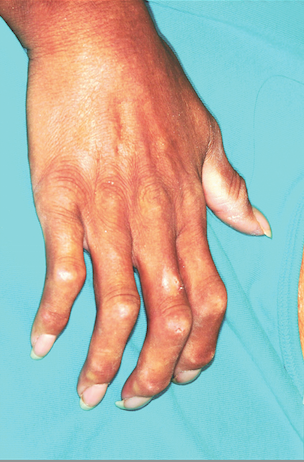
Although this woman may end up having to see all of these specialists, she first needs confirmation of the suspected diagnosis: CREST syndrome, considered a variant of systemic sclerosis (aka scleroderma) and a condition marked by sclerotic transformation of virtually all the organs in and on the body. For more on this condition, see below.
The definitive diagnosis and expert management of this condition are generally considered the province of the rheumatologist, so choice “d” is the correct answer. Any of the other specialists, who often become involved in such cases early on, could likely identify this condition but would defer to rheumatology for definitive diagnosis and management.
DISCUSSION/TREATMENT
Systemic sclerosis (SSc) patients, like those with other rare conditions with protean manifestations, often go through long periods in which they see various specialists for each subset of symptoms—with a delay in arriving at a definitive, unifying diagnosis. This is a good example of the role played in such cases by dermatology, which is often able to “connect the dots” in patients with disparate complaints. Biopsy in such patients, for example, will show increased collagen deposition (among other changes), leading to heightened suspicion and confirmation by specific blood tests and expert clinical corroboration.
SSc affects approximately two million to 10 million people, almost four times as many women as men, and very few Asian patients. There are several variants of the condition, including cases of CREST in which skin changes are limited to the hands and face for decades before internal organs begin to be affected (if ever).
The CREST acronym stands for calcinosis (the scaly foci on our patient’s fingers), Raynaud’s phenomenon, esophageal dysmotility (due to collagen infiltration), sclerodactyly (sclerosis of finger skin), and telangiectasias (often on face and chest). CREST often starts, as in this case, with chronic edema of the hands, which gives way eventually to sclerodactyly and often to reabsorption of the bony tufts on distal digits. Dyschromia, particularly of the hands (as in this patient), is another common finding. SSc can also present with generalized pruritus.
SSc has a poor prognosis, progressing steadily as it begins to affect the lungs, liver, kidneys, and joints. Despite treatment, death may occur (most commonly from renal or respiratory failure).
ANSWER
Although this woman may end up having to see all of these specialists, she first needs confirmation of the suspected diagnosis: CREST syndrome, considered a variant of systemic sclerosis (aka scleroderma) and a condition marked by sclerotic transformation of virtually all the organs in and on the body. For more on this condition, see below.
The definitive diagnosis and expert management of this condition are generally considered the province of the rheumatologist, so choice “d” is the correct answer. Any of the other specialists, who often become involved in such cases early on, could likely identify this condition but would defer to rheumatology for definitive diagnosis and management.
DISCUSSION/TREATMENT
Systemic sclerosis (SSc) patients, like those with other rare conditions with protean manifestations, often go through long periods in which they see various specialists for each subset of symptoms—with a delay in arriving at a definitive, unifying diagnosis. This is a good example of the role played in such cases by dermatology, which is often able to “connect the dots” in patients with disparate complaints. Biopsy in such patients, for example, will show increased collagen deposition (among other changes), leading to heightened suspicion and confirmation by specific blood tests and expert clinical corroboration.
SSc affects approximately two million to 10 million people, almost four times as many women as men, and very few Asian patients. There are several variants of the condition, including cases of CREST in which skin changes are limited to the hands and face for decades before internal organs begin to be affected (if ever).
The CREST acronym stands for calcinosis (the scaly foci on our patient’s fingers), Raynaud’s phenomenon, esophageal dysmotility (due to collagen infiltration), sclerodactyly (sclerosis of finger skin), and telangiectasias (often on face and chest). CREST often starts, as in this case, with chronic edema of the hands, which gives way eventually to sclerodactyly and often to reabsorption of the bony tufts on distal digits. Dyschromia, particularly of the hands (as in this patient), is another common finding. SSc can also present with generalized pruritus.
SSc has a poor prognosis, progressing steadily as it begins to affect the lungs, liver, kidneys, and joints. Despite treatment, death may occur (most commonly from renal or respiratory failure).

A 54-year-old woman presents with changes to the skin on her hands. These began several years ago and have progressed to the point that it is becoming harder and harder for her to use her hands in normal activities of daily living. Several years ago, when the problems began, the first thing the patient noted was bilateral edema. This symptom was always at its worst when the patient woke up and got better as the day went on. It has gradually subsided, giving way to the slow advance of skin hardening on all her fingers. She has also developed a claw-like incurving of the fingers and occasional “ulcers” on the fingertips. Exposure to modest cold turns her fingers quite cold and white. More history taking reveals that during this time, she also began to experience fatigue, shortness of breath, and difficulty swallowing. On examination, the skin on all her fingers is uniformly tight and hard, the tips are all tapered and slender, and all the fingers are firmly fixed in a moderately flexed position. Dermatoscopic exam shows tortuous, dilated capillaries in the cuticular areas of all fingers. Several papulosquamous shallow erosions are seen on the phalanges, ranging in diameter from a pinpoint to 1.0 cm. Groups of telangiectasias are noted on the upper chest and lateral face.
Two Different Diagnoses, But One Location
ANSWER
The white, round spots seen in this lesion were entirely consistent with SK and were not misinterpreted (choice “a”). Since melanoma is not known to display this type of spot, choice “b” is also incorrect. Biopsy reports are occasionally wrong, of course, but the diagnosis of melanoma is fraught with such serious implications that the call is not made if specific criteria are not met. These include specific stains for that diagnosis, histologic findings, and the concurrence of multiple observers, so it is unlikely that the histologic diagnosis is incorrect (choice “d”).
This leaves the correct answer, choice “c,” discussed below.
DISCUSSION
There is truly no law saying that two lesions can’t appear in the same location—and as this case illustrates, it definitely happens. Had we performed additional biopsies, no doubt features of SK would have been seen in corroboration of the dermatoscopic findings.
This case also emphasizes another useful principle: Pay just as much attention to the owner as to the lesion itself. This patient was quite fair and sun-damaged, so any odd lesion he developed would be suspect, let alone one that looked like this.
One final thing to be learned from this case is that as useful as dermatoscopes can be, they still must take a backseat to common sense.

A 57-year-old man self-refers for evaluation of a facial lesion that has been present for several years. As the lesion slowly grew larger, family members became worried and urged him to seek treatment. The man has spent many years in the sun during his adult life, both at work and when engaging in various hobbies, and he burns easily and often. In addition, 20 years ago, he was diagnosed with psoriasis, which was eventually treated with a year’s worth of phototherapy. History also includes a coronary artery bypass graft (undergone 10 years ago) and current smoker status. On examination, the facial lesion proves to be macular, with ominously irregular colors of black and brown, along with ragged borders. The overall size is almost 4 × 2 cm. The lesion is seen in the context of very fair, sun-damaged skin, characterized by widespread actinic keratoses and telangiectasias. Through use of a dermatoscope (a power-viewing instrument that provides 10× magnification, used to examine suspicious lesions for specific changes suggestive of cancer), definite signs of seborrheic keratosis (SK; pseudocysts) are seen. Nonetheless, the overall appearance of the lesion—as well as the patient’s high-risk status—dictates that a punch biopsy be performed. The subsequent report reveals the lesion to be a Clark’s level II superficial melanoma, Breslow 0.6 mm.
If it’s not acne, warts, or molluscum contagiosum, what is this condition?
ANSWER
All of the above choices are correct and, in fact, were seen on this patient. These findings served to confirm the diagnosis of tuberous sclerosis.
DISCUSSION
Also called Bourneville’s disease or epiloia (an acronym for epilepsy, low intelligence, and adenoma sebaceum—all major findings in this disease), tuberous sclerosis (TS) occurs in one of 10,000 births and affects both genders equally. It is considered a genodermatosis, with 75% of cases related to spontaneous mutations and 25% to autosomal dominant transmission. Two separate genes have been identified.
The earliest skin finding in these patients is that of hypopigmented macules, especially the so-called mountain ash leaf spots typically seen on the trunk and extremities—better observed sometimes with the aid of a Wood’s lamp.
Another common early finding is that of seizures, occurring in 75% of TS patients with central nervous system lesions. Facial angiofibromas, called adenoma sebaceum, also appear in early childhood, increase during adolescence, and are permanent. About 80% of affected patients will eventually develop the connective tissue nevus called shagreen patch, which usually occurs on the low back.
Conical smooth papular projections originating from the nail folds of fingers and toes appear at puberty in a high percentage of TS patients and are permanent. Redundant and sometimes papular lesions are seen in the gingival mucosa. Numerous other signs of the disease—in lungs, heart, and bony tissue—have been described.
The acronym mentioned above, epiloia, has been largely abandoned, in part because it suggests that low intelligence with TS patients is inevitable. The truth is, many patients, such as this one, function quite normally, typifying the variable penetrance of the genetic changes so common to TS.
TREATMENT
The main focus for this patient is to get his seizures under control and arrange for proper follow-up, as well as genetic counseling. Unfortunately, there is no effective treatment for the facial lesions.

A 14-year-old boy is seen for “acne” that has been unresponsive to oral and topical antibiotics. There is no family history of acne, but the boy has a history of unexplained grand mal seizures. The dermatologic condition, which has been present since an early age, has been variously diagnosed as warts and molluscum contagiosum, as well as acne. However, since none of the treatments offered has been successful, the patient is referred to dermatology. On examination, you note a dense collection of fleshy papules in bilateral nasolabial areas, thinning out around the mouth.
