User login
Study confirms new mutation, possible therapeutic target in epidermolysis bullosa
PORTLAND, ORE. – New research has uncovered “a new kid on the block” of genes underlying epidermolysis bullosa simplex (EBS), which may someday pave the way for new therapies, John McGrath, MD, said at the annual meeting of the Society for Investigative Dermatology.
The gene encodes kelch-like family member 24 (KLHL24), the substrate receptor of cullin 3-RBX1-KLHL24 complex, explained Dr. McGrath, professor of molecular dermatology, St. John’s Institute of Dermatology, King’s College London, where he studies heritable skin diseases. Keratin 14 is the substrate of this complex. In EBS, KLHL24 mutations affect methionine initiation codons, resulting in the loss of the first 28 amino acids of KLHL24 protein. As a result, the protein becomes overly stable, which leads to excessive ubiquitination and breakdown of keratin 14.
KLHL24 is the 19th gene to be implicated in EBS. News of the finding broke in late 2016, when two publications identified KLHL24 mutations in 19 patients with EBS from 10 different families. The first study, conducted in China, identified start-codon mutations in the KLHL24 gene in five patients, confirmed keratin 14 as the substrate of the protein, and established that KLHL24 mutations induce disproportionate ubiquitination and fragmentation of keratin 14. The researchers confirmed these results by using a knock-in mouse model (Nat Genet. 2016 Dec;48 [12]:1508-16).
Meanwhile, European researchers identified KLHL24 mutations in families from Israel, Germany, Switzerland, Finland, Qatar, and Italy, and confirmed that these mutations affect the equilibrium between intermediate keratin filaments and keratin breakdown that is necessary for skin integrity (Am J Hum Genet. 2016 Dec 1;99[6]:1395-1404).
EBS affects more than 400,000 individuals worldwide, and is usually linked to heterozygous missense mutations in genes encoding keratins 5 and 14, Dr. McGrath noted. “However, EBS is clinically and genetically heterogeneous,” he added. “About 20% of cases have no mutation in known genes.” Pursuing an accurate diagnosis of EBS subtype is important for genetic counseling and clinical care, he emphasized.
Therefore, to help confirm the findings of the studies in China and Europe, Dr. McGrath and his associates performed Sanger sequencing, skin pathology, clinical phenotyping, and electron microscopy of samples from undiagnosed cases of EB simplex that were banked at the U.K.’s National Diagnostic EB Laboratory. Among 183 cases (about 20% of all EBS referrals to the laboratory) that had not been linked to other mutations, 7 from six families had heterozygous KLHL24 mutations.
Together, the three studies name five KLHL24 mutations from 26 individuals, 16 families, and eight countries, Dr. McGrath said. “All these mutations target the initiation codon of methionine,” he added. As has previously been reported in EBS, immunostaining for keratin 14 is positive, outlining areas of cleavage within the basal cell layer of the skin. Semi-thin sections reveal pallor in the basal layer of keratinocytes, and electron microscopy of this layer shows blister formation.
“In nonlesional basal keratinocytes, there are few keratin filaments and mitochondria are prominent,” Dr. McGrath explained. “Higher magnification confirms the lack of identifiable keratin filaments, and reveals prominent microtubules.” Lesional skin shows not only the paucity of keratin filaments, but clear evidence of cytolysis, with numerous autophagosomes, autolysosomes, and disruption of organelles in the cellular cytoplasm. “The keratin has been chopped up, digested, and is being organically recycled. That’s essentially what’s going on here,” he said.
These studies show how clinician scientists can help advance the diagnosis and treatment of skin diseases by linking clinical and molecular pathology, Dr. McGrath said. But just as importantly, they might reveal a new approach to treating EBS. “Instead of gene editing or protein therapy, we have a new mechanism that may be ripe for proteasomal inhibitors or other targets of the ubiquitination pathway,” he added. “It’s an intriguing way forward when thinking about drugs and small molecules for EB therapy.”
The National Institute for Health Research and the NHS Foundation Trust funded the study. Dr. McGrath had no relevant financial disclosures.
AT SID 2017
Key clinical point: Studies that have identified genes related to epidermolysis bullosa may lead to new treatment approaches for patients with epidermolysis bullosa simplex (EBS).
Major finding: Of 183 patients with EBS, not linked to other mutations, testing determined that 7 had heterozygous KLHL24 mutations, recently identified in patients with EB.
Data source: Samples from the 183 patients were banked at the United Kingdom’s National Diagnostic EB laboratory.
Disclosures: The National Institute for Health Research and the NHS Foundation Trust funded the study. Dr. McGrath had no relevant financial disclosures.
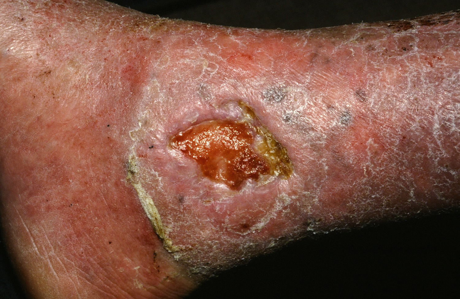
Stem cell therapy significantly improves ulcer healing
PORTLAND, ORE. – Treating chronic venous leg ulcers with mesenchymal stem cells and fibrin spray significantly improved wound healing, compared with vehicle control or saline plus conventional therapy, according to the results of a small randomized, controlled, double-blind pilot trial.
“Topical application of autologous, bone-marrow–derived mesenchymal stem cells may be an effective way to promote healing in patients with difficult-to-heal wounds,” said Ayman Grada, MD, of the department of dermatology at Boston University. “However, larger studies are needed to confirm this finding.”
“Various treatment modalities have been used, but treatment outcomes are not always satisfactory,” said Dr. Grada. “In about 60% of cases, wounds fail to close, and there is also a high rate of recurrence.”
Preclinical work in several animal models indicated that applying mesenchymal stem cells to wounds accelerated healing through a variety of mechanisms, Dr. Grada noted. Based on that premise, he and his associates hypothesized that autologous cultured mesenchymal stem cells could accelerate wound healing in humans.
To test that idea, they randomly assigned the 11 trial participants to one of two control treatments or to the stem cell intervention. Four patients received normal saline with conventional standard care, three patients received fibrin spray plus conventional therapy, and four patients received conventional therapy plus autologous mesenchymal stem cells delivered in fibrin spray at a dose of 1 x 10⁶ cells per square centimeter of wound surface. Patients were treated every 3 weeks, up to three times or until complete wound healing, and were followed for up to 24 weeks.
To acquire the stem cells, the researchers obtained 30- to 50-mL samples of bone marrow aspirate from the iliac crest, then separated and cultured the cells in-house. The controls underwent sham aspiration with needles that did not penetrate the bone, Dr. Grada said. At each 4-week follow-up visit, the investigators measured the perimeter and area of each wound and analyzed the results with public domain software called ImageJ. They calculated the linear advance of the wound margin by dividing change in area by average perimeter.
The healing rate of the intervention group outpaced that of either control group at each time point measured, Dr. Grada said. Average weekly healing rates by time point ranged between –0.002 cm and 0.006 cm for the saline group and between –0.05 cm and 0.01 cm for the fibrin spray group. Neither of these control groups achieved meaningful wound closure by week 24.
In contrast, stem cell recipients experienced consistent wound closure at rates of 0.11-0.13 cm per week. The study was too small for conventional statistical analysis, but a Bayesian time aggregated one-way analysis of variance yielded a statistically significant difference in healing rates among groups (P less than .0005).
Dr. Grada also discussed several case studies. An 82-year-old white woman with a decades-long history of venous ulcers experienced complete wound healing with mesenchymal stem cell therapy, which enabled her to become more independent within her long-term care facility. A 75-year-old African American woman achieved 80% wound healing with stem cell therapy after previously having failed to benefit from two applications of bioengineered skin.
Finally, a 39-year-old man with chronic, treatment-resistant venous ulcers achieved partial wound healing. “He has almost healed, with very thin epidermal coverage, but never to the point of no exudate and complete closure,” Dr. Grada said. “Therefore, we could not declare him healed, even though the ulcer was smaller at the end of the study.”
No patient in the study experienced adverse events from treatment. However, recruiting for the trial was difficult, because patients were reluctant to undergo bone marrow aspiration, Dr. Grada said.
Previous work indicates that the initial rate at which the wound heals dictates its final rate (J Am Acad Dermatol. 1993 Mar;28[3]:418-21), and that 4 weeks is enough to establish a healing trend, he noted. Dr. Grada concluded by quoting Hippocrates: “Natural forces within us are the true healers of disease.”
The National Institutes of Health supported the trial. Dr. Grada had no conflicts of interest.
At SID 2017
Key clinical point: Treating chronic venous leg ulcers with mesenchymal stem cells and fibrin spray significantly improved wound healing, compared with vehicle control or saline plus conventional therapy.
Major finding: Neither control group achieved meaningful wound closure by week 24, while stem cell recipients experienced consistent wound closure at rates of 0.11-0.13 cm per week (P less than .0005 for difference in healing rates among groups).
Data source: A randomized, controlled, double-blind pilot trial of 11 patients.
Disclosures: The National Institutes of Health supported the study. Dr. Grada had no conflicts of interest.
Mole count predicted melanoma death, especially among men
PORTLAND, ORE. – Among white men, the presence of at least one cutaneous nevus measuring 3 mm or more significantly predicted death from melanoma, in an adjusted analysis of a large prospective cohort study.
Mole count also predicted melanoma death among white women, but the association reached statistical significance only when women had at least three cutaneous nevi measuring 3 mm or more, Eunyoung Cho, ScD, said at the annual meeting of the Society for Investigative Dermatology. The reasons why these associations varied by sex is unclear, although previous studies have documented higher rates of melanoma death among men than among women, and even male physicians tend to seek health care less frequently than their female counterparts, Dr. Cho said in her poster presentation.
In the Nurses’ Health Study, white women with at least three moles measuring at least 3 mm in diameter were at significantly increased risk of dying of melanoma, compared with those with no moles that size (hazard ratio, 2.5; 95% confidence interval, 1.5-4.1), even after the investigators controlled for many other potential confounders, including sunburn history, skin reaction to sun during childhood, tanning ability, family history of melanoma, personal history of nonmelanoma skin cancer, age, activity level, smoking, body mass index, alcohol intake, and hair color. Women with one or two moles also showed a trend toward increased risk of melanoma death (HR, 1.4), but the 95% confidence interval for the hazard ratio did not reach statistical significance (0.9-2.3).
The investigators estimated that among white women, each additional mole measuring 3 mm or more conferred about a 12% increase in the melanoma death rate, even after confounders were controlled for.
In the Health Professionals Follow-Up Study, men with one or two moles of at least 3 mm had about twice the melanoma death rate as men without moles of this size (HR, 2.0; 95% CI, 1.3-3.3), even after investigators controlled for potential confounders. The risk of melanoma death was even greater among men with at least three moles (HR, 4.0; 95% CI, 2.5-6.2), and the difference in rates was statistically significant (P less than .0001). After confounders were accounted for, each additional mole measuring at least 3 mm conferred a 20% increase in the rate of melanoma death.
A different picture emerged after narrowing the adjusted analyses to include only people diagnosed with melanoma: In this group, mole count did not predict melanoma death among women, but continued to do so among men with melanoma who had at least three moles at baseline (HR, 1.8; 95% CI, 1.1-3.0), Dr. Cho reported. Among men, higher mole count also predicted melanoma of at least 1-mm Breslow thickness, an important prognostic factor, she added. Hazard ratios for these “thicker melanomas” were 1.9 (95% CI, 1.1-3.3) among men with one or two moles, and 2.5 (95% CI, 1.5-4.4) among men with three or more moles. Among women with melanoma, mole count did not predict Breslow thickness.
The extent to which sex affected trends in this analysis highlights the need for more studies of sex and other phenotypic risk factors for melanoma death, Dr. Cho concluded. She presented on behalf of lead author Wen-Qing Li, PhD, also of Brown University.
The National Institutes of Health and the Dermatology Foundation provided funding. Dr. Cho and Dr. Li had no relevant financial disclosures.
AT SID 2017
Key clinical point: Mole count was an independent risk factor for melanoma death among men and, to a lesser extent, among women.
Major finding: Adjusted hazard ratios were 2.0 among white men with one or two moles at least 3 mm in diameter and 4.0 among those with at least three moles, but among white women, the association was not significant unless they had at least three moles (HR, 2.5).
Data source: Adjusted analyses of 77,288 white women from the Nurses’ Health Study and 32,455 white men from the Health Professionals Follow-Up Study for 1986 through 2012.
Disclosures: The National Institutes of Health and the Dermatology Foundation provided funding for the study. Dr. Cho and Dr. Li had no relevant financial disclosures.
Teletriage cut dermatology wait times ninefold for patients at a free clinic
PORTLAND – For uninsured patients with limited health care access, a teledermatology triage protocol cut average appointment wait times by ninefold, and usually provided adequate dermatologic care without the need for in-person follow-up, Peter B. Chansky reported at the annual meeting of the Society for Investigative Dermatology.
“In our study, teledermatology was sufficient to triage 70% of cases, which significantly reduced time to evaluation, increased the availability of in-person appointments, and provided a new chance for volunteer dermatologists to serve disadvantaged populations that do not have access to specialty providers,” Mr. Chansky, a medical student at the University of Pennsylvania, Philadelphia, said during an oral presentation of his poster.
Puentes de Salud is a nonprofit, multidisciplinary health care clinic that serves uninsured Latino immigrants in southern Philadelphia, explained Mr. Chansky, who conducted the study under the mentorship of Jules B. Lipoff, MD, of the department of dermatology, at the University of Pennsylvania. Volunteer dermatologists hold a clinic at Puentes de Salud once per month, but patients’ need substantially outpaces supply, which has fueled long wait times and delays in care.
To test an alternative, the volunteer dermatologists created a “teletriage” system for primary care providers to turn to first, before attempting to schedule in-person dermatology appointments at Puentes de Salud. The results were striking: Teledermatology cut average wait times by a factor of 9.3, and patients who typically had gone months with unevaluated skin lesions waited an average of 1.4 days (standard deviation, 3.1 days) for a teledermatology consult, instead of 13.4 days (SD, 1.9 days) for an in-person appointment (P less than .0001).
Just as notably, teledermatologists changed or expanded on 70% of primary care providers’ diagnoses and altered their treatment plans 95% of the time. “Teledermatology also reclaimed 18% of monthly in-person clinic appointments for patients who needed face-to-face consultation,” Mr. Chansky said. “Access to dermatologic care is especially limited among uninsured patients, and using teledermatology to triage patients in a volunteer free clinic has never been evaluated,” he noted.
The analysis included 60 teletriage referrals from nurses and physicians over 2.5 years. Patients were usually male, averaged 32 years in age, and reported an average symptom duration of 15 months. Most lesions had not previously been treated. Cases were usually inflammatory in nature (45%), while 18% were neoplastic, 17% were infectious, and 8% were pigmented lesions. Lesions were usually located on visible areas of skin, including the face, hands, and arms.
This protocol relied on volunteer dermatologists, but teletriage repeatedly has been shown to provide effective dermatologic care in a variety of health care settings, Mr. Chansky noted. “Teledermatology is an accurate, cost-effective, and efficient tool for improving access to dermatologic care,” he added.
Mr. Chansky did not acknowledge external funding sources and had no conflicts of interest.
AT SID 2017
Key clinical point: For uninsured patients with limited health care access, teledermatology triage protocol can significantly cut appointment wait times and usually obviates the need for in-person follow-up.
Major finding: Teledermatology triage cut average appointment wait times by a factor of 9.3, and 70% of patients did not need additional in-person care.
Data source: An analysis of 60 referrals to teletriage over 2.5 years, among patients seen at a free clinic in Philadelphia.
Disclosures: Mr. Chansky did not acknowledge external funding sources, and had no conflicts of interest.
Modern estrogen ‘microdoses’ in contraceptives did not increase risk of melanoma
PORTLAND, ORE. – Long-term exposure to commonly used estrogen-based contraceptives was not associated with malignant melanoma in a single-center retrospective study of more than 77,000 women.
This null result belied decades-old studies performed when contraceptives contained much higher doses of estrogen, Kelly A. Mueller of Northwestern University, Chicago, said in a poster presented at the annual meeting of the Society for Investigative Dermatology. Current microdosing of ethinyl estradiol (EE) “is not associated with subsequent diagnosis of melanoma in our population, and is not inconsistent with the full prescribing information for commonly prescribed contraceptives containing EE,” she said. The study also shows how large retrospective analyses of long-term follow-up data can inform pharmacovigilance for rare, serious medical events.
To help clarify whether current microdosing (10-40 mcg/day) of EE can increase melanoma risk, the researchers compared 2,425 women prescribed oral, vaginal ring, or skin patch EE contraceptives for at least 12 months with 74,868 unexposed women. For both groups, initial clinical encounters occurred between 2001 and 2011, women were followed for at least 5 years, and none had a baseline history of melanoma or exogenous estrogen exposure. The data source was the Northwestern Medicine Enterprise Data Warehouse, which integrates electronic medical records from more than 4 million patients in the urban Midwest.
When first seen, patients tended to be in their late 20s and ranged in age between 18 and 40 years. Excluding cutaneous malignant melanomas diagnosed within 12 months of initial contraceptive prescription left three cases in the exposed group and 194 cases in the unexposed group, which translated to statistically similar rates of melanoma (0.1% and 0.3%, respectively; P = 0.3). The three cases in the exposed group were diagnosed between 37 and 92 months after initial prescription of EE contraceptives, but “the limited sample size for the outcome of interest did not allow for further analyses,” she reported. Nevertheless, the findings suggest no link between long-term microdosing of EE exposure and cutaneous melanoma, Ms. Mueller added.
The National Institutes of Health helps support the Northwestern Enterprise Data Warehouse. Ms. Mueller and her associates had no relevant financial conflicts of interest.
AT SID 2017
Key clinical point: Long-term exposure to modern “microdoses” of ethinyl estradiol in contraceptives was not associated with malignant melanoma.
Major finding: Rates were 0.1% in the exposed group and 0.3% in the unexposed group (P = .3).
Data source: A retrospective cohort study of 77,293 women.
Disclosures: The National Institutes of Health helps support the Northwestern Medicine Enterprise Data Warehouse. Ms. Mueller and her associates had no relevant financial conflicts of interest.
Systems modeling advances precision medicine in alopecia
PORTLAND – Alopecia areata can resist treatment stubbornly, but dermatologists might soon have better tools to predict response to therapy.
Personalized gene sequencing is key to this type of precision medicine, but conventional sequencing can be “extremely cumbersome and clinically impractical,” James C. Chen, PhD, said at the annual meeting of the Society for Investigative Dermatology.
During alopecia trials at Columbia, researchers routinely perform RNA sequencing of scalp biopsies to analyze therapeutic response on a molecular level. Using these RNAseq data from patients with untreated alopecia areata and gene regulatory network analysis data from the Algorithm for the Reconstruction of Accurate Cellular Networks, Dr. Chen and his associates modeled the molecular mechanisms of action of the pan–Janus kinase inhibitor tofacitinib, the JAK1/JAK2 inhibitor ruxolitinib, the CTLA4 inhibitor abatacept, and intralesional triamcinolone acetonide (IL-TAC). Heat maps of molecular responses to treatment showed distinct mechanisms of action between IL-TAC and abatacept, Dr. Chen said.
Furthermore, these therapies showed distinct and much less robust molecular effects than either ruxolitinib or tofacitinib. A Venn diagram of the biosignatures and molecular mechanisms of action of all four therapies showed little overlap. In fact, the probability of so little overlap between tofacitinib and IL-TAC occurring by chance was 0.023. The lack of overlap between the two JAK inhibitors was even more pronounced (P = 2.21 x 10⁻¹¹).
Only 5-10 transcription factors are needed to capture these molecular mechanisms of action, which could greatly streamline precision dermatology in the future, according to Dr. Chen. “Systems biology offers a foundation for developing precision medicine strategies and selecting treatments for patients based on their individual molecular pathology,” he concluded. “Even when patients with alopecia areata have the same clinical phenotype, the molecular pathways they take to get there are not necessarily the same. We need to define those paths to maximize our chances of matching drugs to patients.”
Dr. Chen acknowledged support from the National Institutes of Health, epiCURE, and the National Institute of Arthritis and Musculoskeletal and Skin Diseases. He had no relevant financial conflicts of interest.
EXPERT ANALYSIS FROM SID 2017
Topical JAK inhibitor showed promise in facial vitiligo
PORTLAND – Twice-daily topical therapy with the Janus kinase (JAK) inhibitor ruxolitinib led to significant improvements in facial vitiligo in a small, uncontrolled, open-label, proof-of-concept study.
Four patients with significant baseline facial involvement improved by an average of 76% on the facial Vitiligo Area Scoring Index, or VASI (95% confidence interval, 53%-99%, P = .001), Brooke Rothstein reported at the annual meeting of the Society for Investigative Dermatology. The results suggest that topical JAK inhibition might help treat facial vitiligo, while potentially sparing patients from the side effects of oral therapy, said Ms. Rothstein, a medical student at Tufts University, Boston, who conducted the study under the mentorship of David Rosmarin, MD, of the department of dermatology at Tufts.
The study included 11 patients with vitiligo affecting at least 1% of body surface area. In all, 54% were male and the average age was 52 years. Patients applied ruxolitinib 1.5% phosphate cream to affected areas twice daily for 20 weeks. The primary outcome was percent improvement in VASI from baseline, Ms. Rothstein said.
By week 20, eight (73%) patients responded to treatment. Overall VASI scores improved by 23% (95% CI, 4%-43%; P = .02) when considering all patients and affected body regions. Three of eight patients responded on the body, and one of these eight patients also improved on acral surfaces, but these improvements were modest – less than 10%, compared with baseline, which was statistically insignificant.
Adverse events were generally mild and included erythema, hyperpigmentation, and transient acne, Ms. Rothstein reported. Despite the small sample size and open-label design of this study, the findings support further studies of topical JAK inhibition in vitiligo and add to mounting evidence that targeting interferon-gamma and its associated chemokines might stimulate repigmentation of skin in affected patients, she concluded.
This study also was published online in the Journal of the American Academy of Dermatology (J Am Acad Dermatol. 2017 Apr 5. doi: 10.1016/j.jaad.2017.02.049). The work was partially supported by Incyte, manufacturer of ruxolitinib (Jakafi), which supplied the study drug and reviewed the manuscript, but did not have final approval or control over the decision to submit for publication. An Alpha Omega Alpha Carolyn L. Kuckein Student Research Fellowship also helped support the work. Ms. Rothstein and her coinvestigators reported having no financial conflicts of interest.
Ruxolitinib, in a tablet formulation, is approved by the Food and Drug Administration for treating myelofibrosis and polycythemia vera.
AT SID 2017
Key clinical point:
Major finding: Four patients with significant facial vitiligo improved by 76% on the facial Vitiligo Area Scoring Index, from baseline (P = .001).
Data source: An uncontrolled, open-label pilot study of 11 patients with vitiligo affecting more than 1% of body surface area.
Disclosures: The work was partially supported by Incyte, manufacturer of ruxolitinib, which supplied the study drug and reviewed the manuscript, but did not have final approval or control over the decision to submit for publication. An Alpha Omega Alpha Carolyn L. Kuckein Student Research Fellowship also helped support the work. Ms. Rothstein and her coinvestigators reported having no financial conflicts of interest.
Study links photosensitizing antihypertensives to SCC
PORTLAND, ORE. – Patients prescribed photosensitizing antihypertensive drugs had a 16% increase in risk of cutaneous squamous cell carcinoma (cSCC) in a large retrospective cohort study.
These drugs include alpha-2 receptor agonists and loop diuretics, potassium-sparing diuretics, thiazide diuretics, and combination diuretics, Katherine Levandoski said in an oral presentation at the annual meeting of the Society for Investigative Dermatology.
Furthermore, taking antihypertensive drugs of unknown photosensitizing potential conferred a 10% increase in risk of cSCC in the study, she added. Such medications include angiotensin–converting enzyme inhibitors, calcium channel blockers, and vasodilators, she said.
More than 50 million Americans take antihypertensive drugs, many of which are photosensitizing, noted Ms. Levandoski, a research assistant in the Patient Oriented Research on the Epidemiology of Skin Diseases Unit in the department of dermatology, Massachusetts General Hospital, and the department of population medicine, Harvard University, Boston. However, few studies have explored the oncogenic effects of exposure to these drugs, and those that have done so were subject to confounding, small sample sizes, missing data, lack of pathologic verification, and reliance on self-reported medication history, she added.
To help fill this knowledge gap, she and her associates studied 28,357 non-Hispanic whites diagnosed with hypertension and treated at Kaiser Permanente Northern California between 1997 and 2012. They limited the cohort to non-Hispanic whites because they represent the group with most cases of cSCC.
During follow-up, 3,010 patients were diagnosed with new-onset, pathologically verified cSCC, Ms. Levandoski said. Compared with nonusers of antihypertensives, users of photosensitizing antihypertensives had about a 16% increase in the rate of cSCC (hazard ratio, 1.16; 95% confidence interval, 1.06-1.27), even after accounting for age, sex, smoking, comorbidities, health care utilization, skin cancer history, length of health plan membership, and prior exposure to photosensitizing medications.
Strikingly, patients who used antihypertensives of unknown photosensitizing effect had a 10% increase in risk of incident cSCC (RR, 1.10; 95% CI, 1.02-1.19). Some antihypertensive drugs that are classified as unknown photosensitizers “may actually have photosensitizing properties,” Ms. Levandoski commented. Patients taking antihypertensives of known or unknown photosensitizing potential “should be educated on safe sun practices and may benefit from closer screening for cutaneous squamous cell carcinoma,” she added.
The risk of cSCC was not increased among users of nonphotosensitizing antihypertensives (HR, 0.99; 95% CI, 0.91-1.07), including alpha-blockers, beta-blockers, central agonists, and angiotensin receptor blockers, Ms. Levandoski reported.
Patients in the study cohort averaged aged 60 years (standard deviation, 10.6 years), and 56% were female. In all, 1,530 had never been prescribed antihypertensives, while about 17,000-19,000 had been prescribed unknown, known, or nonphotosensitizing antihypertensives.
The work was funded by the National Institutes of Health, a travel award from the Society for Investigative Dermatology, and a Massachusetts General Hospital Medical Student Award. Ms. Levandoski had no conflicts of interest.
AT SID 2017
Key clinical point: Consider skin cancer screening for patients who are taking antihypertensives of known or unknown photosensitizing potential.
Major finding: The risk of cutaneous squamous cell carcinoma associated with photosensitizing antihypertensives was about 16% .
Data source: A retrospective cohort study of 28,357 non-Hispanic whites with hypertension.
Disclosures: The work was funded by the National Institutes of Health, a travel award from the Society for Investigative Dermatology, and a Massachusetts General Hospital Medical Student Award. Ms. Levandoski had no conflicts of interest.
Screen for comorbidities in pyoderma gangrenosum
PORTLAND, ORE. – Comorbidities were common in patients with pyoderma gangrenosum (PG), in a single-center retrospective cohort study of 130 patients.
These comorbid diagnoses were not only clinically significant in themselves, but also were associated with changes in treatment efficacy, highlighting the need for holistic surveillance of patients with PG, Alexander Fischer, MPH, said during an oral presentation of his poster at the annual meeting of the Society for Investigative Dermatology. “We should really be screening patients with PG for multiple comorbid conditions,” he emphasized. 
The study included patients seen for PG at Johns Hopkins Medicine, Baltimore, between 2006 and 2015. The most common comorbidity was inflammatory bowel disease (35%), followed by hidradenitis suppurativa (14%), rheumatoid arthritis (12%), monoclonal gammopathy (12%), leukemia (2%), and lymphoma (2%). “Interestingly, a substantial proportion of patients had multiple systemic diseases,” Mr. Fischer said. “For example, among our PG patients with rheumatoid arthritis, over a third also had comorbid inflammatory bowel disease. We saw this pattern repeat itself in patients with comorbid hidradenitis suppurativa, and also in patients with comorbid monoclonal gammopathy.”
The researchers used rigorous inclusion and exclusion criteria to verify the diagnosis of PG, said Mr. Fischer, a medical student at Johns Hopkins University, Baltimore, who conducted the analysis under the mentorship of Gerald S. Lazarus, MD, professor of dermatology and medicine at Johns Hopkins Bayview Medical Center. A total of 69% of patients were female, 58% were white, and 35% were black. The average age of PG onset was 47 years. Notably, patients with comorbid hidradenitis suppurativa (HS) had an earlier age of PG onset and were more likely to be black than were patients without comorbid HS, and 53% of young black females with PG onset also had HS.
The investigators explored whether the effect of systemic PG therapies varied in the presence of comorbidities. In a crude analysis of 32 patients who received infliximab (Remicade) for PG, those with comorbid HS were significantly more likely to achieve complete healing of PG wounds (83%) than were those without comorbid HS (31%; P = .03), Mr. Fischer reported. “Our sample size was small for this analysis, but we thought this was an interesting finding that perhaps warrants further investigation,” he said. “We need to do larger longitudinal studies that account for wound characteristics and that include more therapies to get a better grasp of this question.”
Only 57% of patients in this study had their diagnosis confirmed by biopsy, Mr. Fischer noted. However, this study generally resembles others in terms of comorbidity prevalence. For example, a single-center study of 103 patients with PG at Brigham and Women’s Hospital, Boston, found that 34% of patients had comorbid IBD compared with 35% in the Hopkins cohort (Br J Dermatol. 2011 Dec;165[6]:1244-50). In a study of 121 patients with PG at three wound care centers in Germany, 10% of patients had IBD, but 14% had rheumatologic conditions and 7% had blood cancers (J Dtsch Dermatol Ges. 2016 Oct;14[10]:1023-30).
Mr. Fischer cited no external funding sources. He had no relevant financial disclosures.
PORTLAND, ORE. – Comorbidities were common in patients with pyoderma gangrenosum (PG), in a single-center retrospective cohort study of 130 patients.
These comorbid diagnoses were not only clinically significant in themselves, but also were associated with changes in treatment efficacy, highlighting the need for holistic surveillance of patients with PG, Alexander Fischer, MPH, said during an oral presentation of his poster at the annual meeting of the Society for Investigative Dermatology. “We should really be screening patients with PG for multiple comorbid conditions,” he emphasized.
The study included patients seen for PG at Johns Hopkins Medicine, Baltimore, between 2006 and 2015. The most common comorbidity was inflammatory bowel disease (35%), followed by hidradenitis suppurativa (14%), rheumatoid arthritis (12%), monoclonal gammopathy (12%), leukemia (2%), and lymphoma (2%). “Interestingly, a substantial proportion of patients had multiple systemic diseases,” Mr. Fischer said. “For example, among our PG patients with rheumatoid arthritis, over a third also had comorbid inflammatory bowel disease. We saw this pattern repeat itself in patients with comorbid hidradenitis suppurativa, and also in patients with comorbid monoclonal gammopathy.”
The researchers used rigorous inclusion and exclusion criteria to verify the diagnosis of PG, said Mr. Fischer, a medical student at Johns Hopkins University, Baltimore, who conducted the analysis under the mentorship of Gerald S. Lazarus, MD, professor of dermatology and medicine at Johns Hopkins Bayview Medical Center. A total of 69% of patients were female, 58% were white, and 35% were black. The average age of PG onset was 47 years. Notably, patients with comorbid hidradenitis suppurativa (HS) had an earlier age of PG onset and were more likely to be black than were patients without comorbid HS, and 53% of young black females with PG onset also had HS.
The investigators explored whether the effect of systemic PG therapies varied in the presence of comorbidities. In a crude analysis of 32 patients who received infliximab (Remicade) for PG, those with comorbid HS were significantly more likely to achieve complete healing of PG wounds (83%) than were those without comorbid HS (31%; P = .03), Mr. Fischer reported. “Our sample size was small for this analysis, but we thought this was an interesting finding that perhaps warrants further investigation,” he said. “We need to do larger longitudinal studies that account for wound characteristics and that include more therapies to get a better grasp of this question.”
Only 57% of patients in this study had their diagnosis confirmed by biopsy, Mr. Fischer noted. However, this study generally resembles others in terms of comorbidity prevalence. For example, a single-center study of 103 patients with PG at Brigham and Women’s Hospital, Boston, found that 34% of patients had comorbid IBD compared with 35% in the Hopkins cohort (Br J Dermatol. 2011 Dec;165[6]:1244-50). In a study of 121 patients with PG at three wound care centers in Germany, 10% of patients had IBD, but 14% had rheumatologic conditions and 7% had blood cancers (J Dtsch Dermatol Ges. 2016 Oct;14[10]:1023-30).
Mr. Fischer cited no external funding sources. He had no relevant financial disclosures.
AT SID 2017
Key clinical point: Carefully screen for comorbidities when patients have pyoderma gangrenosum (PG).
Major finding: The most common comorbidity was inflammatory bowel disease (35%), followed by hidradenitis suppurativa (14%), rheumatoid arthritis (12%), monoclonal gammopathy (12%), leukemia (2%), and lymphoma (2%).
Data source: A single-center retrospective study of 130 patients seen for pyoderma gangrenosum between 2006 and 2015.
Disclosures: Mr. Fischer cited no external funding sources. He had no relevant financial disclosures.
Chronic GVHD linked to fivefold increase in squamous cell skin carcinomas
PORTLAND – Chronic graft versus host disease (GVHD) was associated with a fivefold increase in risk of squamous cell carcinoma and a nearly twofold rise in the rate of basal cell carcinoma, based on a meta-analysis of eight studies.
Acute GVHD was not tied to an increase in secondary nonmelanoma skin cancers, Pooja H. Rambhia and her associates reported in a poster presented at the annual meeting of the Society for Investigative Dermatology. The findings highlight the need for multidisciplinary consults to distinguish malignancies from the cutaneous manifestations of chronic GVHD and for vigorous surveillance for skin cancer even years after hematopoietic stem cell transplantation.
GVHS has been linked to secondary nonmelanoma skin cancers in previous studies, but few have quantified the risk, according to the reviewers, who are from the department of dermatology and dermatopathology at the Cleveland Clinic Foundation. The increased risk may be related to the heavy immunosuppression needed to treat chronic GVHD.
For the meta-analysis, the researchers identified 1,411 studies recorded in academic databases and reviewed those that reported both cases of skin cancers and GVHD. Seven retrospective, and one prospective, studies published between 1997 and 2012 measured both variables in all patients.
The studies included more than 56,000 patients followed for up to 36 years after undergoing allogeneic or syngeneic transplantation, the reviewers reported. During follow-up, between 17% and 73% of patients developed chronic GVHD, and 29% to 67% developed acute GVHD. There were 98 cases of basal cell carcinoma, 49 cases of squamous cell carcinoma, and 34 cases of malignant melanoma. Chronic GVHD was significantly associated with both squamous cell carcinoma (risk ratio, 5.3; 95% confidence interval, 2.4-11.8; P less than .001) and basal cell carcinoma (RR, 2.0; 95% CI, 1.3-3.0; P = .002). In contrast, chronic GVHD showed a nonsignificant trend toward an inverse correlation with the risk of secondary melanoma. Acute GVHD was not linked with squamous cell carcinoma, basal cell carcinoma, or melanoma.
GVHD develops, up to half the time, after hematopoietic stem cell transplantation and often becomes chronic, the reviewers noted. Catching skin cancer early is crucial, and transplant patients should undergo regular skin checks with multidisciplinary consults to promptly, accurately distinguish malignancies from the cutaneous manifestations of GVHD, they added.
The researchers did not report external funding sources. They had no relevant financial conflicts of interest.
AT SID 2017
Key clinical point: Chronic graft versus host disease was associated with a significantly increased risk of squamous cell and basal cell carcinomas.
Major finding: Chronic GVHD was associated with a fivefold increase in squamous cell carcinoma (risk ratio, 5.3; 95% confidence interval, 2.4 to 11.8; P less than .001).
Data source: A meta-analysis of eight cohort studies of 56,000 patients who underwent hematopoietic stem cell transplantation.
Disclosures: The researchers did not report external funding sources. They had no conflicts of interest.