User login
Study: Surgeon post-SG reflux rates vary widely
BALTIMORE – An analysis ofamong surgeons despite similarities in surgeon training, experience, skills, technique, and complication rates, according to findings presented at the annual meeting of the Society of American Gastrointestinal and Endoscopic Surgeons.
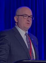
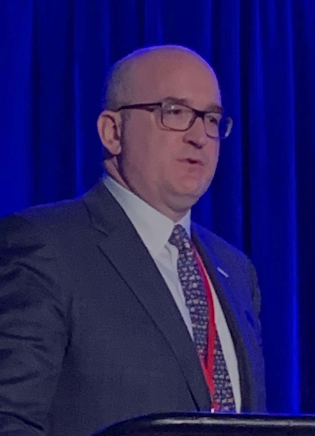
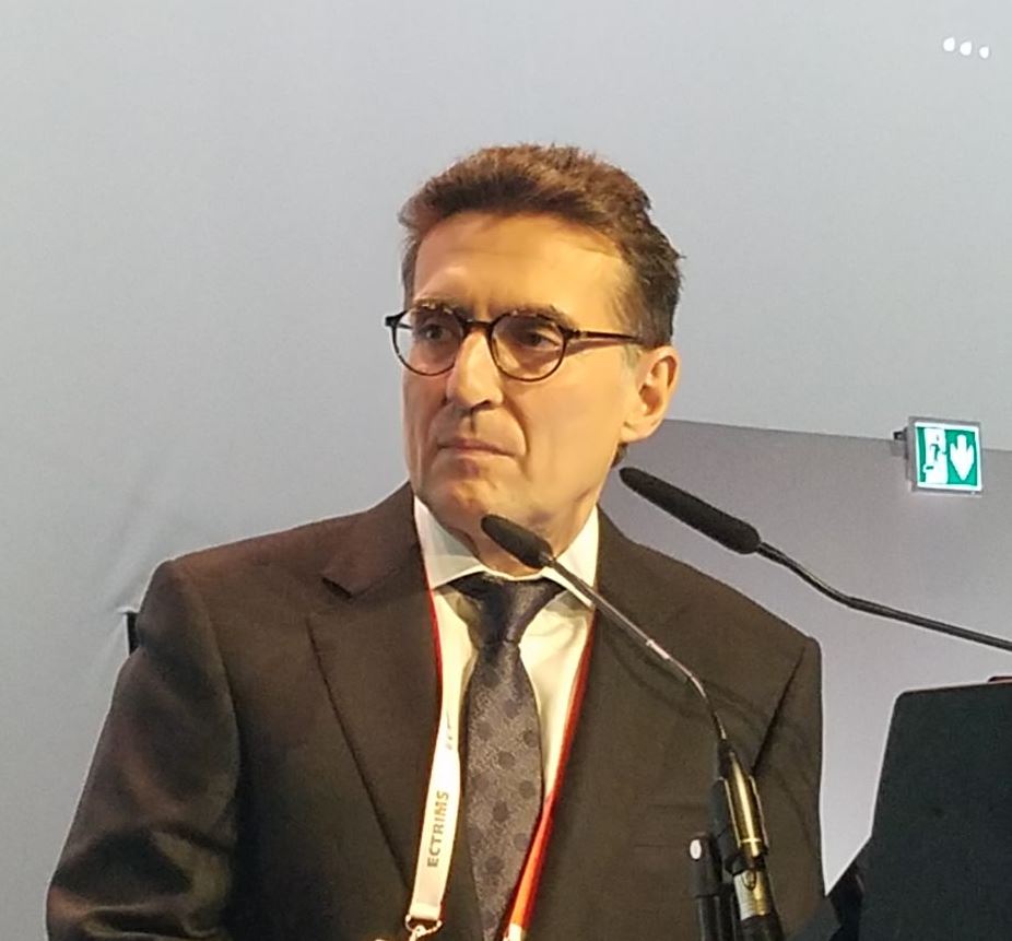
“We found that about a third of patients undergoing sleeve gastrectomy within this data registry developed worsening symptoms after sleeve gastrectomy, and the severity of these symptoms actually varied considerably, from 1 to 13.8 increase in their [GERD–Health Related Quality of Life Questionnaire (HRQL)] score,” said Oliver Varban, MD, of the University of Michigan, Ann Arbor. “Among the surgeons themselves, the rates of severe symptoms varied despite the surgeon’s experience and rate of hiatal hernia repair being similar between the groups.”
This study involved 7,358 patients in the Michigan Bariatric Surgery Collaborative (MBSC) registry who had SG from 2013 to 2017 and 52 surgeons who performed 25 or more SG cases per year. The patients completed the GERD-HRQL survey at baseline and 1 year after SG. The two scores were compared and patients were divided into terciles – mild, moderate, and severe – for worsening of symptoms, then matched with the surgeons who performed the operation. In all, 31.2% of patients (n = 2,294) reported worsening symptoms a year after SG, divided into the following terciles: mild with a 1.4-point increase in GERD-HRQL score (11.7%, n = 866); moderate, a 4.2-point increase (9.7%, n = 716); and severe, 13.8-point increase (9.7%, n = 712).
Among surgeons, the highest rate of patients with severe worsening of GERD was 44.7%, the lowest rate, 18.7%. So the researchers compared characteristics among the surgeons with the highest and lowest rates. “We found that they’re quite similar, actually, in terms of years of bariatric fellowship training, annual sleeve gastrectomy volume, total bariatric annual volume, as well as operative time,” Dr. Varban said. “Interestingly, the rate of concurrent hiatal hernia repair within these two groups is similar as well, which is about one-third for each group” (34.3% for the highest-rate group and 27% for the lowest-rate surgeons).
Likewise, 30-day risk adjusted complication rates were similar between both groups, 3.7% for the high group and 4.3% for the low group.
“Total–body weight loss or excess–body weight loss was actually fairly similar clinically between the two groups, but there was a statistical significance with more weight loss in the GERD patients who had higher severe worsening of symptoms,” Dr. Varban noted.
Surgeons with the highest rates of severe reflux symptoms in their patients tended to operate on more patients with diabetes, hypertension, and cardiovascular disease, whereas the surgeons with the lowest rate of severe symptoms had a higher proportion of patients who were male, white, and had hyperlipidemia and sleep apnea.
Dr. Varban has no financial relationships to disclose. Blue Cross Blue Shield of Michigan provided salary support through the MBSC.
SOURCE: Varban O et al. SAGES 2019; Session SS29, Abstract S139.
REPORTING FROM SAGES 2019
Arsenic exposure increases risk of left ventricular hypertrophy
Young Native Americans had increased left ventricular thickness and left ventricular hypertrophy after being exposed to arsenic, and had a greater risk if they showed signs of prehypertension or hypertension, according to recent research.
“The stronger association in subjects with elevated blood pressure suggests that individuals with preclinical heart disease might be more prone to the toxic effects of arsenic on the heart,” Gernot Pichler, MD, PhD, a cardiologist at Hospital Hietzing/Heart Center Clinic Floridsdorf in Vienna, stated in a press release.
Dr. Pichler and colleagues evaluated 1,337 individuals (mean age, 31 years; 61% female) in the Strong Heart Family Study, an extension of the Strong Heart Study that was designed to study cardiovascular disease in Native Americans. The researchers noted that, while the studies were not originally intended to evaluate arsenic exposure in these populations, “the importance of studying arsenic was recognized overtime as increasing evidence supported the role of arsenic in cardiovascular disease and the relatively high arsenic exposure in tribal communities, compared to other general populations in the United States.”
Arsenic exposure was determined through the sum of inorganic and methylated arsenic concentrations in urine, and researchers used transthoracic ECG at baseline and follow-up to compare left ventricular geometry and function. The mean follow-up was 5.6 years and baseline median sum of inorganic and methylated arsenic concentrations in urine was 4.24 (interquartile range, 2.82-6.90) mcg/g creatinine.
Prevalence of left ventricular hypertrophy was 4.6% overall, and the odds ratio of left ventricular hypertrophy per 100% increase in arsenic in the urine was 1.47 (95% confidence interval, 1.05-2.08) overall and 1.58 for individuals with prehypertension or hypertension (95% CI, 1.04-2.41).
“People drinking water from private wells, which are not regulated, need to be aware that arsenic may increase the risk for cardiovascular disease,” said Dr. Pichler. “Testing those wells is a critical first step to take action and prevent exposure.”
Prospective and cross-sectional analyses both showed changes in left ventricular geometry – such as in the left atrial systolic diameter, interventricular septum, and left ventricular posterior wall thickness and mass index – were associated with exposure to arsenic. In addition, left ventricular function factors such as isovolumic relaxation time and stroke volume were also affected by arsenic exposure. However, the researchers noted that their study was limited by a single method of arsenic exposure and having no long-term follow-up for participants.
“The study raises the question of whether the changes in heart structure are reversible if exposure is reduced. Some changes have occurred in water sources in the study communities, and it will be important to check the potential health impact of reducing arsenic exposure,” said Dr. Pichler.
This study was funded by the National Institute of Health Sciences and grants from the National Heart, Lung, and Blood Institute. The authors reported no relevant conflicts of interest.
SOURCE: Pichler G et al. Circ Cardiovasc Imaging. 2019 May 1. doi: 10.1161/CIRCIMAGING.119.009018.
The results from Pichler et al. show the effects of recent arsenic exposure on left ventricular (LV) hypertrophy, but other questions remain as to the long-term effects of arsenic and its interactions with other environmental metals, Rajiv Chowdhury, MBBS, PhD, and Kim van Daalen, BSc, MPhil, wrote in a related editorial.
It has been established that chronic, low to moderate inorganic arsenic exposure may be linked to cardiovascular disease (CVD) and risk factors such as hypertension and diabetes, but it is unclear how different exposure pathways contribute as well as what pathophysiological mechanisms contribute to CVD, and whether arsenic directly or indirectly contributes to cardiac functioning or a worse cardiometabolic profile, respectively, they wrote.
While the results contribute to the understanding of arsenic exposure through drinking water and its relationship to LV function and geometry, urinary arsenic reflects recent exposure and cannot measure arsenic exposure over a period of time. The study also does not account for an individual’s CVD risk or LV functioning/geometry with daily exposure to co-occurring environmental metals, particularly heavy metals, together with arsenic. Factors such as genetics, age, gender, and nutrition also impact an individual’s reaction to arsenic, and studies should be able to differentiate inorganic arsenic obtained from food and other sources.
While “this elegant analysis ... helps to clarify the observational associations of [arsenic] with LV geometry and function, it stimulates further complimentary work,” they wrote. “Such studies would be essential since CVD remains the single leading cause of adult premature death worldwide, and millions of individuals globally are exposed to arsenic and other metal contaminants.”
Dr. Chowdhury and Dr. Daalen are from the cardiovascular epidemiology unit in the department of public health and primary care at the University of Cambridge (England).
Young Native Americans had increased left ventricular thickness and left ventricular hypertrophy after being exposed to arsenic, and had a greater risk if they showed signs of prehypertension or hypertension, according to recent research.
“The stronger association in subjects with elevated blood pressure suggests that individuals with preclinical heart disease might be more prone to the toxic effects of arsenic on the heart,” Gernot Pichler, MD, PhD, a cardiologist at Hospital Hietzing/Heart Center Clinic Floridsdorf in Vienna, stated in a press release.
Dr. Pichler and colleagues evaluated 1,337 individuals (mean age, 31 years; 61% female) in the Strong Heart Family Study, an extension of the Strong Heart Study that was designed to study cardiovascular disease in Native Americans. The researchers noted that, while the studies were not originally intended to evaluate arsenic exposure in these populations, “the importance of studying arsenic was recognized overtime as increasing evidence supported the role of arsenic in cardiovascular disease and the relatively high arsenic exposure in tribal communities, compared to other general populations in the United States.”
Arsenic exposure was determined through the sum of inorganic and methylated arsenic concentrations in urine, and researchers used transthoracic ECG at baseline and follow-up to compare left ventricular geometry and function. The mean follow-up was 5.6 years and baseline median sum of inorganic and methylated arsenic concentrations in urine was 4.24 (interquartile range, 2.82-6.90) mcg/g creatinine.
Prevalence of left ventricular hypertrophy was 4.6% overall, and the odds ratio of left ventricular hypertrophy per 100% increase in arsenic in the urine was 1.47 (95% confidence interval, 1.05-2.08) overall and 1.58 for individuals with prehypertension or hypertension (95% CI, 1.04-2.41).
“People drinking water from private wells, which are not regulated, need to be aware that arsenic may increase the risk for cardiovascular disease,” said Dr. Pichler. “Testing those wells is a critical first step to take action and prevent exposure.”
Prospective and cross-sectional analyses both showed changes in left ventricular geometry – such as in the left atrial systolic diameter, interventricular septum, and left ventricular posterior wall thickness and mass index – were associated with exposure to arsenic. In addition, left ventricular function factors such as isovolumic relaxation time and stroke volume were also affected by arsenic exposure. However, the researchers noted that their study was limited by a single method of arsenic exposure and having no long-term follow-up for participants.
“The study raises the question of whether the changes in heart structure are reversible if exposure is reduced. Some changes have occurred in water sources in the study communities, and it will be important to check the potential health impact of reducing arsenic exposure,” said Dr. Pichler.
This study was funded by the National Institute of Health Sciences and grants from the National Heart, Lung, and Blood Institute. The authors reported no relevant conflicts of interest.
SOURCE: Pichler G et al. Circ Cardiovasc Imaging. 2019 May 1. doi: 10.1161/CIRCIMAGING.119.009018.
FROM CIRCULATION: CARDIOVASCULAR IMAGING
Robotic and lap surgery achieve similar negative margins for rectal resection
BALTIMORE – A head-to-head comparison of robotic and laparoscopic surgery for locally advanced rectal cancer has found that the operations achieve similar outcomes in terms of composite negative margins, overall survival, and readmissions, but that robotic surgery is associated with lower rates of conversion to open surgery and shorter hospital stays, according to an analysis of cases in the National Cancer Database presented at the annual meeting of the Society of American Gastrointestinal and Endoscopic Surgeons.
“The gold standard for how we’re taking care of our patients, of course, is how we’re doing with the cancer survival, and we found no difference for overall survival in the robotic arm and the laparoscopic arm,” said M. Benjamin Hopkins, MD, of Vanderbilt University, Nashville, Tenn. He reported on a retrospective review of 7,616 operations for rectal adenocarcinoma in the National Cancer Database from 2010 to 2014. The study population included 2,472 (32%) robotic procedures. The hazard ratio for 5-year overall survival was 0.87 (P = .18), Dr. Hopkins noted, with virtually identical Kaplan-Meier survival curves between the two approaches.
The negative margin rates were also similar between the two groups: 93.7% for the robotic patients and 92.9% for the laparoscopic cohort (odds ratio 0.86, P = .23), Dr. Hopkins noted. Readmission rates were 9% and 8%, respectively (odds ratio 1.02, P = .44).
There were two significant differences in outcomes between the two groups: The conversion rate to open surgery for the robotic group “was about half that from what we saw with laparoscopic surgery,” he said – 8% vs. 15%; and “a slightly decreased length of stay” in the robotic group – 6.3 vs. 6.7 days (P less than .001).
Dr. Hopkins noted that the science supporting robotic surgery for rectal resection is still somewhat nascent, pointing to the ROLARR randomized trial, which showed conversion rates similar to those of open between the two minimally invasive approaches (8.1% for robotic and 12.2% for laparoscopy [P = .16] JAMA. 2017; 318[16]:1569-80). “Surgeons in this study were still on their learning curve, and what the results showed was that among surgeons, even with varied experience, robotic surgery was [equal] to laparoscopic surgery in relation to conversion rates and composite margin for cancer specimens,” he said of the ROLARR trial.
That led to the premise for his group’s study. “The hypothesis that we had going into this study was that we are able to get a better composite negative margin with robotic surgery as opposed to laparoscopic surgery, which would then translate into improved cancer survival for our patients,” he said. Like the ROLARR trial, the 2010-2014 National Cancer Database dataset Dr. Hopkins and colleagues used includes a window for the learning curve, he added.
“One of the questions that always comes at us from hospital administration and from insurance companies is whether or not there’s a cost benefit for the patient: What is the value to the patient?” Dr. Hopkins noted. “I’d like to see future studies where we’re looking at this value and, as we decrease our operative time and anesthesia costs, and hopefully some decreased cost in the instrumentation, we start to see some more benefits as we get more facile with robotic surgery.”
Dr. Hopkins has no financial relationships to disclose.
SOURCE: Hopkins B et al. SAGES 2019. Session SS13; abstract S058
CORRECTION 5/14/2109 : The hazard ratio for 5-year overall survival and p-value were updated.
REPORTING FROM SAGES 2019
Type 2 diabetes remission: Reducing excess fat in the liver might be the key
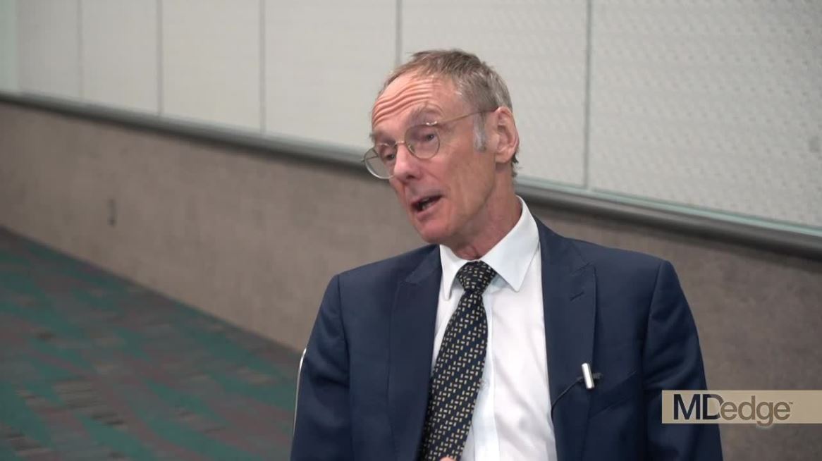
LOS ANGELES – More than 20 years ago, Roy Taylor, MD, began working to further understand the pathogenesis of hepatic insulin resistance in people with type 2 diabetes. It became clear that the main determinant was the amount of fat in the liver.

“If you reduced the amount of fat, the resistance went down,” Dr. Taylor, professor of medicine and metabolism at Newcastle University (England), said at the annual scientific and clinical congress of the American Association of Clinical Endocrinologists. “We had a very clear picture of what might be controlling this awful matter of fasting glucose being too high.”
Then, Dr. Taylor read a study from Caterina Guidone, MD, and colleagues in Italy, which found that 1 week after patients with type 2 diabetes underwent gastric bypass surgery, their fasting plasma glucose levels became normal (Diabetes. 2006;55[7]:2025-31). “I was sitting at my desk and I thought, ‘This really changes type 2 diabetes,’ ” Dr. Taylor said. “It set in process a series of thoughts as to what was controlling what.”
This inspired ongoing work that Dr. Taylor termed the “twin-cycle hypothesis,” which postulates that chronic calorie excess leads to accumulation of liver fat, which spills over into the pancreas (Diabetologia. 2008;51[10]:1781-9).
“People with type 2 diabetes have been in positive calorie balance for a number of years,” he said. “That’s going to lead to an excess of fat in the body, and liver fat levels tend to rise with increasing body weight. If a person has normal insulin sensitivity in muscle tissue, then dealing with a meal is quite easy. Some 30 years ago, we showed using MR spectroscopy that you will have stored the carbohydrate from your breakfast in muscle, to the extent of about one-third of your breakfast, and the peak will be about 5 hours after breakfast. If you had your corn flakes at seven in the morning, by noon there will be peak in muscle, nicely stored away. However, if you happen to be insulin resistant in muscle, that doesn’t happen. There’s only one other pathway that the body can use, and that’s lipogenesis. The body can turn this very toxic substance [glucose] into safe storage [fat]. A lot of that happens in the liver. This means that people with insulin resistance tend to build up liver fat more rapidly than others.”
To test the twin-cycle hypothesis, Dr. Taylor and colleagues launched an 8-week study known as Counterpoint, which set out to induce negative calorie balance using a very low–calorie diet – about one-quarter of an average person’s daily food intake – in 11 people with diabetes (Diabetologia. 2011;54[10]:2506-14). The diet included consuming three packets of liquid formula food each day (46.4% carbohydrate, 32.5% protein, and 20.1% fat; plus vitamins, minerals, and trace elements), supplemented with portions of nonstarchy vegetables such that total energy intake was about 700 calories a day.
“On a liquid-formula diet, hunger is not a problem after the first 36 hours,” Dr. Taylor said. “This is one of the best-kept secrets of the obesity field. Our low-calorie diet was designed as something that people would be able to do in real life. We included nonstarchy vegetables to keep the bowels happy. That was important. It also fulfilled another point. People didn’t want just a liquid diet. They missed the sensation of chewing.”
The researchers also developed three-point Dixon MRI to measure pancreas and liver triacylglycerol content. “The pancreas was particularly challenging, and the full resources of the magnetic resonance physics team were needed to crack the technical problems,” he said.
After just 1 week of restricted energy intake, the fasting plasma glucose level normalized in the diabetic group, going from 9.2 to 5.9 mmol/L (P = .003), while insulin suppression of hepatic glucose output improved from 43% to 74 % (P = .003). By week 8, pancreatic triacylglycerol decreased from 8.0% to 1.1% (P = .03), and hepatic triacylglycerol content fell from 12.8% to 2.9% (P = .003).
“Within 7 days, there was a 30% drop in liver fat, and hepatic insulin resistance had disappeared,” Dr. Taylor said. “This is not a significant change – it’s a disappearance. For one individual, the amount of fat in the liver decreased from 36% to 2%. In fact, 2% [fat in the liver] was the average in the whole group. But what was simply amazing was the change in first-phase insulin response. It gradually increased throughout the 8 weeks of the study to become similar to the normal control group. We knew right away that a low-calorie diet would start correcting this central abnormality of type 2 diabetes.”
After the results from Counterpoint were published, Dr. Taylor received a “tsunami” of emails from researchers and from members of the public. “Some of the medical experts said it was a flash in the pan – interesting, but not relevant,” he said. “People with diabetes learned of it by the media, and it was talked about as a crash diet, which is unfortunate. First, it wasn’t a crash diet. This diet has to be very carefully planned, and people need to think about it in advance. They need to talk about it with their nearest and dearest, because it’s the spouse, the partner, the friends who will be supporting the individual through this journey. That’s critically important. People don’t eat as isolated individuals, they often eat as a family. We’re not talking about cure. We’re talking about reversal of the processes underpinning diabetes, with the aim of achieving remission.”
Dr. Taylor created a website devoted to providing information for clinicians and patients about the low-calorie diet and other tips on how to reverse type 2 diabetes. Soon afterward, he started to receive emails from people telling him about their experiences with the diet. “In the comfort of their own kitchens these people had lost the same amount of weight as in our trial subjects – about 33 pounds,” Dr. Taylor said. “Most of them had gotten rid of their type 2 diabetes. This was not something artificial as part of a research project. This was something that real people would do if the motivation was strong enough.”
To find out if the results from the Counterpoint study were sustainable, Dr. Taylor and his associates launched the Counterbalance study in 30 patients with type 2 diabetes who had a positive calorie imbalance and whom the researchers followed for 6 months. The 8-week diet consisted of consuming three packets of liquid formula a day comprising 43.0% carbohydrates, 34.0% protein, and 19.5% fat, as well as up to 240 g of nonstarchy vegetables (Diabetes Care. 2016;39[5]:808-15). “This was followed for a 6-month period of normal eating: Eating whatever foods they liked but in quantities to keep their weight steady,” Dr. Taylor explained. “These people gained no weight over the 6-month follow-up period. They achieved normalization of liver fat, and it remained normal.”
The patients’ hemoglobin A1c levels fell from an average of 7.1% at baseline to less than 6.0%, and stayed at less than 6.0%. Patients who didn’t respond tended to have a longer duration of diabetes. Their beta cells had fallen to a level beyond that capable of recovery. “So the durability of the return to normal metabolic function was not in question, at least up to 6 months,” he said. “This study also gave us the opportunity to look at changes in pancreas fat. Was it likely that the liver fat was driving the pancreas fat? Yes.”
During the weight-loss period, the researchers found that there was the same degree of reduction of pancreas fat in the Counterbalance study as there’d been in the Counterpoint study. “Remarkably, it decreased slightly during the 6 months of follow-up,” Dr. Taylor said. “Those changes were significant. Type 2 diabetes seems to be caused by about a half a gram of fat within the cells of the pancreas.”
To investigate if a very low–calorie diet could be used as a routine treatment for type 2 diabetes, Dr. Taylor collaborated with his colleague, Mike Lean, MD, in launching the randomized controlled Diabetes Remission Clinical Trial (DiRECT) at 49 primary care practices in the United Kingdom (Diabetologia. 2018;61[3]:589-98). In all, 298 patients were randomized to either best-practice diabetes care alone (control arm) or with an additional evidence-based weight-management program (intervention arm). Remission was defined as having a hemoglobin A1c level of less than 6.5% for at least 2 months without receiving glucose-lowering therapy.
At 1 year, 46% of patients in the intervention arm achieved remission, compared with 4% in the control arm (Lancet Diabetes Endocrinol. 2019;7[5]:344-55). At 2 years, 36% of patients in the intervention arm achieved remission, compared with 2% in the control arm. “The most common comment from study participants was, ‘I feel 10 years younger,’ ” Dr. Taylor said. “That’s important.”
The percentage of patients who achieved remission was 5% in those who lost less than 11 lb (5 kg), 29% in those who lost between 11 lb and 22 lb (5-10 kg), 60% in those who lost between 22 lb and 33 lb (10-15 kg), and 70% in those who lost 33 lb (15 kg) or more.
The researchers found that 62 patients achieved no remission at 12 or 24 months, 15 achieved remission at 12 but not at 24 months, and 48 achieved remission at 12 and 24 months. “We haven’t got this perfectly right yet,” Dr. Taylor said. “There is more work to do in understanding how to achieve prevention of weight gain, maybe with behavioral interventions and/or other agents such as [glucagonlike peptide–1] agonists. This is the start of a story, not the end of it.”
He and his associates also observed that delivery of fat from the liver to the rest of the body was increased in study participants who relapsed. “What effect did that have on the pancreas fat? The people who continued to be free of diabetes showed a slight fall in pancreatic fat between 5 and 24 months,” Dr. Taylor said. “In sharp contrast, the relapsers had a complete increase. Over the whole period of the study, the relapsers had not changed from baseline. It appears beyond reasonable doubt that excess pancreas fat seems to be driving the beta-cell problem underlying type 2 diabetes.”
Dr. Taylor reported that he has received lecture fees from Novartis, Lilly, and Janssen. He has also been an advisory board member for Wilmington Healthcare.
LOS ANGELES – More than 20 years ago, Roy Taylor, MD, began working to further understand the pathogenesis of hepatic insulin resistance in people with type 2 diabetes. It became clear that the main determinant was the amount of fat in the liver.

“If you reduced the amount of fat, the resistance went down,” Dr. Taylor, professor of medicine and metabolism at Newcastle University (England), said at the annual scientific and clinical congress of the American Association of Clinical Endocrinologists. “We had a very clear picture of what might be controlling this awful matter of fasting glucose being too high.”
Then, Dr. Taylor read a study from Caterina Guidone, MD, and colleagues in Italy, which found that 1 week after patients with type 2 diabetes underwent gastric bypass surgery, their fasting plasma glucose levels became normal (Diabetes. 2006;55[7]:2025-31). “I was sitting at my desk and I thought, ‘This really changes type 2 diabetes,’ ” Dr. Taylor said. “It set in process a series of thoughts as to what was controlling what.”
This inspired ongoing work that Dr. Taylor termed the “twin-cycle hypothesis,” which postulates that chronic calorie excess leads to accumulation of liver fat, which spills over into the pancreas (Diabetologia. 2008;51[10]:1781-9).
“People with type 2 diabetes have been in positive calorie balance for a number of years,” he said. “That’s going to lead to an excess of fat in the body, and liver fat levels tend to rise with increasing body weight. If a person has normal insulin sensitivity in muscle tissue, then dealing with a meal is quite easy. Some 30 years ago, we showed using MR spectroscopy that you will have stored the carbohydrate from your breakfast in muscle, to the extent of about one-third of your breakfast, and the peak will be about 5 hours after breakfast. If you had your corn flakes at seven in the morning, by noon there will be peak in muscle, nicely stored away. However, if you happen to be insulin resistant in muscle, that doesn’t happen. There’s only one other pathway that the body can use, and that’s lipogenesis. The body can turn this very toxic substance [glucose] into safe storage [fat]. A lot of that happens in the liver. This means that people with insulin resistance tend to build up liver fat more rapidly than others.”
To test the twin-cycle hypothesis, Dr. Taylor and colleagues launched an 8-week study known as Counterpoint, which set out to induce negative calorie balance using a very low–calorie diet – about one-quarter of an average person’s daily food intake – in 11 people with diabetes (Diabetologia. 2011;54[10]:2506-14). The diet included consuming three packets of liquid formula food each day (46.4% carbohydrate, 32.5% protein, and 20.1% fat; plus vitamins, minerals, and trace elements), supplemented with portions of nonstarchy vegetables such that total energy intake was about 700 calories a day.
“On a liquid-formula diet, hunger is not a problem after the first 36 hours,” Dr. Taylor said. “This is one of the best-kept secrets of the obesity field. Our low-calorie diet was designed as something that people would be able to do in real life. We included nonstarchy vegetables to keep the bowels happy. That was important. It also fulfilled another point. People didn’t want just a liquid diet. They missed the sensation of chewing.”
The researchers also developed three-point Dixon MRI to measure pancreas and liver triacylglycerol content. “The pancreas was particularly challenging, and the full resources of the magnetic resonance physics team were needed to crack the technical problems,” he said.
After just 1 week of restricted energy intake, the fasting plasma glucose level normalized in the diabetic group, going from 9.2 to 5.9 mmol/L (P = .003), while insulin suppression of hepatic glucose output improved from 43% to 74 % (P = .003). By week 8, pancreatic triacylglycerol decreased from 8.0% to 1.1% (P = .03), and hepatic triacylglycerol content fell from 12.8% to 2.9% (P = .003).
“Within 7 days, there was a 30% drop in liver fat, and hepatic insulin resistance had disappeared,” Dr. Taylor said. “This is not a significant change – it’s a disappearance. For one individual, the amount of fat in the liver decreased from 36% to 2%. In fact, 2% [fat in the liver] was the average in the whole group. But what was simply amazing was the change in first-phase insulin response. It gradually increased throughout the 8 weeks of the study to become similar to the normal control group. We knew right away that a low-calorie diet would start correcting this central abnormality of type 2 diabetes.”
After the results from Counterpoint were published, Dr. Taylor received a “tsunami” of emails from researchers and from members of the public. “Some of the medical experts said it was a flash in the pan – interesting, but not relevant,” he said. “People with diabetes learned of it by the media, and it was talked about as a crash diet, which is unfortunate. First, it wasn’t a crash diet. This diet has to be very carefully planned, and people need to think about it in advance. They need to talk about it with their nearest and dearest, because it’s the spouse, the partner, the friends who will be supporting the individual through this journey. That’s critically important. People don’t eat as isolated individuals, they often eat as a family. We’re not talking about cure. We’re talking about reversal of the processes underpinning diabetes, with the aim of achieving remission.”
Dr. Taylor created a website devoted to providing information for clinicians and patients about the low-calorie diet and other tips on how to reverse type 2 diabetes. Soon afterward, he started to receive emails from people telling him about their experiences with the diet. “In the comfort of their own kitchens these people had lost the same amount of weight as in our trial subjects – about 33 pounds,” Dr. Taylor said. “Most of them had gotten rid of their type 2 diabetes. This was not something artificial as part of a research project. This was something that real people would do if the motivation was strong enough.”
To find out if the results from the Counterpoint study were sustainable, Dr. Taylor and his associates launched the Counterbalance study in 30 patients with type 2 diabetes who had a positive calorie imbalance and whom the researchers followed for 6 months. The 8-week diet consisted of consuming three packets of liquid formula a day comprising 43.0% carbohydrates, 34.0% protein, and 19.5% fat, as well as up to 240 g of nonstarchy vegetables (Diabetes Care. 2016;39[5]:808-15). “This was followed for a 6-month period of normal eating: Eating whatever foods they liked but in quantities to keep their weight steady,” Dr. Taylor explained. “These people gained no weight over the 6-month follow-up period. They achieved normalization of liver fat, and it remained normal.”
The patients’ hemoglobin A1c levels fell from an average of 7.1% at baseline to less than 6.0%, and stayed at less than 6.0%. Patients who didn’t respond tended to have a longer duration of diabetes. Their beta cells had fallen to a level beyond that capable of recovery. “So the durability of the return to normal metabolic function was not in question, at least up to 6 months,” he said. “This study also gave us the opportunity to look at changes in pancreas fat. Was it likely that the liver fat was driving the pancreas fat? Yes.”
During the weight-loss period, the researchers found that there was the same degree of reduction of pancreas fat in the Counterbalance study as there’d been in the Counterpoint study. “Remarkably, it decreased slightly during the 6 months of follow-up,” Dr. Taylor said. “Those changes were significant. Type 2 diabetes seems to be caused by about a half a gram of fat within the cells of the pancreas.”
To investigate if a very low–calorie diet could be used as a routine treatment for type 2 diabetes, Dr. Taylor collaborated with his colleague, Mike Lean, MD, in launching the randomized controlled Diabetes Remission Clinical Trial (DiRECT) at 49 primary care practices in the United Kingdom (Diabetologia. 2018;61[3]:589-98). In all, 298 patients were randomized to either best-practice diabetes care alone (control arm) or with an additional evidence-based weight-management program (intervention arm). Remission was defined as having a hemoglobin A1c level of less than 6.5% for at least 2 months without receiving glucose-lowering therapy.
At 1 year, 46% of patients in the intervention arm achieved remission, compared with 4% in the control arm (Lancet Diabetes Endocrinol. 2019;7[5]:344-55). At 2 years, 36% of patients in the intervention arm achieved remission, compared with 2% in the control arm. “The most common comment from study participants was, ‘I feel 10 years younger,’ ” Dr. Taylor said. “That’s important.”
The percentage of patients who achieved remission was 5% in those who lost less than 11 lb (5 kg), 29% in those who lost between 11 lb and 22 lb (5-10 kg), 60% in those who lost between 22 lb and 33 lb (10-15 kg), and 70% in those who lost 33 lb (15 kg) or more.
The researchers found that 62 patients achieved no remission at 12 or 24 months, 15 achieved remission at 12 but not at 24 months, and 48 achieved remission at 12 and 24 months. “We haven’t got this perfectly right yet,” Dr. Taylor said. “There is more work to do in understanding how to achieve prevention of weight gain, maybe with behavioral interventions and/or other agents such as [glucagonlike peptide–1] agonists. This is the start of a story, not the end of it.”
He and his associates also observed that delivery of fat from the liver to the rest of the body was increased in study participants who relapsed. “What effect did that have on the pancreas fat? The people who continued to be free of diabetes showed a slight fall in pancreatic fat between 5 and 24 months,” Dr. Taylor said. “In sharp contrast, the relapsers had a complete increase. Over the whole period of the study, the relapsers had not changed from baseline. It appears beyond reasonable doubt that excess pancreas fat seems to be driving the beta-cell problem underlying type 2 diabetes.”
Dr. Taylor reported that he has received lecture fees from Novartis, Lilly, and Janssen. He has also been an advisory board member for Wilmington Healthcare.
LOS ANGELES – More than 20 years ago, Roy Taylor, MD, began working to further understand the pathogenesis of hepatic insulin resistance in people with type 2 diabetes. It became clear that the main determinant was the amount of fat in the liver.

“If you reduced the amount of fat, the resistance went down,” Dr. Taylor, professor of medicine and metabolism at Newcastle University (England), said at the annual scientific and clinical congress of the American Association of Clinical Endocrinologists. “We had a very clear picture of what might be controlling this awful matter of fasting glucose being too high.”
Then, Dr. Taylor read a study from Caterina Guidone, MD, and colleagues in Italy, which found that 1 week after patients with type 2 diabetes underwent gastric bypass surgery, their fasting plasma glucose levels became normal (Diabetes. 2006;55[7]:2025-31). “I was sitting at my desk and I thought, ‘This really changes type 2 diabetes,’ ” Dr. Taylor said. “It set in process a series of thoughts as to what was controlling what.”
This inspired ongoing work that Dr. Taylor termed the “twin-cycle hypothesis,” which postulates that chronic calorie excess leads to accumulation of liver fat, which spills over into the pancreas (Diabetologia. 2008;51[10]:1781-9).
“People with type 2 diabetes have been in positive calorie balance for a number of years,” he said. “That’s going to lead to an excess of fat in the body, and liver fat levels tend to rise with increasing body weight. If a person has normal insulin sensitivity in muscle tissue, then dealing with a meal is quite easy. Some 30 years ago, we showed using MR spectroscopy that you will have stored the carbohydrate from your breakfast in muscle, to the extent of about one-third of your breakfast, and the peak will be about 5 hours after breakfast. If you had your corn flakes at seven in the morning, by noon there will be peak in muscle, nicely stored away. However, if you happen to be insulin resistant in muscle, that doesn’t happen. There’s only one other pathway that the body can use, and that’s lipogenesis. The body can turn this very toxic substance [glucose] into safe storage [fat]. A lot of that happens in the liver. This means that people with insulin resistance tend to build up liver fat more rapidly than others.”
To test the twin-cycle hypothesis, Dr. Taylor and colleagues launched an 8-week study known as Counterpoint, which set out to induce negative calorie balance using a very low–calorie diet – about one-quarter of an average person’s daily food intake – in 11 people with diabetes (Diabetologia. 2011;54[10]:2506-14). The diet included consuming three packets of liquid formula food each day (46.4% carbohydrate, 32.5% protein, and 20.1% fat; plus vitamins, minerals, and trace elements), supplemented with portions of nonstarchy vegetables such that total energy intake was about 700 calories a day.
“On a liquid-formula diet, hunger is not a problem after the first 36 hours,” Dr. Taylor said. “This is one of the best-kept secrets of the obesity field. Our low-calorie diet was designed as something that people would be able to do in real life. We included nonstarchy vegetables to keep the bowels happy. That was important. It also fulfilled another point. People didn’t want just a liquid diet. They missed the sensation of chewing.”
The researchers also developed three-point Dixon MRI to measure pancreas and liver triacylglycerol content. “The pancreas was particularly challenging, and the full resources of the magnetic resonance physics team were needed to crack the technical problems,” he said.
After just 1 week of restricted energy intake, the fasting plasma glucose level normalized in the diabetic group, going from 9.2 to 5.9 mmol/L (P = .003), while insulin suppression of hepatic glucose output improved from 43% to 74 % (P = .003). By week 8, pancreatic triacylglycerol decreased from 8.0% to 1.1% (P = .03), and hepatic triacylglycerol content fell from 12.8% to 2.9% (P = .003).
“Within 7 days, there was a 30% drop in liver fat, and hepatic insulin resistance had disappeared,” Dr. Taylor said. “This is not a significant change – it’s a disappearance. For one individual, the amount of fat in the liver decreased from 36% to 2%. In fact, 2% [fat in the liver] was the average in the whole group. But what was simply amazing was the change in first-phase insulin response. It gradually increased throughout the 8 weeks of the study to become similar to the normal control group. We knew right away that a low-calorie diet would start correcting this central abnormality of type 2 diabetes.”
After the results from Counterpoint were published, Dr. Taylor received a “tsunami” of emails from researchers and from members of the public. “Some of the medical experts said it was a flash in the pan – interesting, but not relevant,” he said. “People with diabetes learned of it by the media, and it was talked about as a crash diet, which is unfortunate. First, it wasn’t a crash diet. This diet has to be very carefully planned, and people need to think about it in advance. They need to talk about it with their nearest and dearest, because it’s the spouse, the partner, the friends who will be supporting the individual through this journey. That’s critically important. People don’t eat as isolated individuals, they often eat as a family. We’re not talking about cure. We’re talking about reversal of the processes underpinning diabetes, with the aim of achieving remission.”
Dr. Taylor created a website devoted to providing information for clinicians and patients about the low-calorie diet and other tips on how to reverse type 2 diabetes. Soon afterward, he started to receive emails from people telling him about their experiences with the diet. “In the comfort of their own kitchens these people had lost the same amount of weight as in our trial subjects – about 33 pounds,” Dr. Taylor said. “Most of them had gotten rid of their type 2 diabetes. This was not something artificial as part of a research project. This was something that real people would do if the motivation was strong enough.”
To find out if the results from the Counterpoint study were sustainable, Dr. Taylor and his associates launched the Counterbalance study in 30 patients with type 2 diabetes who had a positive calorie imbalance and whom the researchers followed for 6 months. The 8-week diet consisted of consuming three packets of liquid formula a day comprising 43.0% carbohydrates, 34.0% protein, and 19.5% fat, as well as up to 240 g of nonstarchy vegetables (Diabetes Care. 2016;39[5]:808-15). “This was followed for a 6-month period of normal eating: Eating whatever foods they liked but in quantities to keep their weight steady,” Dr. Taylor explained. “These people gained no weight over the 6-month follow-up period. They achieved normalization of liver fat, and it remained normal.”
The patients’ hemoglobin A1c levels fell from an average of 7.1% at baseline to less than 6.0%, and stayed at less than 6.0%. Patients who didn’t respond tended to have a longer duration of diabetes. Their beta cells had fallen to a level beyond that capable of recovery. “So the durability of the return to normal metabolic function was not in question, at least up to 6 months,” he said. “This study also gave us the opportunity to look at changes in pancreas fat. Was it likely that the liver fat was driving the pancreas fat? Yes.”
During the weight-loss period, the researchers found that there was the same degree of reduction of pancreas fat in the Counterbalance study as there’d been in the Counterpoint study. “Remarkably, it decreased slightly during the 6 months of follow-up,” Dr. Taylor said. “Those changes were significant. Type 2 diabetes seems to be caused by about a half a gram of fat within the cells of the pancreas.”
To investigate if a very low–calorie diet could be used as a routine treatment for type 2 diabetes, Dr. Taylor collaborated with his colleague, Mike Lean, MD, in launching the randomized controlled Diabetes Remission Clinical Trial (DiRECT) at 49 primary care practices in the United Kingdom (Diabetologia. 2018;61[3]:589-98). In all, 298 patients were randomized to either best-practice diabetes care alone (control arm) or with an additional evidence-based weight-management program (intervention arm). Remission was defined as having a hemoglobin A1c level of less than 6.5% for at least 2 months without receiving glucose-lowering therapy.
At 1 year, 46% of patients in the intervention arm achieved remission, compared with 4% in the control arm (Lancet Diabetes Endocrinol. 2019;7[5]:344-55). At 2 years, 36% of patients in the intervention arm achieved remission, compared with 2% in the control arm. “The most common comment from study participants was, ‘I feel 10 years younger,’ ” Dr. Taylor said. “That’s important.”
The percentage of patients who achieved remission was 5% in those who lost less than 11 lb (5 kg), 29% in those who lost between 11 lb and 22 lb (5-10 kg), 60% in those who lost between 22 lb and 33 lb (10-15 kg), and 70% in those who lost 33 lb (15 kg) or more.
The researchers found that 62 patients achieved no remission at 12 or 24 months, 15 achieved remission at 12 but not at 24 months, and 48 achieved remission at 12 and 24 months. “We haven’t got this perfectly right yet,” Dr. Taylor said. “There is more work to do in understanding how to achieve prevention of weight gain, maybe with behavioral interventions and/or other agents such as [glucagonlike peptide–1] agonists. This is the start of a story, not the end of it.”
He and his associates also observed that delivery of fat from the liver to the rest of the body was increased in study participants who relapsed. “What effect did that have on the pancreas fat? The people who continued to be free of diabetes showed a slight fall in pancreatic fat between 5 and 24 months,” Dr. Taylor said. “In sharp contrast, the relapsers had a complete increase. Over the whole period of the study, the relapsers had not changed from baseline. It appears beyond reasonable doubt that excess pancreas fat seems to be driving the beta-cell problem underlying type 2 diabetes.”
Dr. Taylor reported that he has received lecture fees from Novartis, Lilly, and Janssen. He has also been an advisory board member for Wilmington Healthcare.
EXPERT ANALYSIS FROM AACE 2109
Evobrutinib demonstrates efficacy, safety in relapsing MS
PHILADELPHIA – Higher doses of evobrutinib significantly improved that study endpoint, and were associated with numerical decreases in the annualized relapse rate versus placebo, according to a report presented at the annual meeting of the American Academy of Neurology.
Some grade 3-4 transaminase elevations were associated with evobrutinib, though these were asymptomatic, reversible, and occurred within the first 24 weeks of the 48-week treatment period, said investigator Xavier Montalban, MD, PhD, chairman and director of the department of neurology-neuroimmunology at Vall d’Hebron University Hospital in Barcelona.
“Overall, we do believe the results of the phase 2 study support further clinical development in relapsing multiple sclerosis,” Dr. Montalban said.
This study builds on previous observations that BTK plays an important role in immune functions related to the pathogenesis of MS, Dr. Montalban said. Evobrutinib in particular impacts B cells and myeloid cells along with pathways involved in MS-related inflammation, he added.
In the current randomized, phase 2, placebo-controlled study, 267 patients with relapsing MS were randomized to one of five arms: placebo, evobrutinib 25-mg daily, 75-mg daily, or 75-mg twice daily, or an open-label reference arm of dimethyl fumarate 240 mg twice daily.
Dr. Montalban presented the results of a 24-week treatment period plus a 24-week blinded extension period, during which placebo-treated patients crossed over to evobrutinib 25 mg daily, for a total of 48 weeks of treatment.
The primary study endpoint was the cumulative number of MRI-assessed T1 Gd+ lesions at weeks 12, 16, 20, and 24. Dr. Montalban said that evobrutinib in the two 75-mg arms significantly reduced enhancing lesions versus placebo over weeks 12-24, with lesion rate ratios of 0.30 for the 75-mg daily arm, and 0.44 for the 75-mg twice-daily arm, with unadjusted P values of .002 and .031, respectively. By contrast, the evobrutinib 25-mg arm did not significantly reduce the cumulative number of enhancing lesions versus placebo over that time period.
There was a rapid reduction in the mean number of enhancing lesions from baseline to the week-12 visit for those 75-mg dosing arms, which was sustained through subsequent visits, Dr. Montalban said.
There were no significant differences between arms in the annualized relapse rate at week 24, a key secondary endpoint of the study, according to the investigators. The annualized relapse rate was 0.37 for placebo, 0.57 for evobrutinib 25 mg daily, 0.13 for the 75-mg daily dose, 0.08 for the 75-mg twice-daily dose, and 0.20 for dimethyl fumarate. The magnitude of reduction was maintained at the 48-week evaluation for evobrutinib 75 mg twice daily, with an annualized relapse rate of 0.11, Dr. Montalban reported.
Most treatment-emergent adverse events were mild or moderate, according to the investigators.
Reported ALT elevations in the evobrutinib arms were mainly grade 1. While some grade 3-4 elevations were seen in the first 24 weeks of the study, they were reversible and did not have clinical consequences, according to Dr. Montalban.
Results of the study were published concurrently in the New England Journal of Medicine.
Dr. Montalban provided disclosures related to Biogen, Merck Serono, Genentech, Genzyme, Novartis, Sanofi-Aventis, Teva Pharmaceuticals, Roche, Celgene, Actelion, National Multiple Sclerosis Society, and Multiple Sclerosis International Federation.
SOURCE: Montalban X et al. AAN 2019. Abstract S56.004.
FROM AAN 2019
Etanercept biosimilar SB4 a cost-effective alternative for psoriasis, PsA
The development of biosimilars such as etanercept SB4 offers a “significant opportunity to decrease medical care cost and increase treatment options,” Alessandro Giunta, MD, of the department of dermatology at the University of Rome Tor Vergata, and associates reported in a letter to the editor in the British Journal of Dermatology.
Dr. Giunta and his associates performed an observational, retrospective, single-center study to investigate etanercept biosimilar SB4 in patients being treated for plaque type psoriasis and psoriatic arthritis (PsA). They evaluated 40 patients – 21 men and 19 women – mean age 55, ranging from 19 to 79 years. The patients received the etanercept biosimilar SB4 between Oct. 21, 2016, and March 31, 2017, at University of Rome Tor Vergata’s department of dermatology. (The etanercept biosimilar SB4 was approved April 29 by the Food and Drug Administration under the brand name Eticovo [etanercept-ykro]. It is also approved in other countries under the names Benepali and Brenzys.)
Accounting for erythrocyte sedimentation rate as a variable, Dr. Giunta and colleagues calculated disease activity scores based on 28 joints; 14 patients (35%) had plaque psoriasis (mean Psoriasis Area Severity Index [PASI] of 9.61 at baseline), while 26 (65%) had psoriatic arthritis (mean PASI, 4.69). All patients reported prior treatment with systemic conventional and biologic treatments. A group of 10 patients (25%) who had been previously treated with etanercept originator underwent an intermittent treatment regimen of 24 weeks with etanercept biosimilar, which was interrupted once clinical resolution was achieved. No treatments were prescribed between etanercept originator and etanercept biosimilar. Mean exposure was 50.4 weeks, ranging from 24 to 96 weeks, with an average washout period of 12.1 weeks from originator to biosimilar (range 8-24).
A significant improvement in mean PASI score was observed in plaque type psoriasis patients as well as psoriatic arthritis patients at week 24 (P less than .0001 and P less than .001, respectively), noted Dr. Giunta and associates.
“All scores achieved a statistical significant improvement with the exception of [swollen joint count] that markedly improved but not significantly,” they added. One patient experienced injection site reaction, but no serious adverse events were observed.
Despite low sample size and limited follow-up time, the authors concluded that etanercept biosimilar achieved effectiveness as a treatment for psoriatic patients even in cases involving previous exposure to originator etanercept. Cost savings of 61.58% for 50-mg treatment and 62.55% for 25-mg treatment respectively guaranteed “the continuity of etanercept-treated patients’ care and gave us the opportunity to allocate patients in innovative but more expensive agents with marginal increase in our annual budget,” they noted.
The authors reported serving as consultants and speakers for AbbVie, Biogen, Eli Lilly, Janssen, Pfizer, and Novartis.
SOURCE: Giunta A et al. Br J Dermatol. 2019 May 3. doi: 10.1111/bjd.18090.
FROM THE BRITISH JOURNAL OF DERMATOLOGY
Fingolimod reduced disease activity more than glatiramer acetate in RRMS: ASSESS study results
PHILADELPHIA – reported at the annual meeting of the American Academy of Neurology. The optimal efficacious dose of fingolimod was 0.5 mg once daily, according to the results of the ASSESS study, which also evaluated a 0.25-mg daily dose of fingolimod versus a 20-mg daily dose of glatiramer acetate.
Adverse events seen with fingolimod were consistent with the established safety profile of the immunomodulatory drug, according to investigator Bruce Cree, MD, PhD, of the UCSF Weill Institute for Neurosciences, department of neurology, University of California, San Francisco.
“I believe this is the first study to go head to head versus glatiramer acetate to show superiority,” Dr. Cree said in a podium presentation of the results.
While fingolimod 0.5 mg has shown superior efficacy over placebo and interferon beta-1a in previous phase 3 trials, head-to-head comparisons versus disease-modifying therapies can help inform treatment decisions in clinical practice, Dr. Cree and coinvestigators noted in the abstract that describes their results.
The randomized, three-armed, phase 3b ASSESS study included 12 months of dose-blinded treatment and 3 months of follow-up. Investigators enrolled 1,054 patients with relapsing-remitting MS, of whom about 74% were women and the average age was 40 years, Dr. Cree said in his presentation.
Annualized relapse rate, the primary endpoint of the study, was 0.153 for the fingolimod 0.5-mg arm, versus 0.258 for the glatiramer acetate 20-mg arm, for a 40.7% relative reduction (P = .013), Dr. Cree reported. By contrast, he said, the annualized relapse rate within the fingolimod 0.25-mg arm was 0.221, which was not statistically different versus glatiramer acetate.
The fingolimod 0.5-mg arm was also superior to glatiramer acetate on a number of radiographic endpoints at month 12, including new or newly enlarged T2 lesion count, change in T2 lesion volume, gadolinium-positive T1 lesion count, and gadolinium-positive T1 lesion volume, Dr. Cree said.
Adverse events and serious adverse events were “much as expected” in all three study groups, Dr. Cree said.
The observed safety with fingolimod was consistent with previously available data on fingolimod 0.5 mg, and the safety profile of the lower 0.25-mg dose seemed to be comparable with the 0.5-mg dose, according to his presentation.
Adverse events with fingolimod were “largely laboratory abnormalities,” that occurred more frequently in the fingolimod arms, he said. Although there was an apparent dose-dependent effect between the 0.25-mg and 0.5-mg doses, the overall proportion of patients experiencing low white blood cell counts or elevations in liver enzymes was low, he added.
Bradycardia was “infrequent” in both fingolimod groups in first-dose observations, though it did again occur with a dose-dependent effect, Dr. Cree said.
He reported bradycardia in two patients (0.5%) in the fingolimod 0.25-mg group and four (1.2%) in the fingolimod 0.5-mg group, while the number of patients requiring overnight hospitalizations was one (0.3%) and five (1.5%) in the 0.25- and 0.5-mg groups, respectively.
Hepatic enzyme abnormalities were the leading reason for discontinuation of fingolimod, while in contrast, drug hypersensitivity and injection site reactions led to discontinuations in the glatiramer acetate arm, he added.
Novartis sponsored the study. Dr. Cree provided disclosures related to Abbvie, Akili, Biogen, EMD Serono, GeNeuro and Novartis.
SOURCE: Cree B et al. AAN 2019, Abstract 56.009.
REPORTING FROM AAN 2019
Ankylosing spondylitis patients taking COX-2 inhibitors may see fewer cardiovascular events
MADISON, WISC. – Patients with ankylosing spondylitis had a small but significant reduction in risk for cardiovascular events if they were taking cyclooxygenase-2 (COX-2) inhibitors, according to a new systematic review and meta-analysis.
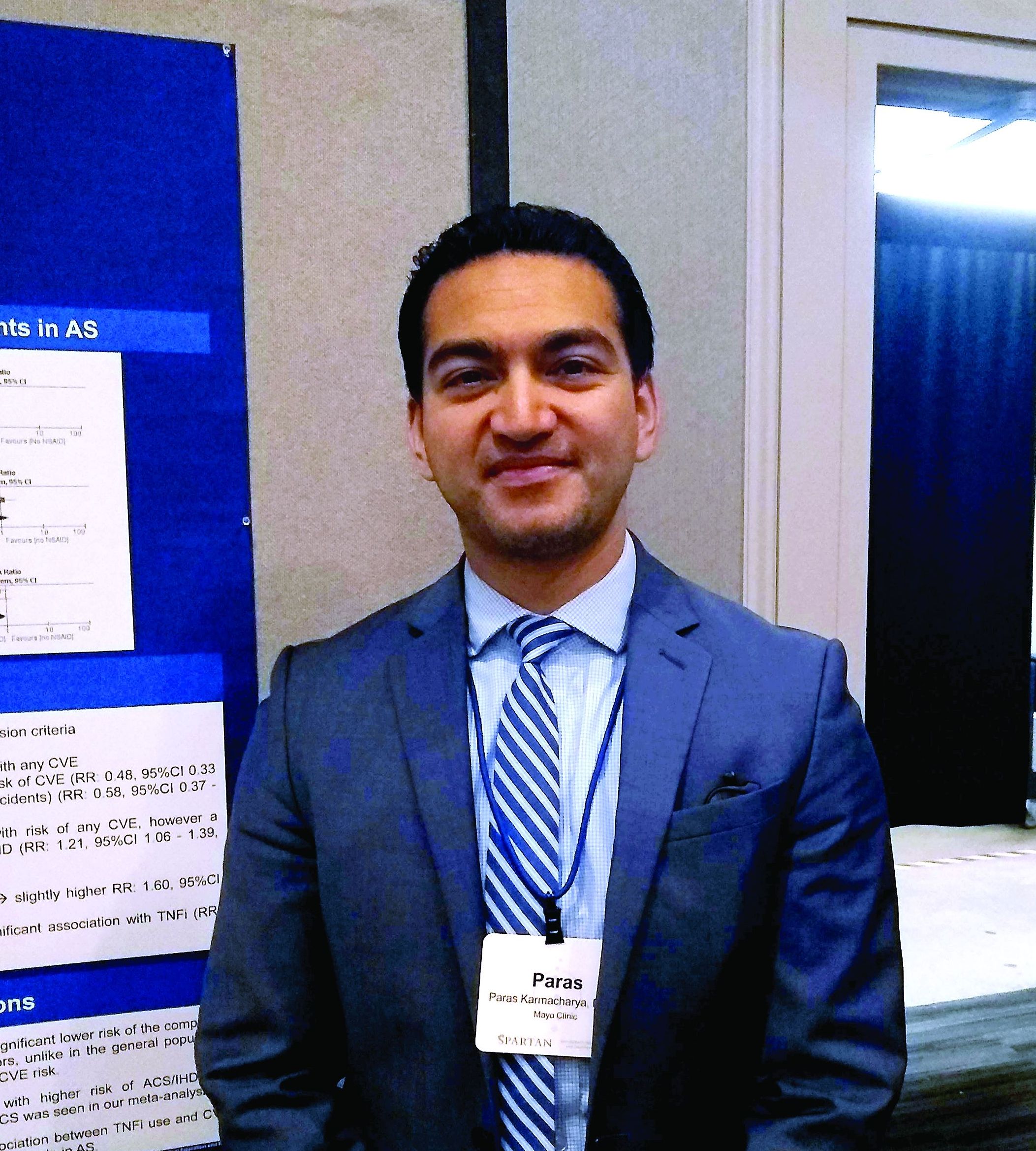
The reduced risk observed in this analysis (risk ratio, 0.48; 95% confidence interval, 0.33-0.70) contrasts with the increased risk for cardiovascular events seen with COX-2 inhibitor use in the general population, said Paras Karmacharya, MBBS, speaking in an interview at the annual meeting of the Spondyloarthritis Research and Treatment Network (SPARTAN). The overall effect was highly statistically significant (P = .0001), and the finding provides “reassuring” data for a population that’s known to have an elevated risk for cardiovascular events, he said.
“[W]e found in the subgroup analysis that COX-2 inhibitors were associated with a reduced risk of cardiovascular events as a whole,” an association also seen when looking just at ischemic stroke, Dr. Karmacharya said. “So that was sort of surprising; in the general population, there are some concerns about using COX-2 inhibitors.”
Looking at data for the nine studies that met criteria for inclusion in the meta-analysis, Dr. Karmacharya, a rheumatology fellow at the Mayo Clinic, Rochester, Minn., and his collaborators calculated risk ratios for a composite outcome of all cardiovascular events (CVE) for all individuals taking NSAIDs, compared with individuals with ankylosing spondylitis (AS) who were not taking NSAIDs. Here, they found a relative risk of 0.94 (95% CI, 0.50-1.75; P = .84).
Next, the investigators calculated a relative risk of 0.78 for the composite CVE outcome just for those taking nonselective NSAIDs (95% CI, 0.44-1.38; P = .40).
Along with NSAIDs, Dr. Karmacharya and his coauthors also looked at the relationship between tumor necrosis factor inhibitors (TNFIs) and cardiovascular events. They found a significantly increased risk for the composite endpoint among AS patients taking TNFIs (RR, 1.60; 95% CI, 1.05-2.41; P = .03), but the comparison was limited to only one study.
In their analysis, the investigators also broke out risk of acute coronary syndrome (ACS)/ischemic heart disease. “The only place where we found some increased risk was ACS and ischemic heart disease, and that was with nonselective NSAIDS,” Dr. Karmacharya said (RR, 1.21; 95% CI, 1.06-1.39; P = .005). No significant changes in relative risk for ACS and ischemic heart disease were seen for the total group of NSAID users, for the subgroups taking COX-2 inhibitors, or for those taking TNFIs.
Finally, the investigators found a relative risk of 0.58 for stroke among the full group of NSAID users and a relative risk of 0.59 for those taking COX-2 inhibitors, but no reduced risk for the subgroup taking nonselective NSAIDs (P = .02, .04, and .37, respectively).
“While NSAIDs are known to be associated with an increased risk of CVE in the general population, whether the anti-inflammatory effects of NSAIDs reduce or modify the CVE risk in AS is controversial,” Dr. Karmacharya and his collaborators wrote. In this context, the meta-analysis provides a useful perspective for rheumatologists who care for AS patients, Dr. Karmacharya said: “I think it’s important, because most of these patients are on NSAIDs long-term.”
However, all of the studies included in the meta-analysis were observational, with no randomized, controlled trials meeting inclusion criteria. Also, some analyses presented in the poster involved as few as two studies, so findings should be interpreted with caution, he added. “We don’t have a lot of studies included in the analysis. ... so we need more data for sure, but I think what data we have so far look reassuring.”
Dr. Karmacharya reported that he had no conflicts of interest, and reported no outside sources of funding.
REPORTING FROM SPARTAN 2019
Key clinical point: Individuals with ankylosing spondylitis (AS) who took cyclooxygenase 2 (COX-2) inhibitors had a reduced risk of cardiovascular events, compared with AS patients who were not using COX-2 inhibitors.
Major finding: Individuals with AS taking COX-2 inhibitors had a risk ratio of 0.48 for cardiovascular events (95% CI, 0.33-0.70; P = .001).
Study details: Systematic review and meta-analysis of nine observational studies that variably examined the association between NSAID use and tumor necrosis factor inhibitor use and cardiovascular events among individuals with AS.
Disclosures: The authors reported no conflicts of interest and no outside sources of funding.
Source: Karmacharya P. et al. SPARTAN 2019.
FDA issues final guidance on seeking licensure for interchangeable biologics
The action provides “clarity for developers who want to demonstrate that their proposed biological product meets the statutory interchangeability standard under the Public Health Service Act,” Norman E. Sharpless, MD, the acting commissioner of the agency, said in a press release.
Biologics deemed “interchangeable” will be able to be substituted without the involvement of the prescriber, similar to how generic drugs are now routinely substituted for brand name drugs. On March 23, 2020, the FDA will be able to license such interchangeable products.
On May 13, the agency will consider what factors need to be weighed when determining whether an insulin product is biosimilar to or interchangeable with a reference product at a public hearing: “The Future of Insulin Biosimilars: Increasing Access and Facilitating the Efficient Development of Biosimilar and Interchangeable Insulin Products.”
“We also expect to hear stakeholder feedback on whether certain insulin products – for example, those that use insulin pumps for continuous subcutaneous infusion among the approved uses – raise unique scientific considerations that we should be considering when evaluating biosimilar or interchangeable insulin products. And importantly, we’ll also be seeking input directly from patients about their experience with insulin products, and this input will inform the FDA’s approach to implementing the regulatory pathway for biosimilar and interchangeable insulin products,” Dr. Sharpless said in the statement.
The ability to substitute an interchangeable insulin product – or any chronically used biologic medication – at the pharmacy could potentially increase access and lower costs for patients.
Dr. Sharpless added that the FDA has developed and is working to implement a Biosimilars Action Plan that includes a suite of ongoing efforts to encourage innovation and competition among biologics and the development of biosimilars.
The final guidance gives an overview of important scientific considerations in demonstrating interchangeability with a reference product and explains the scientific recommendations for an application or a supplement for a proposed interchangeable product. The guidance also explains potential ways to address the Biologics Price Competition and Innovation Act requirement for interchangeability that, for a biological product that is administered more than once to an individual, the risk in terms of safety or diminished efficacy of alternating or switching between use of the biological product and the reference product will not be greater than the risk of using the reference product without alternating or switching.
“Our rigorous scientific standards for approval will be maintained for interchangeable biologics and should serve as assurance to health care professionals and patients that they can be confident in the safety and effectiveness of both interchangeable products and biosimilar products, just as they would be for reference products,” Dr. Sharpless said in the statement.
Medical specialty organizations have begun to weigh in on the FDA final guidance, “Considerations in Demonstrating Interchangeability with a Reference Product.”
The American College of Rheumatology applauded the FDA action.
“Specifically, we are pleased to see that the final guidance expects manufacturers to use robust switching studies. At least three switches with each switch crossing over to the alternate product will be needed to determine whether alternating between a biosimilar and its reference product impacts the safety or efficacy of the drug. The ACR believes these studies will provide an understanding of what patients are likely to experience when changing formularies in a multipayer, multistate market,” Dr. Angus Worthing, chair of the American College of Rheumatology’s Government Affairs Committee, said in a statement.
“We are also pleased to see the FDA finalize its approach to safety, immunogenicity, and efficacy for the demonstration of interchangeability. And we agree with the FDA that postmarketing safety monitoring for an interchangeable product should also have robust pharmacovigilance mechanisms in place. In order to improve clarity, the ACR suggests that FDA prescribing information for all biosimilars include statements about whether each agent is or is not interchangeable to the reference product,” he said. “The ACR shares the FDA’s goal of ensuring that more affordable treatments reach patients as quickly as possible and appreciates the agency’s measured and thoughtful approach throughout this process.”
Relatively high starting infliximab doses recommended for hidradenitis suppurativa
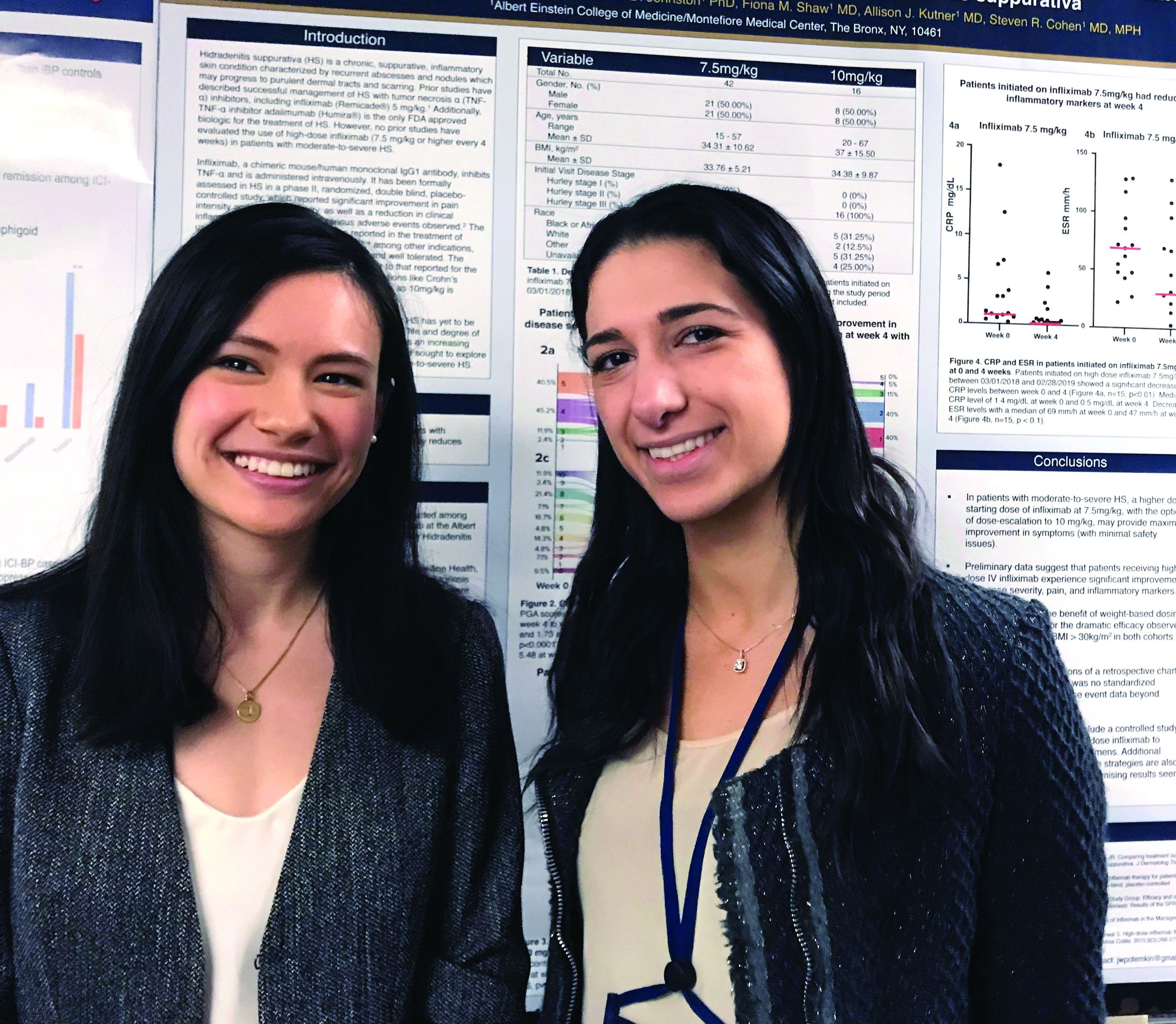
CHICAGO – In patients with hidradenitis suppurativa (HS), infliximab started at 7.5 mg/kg was highly effective and well tolerated and may be a more appropriate starting dose than the 5 mg/kg that is recommended in the treatment of plaque psoriasis, according to a chart review presented as a late breaker at the annual meeting of the Society for Investigative Dermatology.
“Based on these data, we believe that a high starting dose of infliximab should be the new paradigm for this disease,” reported Justine Potemkin, a medical student at Albert Einstein College of Medicine, New York. The analysis was performed under the mentorship of Steven R. Cohen, MD, professor of dermatology and chief of the division of dermatology at Albert Einstein.
Infliximab, a tumor necrosis factor (TNF) blocker, is not approved for the treatment of HS, but it has been widely used following numerous reports of benefit in the literature, including a phase 2 randomized study published several years ago (J Am Acad Dermatol. 2010 Feb;62[2]:205-17), according to Ms. Potemkin.
“Based on dosing recommended for psoriasis, many hidradenitis suppurativa patients are started on 5 mg/kg, but doses of up to 10 mg/kg are within labeling for psoriasis, and our data support more aggressive initial treatment in patients with HS,” said Mondana Ghias, a research assistant in the department of dermatology at Albert Einstein, who was also involved in this study and joined Ms. Potemkin in presenting the data at the meeting.
In the chart review, 58 patients with moderate to severe HS initiated on 7.5 mg/kg of intravenous infliximab were identified. Due to a suboptimal response, 16 of these patients received 10 mg/kg infliximab in subsequent infusions, but the average response scores at 4 weeks compared favorably to previous reports with lower doses of infliximab, without increased toxicity.
“Starting with the 7.5 mg/kg dose with dose escalation if needed appears to provide a maximal response with minimal safety issues, based on our experience,” Ms. Potemkin reported.
At 4 weeks, a 10-point pain scale fell from a baseline mean of 5.65 to 1.34 (P less than .01). It reached 0.52 at week 12. The HS Physician Global Assessment (HS-PGA) fell from a baseline mean of 4.2 to 2.1 at week 4 (P less than .01) and then to 1.9 at week 12. The control of disease activity correlated with significant reductions in C-reactive protein and erythrocyte sedimentation rate.
The mean age in this patient population was 34.6 years. Slightly more than half (53.3%) were female. The average body mass index was 34.
“Obese patients also achieved a good response, which is consistent with weight-based dosing,” Ms. Potemkin reported.
The TNF blocker adalimumab is the only biologic and the only systemic therapy given regulatory approval for the treatment of HS, but Ms. Potemkin indicated that infliximab remains a widely used treatment option. She suggested that the responses observed in this patient series suggest infliximab is effective when used aggressively.
“No previous reports have examined the efficacy of high-dose infliximab,” Ms. Potemkin noted. “We think these findings are relevant to HS management.”
REPORTING FROM SID 2019


