User login
Coffee drinking may cut heart disease risk, prolong survival
A trio of analyses based on the prospective UK Biobank cohort suggest that regular coffee drinking, especially a daily intake of two to three cups, is not only safe for the heart but may be cardioprotective.
People without cardiovascular disease with that level of coffee intake, compared with those who weren’t coffee drinkers, showed significantly reduced risks of death and a range of CVD endpoints, the reductions ranging from 8% to 15% over about 10 years.
In a separate analysis, participants with CVD at baseline also showed significantly improved survival with coffee intake of two to three cups daily, and no increased risk of arrhythmias.
In a third cut of the UK Biobank data, the clinical benefits of the same level of coffee drinking were observed whether the coffee consumed was the “instant” kind for reconstitution with water or brewed from ground whole beans.
Some clinicians advise their patients that coffee drinking may trigger or worsen some types of heart disease, observed Peter M. Kistler, MD, the Alfred Hospital and Baker Heart and Diabetes Institute, Melbourne. But the current analyses suggest that “daily coffee intake should not be discouraged, but rather considered part of a healthy diet.”
Dr. Kistler and colleagues are slated to present the three UK Biobank cohort analyses separately at the annual scientific sessions of the American College of Cardiology. He presented some of the data and commented on them at a press conference held in advance of the meeting.
UK Biobank study participants, who were on average in their late 50s, reported their level of daily coffee intake and preferred type of coffee on questionnaires. The researchers observed generally U-shaped relationships between daily number of cups of coffee and incident CVD, heart failure, coronary heart disease (CHD), stroke, atrial fibrillation, any arrhythmia, and death over 10 years.
“This is music to I think many of our patients’ ears, as well as many in the field of cardiology, as those of us that wake up early and stay up late in the hospital consume a fair amount of coffee,” observed Katie Berlacher, MD, associate chief of cardiology education at the University of Pittsburgh Medical Center.
The analyses were based on a large cohort and saw a consistent pattern for several cardiovascular outcomes, observed Dr. Berlacher, incoming ACC scientific session vice chair.
The findings could have a “profound impact in daily clinical care, as many of us caution patients who have or are at risk for having CV[D] against coffee consumption,” she told this news organization by email.
“These studies suggest that we do not have objective evidence to caution nor ask patients to stop drinking coffee, including patients who have arrhythmias.”
But importantly, “these studies are not causal,” she added. “So we cannot go so far as to recommend coffee consumption, though one could posit that randomized prospective studies should be done to elucidate causation.”
Coffee, Dr. Kistler observed, “is the most common cognitive enhancer. It wakes you up, makes you mentally sharper, and it’s a very important component of many people’s daily lives. The take-home message is that clinicians should NOT advise patients to stop drinking coffee up to three cups per day.”
Also, “in non–coffee drinkers, we do not have the data to suggest they should start drinking coffee,” he said. Moreover, people shouldn’t necessarily increase their coffee intake, particularly if it makes them feel anxious or uncomfortable.
Benefits with or without known heart disease
The researchers identified 382,535 participants in the UK Biobank cohort who were free of CVD at baseline. Their median age was 57, and 52% were women.
Those who reported regular daily intake of two to three cups of coffee, compared with those who were not coffee drinkers, showed significantly reduced risks of CVD (hazard ratio, 0.91; 95% confidence interval, 0.88-0.94), CHD (HR, 0.90; 95% CI, 0.87-0.93), heart failure (HR, 0.85; 95% CI, 0.81-0.90), arrhythmias (HR, 0.92; 95% CI, 0.88-0.95), and death from any cause over 10 years (HR, 0.86; 95% CI, 0.83-0.90) (P < .01 for all endpoints).
The risk of CVD death hit its lowest point at an intake of one cup per day (HR, 0.83; 95% CI, 0.75-0.93). The risk of stroke was lowest at less than one cup per day (HR, 0.85; 95% CI, 0.75-0.96).
A separate analysis found similar outcomes among a different subset of UK Biobank participants with recognized CVD at baseline. Among 34,279 such persons, those who drank two to three cups of coffee per day, compared with non–coffee drinkers, showed a reduced risk of death over 10 years (HR, 0.92; 95% CI, 0.86-0.99; P = .03).
Among the 24,111 persons diagnosed with arrhythmias at baseline, the lowest mortality risk was observed at one cup per day (HR, 0.85; 95% CI, 0.78-0.94; P < .01). Among those with atrial fibrillation or atrial flutter, one cup per day was associated with a mortality HR of 0.82 (95% CI, 0.73-0.93; P < .01).
In still another analysis of UK Biobank cohort, incident CVD and mortality during the 10-year follow-up was similarly reduced among participants who reported consumption of brewed ground coffee and, separately, instant coffee, compared with non–coffee drinkers. Decaffeinated coffee showed a mostly neutral or inconsistent effect on the clinical endpoints.
The lowest CVD risk was observed at two to three cups per day among those regularly drinking ground coffee (HR, 0.83; 95% CI, 0.79-0.87) and those predominantly taking instant coffee (HR, 0.91; 95% CI, 0.88-0.95).
Potential mechanisms, study limitations
“Caffeine blocks adenosine receptors, which may explain its potential mild antiarrhythmic properties,” Dr. Kistler said. “Regular coffee drinkers with supraventricular tachycardia coming to the emergency department often need higher adenosine doses to revert.”

Caffeine has a role in weight loss through inhibition of gut fatty acid absorption and increase in basal metabolic rate, Dr. Kistler added, and coffee has been associated with a significantly reduced risk of new-onset type 2 diabetes.
However, coffee beans contain more than 100 biologically active compounds, he noted. They include antioxidant polyphenols that reduce oxidative stress and modulate metabolism. Better survival with habitual coffee consumption may be related to improved endothelial function, circulating antioxidants, improved insulin sensitivity, or reduced inflammation, the researchers noted.
They acknowledged some limitations to the analyses. Cause and effect can’t be determined from the observational data. Also, a cup of coffee in the United Kingdom means about 200-250 mL of brew, but its actual caffeine content can vary from 90 mg to 250 mg. Also, data regarding added sugar or milk was lacking. And UK Biobank participants are predominantly White, so the findings may not be generalizable to other populations.
A version of this article first appeared on Medscape.com.
A trio of analyses based on the prospective UK Biobank cohort suggest that regular coffee drinking, especially a daily intake of two to three cups, is not only safe for the heart but may be cardioprotective.
People without cardiovascular disease with that level of coffee intake, compared with those who weren’t coffee drinkers, showed significantly reduced risks of death and a range of CVD endpoints, the reductions ranging from 8% to 15% over about 10 years.
In a separate analysis, participants with CVD at baseline also showed significantly improved survival with coffee intake of two to three cups daily, and no increased risk of arrhythmias.
In a third cut of the UK Biobank data, the clinical benefits of the same level of coffee drinking were observed whether the coffee consumed was the “instant” kind for reconstitution with water or brewed from ground whole beans.
Some clinicians advise their patients that coffee drinking may trigger or worsen some types of heart disease, observed Peter M. Kistler, MD, the Alfred Hospital and Baker Heart and Diabetes Institute, Melbourne. But the current analyses suggest that “daily coffee intake should not be discouraged, but rather considered part of a healthy diet.”
Dr. Kistler and colleagues are slated to present the three UK Biobank cohort analyses separately at the annual scientific sessions of the American College of Cardiology. He presented some of the data and commented on them at a press conference held in advance of the meeting.
UK Biobank study participants, who were on average in their late 50s, reported their level of daily coffee intake and preferred type of coffee on questionnaires. The researchers observed generally U-shaped relationships between daily number of cups of coffee and incident CVD, heart failure, coronary heart disease (CHD), stroke, atrial fibrillation, any arrhythmia, and death over 10 years.
“This is music to I think many of our patients’ ears, as well as many in the field of cardiology, as those of us that wake up early and stay up late in the hospital consume a fair amount of coffee,” observed Katie Berlacher, MD, associate chief of cardiology education at the University of Pittsburgh Medical Center.
The analyses were based on a large cohort and saw a consistent pattern for several cardiovascular outcomes, observed Dr. Berlacher, incoming ACC scientific session vice chair.
The findings could have a “profound impact in daily clinical care, as many of us caution patients who have or are at risk for having CV[D] against coffee consumption,” she told this news organization by email.
“These studies suggest that we do not have objective evidence to caution nor ask patients to stop drinking coffee, including patients who have arrhythmias.”
But importantly, “these studies are not causal,” she added. “So we cannot go so far as to recommend coffee consumption, though one could posit that randomized prospective studies should be done to elucidate causation.”
Coffee, Dr. Kistler observed, “is the most common cognitive enhancer. It wakes you up, makes you mentally sharper, and it’s a very important component of many people’s daily lives. The take-home message is that clinicians should NOT advise patients to stop drinking coffee up to three cups per day.”
Also, “in non–coffee drinkers, we do not have the data to suggest they should start drinking coffee,” he said. Moreover, people shouldn’t necessarily increase their coffee intake, particularly if it makes them feel anxious or uncomfortable.
Benefits with or without known heart disease
The researchers identified 382,535 participants in the UK Biobank cohort who were free of CVD at baseline. Their median age was 57, and 52% were women.
Those who reported regular daily intake of two to three cups of coffee, compared with those who were not coffee drinkers, showed significantly reduced risks of CVD (hazard ratio, 0.91; 95% confidence interval, 0.88-0.94), CHD (HR, 0.90; 95% CI, 0.87-0.93), heart failure (HR, 0.85; 95% CI, 0.81-0.90), arrhythmias (HR, 0.92; 95% CI, 0.88-0.95), and death from any cause over 10 years (HR, 0.86; 95% CI, 0.83-0.90) (P < .01 for all endpoints).
The risk of CVD death hit its lowest point at an intake of one cup per day (HR, 0.83; 95% CI, 0.75-0.93). The risk of stroke was lowest at less than one cup per day (HR, 0.85; 95% CI, 0.75-0.96).
A separate analysis found similar outcomes among a different subset of UK Biobank participants with recognized CVD at baseline. Among 34,279 such persons, those who drank two to three cups of coffee per day, compared with non–coffee drinkers, showed a reduced risk of death over 10 years (HR, 0.92; 95% CI, 0.86-0.99; P = .03).
Among the 24,111 persons diagnosed with arrhythmias at baseline, the lowest mortality risk was observed at one cup per day (HR, 0.85; 95% CI, 0.78-0.94; P < .01). Among those with atrial fibrillation or atrial flutter, one cup per day was associated with a mortality HR of 0.82 (95% CI, 0.73-0.93; P < .01).
In still another analysis of UK Biobank cohort, incident CVD and mortality during the 10-year follow-up was similarly reduced among participants who reported consumption of brewed ground coffee and, separately, instant coffee, compared with non–coffee drinkers. Decaffeinated coffee showed a mostly neutral or inconsistent effect on the clinical endpoints.
The lowest CVD risk was observed at two to three cups per day among those regularly drinking ground coffee (HR, 0.83; 95% CI, 0.79-0.87) and those predominantly taking instant coffee (HR, 0.91; 95% CI, 0.88-0.95).
Potential mechanisms, study limitations
“Caffeine blocks adenosine receptors, which may explain its potential mild antiarrhythmic properties,” Dr. Kistler said. “Regular coffee drinkers with supraventricular tachycardia coming to the emergency department often need higher adenosine doses to revert.”
Caffeine has a role in weight loss through inhibition of gut fatty acid absorption and increase in basal metabolic rate, Dr. Kistler added, and coffee has been associated with a significantly reduced risk of new-onset type 2 diabetes.
However, coffee beans contain more than 100 biologically active compounds, he noted. They include antioxidant polyphenols that reduce oxidative stress and modulate metabolism. Better survival with habitual coffee consumption may be related to improved endothelial function, circulating antioxidants, improved insulin sensitivity, or reduced inflammation, the researchers noted.
They acknowledged some limitations to the analyses. Cause and effect can’t be determined from the observational data. Also, a cup of coffee in the United Kingdom means about 200-250 mL of brew, but its actual caffeine content can vary from 90 mg to 250 mg. Also, data regarding added sugar or milk was lacking. And UK Biobank participants are predominantly White, so the findings may not be generalizable to other populations.
A version of this article first appeared on Medscape.com.
FROM ACC 2022
Surgery groups push back on VARC-3 valve trial definitions
Five international cardiac surgery associations have banded together to address “substantive concerns” regarding the recently updated Valve Academic Research Consortium 3 (VARC-3) clinical endpoint definitions for aortic valve research.
The VARC-3 update was a multidisciplinary effort that included more than a dozen new or modified definitions for use in transcatheter and surgical aortic valve replacement (TAVR/SAVR) clinical trials, but drew criticism last year from surgeons that some of its definitions favor TAVR over surgery and that its writing committee had deep ties to industry and lacked diversity.
The new surgical associations’ position statement calls out five specific VARC-3 definitions – rehospitalization, valve thrombosis, bleeding, myocardial infarction (MI), and left bundle-branch block (LBBB).
The statement was jointly issued by the Society of Thoracic Surgeons (STS), the American Association for Thoracic Surgery, the European Association for Cardio-Thoracic Surgery, the Asian Society for Cardiovascular and Thoracic Surgery, and the Latin American Association of Cardiac and Endovascular Surgery.
It was copublished in Annals of Thoracic Surgery, the Journal of Thoracic and Cardiovascular Surgery, the European Journal of Cardio-Thoracic Surgery, and the Asian Cardiovascular and Thoracic Annals.
“We hope that this message can be seen, even if it’s somewhat difficult to hear sometimes, as positive constructive criticism compared to some of the dialogue that we’ve had on social media,” lead author Patrick O. Myers, MD, Lausanne (Switzerland) University Hospital, said in an interview. “It’s not criticizing people or the process but just trying to make these definitions better to ensure the good design of clinical trials.”
The president of each surgical association recommended representatives to help write the position statement, and once completed over Zoom meetings, it received formal endorsement from each association prior to publication, he said.
Reached for comment, VARC-3 lead author Philippe Généreux, MD, Gagnon Cardiovascular Institute, Morristown (N.J.) Medical Center, said, “I was pleasantly surprised that their comments were actually pretty minor and that most of these comments are really more a reflection, not of the validity of the definitions, but rather their applications.”
He noted that all the potential issues with the definitions were already discussed during the making of VARC-3 and resolved by consensus of more than 50 experts including the STS president at the time, Food and Drug Administration officials, and experts from the community.
“To be quite honest, I’m not sure they have consensus,” Dr. Généreux said. He added that the writing committee welcomes input from anyone, but “we’re not going to change the definitions to please eight individuals if we strongly believe by consensus of experts in the field that this is not the right thing to do.”
Rehospitalizations and valve thrombosis
The surgical associations praise VARC-3 for providing a standardized definition of bioprosthetic valve failure, but say they will not endorse the inclusion of rehospitalization as a component of the primary efficacy composite endpoint along with all-cause mortality, stroke, and quality of life.
They note that rehospitalizations outnumber mortality events, especially in short follow-up trials, and that the superiority of TAVR at 1 year in the PARTNER 3 trial of low-risk patients was driven primarily by more rehospitalizations in the surgical arm, but that this superiority was waning at 2 years of follow-up.
“The first thing we are calling for is that it shouldn’t be part of the primary composite outcome measure,” Dr. Myers said. But if it really has to be included, a 30-day blanking period for rehospitalization “would acknowledge that there’s a greater risk of rehospitalization during the acute phase of recovering from surgery.”
Dr. Généreux said that VARC-3 provides granular details for defining the different types of hospitalizations, but that a 30-day blanking period makes no sense. “If you close your eyes to anything within 30 days because you don’t like it, you’re missing the opportunity to improve your procedure, to improve your treatment, and to characterize precisely what happened with your patient.”
The new document lauds VARC-3’s focus on patient-centered and clinically relevant endpoints but questions the definition of valve thrombosis as a “clinically significant” thrombus. It points out that the incidence of valve thrombosis was significantly higher with TAVR versus SAVR in PARTNER 3 using the older VARC-2 definition, which did not require evidence of clinical sequelae (2.6% vs. 0.7%; P = .02). Under the new definition, however, half of the thrombi would be relabeled as “nothing there,” Dr. Myers said.
“As we’re doing this in younger and younger patients who will survive longer, there is a question of thrombus having an effect on the valve and leading to earlier structural valve deterioration,” he added. “All this is conjecture. We don’t have the data. So mainly what we’re advocating is that all thrombi should be reported.”
MIs, bleeding, and LBBB
The policy statement also criticizes VARC-3’s decision to define periprocedural (type 5) MI using a biomarker-only definition without need of clinical confirmation. Such definitions have been shown to have a very poor prognostic significance in surgical series compared with the Universal Definitions of Myocardial Infarction, Dr. Myers said.
“What’s interesting is that for thrombus and bleeding, they require clinical correlation, but on the perioperative MI they now use a definition that does not require clinical significance, meaning no ECG changes, no regional wall motion abnormalities or things like that,” he observed.
The decision also seems to disregard the EXCEL trial controversy that illustrated how outcomes and a trial’s message can change depending on which definition of periprocedural MI is used.
With regard to bleeding, the surgical associations agree with the VARC-3 recommendation to use different thresholds when bleeding is integrated into a composite endpoint (type 2 or greater for TAVR and types 3 or greater for SAVR) but suggest this important point should be featured in the chapter on bleeding rather than the section on composite endpoints.
The surgical associations say VARC-3 also got it right adding the need for a new permanent pacemaker to the early composite safety endpoint, but that it was a “missed opportunity” not to include new left bundle-branch block in the safety composite, despite recognizing that this may become an important endpoint to consider in the future.
Dr. Myers said that left bundle-branch block could have implications for survival as TAVR moves into lower-risk, younger patients, as some data with 1-year follow-up suggest it has a prognostic impact, even in the higher-risk older patients with more competing risks.
Finally, the surgical associations point out that only two of the 23 VARC-3 authors were practicing cardiac surgeons and say that a more diverse writing group “may help mitigate issues related to the duality of interests.”
Dr. Généreux said that the final author list is not a reflection of the rigorous work done by 11 cardiac surgeons including the two surgeon authors. The VARC-3 writing committee also had a good representation of women, unlike the surgical position statement, which was penned by eight men.
Dr. Myers reported no relevant financial relationships. Coauthors disclosed ties with EACTS, Edwards Lifesciences, Medtronic, Abbott Vascular, Boston Scientific, CryoLife, Shockwave, and JenaValve. Dr. Généreux disclosed ties with Abbott Vascular, Abiomed, Boston Scientific, Cardinal Health, Cardiovascular Systems, Edwards Lifesciences, Medtronic, Opsens, Siemens, SoundBite Medical Solutions, Sig.Num, Saranas, Teleflex, Tryton Medical, Pi-Cardia, and Puzzle Medical.
A version of this article first appeared on Medscape.com.
Congress opens investigation into FDA’s handling of a problematic heart device
A congressional oversight subcommittee is investigating the Food and Drug Administration’s regulation of a high-risk heart pump, citing safety issues detailed by ProPublica.
The HeartWare Ventricular Assist Device, created to treat patients with severe heart failure, stopped meeting key federal standards as early as 2014. But the FDA took no decisive action even as those problems persisted, and thousands of Americans continued to be implanted with the pump.
By the end of 2020, the FDA had received more than 3,000 reports of deaths related to the HeartWare device, according to a ProPublica data analysis. A father of four died as his children tried to resuscitate him when his device suddenly stopped. A teenager died after vomiting blood in the middle of the night, while his mother struggled to restart a faulty pump.
“I am concerned by FDA’s slow action, over multiple administrations, to protect patients from this product despite early warning signs,” Rep. Raja Krishnamoorthi, D-Ill., said in a scathing letter sent March 22 to the agency’s commissioner, Robert Califf, MD.
Mr. Krishnamoorthi, the chairman of the U.S. House Committee on Oversight and Reform’s Subcommittee on Economic and Consumer Policy, requested information on how the FDA made regulatory decisions related to the HeartWare device and why it didn’t take further action.
The FDA did not provide comment to ProPublica on the subcommittee’s investigation and said it would respond directly to Mr. Krishnamoorthi. It also reiterated its response to ProPublica’s findings and said the agency had been closely overseeing the HeartWare device since 2012, with patient safety as its “highest priority.”
Medtronic, the company that acquired HeartWare in 2016, took the device off the market in June 2021. The company said that new data showed a competing heart pump had better outcomes. In response to the ProPublica investigation 2 months later, the company said it took the FDA’s inspections seriously and had worked closely with the agency to address issues with the device.
Medtronic declined to comment on the subcommittee’s investigation.
Mr. Krishnamoorthi asked in the letter if any steps were being taken to address how patients, doctors and other federal agencies are notified of problems that the FDA finds with medical devices.
Many patients told ProPublica they were never informed of issues with the HeartWare pump before or after their implants. Some people who still have the device said they weren’t told when it was taken off the market. Medtronic said in December it had confirmed 90% of U.S. patients had received notification of the HeartWare discontinuation, but that it was still working to reach the other 10%.
About 2,000 patients still had HeartWare pumps as of last year. The FDA and Medtronic recommended against removing those devices barring medical necessity because the surgery to do so carries a high risk.
In his letter, Mr. Krishnamoorthi gave the FDA a deadline of April 5 to respond.
This story was originally published on ProPublica. ProPublica is a nonprofit newsroom that investigates abuses of power. Sign up to receive their biggest stories as soon as they’re published.
Sit-to-stand BP spike tied to CV events in young adults
A sudden drop in blood pressure when standing is a common and concerning problem in elderly hypertensive people. Now, research suggests a large BP swing in the opposite direction on standing may be equally concerning in younger hypertensive people.
Young and middle-aged adults with a systolic BP response to standing greater than 6.5 mm Hg had almost double the risk of major adverse cardiovascular events (MACE) during follow-up, compared with other participants.
An exaggerated BP response remained an independent predictor of MACE, even after adjusting for traditional risk factors, including 24-hour BP (hazard ratio, 1.94; 95% confidence interval, 1.10 to 3.44), the study showed.
“The clinical implication is important, because now doctors measure blood pressure in young people in the upright posture, but what we say is it must be measured also while standing,” said Paolo Palatini, MD, a professor of internal medicine at the University of Padova, Italy, who led the study.
Previous studies have found that an exaggerated BP response to standing is a predictor of future hypertension, CV events, and mortality, particularly in older patients, but few prognostic data exist in those who are young to middle age, he noted.
The study, published in Hypertension, included 1,207 participants ages 18-45 years with untreated stage 1 hypertension (systolic BP 140-159 mm Hg or diastolic BP 90-100 mm Hg) in the prospective multicenter HARVEST study that began in Italy in 1990. The average age at enrollment was 33 years.
BP was measured at two visits 2 weeks apart, with each visit including three supine measurements taken after the patient had lain down for a minimum of 5 minutes, followed by three standing measurements taken 1 minute apart.
Based on the average of standing-lying BP differences during the two visits, participants were then classified as having a normal or exaggerated (top decile, lower limit > 6.5 mm Hg) systolic BP response to standing.
The 120 participants classified as “hyper-reactors” averaged an 11.4 mm Hg systolic BP increase upon standing, whereas the rest of the participants averaged a 3.8 mm Hg fall in systolic BP upon standing.
At their initial visit, hyper-reactors were more likely to be smokers (32.1% vs. 19.9%) and coffee drinkers (81.7% vs. 73%) and to have ambulatory hypertension (90.8% vs. 76.4%).
They were, however, no more likely to have a family history of cardiovascular events and had a lower supine systolic BP (140.5 mm Hg vs. 146.0 mm Hg), lower total cholesterol (4.93 mmol/L vs. 5.13 mmol/L), and higher HDL cholesterol (1.42 mmol/L vs. 1.35 mmol/L).
Age, sex, and body mass index were similar between the two groups, as was BP variability, nocturnal BP dip, and the frequency of extreme dippers. Participants with a normal systolic BP response were more likely to be treated for hypertension during follow-up (81.7% vs. 69.7%; P = .003).
In 630 participants who had catecholamines measured from 24-hour urine samples, the epinephrine/creatinine ratio was higher in hyper-reactors than normal responders (118.4 nmol/mol vs. 77.0 nmol/mol; P = .005).
During a median follow-up of 17.3 years, there were 105 major cardiovascular events, broadly defined to include acute coronary syndromes (48), any stroke (13), heart failure requiring hospitalization (3), aortic aneurysms (3), peripheral vascular disease (6), chronic kidney disease (12), and permanent atrial fibrillation (20).
The near doubling of MACE risk among hyper-reactors remained when atrial fibrillation was excluded and when 24-hour ambulatory systolic BP was included in the model, the author reported.
The results are in line with previous studies, indicating that hyper-reactors to standing have normal sympathetic activity at rest but an increased sympathetic response to stressors, observed Dr. Palatini and colleagues. This neurohumoral overshoot seems to be peculiar to young adults, whereas vascular stiffness seems to be the driving mechanism of orthostatic hypertension in older adults.
If a young person’s BP spikes upon standing, “then you have to treat them according to the average of the lying and the standing pressure,” Dr. Palatini said. “In these people, blood pressure should be treated earlier than in the past.”
“The study is important because it identified a new marker for hypertension that is easily evaluated in clinical practice,” Nieca Goldberg, MD, medical director of the Atria Institute, New York, and an associate professor of medicine at New York University Grossman School of Medicine, commented via email.
She noted that standing blood pressures are usually not taken as part of a medical visit and, in fact, seated blood pressures are often taken incorrectly while the patient is seated on the exam table rather than with their feet on the floor and using the proper cuff size.
“By incorporating standing BP, we will improve our diagnosis for hypertension, and with interventions such as diet and exercise, salt reduction, and medication when indicated, lower risk for heart attack, stroke, heart failure, [and] kidney and eye disease,” said Dr. Goldberg, who is also a spokesperson for the American Heart Association.
“The biggest barrier is that office visits are limited to 15 minutes, and not enough time is spent on the vital signs,” she noted. “We need changes to the health care system that value our ability to diagnose BP and take the time to counsel patients and explain treatment options.”
Limitations of the present study are that 72.7% of participants were men and all were White, Dr. Palatini said. Future work is also needed to create a uniform definition of BP hyper-reactivity to standing, possibly based on risk estimates, for inclusion in future hypertension guidelines.
The study was funded by the Association 18 Maggio 1370 in Italy. The authors have disclosed no relevant financial relationships. Dr. Goldberg reported being a spokesperson for the American Heart Association.
A version of this article first appeared on Medscape.com.
Pfizer recalls BP drugs because of potential carcinogen
Pfizer is voluntarily recalling some antihypertensive medications because of unacceptable levels of a potential carcinogen, the company announced.
The affected products are quinapril HCI/hydrochlorothiazide (Accuretic) tablets that Pfizer distributes, and two authorized generics, quinapril plus hydrochlorothiazide and quinapril HCI/hydrochlorothiazide, distributed by Greenstone. The drugs have been withdrawn because of the presence of nitrosamine, N-nitroso-quinapril.
“Although long-term ingestion of N-nitroso-quinapril may be associated with a potential increased cancer risk in humans, there is no immediate risk to patients taking this medication,” Pfizer said in a news release.
The tablets are indicated for the treatment of hypertension. Patients currently taking the products are asked to consult with their doctor about alternative treatment options.
To date, there have been no reports of adverse events related to the recall, the company said.
In all, Pfizer is recalling six lots of Accuretic tablets (two at 10 mg/12.5 mg, three at 20 mg/12.5 mg, and one at 20 mg/25 mg), one lot of quinapril plus hydrochlorothiazide 20-mg/25-mg tablets, and four lots of quinapril HCl/ hydrochlorothiazide tablets (three at 20 mg/12.5 mg and one at 20 mg/25 mg)
The recalled tablets were sold in 90-count bottles distributed in the United States and Puerto Rico between November 2019 and March 2022. Product codes and lot numbers of the recalled medications are listed on the Pfizer website.
Patients who are taking this product should consult with their health care provider or pharmacy to determine if they have the affected product. Those with the affected tablets should contact claims management firm Sedgwick by phone at 888-843-0247 Monday through Friday from 8 a.m. to 5 p.m. ET for instructions on how to return their product and obtain reimbursement.
Health care providers with medical questions regarding the recall can contact Pfizer by telephone at 800-438-1985, option 3, Monday through Friday 8 a.m. to 9 p.m. ET.
Providers should report adverse reactions or quality problems they experience using these tablets to Pfizer either by telephone at 800-438-1985, option 1, by regular mail or by fax, or to the Food and Drug Administration’s MedWatch program.
A version of this article first appeared on Medscape.com.
Empagliflozin scores topline win in EMPA-KIDNEY trial
Researchers running the EMPA-KIDNEY trial that’s been testing the safety and efficacy of the SGLT2 inhibitor empagliflozin (Jardiance) in about 6,600 patients with chronic kidney disease (CKD) announced on March 16 that they had stopped the trial early because of positive efficacy that met the study’s prespecified threshold for early termination.
EMPA-KIDNEY is the third major trial of an agent from the sodium-glucose cotransport 2 (SGLT2) inhibitor class tested in patients with CKD to be stopped early because of positive results that met a prespecified termination rule.
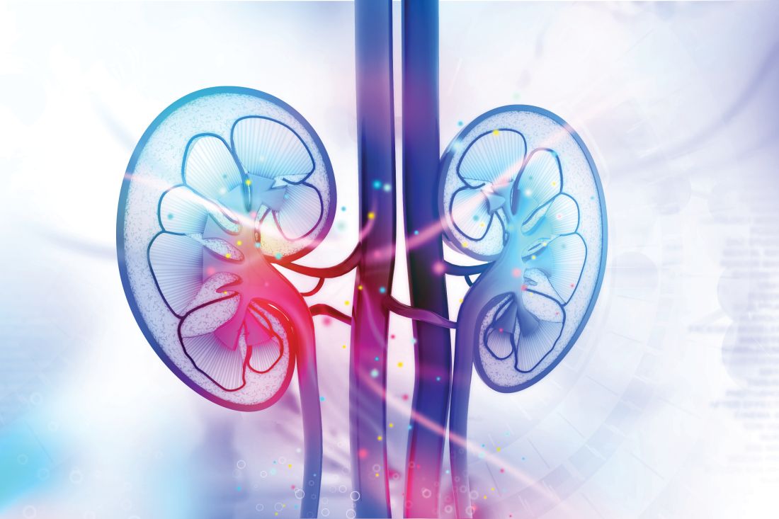
In 2020, the DAPA-CKD trial of dapagliflozin (Farxiga) stopped early, after a median follow-up of 2.4 years, because of positive efficacy results. In 2019, the same thing happened in the CREDENCE trial of canagliflozin (Invokana), with the unexpected halt coming after a median follow-up of 2.62 years.
The announcement about EMPA-KIDNEY did not include information on median follow-up, but enrollment into the trial ran from May 2019 to April 2021, which means that the longest that enrolled patients could have been in the study was about 2.85 years.
The primary efficacy endpoint in EMPA-KIDNEY was a composite of a sustained decline in estimated glomerular filtration rate (eGFR) to less than 10 mL/min/1.73 m2, renal death, a sustained decline of at least 40% in eGFR from baseline, or cardiovascular death. The announcement of the trial’s early termination provided no details on the efficacy results.
EMPA-KIDNEY enrolled a wider range of patients
EMPA-KIDNEY expands the scope of types of patients with CKD now shown to benefit from treatment with an SGLT2 inhibitor. CREDENCE tested canagliflozin only in patients with type 2 diabetes and diabetic nephropathy, and in DAPA-CKD, two-thirds of enrolled patients had type 2 diabetes, and all had CKD. In EMPA-KIDNEY, 46% of the 6,609 enrolled patients had diabetes (including a very small number with type 1 diabetes).
Another departure from prior studies of an SGLT2 inhibitor for patients selected primarily for having CKD was that in EMPA-KIDNEY, 20% of patients did not have albuminuria, and for 34%, eGFR at entry was less than 30 mL/min/1.73 m2, with all enrolled patients required to have an eGFR at entry of greater than or equal to 20 mL/min/1.73 m2. Average eGFR in EMPA-KIDNEY was about 38 mL/min/1.73 m2. To be included in the trial, patients were not required to have albuminuria, except those whose eGFR was greater than or equal to 45 mL/min/1.73 m2.
In DAPA-CKD, the minimum eGFR at entry had to be greater than or equal to 25 mL/min/1.73 m2, and roughly 14% of enrolled patients had an eGFR of less than 30 mL/min/1.73 m2. The average eGFR in DAPA-CKD was about 43 mL/min/1.73 m2. In addition, all patients had at least microalbuminuria, with a minimum urinary albumin-to-creatinine ratio of 200. In CREDENCE, the minimum eGFR for enrollment was 30 mL/min/1.73 m2, and the average eGFR was about 56 mL/min/1.73 m2. All patients in CREDENCE had to have macroalbuminuria, with a urinary albumin-to-creatinine ratio of more than 300.
According to the researchers who designed EMPA-KIDNEY, the trial enrollment criteria aimed to include adults with CKD “who are frequently seen in practice but were under-represented in previous SGLT2 inhibitor trials.”
Indications for empagliflozin are expanding
The success of empagliflozin in EMPA-KIDNEY follows its positive results in both the EMPEROR-Reduced and EMPEROR-Preserved trials, which collectively proved the efficacy of the agent for patients with heart failure regardless of their left ventricular ejection fraction and regardless of whether they also had diabetes.
These results led the U.S. Food and Drug Administration to recently expand the labeled indication for empagliflozin to all patients with heart failure. Empagliflozin also has labeled indications for glycemic control in patients with type 2 diabetes and to reduce the risk of cardiovascular death in adults with type 2 diabetes and established cardiovascular disease.
As of today, empagliflozin has no labeled indication for treating patients with CKD. Dapagliflozin received that indication in April 2021, and canagliflozin received an indication for treating patients with type 2 diabetes, diabetic nephropathy, and albuminuria in September 2019.
EMPA-KIDNEY is sponsored by Boehringer Ingelheim and Lilly, the two companies that jointly market empagliflozin (Jardiance).
A version of this article first appeared on Medscape.com.
Researchers running the EMPA-KIDNEY trial that’s been testing the safety and efficacy of the SGLT2 inhibitor empagliflozin (Jardiance) in about 6,600 patients with chronic kidney disease (CKD) announced on March 16 that they had stopped the trial early because of positive efficacy that met the study’s prespecified threshold for early termination.
EMPA-KIDNEY is the third major trial of an agent from the sodium-glucose cotransport 2 (SGLT2) inhibitor class tested in patients with CKD to be stopped early because of positive results that met a prespecified termination rule.
In 2020, the DAPA-CKD trial of dapagliflozin (Farxiga) stopped early, after a median follow-up of 2.4 years, because of positive efficacy results. In 2019, the same thing happened in the CREDENCE trial of canagliflozin (Invokana), with the unexpected halt coming after a median follow-up of 2.62 years.
The announcement about EMPA-KIDNEY did not include information on median follow-up, but enrollment into the trial ran from May 2019 to April 2021, which means that the longest that enrolled patients could have been in the study was about 2.85 years.
The primary efficacy endpoint in EMPA-KIDNEY was a composite of a sustained decline in estimated glomerular filtration rate (eGFR) to less than 10 mL/min/1.73 m2, renal death, a sustained decline of at least 40% in eGFR from baseline, or cardiovascular death. The announcement of the trial’s early termination provided no details on the efficacy results.
EMPA-KIDNEY enrolled a wider range of patients
EMPA-KIDNEY expands the scope of types of patients with CKD now shown to benefit from treatment with an SGLT2 inhibitor. CREDENCE tested canagliflozin only in patients with type 2 diabetes and diabetic nephropathy, and in DAPA-CKD, two-thirds of enrolled patients had type 2 diabetes, and all had CKD. In EMPA-KIDNEY, 46% of the 6,609 enrolled patients had diabetes (including a very small number with type 1 diabetes).
Another departure from prior studies of an SGLT2 inhibitor for patients selected primarily for having CKD was that in EMPA-KIDNEY, 20% of patients did not have albuminuria, and for 34%, eGFR at entry was less than 30 mL/min/1.73 m2, with all enrolled patients required to have an eGFR at entry of greater than or equal to 20 mL/min/1.73 m2. Average eGFR in EMPA-KIDNEY was about 38 mL/min/1.73 m2. To be included in the trial, patients were not required to have albuminuria, except those whose eGFR was greater than or equal to 45 mL/min/1.73 m2.
In DAPA-CKD, the minimum eGFR at entry had to be greater than or equal to 25 mL/min/1.73 m2, and roughly 14% of enrolled patients had an eGFR of less than 30 mL/min/1.73 m2. The average eGFR in DAPA-CKD was about 43 mL/min/1.73 m2. In addition, all patients had at least microalbuminuria, with a minimum urinary albumin-to-creatinine ratio of 200. In CREDENCE, the minimum eGFR for enrollment was 30 mL/min/1.73 m2, and the average eGFR was about 56 mL/min/1.73 m2. All patients in CREDENCE had to have macroalbuminuria, with a urinary albumin-to-creatinine ratio of more than 300.
According to the researchers who designed EMPA-KIDNEY, the trial enrollment criteria aimed to include adults with CKD “who are frequently seen in practice but were under-represented in previous SGLT2 inhibitor trials.”
Indications for empagliflozin are expanding
The success of empagliflozin in EMPA-KIDNEY follows its positive results in both the EMPEROR-Reduced and EMPEROR-Preserved trials, which collectively proved the efficacy of the agent for patients with heart failure regardless of their left ventricular ejection fraction and regardless of whether they also had diabetes.
These results led the U.S. Food and Drug Administration to recently expand the labeled indication for empagliflozin to all patients with heart failure. Empagliflozin also has labeled indications for glycemic control in patients with type 2 diabetes and to reduce the risk of cardiovascular death in adults with type 2 diabetes and established cardiovascular disease.
As of today, empagliflozin has no labeled indication for treating patients with CKD. Dapagliflozin received that indication in April 2021, and canagliflozin received an indication for treating patients with type 2 diabetes, diabetic nephropathy, and albuminuria in September 2019.
EMPA-KIDNEY is sponsored by Boehringer Ingelheim and Lilly, the two companies that jointly market empagliflozin (Jardiance).
A version of this article first appeared on Medscape.com.
New ACC guidance on cardiovascular consequences of COVID-19
The American College of Cardiology has issued an expert consensus clinical guidance document for the evaluation and management of adults with key cardiovascular consequences of COVID-19.
The document makes recommendations on how to evaluate and manage COVID-associated myocarditis and long COVID and gives advice on resumption of exercise following COVID-19 infection.
The clinical guidance was published online March 16 in the Journal of the American College of Cardiology.
“The best means to diagnose and treat myocarditis and long COVID following SARS-CoV-2 infection continues to evolve,” said Ty Gluckman, MD, MHA, cochair of the expert consensus decision pathway. “This document attempts to provide key recommendations for how to evaluate and manage adults with these conditions, including guidance for safe return to play for both competitive and noncompetitive athletes.”
The authors of the guidance note that COVID-19 can be associated with various abnormalities in cardiac testing and a wide range of cardiovascular complications. For some patients, cardiac symptoms such as chest pain, shortness of breath, fatigue, and palpitations persist, lasting months after the initial illness, and evidence of myocardial injury has also been observed in both symptomatic and asymptomatic individuals, as well as after receipt of the COVID-19 mRNA vaccine.
“For clinicians treating these individuals, a growing number of questions exist related to evaluation and management of these conditions, as well as safe resumption of physical activity,” they say. This report is intended to provide practical guidance on these issues.
Myocarditis
The report states that myocarditis has been recognized as a rare but serious complication of SARS-CoV-2 infection as well as COVID-19 mRNA vaccination.
It defines myocarditis as: 1.cardiac symptoms such as chest pain, dyspnea, palpitations, or syncope; 2. elevated cardiac troponin; and 3. abnormal electrocardiographic, echocardiographic, cardiac MRI, and/or histopathologic findings on biopsy.
The document makes the following recommendations in regard to COVID-related myocarditis:
When there is increased suspicion for cardiac involvement with COVID-19, initial testing should consist of an ECG, measurement of cardiac troponin, and an echocardiogram. Cardiology consultation is recommended for those with a rising cardiac troponin and/or echocardiographic abnormalities. Cardiac MRI is recommended in hemodynamically stable patients with suspected myocarditis.
Hospitalization is recommended for patients with definite myocarditis, ideally at an advanced heart failure center. Patients with fulminant myocarditis should be managed at centers with an expertise in advanced heart failure, mechanical circulatory support, and other advanced therapies.
Patients with myocarditis and COVID-19 pneumonia (with an ongoing need for supplemental oxygen) should be treated with corticosteroids. For patients with suspected pericardial involvement, treatment with NSAIDs, colchicine, and/or prednisone is reasonable. Intravenous corticosteroids may be considered in those with suspected or confirmed COVID-19 myocarditis with hemodynamic compromise or MIS-A (multisystem inflammatory syndrome in adults). Empiric use of corticosteroids may also be considered in those with biopsy evidence of severe myocardial infiltrates or fulminant myocarditis, balanced against infection risk.
As appropriate, guideline-directed medical therapy for heart failure should be initiated and continued after discharge.
The document notes that myocarditis following COVID-19 mRNA vaccination is rare, with highest rates seen in young males after the second vaccine dose. As of May 22, 2021, the U.S. Vaccine Adverse Event Reporting System noted rates of 40.6 cases per million after the second vaccine dose among male individuals aged 12-29 years and 2.4 cases per million among male individuals aged 30 and older. Corresponding rates in female individuals were 4.2 and 1 cases per million, respectively.
But the report says that COVID-19 vaccination is associated with “a very favorable benefit-to-risk ratio” for all age and sex groups evaluated thus far.
In general, vaccine-associated myocarditis should be diagnosed, categorized, and treated in a manner analogous to myocarditis following SARS-CoV-2 infection, the guidance advises.
Long COVID
The document refers to long COVID as postacute sequelae of SARS-CoV-2 infection (PASC), and reports that this condition is experienced by up to 10%-30% of infected individuals. It is defined by a constellation of new, returning, or persistent health problems experienced by individuals 4 or more weeks after COVID-19 infection.
Although individuals with this condition may experience wide-ranging symptoms, the symptoms that draw increased attention to the cardiovascular system include tachycardia, exercise intolerance, chest pain, and shortness of breath.
Nicole Bhave, MD, cochair of the expert consensus decision pathway, says: “There appears to be a ‘downward spiral’ for long-COVID patients. Fatigue and decreased exercise capacity lead to diminished activity and bed rest, in turn leading to worsening symptoms and decreased quality of life.” She adds that “the writing committee recommends a basic cardiopulmonary evaluation performed up front to determine if further specialty care and formalized medical therapy is needed for these patients.”
The authors propose two terms to better understand potential etiologies for those with cardiovascular symptoms:
PASC-CVD, or PASC-cardiovascular disease, refers to a broad group of cardiovascular conditions (including myocarditis) that manifest at least 4 weeks after COVID-19 infection.
PASC-CVS, or PASC-cardiovascular syndrome, includes a wide range of cardiovascular symptoms without objective evidence of cardiovascular disease following standard diagnostic testing.
The document makes the following recommendations for the management of PASC-CVD and PASC-CVS.
For patients with cardiovascular symptoms and suspected PASC, the authors suggest that a reasonable initial testing approach includes basic laboratory testing, including cardiac troponin, an ECG, an echocardiogram, an ambulatory rhythm monitor, chest imaging, and/or pulmonary function tests.
Cardiology consultation is recommended for patients with PASC who have abnormal cardiac test results, known cardiovascular disease with new or worsening symptoms, documented cardiac complications during SARS-CoV-2 infection, and/or persistent cardiopulmonary symptoms that are not otherwise explained.
Recumbent or semirecumbent exercise (for example, rowing, swimming, or cycling) is recommended initially for PASC-CVS patients with tachycardia, exercise/orthostatic intolerance, and/or deconditioning, with transition to upright exercise as orthostatic intolerance improves. Exercise duration should also be short (5-10 minutes/day) initially, with gradual increases as functional capacity improves.
Salt and fluid loading represent nonpharmacologic interventions that may provide symptomatic relief for patients with tachycardia, palpitations, and/or orthostatic hypotension.
Beta-blockers, nondihydropyridine calcium-channel blockers, ivabradine, fludrocortisone, and midodrine may be used empirically as well.
Return to play for athletes
The authors note that concerns about possible cardiac injury after COVID-19 fueled early apprehension regarding the safety of competitive sports for athletes recovering from the infection.
But they say that subsequent data from large registries have demonstrated an overall low prevalence of clinical myocarditis, without a rise in the rate of adverse cardiac events. Based on this, updated guidance is provided with a practical, evidence-based framework to guide resumption of athletics and intense exercise training.
They make the following recommendations:
- For athletes recovering from COVID-19 with ongoing cardiopulmonary symptoms (chest pain, shortness of breath, palpitations, lightheadedness) or those requiring hospitalization with increased suspicion for cardiac involvement, further evaluation with triad testing – an ECG, measurement of cardiac troponin, and an echocardiogram – should be performed.
- For those with abnormal test results, further evaluation with cardiac MRI should be considered. Individuals diagnosed with clinical myocarditis should abstain from exercise for 3-6 months.
- Cardiac testing is not recommended for asymptomatic individuals following COVID-19 infection. Individuals should abstain from training for 3 days to ensure that symptoms do not develop.
- For those with mild or moderate noncardiopulmonary symptoms (fever, lethargy, muscle aches), training may resume after symptom resolution.
- For those with remote infection (≥3 months) without ongoing cardiopulmonary symptoms, a gradual increase in exercise is recommended without the need for cardiac testing.
Based on the low prevalence of myocarditis observed in competitive athletes with COVID-19, the authors note that these recommendations can be reasonably applied to high-school athletes (aged 14 and older) along with adult recreational exercise enthusiasts.
Future study is needed, however, to better understand how long cardiac abnormalities persist following COVID-19 infection and the role of exercise training in long COVID.
The authors conclude that the current guidance is intended to help clinicians understand not only when testing may be warranted, but also when it is not.
“Given that it reflects the current state of knowledge through early 2022, it is anticipated that recommendations will change over time as our understanding evolves,” they say.
The 2022 ACC Expert Consensus Decision Pathway on Cardiovascular Sequelae of COVID-19: Myocarditis, Post-Acute Sequelae of SARS-CoV-2 Infection (PASC), and Return to Play will be discussed in a session at the American College of Cardiology’s annual scientific session meeting in Washington in April.
A version of this article first appeared on Medscape.com.
High-intensity exercise vs. omega-3s for heart failure risk reduction
A year of high-intensity interval training seemed to benefit obese middle-aged adults at a high risk of heart failure, but omega-3 fatty acid supplementation didn’t have any effect on cardiac biomarkers measured in a small, single-center, prospective study.
“One year of HIIT training reduces adiposity but had no consistent effect on myocardial triglyceride content or visceral adiposity,” wrote lead author Christopher M. Hearon Jr., PhD, and colleagues in JACC: Heart Failure. “However, long-duration HIIT improves fitness and induces favorable cardiac remodeling.” Omega-3 supplementation, however, had “no independent or additive effect.” Dr. Hearon is an instructor of applied clinical research at University of Texas Southwestern Medical Center in Dallas.
Investigators there and at the Institute for Exercise and Environmental Medicine at Texas Health Presbyterian Hospital Dallas studied 80 patients aged 40-55 years classified as high risk for HF and obese, randomizing them to a year of high-intensity interval training (HIIT) with supplementation of either 1.6 g omega-3 FA or placebo daily; or to a control group split between supplementation or placebo. Fifty-six patients completed the 1-year study, with a compliance rate of 90% in the HIIT group and 92% in those assigned omega-3 FA supplementation.
Carl J. “Chip” Lavie, MD, of the John Ochsner Heart and Vascular Institute in New Orleans, commented that, although the study was “extremely well done from an excellent research group,” it was limited by its small population and relatively short follow-up. Future research should evaluate HIIT and moderate exercise on clinical events over a longer term as well as different doses of omega-3 “There is tremendous potential for omega-3 in heart failure prevention and treatment.”
HIIT boosts exercise capacity, more
In the study, the HIIT group showed improvement in a number of cardiac markers: around a 22% improvement in exercise capacity as measured by absolute peak and relative peak oxygen uptake (VO2), even without significant weight loss. They improved an average of 0.43 L/min (0.32-0.53; P < .0001) and 4.46 mL/kg per minute (3.18-5.56; P < .0001), respectively.
The researchers attributed the increase in peak VO2 to an increase in peak cardiac output averaging 2.15 L/min (95% confidence interval, 0.90-3.39; P = .001) and stroke volume averaging 9.46 mL (95% CI, 0.65-18.27; P = .04). A year of exercise training also resulted in changes in cardiac remodeling, including increases in left ventricle mass and LV end diastolic volume, averaging 9.4 g (95% CI, 4.36-14.44; P < .001) and 12.33 mL (95% CI, 5.61-19.05; P < .001), respectively.
The study also found that neither intervention had any appreciable impact on body weight, body mass index, body surface area or lean mass, or markers of arterial or local carotid stiffness. The exercise group had a modest decrease in fat mass, averaging 2.63 kg (95% CI,–4.81 to –0.46; P = .02), but without any effect from omega-3 supplementation.
The study acknowledged that high-dose omega-3 supplements have been found to lower triglyceride levels in people with severe hypertriglyceridemia, and hypothesized that HIIT alone or with omega-3 supplementation would improve fitness and biomarkers in people with stage A HF. “Contrary to our hypothesis, we found that one year of n-3FA [omega-3 FA] supplementation had no detectable effect on any parameter related to cardiopulmonary fitness, cardiovascular remodeling/stiffness, visceral adiposity, or myocardial triglyceride content,” Dr. Hearon and colleagues wrote.
The study “shows that obese middle-aged patients with heart failure with preserved ejection fraction [HFpEF] can markedly improve their fitness with HIIT and, generally, fitness is one of the strongest if not the strongest predictor of prognosis and survival,” said Dr. Lavie.
“Studies are needed on exercise that improves fitness in both HF with reduced ejection fraction and HFpEF, but especially HFpEF,” he said.
The study received funding from the American Heart Association Strategically Focused Research Network. Dr. Hearon and coauthors have no relevant disclosures. Dr. Lavie is a speaker and consultant for PAI Health, the Global Organization for EPA and DHA Omega-3s and DSM Nutritional Products.
FROM JACC: HEART FAILURE
Cancer increases patients’ risk for cardiovascular deaths
and irrespective of cancer type, according to a population-based study.
The retrospective analysis, which included data from more than 200,000 patients with cancer, found that a new cancer diagnosis significantly increased the risk of cardiovascular (CV) death (hazard ratio [HR], 1.33) as well as other CV events, including stroke (HR, 1.44), heart failure (HR, 1.62) and pulmonary embolism (HR, 3.43).
From the results, the researchers concluded that a “new cancer diagnosis is independently associated with a significantly increased risk for cardiovascular death and nonfatal morbidity regardless of cancer site.”
The findings were published in the Journal of the American College of Cardiology: CardioOncology (2022 Mar;4[1]:85-94).
Patients with cancer and cancer survivors are known to have an increased risk for heart failure, but evidence on the risk for other CV outcomes remains less clear. In addition, the authors noted, many cancer therapies – including chest irradiation and chemotherapy – can increase a person’s risk of incident CV disease during treatment and after, but data on the long-term CV risk among cancer survivors conflict.
D. Ian Paterson, MD, of the University of Alberta, Edmonton, and coauthors wanted to clarify how a new cancer diagnosis at various sites and stages might affect a person’s risk for fatal and nonfatal CV events over the long term.
The current analysis included data from 224,016 patients with a new cancer diagnosis identified from an administrative database of more than 4.5 million adults residing in Alberta. The researcher identified 73,360 CV deaths and 470,481 nonfatal CV events between April 2007 and December 2018.
Comparing CV events in those with and in those without cancer, the authors found that patients with cancer had a 33% increased risk for CV mortality over the 12-year study follow-up, after adjusting for sociodemographic data and comorbidities (HR, 1.33; 95% confidence interval [CI], 1.29-1.37). Patients with cancer also had an increased risk for stroke (HR, 1.44), heart failure (HR, 1.62) and pulmonary embolism (HR, 3.43), though not myocardial infarction (HR, 1.01; 95% CI, 0.97 – 1.05), compared to those without cancer.
The extent of the risk varied somewhat by cancer stage, time from diagnosis, and cancer type.
A new cancer diagnosis put patients at a significantly higher risk of CV mortality, heart failure, stroke, or pulmonary embolism, regardless of the cancer site, but the risk of CV events was highest for patients with genitourinary, gastrointestinal, thoracic, nervous system, and hematologic malignancies. These patients accounted for more than half of the cancer cohort and more than 70% of the incident CV burden.
Patients with more advanced cancer were at the highest risk for poor CV outcomes, but even those with very early-stage disease faced an elevated risk.
The risk for CV events was greatest in the first year following a cancer diagnosis for all outcomes (HRs, 1.24-8.36) but remained significantly elevated for CV death, heart failure, and pulmonary embolism a decade later.
Overall, the authors concluded that “patients with cancer constitute a high-risk population for CV disease” over the long term and suggested that those with cancer “may benefit from comanagement that includes cardiologists as well as stroke and thrombosis specialists.”
In an accompanying editorial, Hiroshi Ohtsu of Juntendo University in Tokyo, and colleagues concluded that the work “has remarkable strengths” and important clinical implications. However, they said that additional steps may be warranted before translating these findings to clinical practice.
For example, the study is limited by its retrospective population-based design and the lack of data on cancer therapy as well as on several patient factors, including ethnicity, smoking, and physical activity.
The study authors agreed, noting that future work should evaluate how cancer therapies and other potential contributors to poor CV outcomes influence patients’ risk.
“Such work would potentially lead to better prediction of CV risk for patients with cancer and survivors and improved prevention and treatment strategies,” they wrote.
The study was supported by a foundation grant from the Canadian Institutes of Health Research. The authors have disclosed no relevant financial relationships. The editorial was supported in part by funding to individual authors from the Japan Society for the Promotion of Science/Ministry of Education, Culture, Sports, Science and Technology, the Ministry of Health, Labour and Welfare, and the Agency for Medical Research and Development.
A version of this article first appeared on Medscape.com.
FROM JOURNAL OF THE AMERICAN COLLEGE OF CARDIOLOGY
Death of pig heart transplant patient is more a beginning than an end
The genetically altered pig’s heart “worked like a rock star, beautifully functioning,” the surgeon who performed the pioneering Jan. 7 xenotransplant procedure said in a press statement on the death of the patient, David Bennett Sr.
“He wasn’t able to overcome what turned out to be devastating – the debilitation from his previous period of heart failure, which was extreme,” said Bartley P. Griffith, MD, clinical director of the cardiac xenotransplantation program at the University of Maryland, Baltimore.
Representatives of the institution aren’t offering many details on the cause of Mr. Bennett’s death on March 8, 60 days after his operation, but said they will elaborate when their findings are formally published. But their comments seem to downplay the unique nature of the implanted heart itself as a culprit and instead implicate the patient’s diminished overall clinical condition and what grew into an ongoing battle with infections.
The 57-year-old Bennett, bedridden with end-stage heart failure, judged a poor candidate for a ventricular assist device, and on extracorporeal membrane oxygenation (ECMO), reportedly was offered the extraordinary surgery after being turned down for a conventional transplant at several major centers.
“Until day 45 or 50, he was doing very well,” Muhammad M. Mohiuddin, MD, the xenotransplantation program’s scientific director, observed in the statement. But infections soon took advantage of his hobbled immune system.
Given his “preexisting condition and how frail his body was,” Dr. Mohiuddin said, “we were having difficulty maintaining a balance between his immunosuppression and controlling his infection.” Mr. Bennett went into multiple organ failure and “I think that resulted in his passing away.”
Beyond wildest dreams
The surgeons confidently framed Mr. Bennett’s experience as a milestone for heart xenotransplantation. “The demonstration that it was possible, beyond the wildest dreams of most people in the field, even, at this point – that we were able to take a genetically engineered organ and watch it function flawlessly for 9 weeks – is pretty positive in terms of the potential of this therapy,” Dr. Griffith said.
But enough questions linger that others were more circumspect, even as they praised the accomplishment. “There’s no question that this is a historic event,” Mandeep R. Mehra, MD, of Harvard Medical School, and director of the Center for Advanced Heart Disease at Brigham and Women’s Hospital, both in Boston, said in an interview.
Still, “I don’t think we should just conclude that it was the patient’s frailty or death from infection,” Dr. Mehra said. With so few details available, “I would be very careful in prematurely concluding that the problem did not reside with the heart but with the patient. We cannot be sure.”
For example, he noted, “6 to 8 weeks is right around the time when some cardiac complications, like accelerated forms of vasculopathy, could become evident.” Immune-mediated cardiac allograft vasculopathy is a common cause of heart transplant failure.
Or, “it could as easily have been the fact that immunosuppression was modified at 6 to 7 weeks in response to potential infection, which could have led to a cardiac compromise,” Dr. Mehra said. “We just don’t know.”
“It’s really important that this be reported in a scientifically accurate way, because we will all learn from this,” Lori J. West, MD, DPhil, said in an interview.
Little seems to be known for sure about the actual cause of death, “but the fact there was not hyperacute rejection is itself a big step forward. And we know, at least from the limited information we have, that it did not occur,” observed Dr. West, who directs the Alberta Transplant Institute, Edmonton, and the Canadian Donation and Transplantation Research Program. She is a professor of pediatrics with adjunct positions in the departments of surgery and microbiology/immunology.
Dr. West also sees Mr. Bennett’s struggle with infections and adjustments to his unique immunosuppressive regimen, at least as characterized by his care team, as in line with the experience of many heart transplant recipients facing the same threat.
“We already walk this tightrope with every transplant patient,” she said. Typically, they’re put on a somewhat standardized immunosuppressant regimen, “and then we modify it a bit, either increasing or decreasing it, depending on the posttransplant course.” The regimen can become especially intense in response to new signs of rejection, “and you know that that’s going to have an impact on susceptibility to all kinds of infections.”
Full circle
The porcine heart was protected along two fronts against assault from Mr. Bennett’s immune system and other inhospitable aspects of his physiology, either of which could also have been obstacles to success: Genetic modification (Revivicor) of the pig that provided the heart, and a singularly aggressive antirejection drug regimen for the patient.
The knockout of three genes targeting specific porcine cell-surface carbohydrates that provoke a strong human antibody response reportedly averted a hyperacute rejection response that would have caused the graft to fail almost immediately.
Other genetic manipulations, some using CRISPR technology, silenced genes encoded for porcine endogenous retroviruses. Others were aimed at controlling myocardial growth and stemming graft microangiopathy.
Mr. Bennett himself was treated with powerful immunosuppressants, including an investigational anti-CD40 monoclonal antibody (KPL-404, Kiniksa Pharmaceuticals) that, according to UMSOM, inhibits a well-recognized pathway critical to B-cell proliferation, T-cell activation, and antibody production.
“I suspect the patient may not have had rejection, but unfortunately, that intense immunosuppression really set him up – even if he had been half that age – for a very difficult time,” David A. Baran, MD, a cardiologist from Sentara Advanced Heart Failure Center, Norfolk, Va., who studies transplant immunology, said in an interview.
“This is in some ways like the original heart transplant in 1967, when the ability to do the surgery evolved before understanding of the immunosuppression needed. Four or 5 years later, heart transplantation almost died out, before the development of better immunosuppressants like cyclosporine and later tacrolimus,” Dr. Baran said.
“The current age, when we use less immunosuppression than ever, is based on 30 years of progressive success,” he noted. This landmark xenotransplantation “basically turns back the clock to a time when the intensity of immunosuppression by definition had to be extremely high, because we really didn’t know what to expect.”
Emerging role of xeno-organs
Xenotransplantation has been touted as potential strategy for expanding the pool of organs available for transplantation. Mr. Bennett’s “breakthrough surgery” takes the world “one step closer to solving the organ shortage crisis,” his surgeon, Dr. Griffith, announced soon after the procedure. “There are simply not enough donor human hearts available to meet the long list of potential recipients.”
But it’s not the only proposed approach. Measures could be taken, for example, to make more efficient use of the human organs that become available, partly by opening the field to additional less-than-ideal hearts and loosening regulatory mandates for projected graft survival.
“Every year, more than two-thirds of donor organs in the United States are discarded. So it’s not actually that we don’t have enough organs, it’s that we don’t have enough organs that people are willing to take,” Dr. Baran said. Still, it’s important to pursue all promising avenues, and “the genetic manipulation pathway is remarkable.”
But “honestly, organs such as kidneys probably make the most sense” for early study of xenotransplantation from pigs, he said. “The waiting list for kidneys is also very long, but if the kidney graft were to fail, the patient wouldn’t die. It would allow us to work out the immunosuppression without putting patients’ lives at risk.”
Often overlooked in assessments of organ demand, Dr. West said, is that “a lot of patients who could benefit from a transplant will never even be listed for a transplant.” It’s not clear why; perhaps they have multiple comorbidities, live too far from a transplant center, “or they’re too big or too small. Even if there were unlimited organs, you could never meet the needs of people who could benefit from transplantation.”
So even if more available donor organs were used, she said, there would still be a gap that xenotransplantation could help fill. “I’m very much in favor of research that allows us to continue to try to find a pathway to xenotransplantation. I think it’s critically important.”
Unquestionably, “we now need to have a dialogue to entertain how a technology like this, using modern medicine with gene editing, is really going to be utilized,” Dr. Mehra said. The Bennett case “does open up the field, but it also raises caution.” There should be broad participation to move the field forward, “coordinated through either societies or nationally allocated advisory committees that oversee the movement of this technology, to the next step.”
Ideally, that next step “would be to do a safety clinical trial in the right patient,” he said. “And the right patient, by definition, would be one who does not have a life-prolonging option, either mechanical circulatory support or allograft transplantation. That would be the goal.”
Dr. Mehra has reported receiving payments to his institution from Abbott for consulting; consulting fees from Janssen, Mesoblast, Broadview Ventures, Natera, Paragonix, Moderna, and the Baim Institute for Clinical Research; and serving on a scientific advisory board NuPulseCV, Leviticus, and FineHeart. Dr. Baran disclosed consulting for Getinge and LivaNova; speaking for Pfizer; and serving on trial steering committees for CareDx and Procyrion, all unrelated to xenotransplantation. Dr. West has declared no relevant conflicts.
A version of this article first appeared on Medscape.com.