User login
RA unlikely to be transmitted through blood transfusions
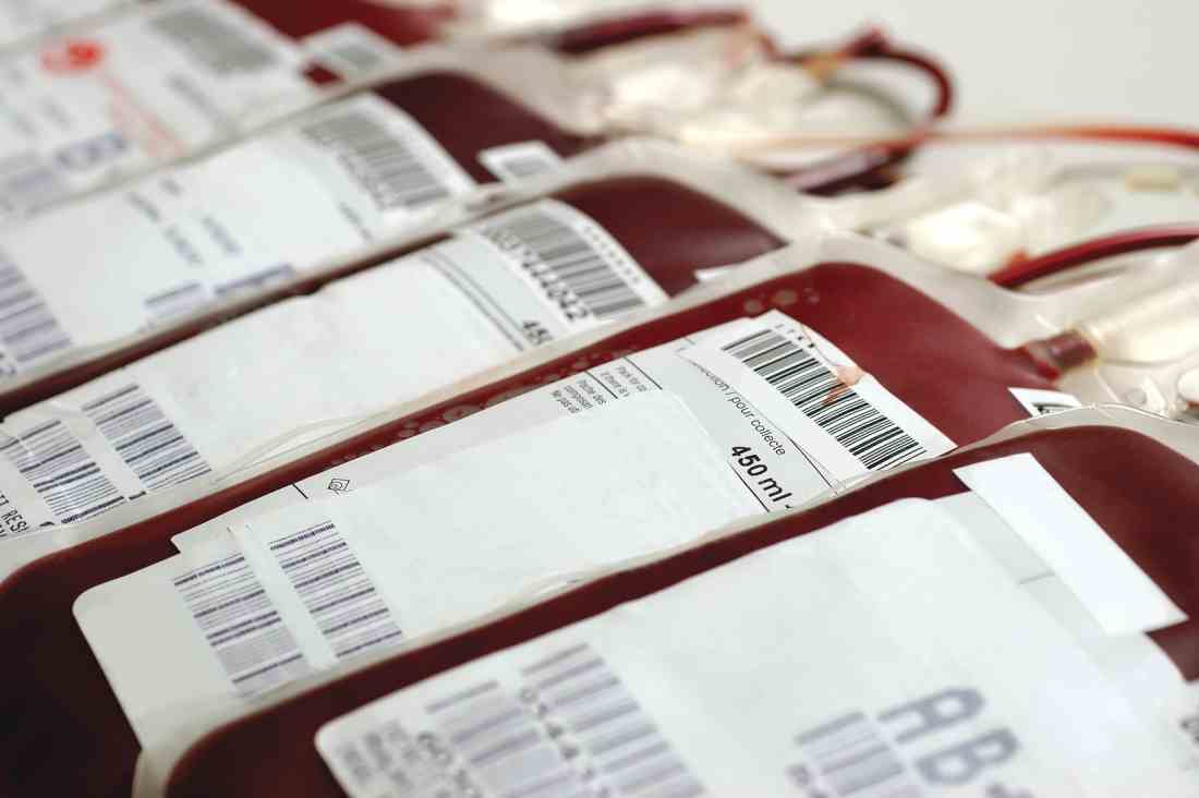
Rheumatoid arthritis does not get transmitted through blood transfusions, according to findings from a large retrospective study of blood transfusions in Denmark and Sweden.
Two separate analyses showed that the risk of developing RA among transfusion recipients was not correlated to the presence of RA in the blood donor.
There had been concern about risks of transfusion because of RA’s long preclinical phase, in which RA pathogenesis factors circulate in the periphery and could potentially be transmitted in a transfusion. Mouse models had suggested that anti–citrullinated peptide/protein antibodies could spark or worsen arthritis, and RA fibroblast-like synoviocyte cell precursors could spread RA between joints.
Two previous studies, based on self-reported history of blood transfusion, reached the opposite conclusion regarding the risk of RA transmission.
The latest findings, published online in Annals of the Rheumatic Diseases, involved an analysis of data from the Danish–Swedish population-based research donations and transfusions database (SCANDAT2). In one model, they looked at 938,942 blood donors, 2,412 of whom were diagnosed with RA during the follow-up period. The researchers then analyzed data from 13,369 subjects who had been exposed to blood from donors who went on to develop RA, and compared them to 139,470 recipients who received blood from donors who did not develop RA. There was no statistically significant correlation between risk of RA among recipients by RA status of the donors, RA serotype in the donors, donor age at RA diagnosis, or elapsed time between donation and RA diagnosis.
In a second analysis, the researchers looked for RA clusters among recipients who might have received blood from a donor with RA. They found no association between RA risk for a given recipient based on whether other recipients from the same donor had gone on to develop RA.
“In light of the study’s strengths, including low likelihood of confounding and large study size ensuring meaningful statistical power, we believe the possibility of RA transmission is unlikely to be clinically relevant,” the authors wrote.
The study was funded by the Danish Rheumatism Association, the Odense University Hospital PhD Fund and Fund for clinical research, the Nordic Cancer Union, the Swedish Foundation for Strategic Research, the Swedish Research Council, and ALF funds. One author reported receiving grants from AbbVie, Bristol-Myers Squibb, Merck, Pfizer, Roche, Samsung, and UCB.
SOURCE: Just SA et al. Ann Rheum Dis. 2018 Mar 1. doi: 10.1136/annrheumdis-2017-212844
FROM ANNALS OF THE RHEUMATIC DISEASES
Key clinical point: Blood transfusion is unlikely to be a RA transmission route.
Major finding: Recipients had similar risk of RA whether or not the donor later developed RA.
Data source: Retrospective analysis of 938,942 blood donors and 152,839 recipients.
Disclosures: The study was funded by the Danish Rheumatism Association, the Odense University Hospital PhD Fund and Fund for clinical research, the Nordic Cancer Union, the Swedish Foundation for Strategic Research, the Swedish Research Council, and ALF funds. One author reported receiving grants from AbbVie, Bristol-Myers Squibb, Merck, Pfizer, Roche, Samsung, and UCB.
Source: Just SA et al. Ann Rheum Dis. 2018 Mar 1. doi: 10.1136/annrheumdis-2017-212844.
Blood transfusions are dropping in U.S. hospitals
The number of red blood cell (RBC) and plasma transfusions conducted in U.S. hospitals has declined steadily since 2011, perhaps as a result of hospitals instituting new blood management programs after randomized trials showed the safety of restrictive transfusion strategies.
There has been no change in the frequency of platelet transfusions since 2011.
The researchers analyzed data from the National Inpatient Sample, using ICD-9-CM procedure codes to identify transfusion procedures. They examined the percentage of hospitalizations with one or more RBC transfusions, since these represent the majority of transfusions. Secondary outcomes included hospitalizations with one or more plasma or one or more platelet transfusions. The findings were published in a research letter in JAMA.
The study included data from the period of 1993-2014. The frequency of transfusions has trended upward since 1993, but a joinpoint analysis found an inflection point at 2011. The researchers then focused their analysis on the period from 2011 to 2014.
The researchers found reductions in RBC transfusions among all sexes, race/ethnicities, patient risk severities, payer types, and admission types. They found no statistically significant reductions in RBC transfusions in private investor–owned hospitals or in patients under the age of 18, though they noted that there is limited evidence to guide clinical practice in the pediatric population.
The decline in RBC transfusions was greater for elective admissions (aRR, 0.74, 95% CI, 0.67-0.80) than it was for nonelective admissions (aRR, 0.86; 95% CI, 0.81-0.91; P for interaction less than .001).
“The observed decreases in RBC and plasma transfusions from 2011 to 2014 may reflect evidence demonstrating the safety of restricting RBC transfusions, patient blood management programs, conservation initiatives (e.g., cell salvage, pharmacotherapy, improved surgical techniques), advocacy from medical organizations, and publication of transfusion guidelines,” the researchers wrote.
The study is limited by its retrospective design and may not be generalizable to outpatient settings.
The study was supported by grants from the National Institutes of Health and Weill Cornell Medical College. Two of the study authors reported personal fees from Terumo BCT, Haemonetics, and Octapharma. No other disclosures were reported.
hematologynews@frontlinemedcom.com
SOURCE: Goel R et al. JAMA. 2018 Feb 27;319(8):825-7.
FROM JAMA
Key clinical point:
Major finding: The frequency of red blood cell transfusions among hospital inpatients dropped from 6.8% to 5.7% from 2011 to 2014.
Study details: A retrospective analysis of procedures codes at U.S. hospitals from 1993 to 2014.
Disclosures: The study was supported by grants from the National Institutes of Health and Weill Cornell Medical College. Two of the study authors reported personal fees from Terumo BCT, Haemonetics, and Octapharma. No other disclosures were reported.
Source: Goel R et al. JAMA. 2018 Feb 27;319(8):825-7.
Spray-dried plasma inches toward clinical trials
SAN DIEGO – Spray-dried plasma compared well with fresh frozen plasma in two in vitro studies, but clinical studies are needed to confirm the findings, researchers reported at the annual meeting of the American Association of Blood Banks.
The product’s logistical benefits include ease of transport, stability at room temperature, and the ability to be rapidly reconstituted – attributes that make it particularly useful in combat situations and prehospital settings where it is impractical to administer fresh frozen plasma (FFP).
The advantages of reconstituted blood products in combat settings have prompted recent efforts to speed their availability. The Food and Drug Administration and the Department of Defense recently announced a joint program to expedite the FDA’s review of products that could diagnose, treat, or prevent life-threatening conditions facing U.S. military personnel. It would be a fast-track process similar to how the FDA handles the breakthrough designation program.
In the first study, the investigators compared spray-dried plasma (SpDP) and FFP in reconstituted whole blood to test their hypothesis that SpDP is not inferior to FFP in facilitating platelet adhesion and thrombus formation, as evaluated by using a microfusion assay.
“Trauma is frequently associated with the use of plasma,” said Rachel S. Bercovitz, MD, MS, of the BloodCenter of Wisconsin and associate professor of pediatrics (hematology, oncology, and stem cell transplantation) at Northwestern University, Chicago.
Compared with FFP, SpDP can be reconstituted in 5 minutes and has more than 80% of the procoagulation and anticoagulation proteins, she explained. “Factor 8 levels were lower in the spray-dried plasma and were about at the 70% level of FFP. The other factor that was reduced, as compared to the FFP, was the von Willebrand factor (vWF), which was about 60% in SpDP compared to FFP.”
Whole blood was obtained from healthy volunteers and red blood cells (RBCs) were separated from platelet-rich plasma, and following standard procedures, resuspended in either SpDP or FFP and recombined with the packed red blood cells to create reconstituted whole blood with hematocrit of 34%-40% and 150,000-250,000 platelets per mcL.
After fluorescent labeling, the samples were flowed through a type I collagen-coated microchannel and still images of adherent platelets and thrombi were captured in order to calculate surface area coverage along the length of the channel. Next, the investigators used a ratio paired t-test to compare surface area coverage in SpDP versus FFP. The margin of noninferiority was 20% (SpDP/FFP greater than 0.8).
A total of six batches of SpDP and FFP were evaluated with 17 donors, and there was no statistical difference between the SpDP versus FFP pairs (P = .7558).
The mean ratio of SpDP versus FFP was 1.21 with a 95% confidence interval of 0.84-1.57. The surface area coverage in samples that were reconstituted with SpDP were, on average, 20% greater than in samples reconstituted with FFP. The lower limit of the 95% confidence interval was a difference of 16%, and therefore lower than the a priori determined margin of noninferiority of 20%.
“We found that SpDP is not inferior to FFP in supporting platelet adhesion and thrombus formation in our in vitro model,” Dr. Bercovitz said. “We feel that these in vitro assays support further in vivo studies of safety and efficacy of spray dried plasma.”
In a second study, Michael A. Meledeo, PhD, of the U.S. Army Institute of Surgical Research (coagulation and blood research), and his colleagues examined methods of reconstituting SpDP. They noted that a single unit process has been developed that produces a long-lived and readily stored SpDP product, which decreased high-molecular-weight multimers of vWF but increased low-molecular-weight multimers. vWF is critical in the process of platelet adhesion and thrombus formation, Dr. Meledeo said.
The researchers examined different reconstitution solutions: FFP, FFP with glycine, regular SpDP without pretreatment and rehydrated with glycine-hydrochloride:glycine, SpDP pretreated with glycine-HCl, or glycine-HCl:glycine and rehydrated with water.
Several in vitro analyses were performed, including measurement of vWF activity, fibrin polymerization kinetics, thrombin generation, coagulation properties and platelet adhesion to collagen.
Pretreated SpDP had better vWF activity, compared with regular SpDP (P less than .05). As compared with FFP, fibrin polymerization density was slightly lower in regular SpDP (0.879 vs. 0.742 optical density; P less than .01), although generation of thrombin was similar.
The researchers also found that the bicarbonate/base excess were lower in SpDP samples versus FFP (P less than .001). Thromboelastography results (used to measure coagulation properties) remained unchanged in plasma-only samples, but clot strength in reconstructed whole blood was reduced in all SpDP samples, compared with FFP (63.82 vs. 55-59.38; P less than .01).
Finally, platelet adhesion was equivalent in pretreated SpDP samples and FFP, while with regular SpDP, it was improved as compared with all other samples (71.53% surface coverage vs. 30.26%-43.87%; P less than .05).
“Based on these results, spray dried plasma was equivalent or superior to FFP in most of the in vitro hemostasis assays,” Dr. Meledeo said. “Reconstitution with glycine-HCl or glycine-HCl:glycine induced a superior von Willebrand function, but it was inferior in terms of supporting a flowing platelet adhesion to collagen.”
Dr. Bercovitz and Dr. Meledeo reported having no financial disclosures.
hematologynews@frontlinemedcom.com
SOURCES: Bercovitz R et al. AABB 17 Abstract C20-A02B; Meledeo M et al. AABB 17 Abstract C21-A02B.
REPORTING FROM AABB 17
Key clinical point:
Major finding: Spray-dried plasma was equal to, or superior to, fresh frozen plasma in many of the in vitro assays utilized, especially when pretreated in glycine solutions.
Study details: Two in vitro assays that compared spray-dried plasma with fresh frozen plasma.
Disclosures: Dr. Bercovitz and Dr. Meledeo reported having no financial disclosures.
Sources: Bercovitz R et al. AABB 17 Abstract C20-A02B; Meledeo M et al. AABB 17 Abstract C21-A02B.
Zika RNA persists in blood components after clearance from plasma
SAN DIEGO – Zika virus can persist in blood components for several months, long after it is no longer detectable in plasma and other body fluids, based on data reported at the annual meeting of the American Association of Blood Banks.
The presence of Zika virus in plasma declined rapidly following donation, but could be detected in red blood cells (RBCs) and whole blood for up to 3 months. In addition, the virus was also detected intermittently in peripheral blood mononuclear cells (PBMCs) at low levels after clearance from plasma.
“There was longer persistence of Zika RNA in whole blood and RBC blood components than in plasma and other body fluids. It was detected at high levels and persists for about 6 weeks,” Mars Stone, PhD said in presenting the findings.
The findings were based on 2016 data collected in Puerto Rico, which began screening blood donations for Zika virus RNA under an investigational protocol by using a nucleic acid test; they began doing so as a result of guidance from the U.S. Food and Drug Administration. Approximately 350 confirmed infected – that is, nucleic acid test positive (NAT+) – donations were detected through December 2016.
The FDA approved the cobas Zika test used in the study on October 5. Intended for use by blood collection establishments to detect Zika virus in blood donations, the test is manufactured by Roche Molecular Systems. Dr. Stone is an employee of Blood Systems Research Institute, a confirmatory laboratory for Roche.
Of the 52,942 donations collected between April 3 and Dec. 31, 352 were reactive for ZIKV RNA. Plasma from blood donors was screened by individual donation NAT for the presence of ZIKV RNA with the cobas Zika test. At an interval of between 57 and 120 days, no Zika RNA was found in 350 samples of plasma, but it was found in 38.2% (34 samples) of RBCs and 40.0% (35 samples) of whole blood.
In urine and saliva, the virus was detected in high levels at 1-2 weeks, but then rapidly waned. In semen, it was detected at high levels and persisted for about 6 weeks.
“But despite the huge epidemics in Latin America, Puerto Rico, and the other Caribbean islands, there have been no cases of transfusion-related infections linked to RBC transfusions when plasma NAT screening was negative,” said Dr. Stone. “So we are tentatively confirming that red cell–associated virus is not infectious and that plasma NAT screening is likely sufficient.”
The authors point out that RNA persistence has been reported in whole blood long after the virus cleared from plasma and therefore have raised concerns about the risk of transfusion-related viral transmission. The goal of the current study was to characterize the dynamics of infection using donors who were infected with Zika virus.
To date there are 56 donors enrolled in the study, primarily male and from Puerto Rico. Most of them are also positive for dengue fever, Dr. Stone pointed out. “The epidemic in Puerto Rico reached peak in June 2016 but has very little activity this year.”
Plasma and RBCs were collected from index donations, while blood, urine, saliva, and semen samples were collected prospectively at weeks 1, 3, 6, 12, and 24 following index donations. Blood compartments and body fluids were tested for Zika RNA by real time reverse transcription polymerase chain reaction testing, and plasma samples were tested for Zika-specific immunoglobulin M and immunoglobulin G antibodies.
In plasma, Zika virus RNA rapidly decreased after index donations but persisted for up to 3 months in RBCs and whole blood. In peripheral blood mononuclear cells, Zika virus was detected intermittently at low levels but waned by 3 months. In peripheral blood mononuclear cells, Zika RNA was detected in 5.9% (17 samples) at 121-196 days.
Among donors who entered the study while in the acute preseroconversion stage of infection, 65% developed Zika virus symptoms at 1 week post index donation, compared with 30% of donors detected after seroconversion.
The study was funded by the U.S. Department of Health & Human Services.
AT AABB17
Key clinical point: Zika virus can persist in cellular blood components for months after clearance from plasma.
Major finding: Plasma viremia rapidly declined after index donations, but
Data source: Zika RNA persistence in blood components after clearance of viremia in plasma was evaluated in 56 donors positive for Zika virus.
Disclosures: Dr. Stone is an employee of Blood Systems Research Institute in San Francisco, a confirmatory laboratory for Roche Molecular Systems, which manufactures the testing device used. The study was funded by the U.S. Department of Health & Human Services.
Source: Stone M et al. AABB 2017 Abstract C9-A01AC.
Zika virus testing shows low incidence in donor blood outside of high-infection areas
SAN DIEGO – The Zika virus is primarily transmitted via the Aedes mosquitoes, most commonly by A. aegypti, but recent outbreaks have revealed that nonvector transmission routes may also spread the infection. Some data suggest that blood transfusion can be a source of transmission.
While the number of contaminated blood donations remains very small, three studies presented at the American Association of Blood Banks annual meeting confirmed the ability of new investigational assays to detect Zika virus in donated blood.
There have been no confirmed transfusion-transmission cases of Zika virus in the United States, but as cases have now been documented in Brazil, the Food and Drug Administration issued revised guidance in August 2016 recommending that blood centers in all states and U.S. territories screen individual units of donated whole blood and blood components.
In the first report (C7-A01C), Paula Saá, PhD, and her colleagues at the American Red Cross initially investigated the use of mini-pool (MP)- nucleic acid testing (NAT) using the Procleix Zika Virus Assay (TMA). Testing was initially implemented on blood collections from Florida, Georgia, South Carolina, Mississippi, and Alabama – five states that were presumed to be at high risk of Zika virus infection. After the FDA revised its guidance, the protocol changed and testing was extended to all blood donations. The use of the MP-NAT was also converted to individual donation (ID)-NAT, and questions concerning travel history was also eventually discontinued.
However, even with the use of ID-NAT, the rate of confirmed positive donations was quite small but the associated cost was quite high, the researchers pointed out. “In the first year of testing at the American Red Cross, we identified nine confirmed positive donations,” said Dr. Saá.
The rate of confirmed positive donations was 1:354, 602 during the study period, but if the period up until September 2017 is taken into account (no additional cases were identified), the rate increases to 1:514,266. “This is a very low rate,” Dr. Saá said. “If there are no changes to the current guidelines, we have estimated that the yearly cost for the American Red Cross of testing will exceed $48 million.”
These figures extrapolate to approximately $6 million per confirmed case, according to the results of this study sponsored by the American Red Cross.
Confirmatory testing included repeat TMA; in addition, RT-PCR, serology and red blood cell count (RBC) TMA were performed. Estimates of viral loads were performed by endpoint TMA on plasma and RBCs.
A total of 2,288,855 blood donations had been tested as of April 2017, including 393,713 (17%) in 24,611 MPs, which did not detect any reactive donations.
Of the confirmed positive blood donors, three lived in Florida and two of those were from local transmission. Six individuals had traveled to a region highly active for Zika virus, and returned to the United States between 2 and 73 days before donating blood. Clinical symptoms were reported in two individuals with a travel risk; the other donors with a confirmed positive test (75%) remained asymptomatic. The longest period for detection in RBCs was 91 days thus far, but in the same person, detection in plasma was only 17 days.
“The data that we are showing here recommends a testing strategy with mini pool testing in areas at low risk of Zika transmission,” said Dr. Saá.
A second related study (C9-A01C), described the detection of ZIKV RNA in blood donations collected in U.S. states between April 3, 2016, and September 23, 2017, using the cobas Zika test, to be used on the cobas 6800/8800 Systems.
Although the test was investigational during the study period, it has just been approved by the FDA, said study author Lisa Pate, MD, who is with Roche Molecular Systems, the manufacturer of the cobas Zika test and cobas 6800/8800 systems. “This is now the first licensed test for screening blood donations for Zika virus.”
Overall, testing showed that Zika contamination in the U.S. blood supply was quite low. Only 0.001% of screened blood donations in United States were confirmed as true positives.
The development of this test came about after the first cases of Zika virus in the United States were detected in Puerto Rico in December 2015, explained Dr. Pate. Shortly after that, the FDA issued guidance prohibiting the use of blood collected in Zika active areas, unless the donations were screened.
“The impact was significant in Puerto Rico, as blood donations were halted, which then forced Puerto Rico to rely on imported blood,” she said.
About that time the FDA reached out to Roche and competitors to see if a test could be developed to screen for Zika.
The cobas Zika test was approved under an investigational new drug application on March 30, 2016, and although initially used to test blood samples in Puerto Rico, testing was expanded to include donor blood from all over the country.
Screening was conducted by individual donation testing, with all initial reactive results repeated in duplicate. Supplemental testing was also done, and included an alternative NAT (AltNAT) assay which was considered to be less sensitive than cobas Zika and serology testing for anti-Zika IgM and IgG. A donor confirmed Zika confirmed positive if at least one replicate of the repeat testing by cobas Zika was reactive on index donation or follow-up, reactive by AltNAT on the index donation, or positive for anti-Zika IgM on index or follow-up.
Screening was conducted at 12 testing labs in the United States, and more than 4 million donations were screened and 27 positive donations were confirmed. Overall, that amounted to less than 1 in 100,000.
“For donors in the U.S. with confirmed positive results, and for whom follow-up information is available, 84% of them report recent travel to Zika active areas,” noted Dr. Pate.
For Puerto Rico, 111,842 blood donations were screened and there were 356 confirmed positive results. The incidence is much higher than in United States, and was 1.27% during peak incidence in July 2016.
A third paper (C12-A01C), also reported on testing the blood supply in Singapore, which had reported its first locally transmitted Zika case last August, using the investigational Procleix ZIKV nucleic acid technology (NAT) assay.
The presence of Zika virus in screened blood was also quite low, with an incidence of only 0.0032%. The Procleix ZIKV assay was found to suitable for screening for Zika infection in an asymptomatic population, as it showed good analytical sensitivity and clinical performance.
The Zika virus came to Singapore in May 2016, imported by an individual who had recently traveled to Brazil, said Sally Lam, laboratory director, Blood Services Group, Health Sciences Authority, Singapore.
“Then in August we had 41 confirmed local Zika virus cases,” she said.
In 2016, there were 458 clinical Zika cases reported, with 8 clusters identified. This year, 63 cases have been reported to date, she said.
Mandatory Zika virus screening in donor blood with ID-NAT began after the onset of local outbreaks, and was implemented in January 2017. A total of 126,906 blood donations were screened.
Researchers in Singapore assessed the performance of the Procleix ZIKV NAT assay for universal blood donation screening. They screened all blood that was donated, beginning Oct. 1, 2016, a confirmed case was defined as having Zika RNA by PCR and/or Zika antibodies. Analytical sensitivity was assessed by use of 300 blinded frozen samples containing Zika virus and 25 negative controls. The performance of the Procleix ZIKV assay was also evaluated by use of samples from the local patient population.
Of four confirmed positive cases, only one was available for follow-up. “In the index donation, the viral load was quite high in the plasma but at 10 days, it was reduced to about 400 copies/mL in the plasma,” said Ms. Lam. “The donor did not develop any symptoms.”
The analytical sensitivity for the Procleix ZIKV assay was determined to be 2.1 copies/mL at 50% LOD and 10.0 copies/mL at 95% LOD, and it detected RNA in six out of nine patient samples for an 85.7% agreement with reference material, according to the researchers.
AT AABB17
Denmark reinstates ID NAT screening for blood donations
SAN DIEGO – After funding was discontinued for individual donation (ID) nucleic acid testing (NAT) of blood donations, the risk of transfusion-related infections in Denmark increased. But according to new findings presented here at the American Association of Blood Banks annual meeting, that policy was short lived.
When ID NAT was removed from the screening process, the estimated increase in the risk for transfusion-transmitted HIV went from one in 80 years to one in 18 years; for hepatitis B virus (HBV), it went from one in 34 years to one in 17 years; and for hepatitis C virus (HCV), the risk increased from one in 250 years to one in 8 years.
“Between 2009 and 2016, we had 14 NAT reactive/seronegative cases among the repeat donors, and one was due to an acute hepatitis C infection,” said study author Leen Baudewijn, MD, of Odense (Denmark) University Hospital. “This would have been missed without NAT testing.”
Dr. Baudewijn explained that Denmark has since reversed this new policy. “The Danish government decided not to discontinue NAT because there were almost no savings,” she said during her presentation. “We had the extra costs for anti-HBc [anti-Hepatitis B core] testing for repeat donors, and extra cost due to mandatory NAT testing for plasma for manufacturing medicinal products. So it was not possible to abrogate NAT testing because of contracts with the industry.”
However, she added that “one of the most important reasons was that one of the vendors for NAT screening reduced the price substantially by 45%.”
The majority of industrialized countries have implemented the use of increasingly sensitive assays for screening donated blood, although to date, there is no technology that can guarantee zero-risk blood products. However, the risk for transmission of a viral infection via transfusion is low in Northern Europe.
But after a case of HIV infection linked to a blood transfusion occurred, Denmark mandated ID NAT in 2009 for serologic-based screening assays for HIV, HBV, and HCV. In July 2017, the government changed its policy and discontinued funding for ID NAT screening and, instead, mandated anti-Hepatitis B core screening be added to the serologic screening assays for repeat donors.
In this study, Dr. Baudewijn and her colleagues used an incidence/window model to estimate the residual risk (RR) of transfusion-transmitted viral infections after ID NAT was halted in Denmark. They estimated incidence rates for blood and plasma donations obtained from repeat donors during 2006-2016 based on the number of positive tests after a negative donation. The residual risk was estimated as the incidence rate multiplied by the average window period for HIV, HBV, and HCV, with and without ID NAT testing.
A total of 3.5 million donations were screened during the study’s time period, with donors averaging two blood donations per year. The researchers estimated the RR for each donation with and without ID NAT.
For HIV, the RR without ID NAT was 1/3,647,888 and with ID NAT it was 1/16,436,213; for HBV, those numbers were 1/3,352,628 without and 1/6,806,407 with; and for HCV, 1/1,573,213 without and 1/50,429,289, with.
The overall probability of missing an infectious red blood cell unit was 35/1,000 in a population of 6 million, Dr. Baudewijn noted.
The authors had no relevant financial disclosures
SOURCE: Baudewijn L et al. AABB 2017. Abstract P4-A03A.
AT AABB17
Key clinical point:
Major finding: Without ID NAT, the estimated increase in the risk for transfusion-transmitted HIV went from 1 patient per 80 years to 1 per 18; for HBV, from 1 per 34 to 1 per 17; and for HCV, the risk increased from 1 per 250 to 1 per 8.
Data source: An incidence/window model estimating the residual risk of transfusion-transmitted viral infections following Denmark’s decision not to fund ID NAT testing.
Disclosures: The authors had no relevant financial disclosures.
SOURCE: Baudewijn L et al. AABB 2017. Abstract P4-A03A.
Three-month response to CAR T-cells looks durable in DLBCL
ATLANTA – Responses 3 months after chimeric antigen receptor (CAR) T-cell therapy look durable in adults with transplant-ineligible relapsed/refractory diffuse large B-cell lymphoma (DLBCL), according to updated results from the single-arm, global, phase 2 JULIET trial.
Fully 95% of patients who had a complete response to CTL019 (tisagenlecleucel; Kymriah) at 3 months maintained that complete response at 6 months, Stephen J. Schuster, MD, said at the annual meeting of the American Society of Hematology.
Patients with relapsed/refractory DLBCL tend to face a very poor prognosis, noted Dr. Schuster of Perelman School of Medicine and Abramson Cancer Center, University of Pennsylvania, Philadelphia. High-dose chemotherapy followed by autologous stem cell transplantation “is capable of long-term survival, but in very few patients,” he said. A dismal 8% of patients completely respond to salvage treatment and only about one in five partially respond. Both levels of response are short-lived, with a median survival of about 4 months.
Meanwhile, CTL019 therapy has produced durable complete remissions in children with lymphoblastic leukemia and in adults with chronic lymphocytic leukemia, Dr. Schuster and his associates wrote in an article simultaneously published in the New England Journal of Medicine (2017 Dec 10. doi: 10.1056/NEJMoa1708566).
To test the CAR T-cell therapy in relapsed/refractory DLBCL, they enrolled affected adults who had received at least two prior lines of antineoplastic treatment and who were not candidates for autologous stem cell transplantation.
Treatment consisted of a single CTL019 infusion (median dose, 3.1 × 10⁸ cells; range, 0.1 × 10⁸ to 6.0 × 10⁸ cells), usually after lymphodepleting chemotherapy. Previously, patients had received a median of three lines of therapy, and about half had undergone autologous stem cell transplantation.
Median time from infusion to data cutoff in March 2017 was 5.6 months. Among 81 patients followed for at least 3 months before data cutoff, best overall response rate was 53% and 40% had a complete response. Overall response rates were 38% at 3 months and 37% at 6 months. Rates of complete response as confirmed by 18F-fluorodeoxyglucose–positron-emission tomography (PET) were 32% at 3 months and 30% at 6 months.These findings highlight the predictive power of 3-month response to CTL019 therapy in relapsed/refractory DLBCL, Dr. Schuster said. Among all responders, 74% remained relapse free at 6 months, meaning that median duration of response and median overall survival were not reached at data cutoff.
Dr. Schuster also reported that 26% of patients were infused as outpatients, which he called “easy to do” and appropriate as long as patients who become febrile are admitted and monitored for cytokine release syndrome. Three-quarters of patients who were infused as outpatients were able to remain home for at least 3 days afterward, he said.
Adverse events typified those of CAR T-cell therapy, including cytokine release syndrome (all grades: 58%; grade 3-4: 23%) and neurological toxicities (all grades: 21%; grade 3-4: 12%). The current labeling for CTL019 in children and young adults with acute lymphoblastic leukemia also includes a boxed warning for these toxicities.Tisagenlecleucel, the first-ever approved CAR T-cell therapy, is made by using a lentiviral vector to genetically engineer a patient’s own T-cells to express a CAR for the pan-B-cell CD19 antigen. These anti-CD19 CAR T-cells are then expanded in the laboratory, frozen for shipping purposes, and infused back into patients. In October 2017, Novartis submitted a biologics license application to the Food and Drug Administration to expand the label for CTL019 to include transplant-ineligible relapsed/refractory DLBCL.
Novartis Pharmaceuticals anticipates large-scale production in 2018, Dr. Schuster said. Manufacturing time has been cut to 22 days from the 30-day turnaround used in the trial, he reported.
Dr. Schuster also said that he sees no point in retreating patients whose relapsed/refractory DLBCL doesn’t respond to tisagenlecleucel, and that JULIET did not test this approach. “If someone fails therapy and you retreat, you don’t see success, in my experience,” he said. “If patients respond and then fail later, then you retreat and you may succeed.”
Novartis Pharmaceuticals sponsored JULIET. Dr. Schuster disclosed consultancy and research funding from Novartis and ties to Celgene, Gilead, Genentech, and several other pharmaceutical companies.
SOURCE: Schuster S et al. ASH 2017 Abstract 577.
REPORTING FROM ASH 2017
Key clinical point:
Major finding: Among 81 patients with at least 3 months of follow-up, best overall response rate was 53% (95% CI, 42%-64%; P less than .0001) and rates of complete response were 32% at 3 months and 30% at 6 months.
Study details: JULIET is an international, single-arm, phase 2 study of adults with relapsed/refractory DLBCL.
Disclosures: Novartis Pharmaceuticals sponsored JULIET. Dr. Schuster reported consultancy and research funding from Novartis and ties to Celgene, Gilead, Genentech, and several other pharmaceutical companies.
Source: Schuster S et al. ASH 2017 Abstract 577.
ASCO platelet transfusion guidelines updated
A lot has happened in oncology since the American Society of Clinical Oncology (ASCO) first issued its guidelines on platelet transfusion for patients with cancer in 2001, noted the authors of the updated recommendations.
“The expense of platelet transfusions, coupled with potential adverse effects such as febrile and allergic reactions, transfusion-related acute lung injury, and bacterial contamination point to the importance of evidence-based transfusion practice,” wrote Charles A Schiffer, MD, of Wayne State Michigan, Detroit, and his colleagues in the updated guidelines.
Many of the original recommendations remain unchanged, but there are updated evidence-based recommendations in five key areas.
For example, regarding platelet transfusion thresholds in the setting of hematologic stem-cell transplantation in adults, the guidelines incorporate evidence from randomized clinical trials showing that among adults who have received autologous hematologic stem-cell transplantation, bleeding rates with decreased use of platelets are similar whether patients are treated prophylactically or at the first sign of bleeding, “and this approach may be used in experienced centers,” wrote Dr. Schiffer and his colleagues (J Clin Oncol. 2017 Nov 28. doi: 10.1200/JCO.2017.76.1734).
The authors caution, however, that the recommendation applies to adults only.
Other updated recommendations include:
• Rhesus D alloimmunization from platelet transfusions to RhD-negative patients can be prevented through either exclusive use of platelet products from RhD-negative donors or immunoprophylaxis. The guidelines note that there is a low rate of RhD alloimmunization in cancer patients in general, but state that prevention may be used in girls and in women of child-bearing age who are being treated with curative intent.
• For patients with acute myeloid leukemia, receiving induction chemotherapy, the use of leukoreduced platelet and red blood cell products can reduce the likelihood that patients will develop alloantibody-mediated refractory reactions to plate transfusions.
“It is therefore appropriate to provide leukoreduced blood products to patients with [acute myeloid leukemia] from the time of diagnosis to ameliorate this important clinical problem,” the investigators wrote.
They noted that leukoreduction to prevent alloimmunization might benefit patients with other leukemia histologies and with other types of cancer who are undergoing chemotherapy, but added that there is a lack of evidence to support this as a recommendation outside of acute myeloid leukemia.
To reduce the risk of bleeding due to thrombocytopenia in patients with solid tumors who are undergoing chemotherapy, the panelists recommend transfusing patients when their platelet levels fall below 10 x 109 per liter. It is appropriate to give platelet transfusions to patients with higher levels when there is active localized bleeding, they stated.
The guideline authors also recommend that when refractoriness to platelet infusions is suspected, clinicians should perform platelet counts from 10 to 60 minutes after the transfusion is completed. A refractoriness determination should be made only after two or more infusions of ABO-compatible units that have been stored for less than 72 hours result in poor increments, they advised.
The guideline development process is supported by ASCO. Dr. Schiffer and six other guideline authors disclosed consulting or advisory roles, research funding, honoraria and/or fees with various pharmaceutical companies or other corporate entities.
FROM THE JOURNAL OF CLINICAL ONCOLOGY
Panel votes against universal blood donor screens for Zika virus
SILVER SPRING, MD – Universal testing of individual blood donations for the presence of Zika virus was unanimously rejected by voting members of the Food and Drug Administration’s Blood Products Advisory Committee at a December 1 meeting.
Universal individual donor testing, while comprehensive, is resource intensive and places a burden on the blood system that is not outweighed by the benefits, 10 of the 11 committee members concluded. The other committee member could not be reached by phone for this vote.
The committee instead recommended by a vote of 10 to 1 that mini-pool nucleic acid testing (MP-NAT) be performed in all states and territories with known cases of Zika virus infection and the presence of A. aegypti mosquitoes, as well as in states and territories with a high number of travelers from areas with Zika virus infections. Also, the committee members agreed that a trigger needs to be defined for when to undertake universal individual donor nucleic acid testing (ID-NAT) in those areas.
Additionally, the committee agreed that it was not necessary to maintain a Zika virus-negative blood inventory for at-risk patients, such as pregnant women and newborns. Zika virus, a vector-borne disease carried by the Aedes aegypti and Aedes albopictus mosquitoes, has also been transmitted via sexual contact and blood transfusion. Infection has been linked to fetal loss and microcephaly in the offspring of infected pregnant women. Other neurological disorders, including Guillain-Barré Syndrome, also have been linked to Zika virus infection.
Noting the complexity of managing an inventory of tested and non-tested blood, the committee rejected the separate inventory approach by a vote of 9 to 2.*
The panel was clearly divided on the possibility of eliminating all Zika virus testing in some states and territories; 5 members supported this measure, 4 opposed it, and 2 abstained from voting.
The panel unanimously rejected using screening questionnaires to determine whether to selectively test individual at-risk donors in areas with active vector-borne Zika virus infections. This option was considered particularly troublesome, they agreed, because it relies on the use of nonspecific, insensitive, and error-prone questionnaires.
Some level of Zika virus testing is needed to safeguard blood products, the committee said. Eliminating all Zika virus testing of blood products would open the door for infections via transfusion and would diminish preparedness against a potential epidemic, they unanimously determined.
Prior to voting, the committee listened to presentations on the epidemiology of Zika virus infections, the effectiveness of screening tests, and the risk for transmission via transfusion.
Carolyn Gould, MD, of the Centers for Disease Control and Prevention, Atlanta, reported that the number of laboratory-confirmed Zika virus infections in 2016 was 4,830 for travelers to endemic areas and 224 for locally-acquired cases. In 2017, those numbers dropped to 344 confirmed cases for travelers and 2 for locally-acquired cases.
More than 4 million blood donations in the United States and Puerto Rico have been screened for Zika virus RNA using the cobas assay, which is now FDA approved. The overall confirmed positive rate of Zika virus is 0.0007% in donations from the continental United States (29 positive results in 4,341,770 donations) and 0.326% in donations from Puerto Rico (356 out of 111,808 donations) based on data obtained from May 23, 2016 to October 7, 2017, according to Tony Hardiman, Blood Screening, Life Cycle Leader at Roche Molecular Systems.
Of the Zika virus-positive donors who were available for follow up, 23 of 27 had traveled to Zika-active areas, including 3 cases associated with domestic travel to Florida. “I was surprised that 4 of the 29 were from Cuba, but it does seem, as we just saw, (that) an increasing number are coming out from Cuba, from travel to Cuba,” Mr. Hardiman said during his presentation to the committee.
Findings concerning viral RNA duration in blood and other body fluids were presented by Michael Busch, MD of the University of California, San Francisco, who spoke during the public hearing portion of the meeting.**
According to Dr. Busch, blood is likely not infectious to others once donors develop Zika virus-neutralizing antibodies and their viral load levels become very low.
Based on his review of various studies, Dr. Busch concluded that mini-pool testing options with triggers for individual testing are appropriate and effective for detecting Zika virus in endemic areas.
“In Puerto Rico, within a day of picking up a mini-pool positive [result}, we would have ID-NAT in place,” Dr. Busch said. “The mini-pool testing is picking up 90% of those at highest risk.”
The committee recommendations serve as guidance to the FDA, which is not obligated to follow the committee’s recommendations.
*Correction 12/14/17: An earlier version of this story misstated the vote on maintaining a Zika virus-negative blood inventory for at-risk patients. The advisory committee voted against that approach.
**Correction 12/14/17: Dr. Michael Busch's name was misstated in an earlier version of this article.
AT AN FDA ADVISORY COMMITTEE MEETING
Universal paternal Rh screening is cost effective in IVF
SAN ANTONIO – Implementing universal paternal Rh screening would be a cost-effective safety strategy among patients receiving in vitro fertilization, according to a model that used up-to-date data and accounted for ethnic variations in the prevalence of the Rh (D) antibody.
Using a universal Rh screening strategy for semen donors to Rh (D) negative women undergoing in vitro fertilization would result in a cost savings of $11.01 per patient, or $1,120,000 per 100,000 Rh negative IVF pregnancies, according to Pietro Bortoletto, MD, and his coauthors. Their findings were presented during a poster session at the annual meeting of the American Society for Reproductive Medicine.
If paternal Rh factor status is unknown, vaginal bleeding during pregnancy prompts maternal administration of anti-D immune globulin if the mother is Rh (D) negative to prevent hemolytic disease of the fetus and newborn. However, wrote Dr. Bortoletto and his colleagues, “in the IVF population, where paternity status is presumed to be certain, the Rh (D) status of the male partner can be used to triage Rh (D) negative women to more appropriate administration of anti-D globulin.”
To see whether a universal paternal Rh screening strategy would be cost effective, Dr. Bortoletto, a resident physician in obstetrics and gynecology at Harvard Medical School and Brigham and Women’s Hospital, both in Boston, and his collaborators constructed a decision tree to estimate cost savings. The model compared a universal paternal-screening strategy for Rh (D) negative women undergoing IVF with the current standard of practice, which does not involve routine Rh (D) screening for the sperm donor.
In constructing the model, the investigators drew on published data showing that first trimester bleeding is more common in women who undergo IVF: It occurs in one-third of these women, compared with about 20% of the general pregnant population.
They also established probability estimates of pregnancy loss before 20 weeks, with and without first trimester bleeding (0.34 and 0.18, respectively); third trimester bleeding, with and without first trimester bleeding (0.05 and 0.02); and trauma in pregnancy (0.08). They estimated the overall probability of the pregnancy producing an Rh positive neonate at 0.62.
An additional factor that Dr. Bortoletto and his collaborators took into account was the variable prevalence of Rh factor by ethnicity; in the United States, it’s most common in white men and least common in Asian men, with intermediate prevalence in African American and Hispanic men.
When paternal ethnicity was included in the analysis, savings were greatest with white sperm donors, at $1,889,000 per 100,000 Rh negative IVF pregnancies. The lower prevalence of Rh factor in Asian men meant that the strategy was not cost effective in this population since it would cost a net $2,323,000 per 100,000 Rh negative IVF pregnancies.
“A targeted screening approach, by paternal ethnicity, may be a targeted strategy for cost reduction,” wrote Dr. Bortoletto and his colleagues.
Figures for cost estimates were drawn from data from the Centers for Medicare & Medicaid (CMS) using 2017 dollars. The cost for tests to determine blood type and Rh status ranged from $6 to $11, so the investigators set the cost estimate of $8.20. The cost for an antibody screen was estimated at $5.25 (range, $4-$7).
The cost for a 300 mcg dose of anti-D immune globulin was estimated at $93.93 (range, $79-$109), and administration costs were $27.04 (range, $25-$28). Kleihauer-Betke testing to determine the amount of fetal blood in maternal circulation was pinned at $10.61 (range, $8-$14).
Even when the lowest end of the cost range of anti-D immune globulin was used, a universal screening model would still realize a cost savings of $820,000 per 100,000 Rh negative IVF pregnancies. When Rh screening cost was set at $11 – at the high end of the range – “the strategy still remained favorable at $981,000,” wrote Dr. Bortoletto and his colleagues.
“Universal paternal Rh screening provides a cost saving intervention by preventing nonindicated and costly administration of anti-D immune globulin in the IVF population presenting with bleeding or trauma in pregnancy,” they wrote.
Dr. Bortoletto reported no outside sources of funding and no conflicts of interest.
koakes@frontlinemedcom.com
On Twitter @karioakes
At ASRM 2017
Key clinical point:
Major finding: Universal screening would save $1,120,000 per 100,000 Rh negative IVF pregnancies.
Data source: Decision tree incorporating Rh (D) prevalence, bleeding risk, and cost data.
Disclosures: Dr. Bortoletto reported having no relevant disclosures and no outside funding.