User login
One HCV infection leads to another in HIV+ MSM
SEATTLE – Once HIV positive men who have sex with men contract the hepatitis C virus, they are more likely to get it again, according a study of 305 men in New York.

Overall, 38 men (12%) picked up another HCV infection a median of 1.9 years after clearance of their first, yielding a reinfection rate was 4.4/100 person-years, “a solid seven times higher than the primary infection rate” among HIV-positive men who have sex with men (MSM), said senior investigator Daniel Fierer, MD, an associate professor of infectious diseases at Mount Sinai Hospital, New York.
Thirty-three men cleared their second infection. Of those, six picked up a third infection at a median of 1.1 years, yielding an overall third infection incidence of 8.7/100 person-years.
The results held no matter how the men cleared HCV, whether spontaneously, as in about 10%, or by interferon before 2013, and direct-acting antivirals (DAAs) after.
Most reinfections occurred within 2 years of initial clearance, but some occurred more than a decade later.
The results suggest that there’s a particular need for HCV prevention efforts among men who have previously cleared the infection. For those patients, testing for HCV at an annual HIV checkup might not be frequent enough, Dr. Fierer said at the Conference on Retroviruses and Opportunistic Infections.
“Long-term surveillance is warranted for all HIV-infected MSM after clearance of HCV infection. Further, strategies to reduce HCV reinfections are needed to meet the goal of eliminating HCV in these men,” he said.
Also, “the large difference between primary” and secondary infection “rates suggests HCV risk is not distributed evenly between HIV-infected MSM, but concentrated among a small subpopulation. By definition, this subpopulation would have a higher prevalence” of risky behavior, such as condomless receptive anal sex and sexualized injection methamphetamine use, he said.
The high reinfection rate “tells us basically that we have not done a good job of” preventing infection and reinfection among at risk, HIV-positive men. There’s an “inadequate level of HCV treatment ... we need to eliminate restrictions on DAA” access, Dr. Fierer said.
As far as prevention goes, “I believe we just don’t know what to do. I tell all of my patients about the body fluids that have HCV in them,” which is a good start, he said.
The median age at first clearance was about 45 years, 82% of the men were white, and there was about a 50-50 split between people with private and public insurance.
The work was funded by Gilead. Dr. Fierer did not mention any disclosures.
SOURCE: Carollo JR et al. CROI 2019, Abstract 86
SEATTLE – Once HIV positive men who have sex with men contract the hepatitis C virus, they are more likely to get it again, according a study of 305 men in New York.
Overall, 38 men (12%) picked up another HCV infection a median of 1.9 years after clearance of their first, yielding a reinfection rate was 4.4/100 person-years, “a solid seven times higher than the primary infection rate” among HIV-positive men who have sex with men (MSM), said senior investigator Daniel Fierer, MD, an associate professor of infectious diseases at Mount Sinai Hospital, New York.
Thirty-three men cleared their second infection. Of those, six picked up a third infection at a median of 1.1 years, yielding an overall third infection incidence of 8.7/100 person-years.
The results held no matter how the men cleared HCV, whether spontaneously, as in about 10%, or by interferon before 2013, and direct-acting antivirals (DAAs) after.
Most reinfections occurred within 2 years of initial clearance, but some occurred more than a decade later.
The results suggest that there’s a particular need for HCV prevention efforts among men who have previously cleared the infection. For those patients, testing for HCV at an annual HIV checkup might not be frequent enough, Dr. Fierer said at the Conference on Retroviruses and Opportunistic Infections.
“Long-term surveillance is warranted for all HIV-infected MSM after clearance of HCV infection. Further, strategies to reduce HCV reinfections are needed to meet the goal of eliminating HCV in these men,” he said.
Also, “the large difference between primary” and secondary infection “rates suggests HCV risk is not distributed evenly between HIV-infected MSM, but concentrated among a small subpopulation. By definition, this subpopulation would have a higher prevalence” of risky behavior, such as condomless receptive anal sex and sexualized injection methamphetamine use, he said.
The high reinfection rate “tells us basically that we have not done a good job of” preventing infection and reinfection among at risk, HIV-positive men. There’s an “inadequate level of HCV treatment ... we need to eliminate restrictions on DAA” access, Dr. Fierer said.
As far as prevention goes, “I believe we just don’t know what to do. I tell all of my patients about the body fluids that have HCV in them,” which is a good start, he said.
The median age at first clearance was about 45 years, 82% of the men were white, and there was about a 50-50 split between people with private and public insurance.
The work was funded by Gilead. Dr. Fierer did not mention any disclosures.
SOURCE: Carollo JR et al. CROI 2019, Abstract 86
REPORTING FROM CROI 2019
Do Collaborative Models Work for Mental Health in a General Clinical Setting?
Collaborative chronic care models (CCMs) are effective in serious mental illnesses, which has been shown in extensive randomized clinical trials. Much of their effectiveness comes from the emphasis on flexibility: They are implemented according to local needs, capabilities, and priorities. Collaborative chronic care models also provide support: for redesigned work roles that promote “anticipatory” continuous care, for self-management, and for clinical decision making at a local level.
In 2013, the VA Office of Mental Health and Suicide Prevention (OMHSP) began an initiative to enhance care coordination in general mental health clinics with mixed-diagnosis populations. It established interdisciplinary teams in each VA medical center throughout the US. Although providing centrally developed guidance, VAOMHSP gave facilities “broad latitude” to develop their team processes. In 2015, VAOMHSP adopted the CCM.
But most of the data on how well CCMs work for mental health conditions come from depression treatment in primary care—and the effects seem to be inconsistent. So researchers from Veterans Affairs Boston Healthcare System and others partnered with VAOMHSP to find out whether the CCM model would be effective in a general clinical setting.
They recruited 9 VA facilities for a 2-year study conducted in 3 waves. The implementation strategy was based on the premise that “health care is a complex adaptive system rather than a highly controlled machine.” That is, it would work best if local solutions for local challenges could be developed in accordance with evidence-based guidance. The multifaceted approach included an external facilitator who provided guidance and quality improvement expertise and an on-site internal facilitator to direct the implementation.
In the study, 5,596 veterans treated by outpatient general mental health teams were included in hospitalization analyses. A randomly selected sample of 1,050 (including 210 women) was identified for health status interviews.
The researchers found a “robust” and sustained reduction in mental health hospitalization. However, the effects on self-reported health outcomes were “limited,” the researchers say. The mental component score (the primary intervention outcome) did not change statistically significantly with implementation support in adjusted or unadjusted models, nor did other interview measures. The researchers say they saw no difference in the way veterans were treated between higher and lower implementing teams.
In post hoc analyses, though, patients with more complex problems, defined as receiving treatment for ≥ 3 mental health diagnoses in the previous year, did show statistically significant improvements in the facilitation year (by a magnitude of 0.31 SD). By contrast, those with ≤ 2 diagnoses declined nonsignificantly during the same time. The researchers note that other studies have found that CCM-based teams in patient-centered medical homes have also shown more benefit among higher morbidity patients.
Overall, the model was shown to be effectively implemented with “practical, scalable support” for clinicians. Another benefit was that teams performed better, the researchers found. They assessed team function at baseline and during the second 6 months on measures, including communication, cohesion, role clarity, and team primacy (prioritizing team over individual goals). The subscales showed high ratings for cohesion and communication at baseline, which did not change with implementation support. However, role clarity and team primacy improved significantly. The researchers conclude that under typical practice conditions CCMs can help the clinicians help the sickest patients.
Imaging mass cytometry sheds light on inflammation, demyelination in MS
DALLAS – Imaging mass cytometry is helping researchers to better understand how meningeal inflammation relates to cortical pathology in a subset of multiple sclerosis patients.

This technique for examining multiple proteins within intact tissue and distinguishing cell types based on complex combinations of markers has helped to spot evidence of meningeal inflammation in areas of patient brain samples with cortical gray matter lesions, confirming demyelination and meningeal inflammation observed in experimental allergic encephalomyelitis mouse models.
Imaging mass cytometry “allows us to potentially discriminate microglia from macrophages within brain lesions,” Jennifer Gommerman, PhD, said at a meeting held by the Americas Committee for Treatment and Research in Multiple Sclerosis. “We can look at lymphocytes as well. There’s a lot of potential with this technique, and we’re excited to apply it to the meningeal inflammatory sections of the brain.”
Dr. Gommerman, professor of immunology at the University of Toronto, noted that multiple sclerosis (MS) begins often with a relapsing form of the disease, which tracks with deep white matter lesions that clinicians can image with MRI. “But as the disease progresses, we know that pathology can change, and we can see more pathology in the cortex, including varied bands of demyelination that are adjacent to the meninges,” she said. “This phase of the disease is not effectively treated by therapeutics.”
In this later phase of the disease, she continued, “it’s thought perhaps that the immune system isn’t playing such a big role, but there has been a fair bit of evidence in the literature in recent years that there are in fact immune cells in the brains of people with progressive MS. You can find them in the meninges. They can form clusters of cells within the meninges and they tend to be adjacent to areas of cortical demyelination, suggesting they might be involved in this pathology.”
Dr. Gommerman and her colleagues developed an animal model to evaluate meningeal inflammation in an effort to determine if they can model cortical injury and disease progression. They used an adoptive transfer form of experimental allergic encephalomyelitis in which they prime T cells in SJL mice, remove them, polarize them toward a Th17 phenotype, and transfer them into naive recipients. “When we do this we can see clusters of immune cells forming in the meninges,” Dr. Gommerman said. “They start with T cells but then become overwhelmingly populated by B cells.” Adjacent to these clusters they noted disruption of the glia limitans and demyelination in the cortex. “There’s clearly something going on in the cortex of these animals.”
Mindful that age is one of the most significant predictors of disease progression, the researchers transferred young T cells into mice that were 6-8 months old in addition to animals that were 6-8 weeks old. “Upon sacrifice, the younger mice that got the young T cells had largely resolved their cortical pathology, while the old mice that got the young T cells still showed evidence of demyelination, very angry microglia, and a continual disruption of the glial limitans,” Dr. Gommerman said. “We were able to see axonal stress in comparison to the young mice. We also saw some evidence of synapse loss. It seems that these animals not only have demyelination in the cortex, but there are problems with the axons and the synapses.”
To apply this model in humans, she and her colleagues collaborated with Netherlands Brain Bank in order to obtain brain samples for analysis with imaging mass cytometry, which provides a time-of-flight mass spectrometry readout of the staining pattern of heavy metal ion–tagged antibodies on a single slide-mounted tissue section.
“We really have to be careful which [brain] samples we choose, because not all samples from progressive MS patients have evidence of meningeal inflammation,” she noted. So far, they have observed that meningeal inflammation is associated with gray matter lesions, rather than with normal-appearing gray matter.
“We also need to look at appropriate controls, so our plan is to look at patients who have meningeal inflammation but do not have MS,” she said.
Dr. Gommerman reported having received grants from Novartis, Roche, and Merck, as well as a consulting agreement with Roche.
DALLAS – Imaging mass cytometry is helping researchers to better understand how meningeal inflammation relates to cortical pathology in a subset of multiple sclerosis patients.
This technique for examining multiple proteins within intact tissue and distinguishing cell types based on complex combinations of markers has helped to spot evidence of meningeal inflammation in areas of patient brain samples with cortical gray matter lesions, confirming demyelination and meningeal inflammation observed in experimental allergic encephalomyelitis mouse models.
Imaging mass cytometry “allows us to potentially discriminate microglia from macrophages within brain lesions,” Jennifer Gommerman, PhD, said at a meeting held by the Americas Committee for Treatment and Research in Multiple Sclerosis. “We can look at lymphocytes as well. There’s a lot of potential with this technique, and we’re excited to apply it to the meningeal inflammatory sections of the brain.”
Dr. Gommerman, professor of immunology at the University of Toronto, noted that multiple sclerosis (MS) begins often with a relapsing form of the disease, which tracks with deep white matter lesions that clinicians can image with MRI. “But as the disease progresses, we know that pathology can change, and we can see more pathology in the cortex, including varied bands of demyelination that are adjacent to the meninges,” she said. “This phase of the disease is not effectively treated by therapeutics.”
In this later phase of the disease, she continued, “it’s thought perhaps that the immune system isn’t playing such a big role, but there has been a fair bit of evidence in the literature in recent years that there are in fact immune cells in the brains of people with progressive MS. You can find them in the meninges. They can form clusters of cells within the meninges and they tend to be adjacent to areas of cortical demyelination, suggesting they might be involved in this pathology.”
Dr. Gommerman and her colleagues developed an animal model to evaluate meningeal inflammation in an effort to determine if they can model cortical injury and disease progression. They used an adoptive transfer form of experimental allergic encephalomyelitis in which they prime T cells in SJL mice, remove them, polarize them toward a Th17 phenotype, and transfer them into naive recipients. “When we do this we can see clusters of immune cells forming in the meninges,” Dr. Gommerman said. “They start with T cells but then become overwhelmingly populated by B cells.” Adjacent to these clusters they noted disruption of the glia limitans and demyelination in the cortex. “There’s clearly something going on in the cortex of these animals.”
Mindful that age is one of the most significant predictors of disease progression, the researchers transferred young T cells into mice that were 6-8 months old in addition to animals that were 6-8 weeks old. “Upon sacrifice, the younger mice that got the young T cells had largely resolved their cortical pathology, while the old mice that got the young T cells still showed evidence of demyelination, very angry microglia, and a continual disruption of the glial limitans,” Dr. Gommerman said. “We were able to see axonal stress in comparison to the young mice. We also saw some evidence of synapse loss. It seems that these animals not only have demyelination in the cortex, but there are problems with the axons and the synapses.”
To apply this model in humans, she and her colleagues collaborated with Netherlands Brain Bank in order to obtain brain samples for analysis with imaging mass cytometry, which provides a time-of-flight mass spectrometry readout of the staining pattern of heavy metal ion–tagged antibodies on a single slide-mounted tissue section.
“We really have to be careful which [brain] samples we choose, because not all samples from progressive MS patients have evidence of meningeal inflammation,” she noted. So far, they have observed that meningeal inflammation is associated with gray matter lesions, rather than with normal-appearing gray matter.
“We also need to look at appropriate controls, so our plan is to look at patients who have meningeal inflammation but do not have MS,” she said.
Dr. Gommerman reported having received grants from Novartis, Roche, and Merck, as well as a consulting agreement with Roche.
EXPERT ANALYSIS FROM ACTRIMS FORUM 2019
Novel assay unable to predict clinical severity in type 1 VWD
A novel whole blood assay was unable to predict clinical severity in patients with type 1 von Willebrand disease (VWD).
The whole blood ristocetin-induced platelet agglutination (WB-RIPA) assay has shown potential for use as a diagnostic test for patients with type 1 VWD. However, the ability of the assay to predict clinical severity of the disease is unknown.
Yuto Nakajima, MD, of the Nara (Japan) Medical University and his colleagues studied the diagnostic and clinical capabilities of the assay in 55 patients with type 1 VWD and 20 healthy controls. The results of the study were published in Haemophilia.
The team examined relationships between WB-RIPA levels, VWF-associated measurements, and bleeding scores using whole blood samples. Bleeding severity was assessed using scores from a standardized assessment tool.
After analysis, the team found that assay values were significantly lower in patients with type 1 VWD versus healthy controls (P less than .0001), which validated the detection abilities of the test.
However, correlations between WB-RIPA levels and specific VWF-associated measurements were found to be weak. Similar results were reported with assay levels and bleeding scores. There were no significant differences seen in WB-RIPA between patients with a bleeding score of 4 or greater (abnormal bleeding tendency) and less than 4 (no abnormal bleeding tendency).
“Our study indicated that the WB-RIPA assay would not be useful for assessing and predicting the clinical severity in type 1 VWD,” they wrote.
The authors acknowledged that a key limitation was that the definition of type 1 VWD used in the study was not based on the most current classification.
The study was supported by grant funding from the Ministry of Education, Culture, Sports, Science and Technology (Japan). The authors reported having no conflicts of interest.
SOURCE: Nakajima Y et al. Haemophilia. 2019 Mar 13. doi: 10.1111/hae.13725.
FROM HAEMOPHILIA
FDA approves Jatenzo for treatment of male hypogonadism
The Food and Drug Administration has approved testosterone undecanoate (Jatenzo), an oral capsule for the treatment of male hypogonadism caused by certain conditions, such as genetic disorders or tumors that have damaged the pituitary gland.

The approval is based on results from a 4-month clinical trial involving 166 men with hypogonadism. Patients received 237 mg testosterone undecanoate twice a day initially, a dosage that was adjusted downward or upward to a maximum of 396 mg twice a day, based on testosterone levels. At the end of the trial, 87% of men had achieved testosterone levels within the normal range.
The label for testosterone undecanoate contains a warning that the drug can cause an increase in blood pressure, raising the risk of heart attack, stroke, and cardiovascular death. Other common adverse events associated with testosterone undecanoate include headache, an increase in hematocrit, a decrease in HDL cholesterol, and nausea.
“[The] oral route of administration provides an important addition to current treatment options available for men with certain hypogonadal conditions who, until now, have most commonly been treated with testosterone products that are applied to the skin or injected,” said Hylton V. Joffe, MD, MMSc, director of the division of bone, reproductive, and urologic products at the FDA’s Center for Drug Evaluation and Research, in the press release.
Testosterone undecanoate is not approved to treat age-related hypogonadism, even in cases in which men have symptoms caused by low testosterone. The drug’s benefits do not outweigh its risks in those cases, the agency noted.
Find the full press release on the FDA website.
lfranki@mdedge.com
The Food and Drug Administration has approved testosterone undecanoate (Jatenzo), an oral capsule for the treatment of male hypogonadism caused by certain conditions, such as genetic disorders or tumors that have damaged the pituitary gland.
The approval is based on results from a 4-month clinical trial involving 166 men with hypogonadism. Patients received 237 mg testosterone undecanoate twice a day initially, a dosage that was adjusted downward or upward to a maximum of 396 mg twice a day, based on testosterone levels. At the end of the trial, 87% of men had achieved testosterone levels within the normal range.
The label for testosterone undecanoate contains a warning that the drug can cause an increase in blood pressure, raising the risk of heart attack, stroke, and cardiovascular death. Other common adverse events associated with testosterone undecanoate include headache, an increase in hematocrit, a decrease in HDL cholesterol, and nausea.
“[The] oral route of administration provides an important addition to current treatment options available for men with certain hypogonadal conditions who, until now, have most commonly been treated with testosterone products that are applied to the skin or injected,” said Hylton V. Joffe, MD, MMSc, director of the division of bone, reproductive, and urologic products at the FDA’s Center for Drug Evaluation and Research, in the press release.
Testosterone undecanoate is not approved to treat age-related hypogonadism, even in cases in which men have symptoms caused by low testosterone. The drug’s benefits do not outweigh its risks in those cases, the agency noted.
Find the full press release on the FDA website.
lfranki@mdedge.com
Increased Incidence of Myocardial Infarction in MS
In patients with multiple sclerosis (MS), the risk of acute myocardial infarction (AMI) is elevated but may not be accounted for by traditional vascular risk factors, a new study found. Researchers conducted a retrospective matched cohort study using population-based administrative data in 2 Canadian provinces. Incident MS cases were identified and for each case, up to 5 controls without MS were matched on age, sex, and region. The incidence of AMI between cohorts was compared using incidence rate ratios (IRRs). Among the findings:
- 14,565 persons with MS and 72,825 matched controls were identified.
- The crude incidence of AMI per 100,000 population was 146.2 in the MS population vs 128.8 in the matched population.
- After age standardization, the incidence of AMI was higher in the MS population vs the matched population (IRR 1.18).
- After adjustment, the hazard of AMI was 60% higher in the MS population vs the matched population (hazard ratio 1.63).
Marrie RA, Garland A, Schaffer SA. Traditional risk factors may not explain increased incidence of myocardial infarction in MS. [Published online ahead of print March 6, 2019]. Neurology. doi:10.1212/WNL.0000000000007251.
Risk Tolerance in MS Therapies
In a recent survey, patients with multiple sclerosis (MS) displayed a wide range of risk tolerance (RT) to MS therapies. People with MS from the North American Research Committee on Multiple Sclerosis Registry’s online cohort and the National Multiple Sclerosis Society were invited to complete a questionnaire on tolerance to real-world risks associated with a hypothetical therapy. Multiple risk levels were presented, including skin rash, infection, kidney injury, thyroid injury, liver injury, and progressive multifocal leukoencephalopathy (PML). Researchers found:
- Both PML and kidney injury had the lowest RT; thyroid and infection risk had the highest tolerance.
- Men, younger individuals, and those with greater disability reported a higher tolerance to all risk scenarios.
- Participants currently taking an MS therapy reported higher tolerance vs those not taking any therapy.
Fox RJ, Cosenza C, Cripps L, et al. A survey of risk tolerance to multiple sclerosis therapies. [Published online ahead of print March 13, 2019]. Neurology. doi:10.1212/WNL.0000000000007245.
30-Day Readmissions in Multiple Sclerosis
In an age and gender-based US national retrospective analysis, overall 30-day readmission in multiple sclerosis (MS) was ∼10%, with higher readmission rates observed in older patients. The retrospective observational cohort study included patients hospitalized with primary discharge diagnosis of MS using 2013 Nationwide Readmission Database (NRD). Age (<40 vs >40 years) and gender-based analyses were performed using multivariable logistic regression adjusting co-variables to identify the patient/system-specific factors associated with 30-day readmission. Researchers found:
- 30-day readmission rate in MS was 10.2% in the cohort.
- Higher 30-day readmission was observed in patients aged >40 years due to burden of comorbidities.
- Readmission causes were MS exacerbations, sepsis, and respiratory complications.
- Readmission was associated with higher cost of care and longer length of stay compared to index admissions.
Patel S, SirDeshpande P, Desai R, et al. Thirty-day readmissions in multiple sclerosis: An age and gender-based US national retrospective analysis. [Published online ahead of print March 20, 2019]. Mult Scler Relat Disord. doi:10.1016/j.msard.2019.03.012.
Pathogenic ball pits, zombie bacteria, and OTC beer
It’s the pits
Physical therapists sometimes use therapeutic ball pits to provide stimulation to pediatric patients with sensory impairments. However, anyone who’s ever been to a fast food joint or a Chuck E. Cheeses in the last 30 years knows that ball pits are not as innocent as they seem – instead of being simply a joyous sunken hole of fun, they are a prime breeding ground for all sorts of bacteria. And adhesive bandages. Why are there always bandages??

Researchers recently took a deep, disgusting dive into the underworld of clinical ball pits, which do not seem to be regulated by any governing body. There are no clinical guidelines from the Ball Pit Association of America on how or when to clean these cesspools of spheres. In fact, there’s no Ball Pit Association of America at all! Shocking.
Unsurprisingly (for any parent or person older than 14), the ball pits sampled were teeming with human and zoonotic-associated microorganisms. Researchers found that “bacterial colonization was found to be as high as thousands of cells per ball” (gag), exposing children to a lot of opportunity for infections. In case the diapers and bandages swimming in the McDonald’s ball pit didn’t turn you off them, this certainly should.
Zom-bacteria are coming
Special edition: Zombie Bacteria vs. The World. Recent research reveals that one type of bacteria, Bacillus subtilis, enters an oligotrophic state when nutrient starved, in which they are not fully dormant but verrrrrry slowly continue to grow and divide. Much like zombies verrrrry slowly continue to walk and eat flesh.

This research fundamentally changes the way we look at bacteria’s ability to survive. Previously, all bacteria were thought to go into a state of complete dormancy within a protective endospore. It appears that the zombie-like bacteria can awaken more easily from their oligotrophic state than bacteria using an endospore, which may shed light on how bacteria can escape antibiotic treatments. As if superbugs weren’t enough to deal with already.
An OTC beer keeps the doctor away
There’s plenty of evidence that a beer or two every now and again is good for your health. But the owner of the Seery Athlone Brewing Company in Addison, Ill., may have taken things too far.

While the reason of its closure just a few months after its grand opening in December 2018 is technically still a mystery, we feel fairly safe in speculating that it had something to do with the fact that brewery owner James Stephen was making his beer at the same location as his main business – a pharmaceutical company engaging in testing of over-the-counter drugs.
In fact, the Food and Drug Administration sent Mr. Stephen a letter in August 2018 warning him that, among numerous other transgressions, fermenting beer literally 10 feet away from where drugs were being tested is a health hazard, for both the beer and the drugs. The FDA ordered the brewery shut down, but the intrepid Mr. Stephen soldiered on, to obviously limited success.
Our advice to Mr. Stephen? Go all in with the pharmaceutical theme. Who wouldn’t want to drink an ibuprofen IPA, ranitidine red ale, loratadine lambic, pseudoephedrine porter, or diphenhydramine doppelbock? You’ll make millions!
A seat in the oval office
No, not that seat, and not the Oval Office. This is LOTME, after all, not Politico. The “seat” we’re talking about is a cardiovascular monitoring system, and “oval office” is just a euphemism for a toilet.
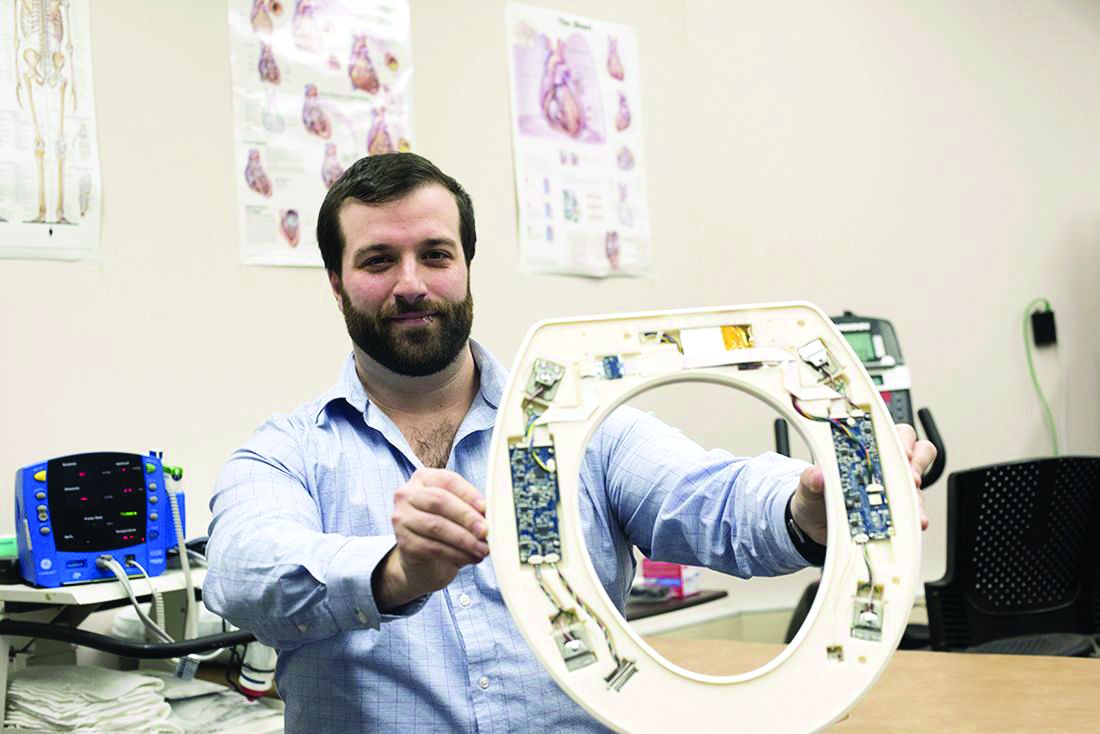
Here’s the deal: A company called Heart Health Intelligence (See? Intelligence is involved, so clearly we’re not talking about politics) has developed a toilet seat equipped “with an integrated electrocardiogram, ballistocardiogram , and photoplethysmogram … capable of clinical-grade measurements of systolic and diastolic blood pressure, stroke volume, and peripheral blood oxygenation.” Wait, ballistocardiogram? That sounds like something from that show House; it sounds too cool to pass up: “Chase, you idiot, why didn’t you do that ballistocardiogram I ordered?”
The idea is to prevent expensive readmissions by keeping track of patients with heart failure after they leave the hospital in a way that fits into their daily routine and ensures adherence.
It would cost about $200,000 to provide 150 heart failure patients with the toilet seats when they left the hospital, the company estimated, but the penalties from the Centers for Medicare & Medicaid Services for readmitting 150 patients come to $500,000 a year. Thus saving money by flushing it down the toilet.
So it looks like we were talking about politics after all. An intelligent toilet seat is like the Oval Office because, in both cases, the buck stops here.

It’s the pits
Physical therapists sometimes use therapeutic ball pits to provide stimulation to pediatric patients with sensory impairments. However, anyone who’s ever been to a fast food joint or a Chuck E. Cheeses in the last 30 years knows that ball pits are not as innocent as they seem – instead of being simply a joyous sunken hole of fun, they are a prime breeding ground for all sorts of bacteria. And adhesive bandages. Why are there always bandages??
Researchers recently took a deep, disgusting dive into the underworld of clinical ball pits, which do not seem to be regulated by any governing body. There are no clinical guidelines from the Ball Pit Association of America on how or when to clean these cesspools of spheres. In fact, there’s no Ball Pit Association of America at all! Shocking.
Unsurprisingly (for any parent or person older than 14), the ball pits sampled were teeming with human and zoonotic-associated microorganisms. Researchers found that “bacterial colonization was found to be as high as thousands of cells per ball” (gag), exposing children to a lot of opportunity for infections. In case the diapers and bandages swimming in the McDonald’s ball pit didn’t turn you off them, this certainly should.
Zom-bacteria are coming
Special edition: Zombie Bacteria vs. The World. Recent research reveals that one type of bacteria, Bacillus subtilis, enters an oligotrophic state when nutrient starved, in which they are not fully dormant but verrrrrry slowly continue to grow and divide. Much like zombies verrrrry slowly continue to walk and eat flesh.
This research fundamentally changes the way we look at bacteria’s ability to survive. Previously, all bacteria were thought to go into a state of complete dormancy within a protective endospore. It appears that the zombie-like bacteria can awaken more easily from their oligotrophic state than bacteria using an endospore, which may shed light on how bacteria can escape antibiotic treatments. As if superbugs weren’t enough to deal with already.
An OTC beer keeps the doctor away
There’s plenty of evidence that a beer or two every now and again is good for your health. But the owner of the Seery Athlone Brewing Company in Addison, Ill., may have taken things too far.
While the reason of its closure just a few months after its grand opening in December 2018 is technically still a mystery, we feel fairly safe in speculating that it had something to do with the fact that brewery owner James Stephen was making his beer at the same location as his main business – a pharmaceutical company engaging in testing of over-the-counter drugs.
In fact, the Food and Drug Administration sent Mr. Stephen a letter in August 2018 warning him that, among numerous other transgressions, fermenting beer literally 10 feet away from where drugs were being tested is a health hazard, for both the beer and the drugs. The FDA ordered the brewery shut down, but the intrepid Mr. Stephen soldiered on, to obviously limited success.
Our advice to Mr. Stephen? Go all in with the pharmaceutical theme. Who wouldn’t want to drink an ibuprofen IPA, ranitidine red ale, loratadine lambic, pseudoephedrine porter, or diphenhydramine doppelbock? You’ll make millions!
A seat in the oval office
No, not that seat, and not the Oval Office. This is LOTME, after all, not Politico. The “seat” we’re talking about is a cardiovascular monitoring system, and “oval office” is just a euphemism for a toilet.
Here’s the deal: A company called Heart Health Intelligence (See? Intelligence is involved, so clearly we’re not talking about politics) has developed a toilet seat equipped “with an integrated electrocardiogram, ballistocardiogram , and photoplethysmogram … capable of clinical-grade measurements of systolic and diastolic blood pressure, stroke volume, and peripheral blood oxygenation.” Wait, ballistocardiogram? That sounds like something from that show House; it sounds too cool to pass up: “Chase, you idiot, why didn’t you do that ballistocardiogram I ordered?”
The idea is to prevent expensive readmissions by keeping track of patients with heart failure after they leave the hospital in a way that fits into their daily routine and ensures adherence.
It would cost about $200,000 to provide 150 heart failure patients with the toilet seats when they left the hospital, the company estimated, but the penalties from the Centers for Medicare & Medicaid Services for readmitting 150 patients come to $500,000 a year. Thus saving money by flushing it down the toilet.
So it looks like we were talking about politics after all. An intelligent toilet seat is like the Oval Office because, in both cases, the buck stops here.
PK analysis shows benefit for rFVIII product in hemophilia A
Switching from the sucrose‐formulated recombinant factor VIII replacement therapy rFVIII‐FS (Kogenate) to the rFVIII product BAY 81‐8973 (Kovaltry) resulted in a longer half-life and improved bleeding rates in patients with hemophilia A, according to a recent report.
Juan Eduardo Megías‐Vericat, PharmD, of the Hospital Universitari i Politècnic La Fe, Valencia, Spain, and his colleagues conducted a comparative, cross‐sectional, pharmacokinetic analysis of switching from rFVIII‐FS to BAY 81‐8973 in patients with hemophilia A. The results of the analysis were reported in a letter to the editor published in Haemophilia.
“All the patients of this cohort were switched in a short period of time, while maintaining the same dose and frequency of infusions,” the researchers wrote.
The study included 14 patients who had moderate (n = 3) or severe (n = 11) hemophilia A and received pharmacokinetic-guided prophylaxis with rFVIII. The median weekly doses were 60.6 IU/kg and 61.6 IU/kg for rFVIII‐FS and BAY 81‐8973, respectively.
After analysis, the researchers found that the half-life of BAY 81‐8973 was significantly longer than rFVIII‐FS (median half-life, 16.9 hours vs. 15.4 hours; P = .001), with a median improvement of 3.0 hours (range, 1.5-4.0 hours). No significant differences were seen in any other pharmacokinetic factors.
With respect to bleeding, significant reductions were seen in annualized joint bleeding rate (P = .021), annualized bleeding rate (P = .038), and the number of spontaneous bleeds (P = .028) after the switch to BAY 81-8973.
The researchers acknowledged that the external validity of the study was a key limitation. As a result, Dr. Megías‐Vericat and his colleagues recommended that the findings be interpreted with discretion.
“The results of this study should be validated with an independent and larger cohort of patients, analyzing also the differences between adult and pediatric patients,” they concluded.
The study was partially funded by an unrestricted grant from Bayer. The authors reported having no conflicts of interest.
SOURCE: Megías-Vericat JE et al. Haemophilia. 2019 Mar 13. doi: 10.1111/hae.13733.
FROM HAEMOPHILIA