User login
Guidelines on Integrating New Migraine Treatments
In order to provide healthcare professionals with updated guidance in the use of novel preventive and acute treatments for migraine in adults, a recent position statement released by the American Headache Society updates prior recommendations and outlines the indications for initiating, continuing, combining, and switching preventive and acute treatments. Input was sought from health insurance providers, employers, pharmacy benefit service companies, device manufacturers, pharmaceutical and biotechnology companies, patients, and patient advocates. In addition, expert clinicians and researchers in the field of headache medicine from across North America and the European Union provided input and feedback.
The principles of pharmacologic preventive treatment of migraine with oral treatments have been as follows:
- Use evidence‐based treatments when possible and appropriate.
- Start with a low dose and titrate slowly; newer injectable treatments may work faster and may not need titration.
- Reach a therapeutic dose if possible.
- Allow for an adequate treatment trial duration.
- Establish expectations of therapeutic response and adverse events and maximize adherence.
The principles of acute treatment include:
- Use evidence‐based treatments when possible and appropriate.
- Treat early after the onset of a migraine attack.
- Choose a non-oral route of administration for selected patients.
- Account for tolerability and safety issues.
- Consider self‐administered rescue treatments.
- Avoid overuse of acute medications.
American Headache Society. The American Headache Society position statement on integrating new migraine treatments into clinical practice. [Published online ahead of print December 10, 2018]. Headache. doi:10.1111/head.13456.
Migraine with Visual Aura a Risk Factor for AF
Migraine with aura was associated with increased risk of incident atrial fibrillation (AF), according to a recent study, which may potentially lead to ischemic strokes. In the Atherosclerosis Risk in Communities (ARIC) study, participants were interviewed for migraine history from 1993 through 1995 and were followed for incident AF through 2013. AF was adjudicated using electrocardiograms (ECGs), discharge codes, and death certificates. Multivariable Cox proportional hazards models were used to study the relation between migraine and its subtypes with incident AF, compared with controls without headaches. Researchers found:
- Of 11,939 participants assessed for headache and without prior AF or stroke, 426 reported migraine with visual aura, 1090 migraine without visual aura, 1018 non-migraine headache, and 9405 no headache.
- Over a 20-year follow-up period, incident AF was noted in 232 (15%) of 1516 with migraine and 1623 (17%) of 9405 without headache.
- After adjustment for multiple confounders, migraine with visual aura was associated with increased risk of AF compared to no headache as well as when compared to migraine without visual aura.
Sen S, Androulakis XM, Duda V, et al. Migraine with visual aura is a risk factor for incident atrial fibrillation. A cohort study. [Published online ahead of print December 11, 2018]. Neurology. doi:10.1212/WNL.0000000000006650.
Women with Migraine Have Lower T2D Risk
Women with active migraine have a lower risk of developing type 2 diabetes (T2D), according to a recent study, and a decrease in active migraine prevalence prior to diabetes diagnosis. Researchers used data from a prospective population-based study initiated in 1990 on a cohort of women born between 1925 and 1950. From eligible women in the study, researchers included those who completed a 2002 follow-up questionnaire with information available on migraine. They then excluded prevalent cases of T2D, leaving a final sample of women who were followed up between 2004 and 2014. All potential occurrences of T2D were identified through a drug reimbursement database. They found:
- From the 98,995 women in the study, 76,403 women completed the 2002 follow-up survey.
- Of these, 2156 were excluded because they had T2D, leaving 74,247 women.
- During 10 years of follow-up, 2372 incident T2D cases occurred.
- A lower risk of T2D was observed for women with active migraine compared with women with no migraine history (univariate hazard ratio, 0.80, multivariable-adjusted hazard ratio, 0.7).
Fagherazzi G, El Fatouhi D, Fournier A, et al. Associations between migraine and type 2 diabetes in women: Findings from the E3N Cohort Study. [Published online ahead of print December 17, 2018]. JAMA Neurology. doi:10.1001/jamaneurol.2018.3960.
Back pain criteria perform well in patients with active axial psoriatic arthritis
A back pain screening questionnaire developed for ankylosing spondylitis performs well for identifying the subset of axial psoriatic arthritis patients who have active symptoms, according to researchers.
The inflammatory back pain criteria didn’t perform as well when patients with established disease but no symptoms were included, though using a lower cutoff point for the questionnaire improved its sensitivity, the researchers reported in the Annals of the Rheumatic Diseases.
Previous investigations showed that the inflammatory back pain criteria, as defined by the Assessment of Spondyloarthritis International Society (ASAS), had low sensitivity and high specificity for axial involvement in psoriatic arthritis.
Those earlier studies may have registered suboptimal performance of the inflammatory back pain criteria by not distinguishing between patients with axial disease in remission and those with active symptoms, according to Muhammad Haroon, PhD, of the division of rheumatology at University Hospital Kerry in Tralee, Ireland, and his coinvestigators.
The present study, which they said represents a much larger cohort than earlier investigations, included 406 patients with psoriatic arthritis, about one-quarter of whom had rheumatologist-diagnosed axial psoriatic arthritis. The mean age of the axial psoriatic arthritis patients was 51 years and 53% were male.
The researchers found that the inflammatory back pain criteria had poor sensitivity but good specificity at 59% and 84%, respectively, in patients with established axial psoriatic arthritis, defined as axial disease regardless of whether it was active or in remission.
By contrast, the criteria had good sensitivity and good specificity at 82% and 88%, respectively, in patients who had active axial psoriatic arthritis, according to the investigators.
The standard cutoff points used by the ASAS inflammatory back pain criteria may be too high for screening for early disease or for evaluating patients already receiving systemic therapies for psoriatic disease, the investigators said.
Looking at a lower cutoff point of three of five ASAS criteria, sensitivity was “quite high” for detecting established axial psoriatic arthritis, they said, increasing from 59% to 84%, while specificity remained relatively high, decreasing from 84% to 80%.
“We suggest that the standard cutoffs for this questionnaire be used for patients with active axial psoriatic arthritis, and the lower cutoffs should be used among patients with established axial psoriatic arthritis, where patients can potentially be in remission or partial remission,” they wrote in their report.
These findings could have important implications for the use of this screening tool in patients with psoriatic arthritis; however, more research is needed to validate the observations, the researchers cautioned.
Dr. Haroon reported competing interests related to AbbVie, Pfizer, and Celgene.
SOURCE: Haroon M et al. Ann Rheum Dis. 2018 Dec 14. doi: 10.1136/annrheumdis-2018-214583.
FROM ANNALS OF THE RHEUMATIC DISEASES
Key clinical point: An inflammatory back pain screening questionnaire, developed for ankylosing spondylitis, performed well in identifying axial psoriatic arthritis in patients with active symptoms.
Major finding: The tool performed suboptimally when patients without active symptoms were included, but had good sensitivity (82%) and specificity (88%) in patients with active axial psoriatic arthritis.
Study details: A study including more than 400 patients with psoriatic arthritis.
Disclosures: The corresponding author reported competing interests related to AbbVie, Pfizer, and Celgene.
Source: Haroon M et al. Ann Rheum Dis. 2018 Dec 14. doi: 10.1136/annrheumdis-2018-214583.
Continuous certification – Not just one more hoop to jump through
Maintenance of Certification (MOC) is an American Board of Medical Specialties (ABMS) requirement for their 24 member boards. The MOC process has received much criticism, especially in recent years. To date, a 5-hour exam at a secure testing center every 10 years covering comprehensive vascular surgery knowledge has been the routine. This requirement had the surgeon take off a day from work for the exam, in addition to the time it took to prepare. Burnout, at least in part, is related to the sheer volume of busywork not directly relevant to being a practicing surgeon.

The American Board of Surgery is sensitive to both the relevance of MOC and needs of the diplomate, and is striving to make appropriate changes. Diplomates were surveyed regarding MOC and the accompanying exam in both 2016 and 2017. Using this input, the development of the 10-year exam format was studied carefully by the board directors and executive staff, all of whom are active in the clinical practice of surgery, and a new process now known as Continuous Certification was introduced. The intent of the new Continuous Certification Assessment (to replace the every-10-year MOC exam) is to be an activity that is convenient, timely, and more reflective of the surgeon’s daily practice. The assessment is to be done every 2 years and is online, open book, and taken at a place of the examinee’s choosing, such as the home or in the office. Another key feature of the continuous certification process is that the total number of CME required is decreased and the self-assessment requirement is eliminated.
In November 2018, I took the first General Surgery Continuous Certification Assessment. There was approximately a 2-month window to register, and online registration was simple, taking only about 15 minutes to complete. All the references were listed on the ABS website and the vast majority were open access and directly linked to the article. For those articles that were not open access, there was a link to the PubMed abstract. I downloaded all of the articles (actually this part my assistant did) and requested five articles from the library. I did not review the articles in advance, but used them when going sequentially through the assessment questions. Depending on the article, I read it or looked up the specific aspect I was looking for. I worked on the test three different times – at the airport during a long layover, at home, and at my office. After answering each question, I received feedback on what was the correct answer and a one-paragraph explanation which I read completely. After completing all 40 questions, each question for which I had an incorrect answer (not more than one or two of course, Ha!) was shown again with the opportunity to answer the question. The total time took me was about 4.5 hours. All in all, it was a good experience, and I learned something.
The general surgery assessment is modular. Twenty questions (half) were core surgery topics, and the other twenty questions came from one of four specialty modules of the examinee’s choice – breast, abdomen, alimentary tract, or comprehensive general surgery. I took the core and the abdomen modules. The core topics were, for the most part, areas that a surgeon who does patient care would find relevant (for example, perioperative management of a patient on corticosteroids, postoperative delirium, and prophylaxis for venous thromboembolism).
A couple of other details should be mentioned about this new process. From the time of initiation of the assessment, there are 2 weeks allocated for completion. One needs 80% correct to pass. If the examinee receives less than 80% but higher than 40% on the first assessment attempt, he/she will have a second attempt to answer the questions that were incorrect on the first try. If a cumulative score of less than 80% is achieved after the second attempt, a grace year will be provided, which is an extension of certification for 1 year with the opportunity to take the next year’s assessment. If after the grace year (four attempts) the diplomate is unsuccessful, then a secure exam is required to regain certification.
Overall, there has been much positive feedback. Of the 2,164 diplomates taking the Continuous Certification Assessment, only 21 were unsuccessful. Therefore, the pass rate was over 99% for the inaugural year. The average examinee took just over 3 hours to complete the assessment.
In 2018, the 10-year recertification examination in vascular surgery with 10 years of credit was given for the last time. The Vascular Surgery Continuous Certification Assessment is in preparation now and will roll out in the fall of 2019. It will follow a format similar to general surgery with 40 questions on a number of topics in vascular surgery. However, the vascular surgery assessment will not be modular. This activity will incorporate general knowledge (for example, from consensus guidelines), as well as late breaking trials. So far, this process looks to be a better one, as well as more efficient and relevant for the busy surgeon.
Dr. Gahtan is professor and chief, division of vascular surgery and endovascular services, State University of New York Upstate Medical University, Syracuse.
Maintenance of Certification (MOC) is an American Board of Medical Specialties (ABMS) requirement for their 24 member boards. The MOC process has received much criticism, especially in recent years. To date, a 5-hour exam at a secure testing center every 10 years covering comprehensive vascular surgery knowledge has been the routine. This requirement had the surgeon take off a day from work for the exam, in addition to the time it took to prepare. Burnout, at least in part, is related to the sheer volume of busywork not directly relevant to being a practicing surgeon.
The American Board of Surgery is sensitive to both the relevance of MOC and needs of the diplomate, and is striving to make appropriate changes. Diplomates were surveyed regarding MOC and the accompanying exam in both 2016 and 2017. Using this input, the development of the 10-year exam format was studied carefully by the board directors and executive staff, all of whom are active in the clinical practice of surgery, and a new process now known as Continuous Certification was introduced. The intent of the new Continuous Certification Assessment (to replace the every-10-year MOC exam) is to be an activity that is convenient, timely, and more reflective of the surgeon’s daily practice. The assessment is to be done every 2 years and is online, open book, and taken at a place of the examinee’s choosing, such as the home or in the office. Another key feature of the continuous certification process is that the total number of CME required is decreased and the self-assessment requirement is eliminated.
In November 2018, I took the first General Surgery Continuous Certification Assessment. There was approximately a 2-month window to register, and online registration was simple, taking only about 15 minutes to complete. All the references were listed on the ABS website and the vast majority were open access and directly linked to the article. For those articles that were not open access, there was a link to the PubMed abstract. I downloaded all of the articles (actually this part my assistant did) and requested five articles from the library. I did not review the articles in advance, but used them when going sequentially through the assessment questions. Depending on the article, I read it or looked up the specific aspect I was looking for. I worked on the test three different times – at the airport during a long layover, at home, and at my office. After answering each question, I received feedback on what was the correct answer and a one-paragraph explanation which I read completely. After completing all 40 questions, each question for which I had an incorrect answer (not more than one or two of course, Ha!) was shown again with the opportunity to answer the question. The total time took me was about 4.5 hours. All in all, it was a good experience, and I learned something.
The general surgery assessment is modular. Twenty questions (half) were core surgery topics, and the other twenty questions came from one of four specialty modules of the examinee’s choice – breast, abdomen, alimentary tract, or comprehensive general surgery. I took the core and the abdomen modules. The core topics were, for the most part, areas that a surgeon who does patient care would find relevant (for example, perioperative management of a patient on corticosteroids, postoperative delirium, and prophylaxis for venous thromboembolism).
A couple of other details should be mentioned about this new process. From the time of initiation of the assessment, there are 2 weeks allocated for completion. One needs 80% correct to pass. If the examinee receives less than 80% but higher than 40% on the first assessment attempt, he/she will have a second attempt to answer the questions that were incorrect on the first try. If a cumulative score of less than 80% is achieved after the second attempt, a grace year will be provided, which is an extension of certification for 1 year with the opportunity to take the next year’s assessment. If after the grace year (four attempts) the diplomate is unsuccessful, then a secure exam is required to regain certification.
Overall, there has been much positive feedback. Of the 2,164 diplomates taking the Continuous Certification Assessment, only 21 were unsuccessful. Therefore, the pass rate was over 99% for the inaugural year. The average examinee took just over 3 hours to complete the assessment.
In 2018, the 10-year recertification examination in vascular surgery with 10 years of credit was given for the last time. The Vascular Surgery Continuous Certification Assessment is in preparation now and will roll out in the fall of 2019. It will follow a format similar to general surgery with 40 questions on a number of topics in vascular surgery. However, the vascular surgery assessment will not be modular. This activity will incorporate general knowledge (for example, from consensus guidelines), as well as late breaking trials. So far, this process looks to be a better one, as well as more efficient and relevant for the busy surgeon.
Dr. Gahtan is professor and chief, division of vascular surgery and endovascular services, State University of New York Upstate Medical University, Syracuse.
Knee and elbow rejuvenation
The cosmetic industry improves techniques for tightening faces, hands, necks, and decolletes; meanwhile, sagging elbows and knees, once ignored, also are a visible sign of aging. Modifying techniques commonly used for the face and neck can yield significant improvements in the elbows and knees. The elbows and knees naturally have looser skin to allow for joint movement; over time, the skin over these joints is exposed to sun damage, friction, and recurrent extension and flexion, which cause skin laxity and aging.

A combination approach addressing skin texture, collagen damage, rhytides, and fat deposition is the most effective method for knee and elbow rejuvenation.
For knees and elbows with loose skin and rhytides, in-office noninvasive and minimally invasive radio-frequency and light energy treatments are helpful in increasing collagen production and tissue tightening. Similarly, microfocused ultrasound has been shown to be a safe and effective skin tightening treatment for the knees. In comparison to the face, however, the skin around the elbows and knees can be thinner and has fewer sebaceous glands. Caution should be used particularly with minimally invasive radio-frequency techniques in order to protect the epidermal skin. Often, treatments have to be repeated to give optimal results, which are not apparent until 3-6 months after the initial procedure.
For knee skin with severe laxity, a comprehensive approach using polydioxanone (PDO) or poly-l-lactic acid (PLLA) threads in both the upper thighs and circumferentially around the knees provides collagen production and tightening of the loose skin. Treatment of the upper thighs is essential in providing a vector that lifts the skin of the knees. Treatments can be repeated, with results seen after 90 days. Thread lifts of the knees and thighs are highly effective, noninvasive procedures with little to no downtime and can be used for severe skin laxity, wrinkling, and thinning of the knee skin.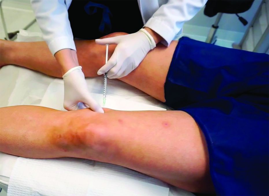
Loose, roughened knee and elbow skin can also be treated with nonablative factional resurfacing, radio-frequency microneedling, or a series of monthly treatments with PLLA and hyaluronic acid fillers injected in the superficial to mid-dermis. Both fractional resurfacing and dermal filler injections help stimulate collagen production and improve both fine rhytides and dermatoheliosis.
Adipose tissue around the knees can be treated with monthly deoxycholic acid injections (for a video of this procedure, go to https://drive.google.com/file/d/1rhw-nESy15AoDhKUrc25DDjKEun7RL4i/view). The volume of injection, however, is significantly higher than that recommended in the submental area. Two to four times the volume is needed per knee over a series of 3-6 treatments, depending on the amount of fat in the knees.
Cryolipolysis is also an effective option for fat pockets around the knees; however, in my experience, it can be difficult to fit the applicators onto the area of concern appropriately unless smaller applicators are applied.
With the increasing demand for body rejuvenation techniques, providers are adapting techniques used for the face and neck to lift, tighten, thin, and sculpt the knees and elbows. A combination approach using lasers, ultrasound, fillers, threads, and cryolipolysis can be effective for these areas. Results are obtainable when repeat treatments are performed; however, one must be patient because results are not seen for 6 months or more.
Dr. Lily Talakoub and Dr. Naissan Wesley are cocontributors to this column. Dr. Talakoub is in private practice in McLean, Va. Dr. Wesley practices dermatology in Beverly Hills, Calif. This month’s column is by Dr. Talakoub. Write to them at dermnews@mdedge.com. They had no relevant disclosures.
References
Macedo O. et al. J Am Acad Dermatol. 2014;70(Suppl 1), Abstract P800, page AB193.
The cosmetic industry improves techniques for tightening faces, hands, necks, and decolletes; meanwhile, sagging elbows and knees, once ignored, also are a visible sign of aging. Modifying techniques commonly used for the face and neck can yield significant improvements in the elbows and knees. The elbows and knees naturally have looser skin to allow for joint movement; over time, the skin over these joints is exposed to sun damage, friction, and recurrent extension and flexion, which cause skin laxity and aging.
A combination approach addressing skin texture, collagen damage, rhytides, and fat deposition is the most effective method for knee and elbow rejuvenation.
For knees and elbows with loose skin and rhytides, in-office noninvasive and minimally invasive radio-frequency and light energy treatments are helpful in increasing collagen production and tissue tightening. Similarly, microfocused ultrasound has been shown to be a safe and effective skin tightening treatment for the knees. In comparison to the face, however, the skin around the elbows and knees can be thinner and has fewer sebaceous glands. Caution should be used particularly with minimally invasive radio-frequency techniques in order to protect the epidermal skin. Often, treatments have to be repeated to give optimal results, which are not apparent until 3-6 months after the initial procedure.
For knee skin with severe laxity, a comprehensive approach using polydioxanone (PDO) or poly-l-lactic acid (PLLA) threads in both the upper thighs and circumferentially around the knees provides collagen production and tightening of the loose skin. Treatment of the upper thighs is essential in providing a vector that lifts the skin of the knees. Treatments can be repeated, with results seen after 90 days. Thread lifts of the knees and thighs are highly effective, noninvasive procedures with little to no downtime and can be used for severe skin laxity, wrinkling, and thinning of the knee skin.
Loose, roughened knee and elbow skin can also be treated with nonablative factional resurfacing, radio-frequency microneedling, or a series of monthly treatments with PLLA and hyaluronic acid fillers injected in the superficial to mid-dermis. Both fractional resurfacing and dermal filler injections help stimulate collagen production and improve both fine rhytides and dermatoheliosis.
Adipose tissue around the knees can be treated with monthly deoxycholic acid injections (for a video of this procedure, go to https://drive.google.com/file/d/1rhw-nESy15AoDhKUrc25DDjKEun7RL4i/view). The volume of injection, however, is significantly higher than that recommended in the submental area. Two to four times the volume is needed per knee over a series of 3-6 treatments, depending on the amount of fat in the knees.
Cryolipolysis is also an effective option for fat pockets around the knees; however, in my experience, it can be difficult to fit the applicators onto the area of concern appropriately unless smaller applicators are applied.
With the increasing demand for body rejuvenation techniques, providers are adapting techniques used for the face and neck to lift, tighten, thin, and sculpt the knees and elbows. A combination approach using lasers, ultrasound, fillers, threads, and cryolipolysis can be effective for these areas. Results are obtainable when repeat treatments are performed; however, one must be patient because results are not seen for 6 months or more.
Dr. Lily Talakoub and Dr. Naissan Wesley are cocontributors to this column. Dr. Talakoub is in private practice in McLean, Va. Dr. Wesley practices dermatology in Beverly Hills, Calif. This month’s column is by Dr. Talakoub. Write to them at dermnews@mdedge.com. They had no relevant disclosures.
References
Macedo O. et al. J Am Acad Dermatol. 2014;70(Suppl 1), Abstract P800, page AB193.
DRd improves PFS in transplant-ineligible MM

SAN DIEGO—An interim analysis from the MAIA trial showed that adding daratumumab to lenalidomide and dexamethasone could significantly improve progression-free survival (PFS) in older patients with multiple myeloma (MM) who were ineligible for transplant.
The 30-month PFS rate was 71% in patients who received daratumumab plus lenalidomide and dexamethasone (DRd) and 56% in patients who received only lenalidomide and dexamethasone (Rd).
“These results support DRd as a new standard of care for elderly patients with myeloma who are ineligible for transplant,” said Thierry Facon, MD, of Hôpital Claude Huriez and the University of Lille in France.
Dr. Facon presented results from MAIA during the late-breaking abstracts session at the 2018 ASH Annual Meeting (abstract LBA-2).
The phase 3 trial (NCT02252172) enrolled 737 transplant-ineligible patients with newly diagnosed MM.
The patients were randomly assigned to DRd or Rd. Daratumumab was given at 16 mg/kg weekly for cycles 1 and 2, every other week for cycles 3 through 6, and every 4 weeks from cycle 7 until disease progression.
Lenalidomide was given at 25 mg orally per day on days 1-21 until disease progression, and dexamethasone was given at 40 mg orally or intravenously weekly until disease progression.
The median patient age was 73 years, and 99% of all patients were 65 or older. Demographic and clinical characteristics were well balanced between the treatment arms.
Results
The primary endpoint of PFS was superior with DRd.
At a median follow-up of 28 months, the median PFS had not been reached in the DRd arm and was 31.9 months in the Rd arm.
The 30-month PFS rate was 71% in the DRd arm and 56% in the Rd arm (hazard ratio [HR]=0.56; P<0.0001).
DRd was associated with a significantly higher overall response rate than Rd—93% and 81%, respectively (P<0.0001).
The complete response rates were 48% and 25%, respectively (P<0.0001). The rates of very good partial response or better were 79% and 53%, respectively (P<0.0001). And the rates of minimal residual disease negativity were 24% and 7%, respectively (P<0.0001).
DRd was associated with infusion-related reactions in 41% of patients, and 3% were grade 3 or 4 in severity.
Hematologic treatment-emergent adverse events of grade 3 or higher that were more common with DRd than Rd included neutropenia (50% vs. 35%) and lymphopenia (15% vs. 11%).
Conversely, grade 3/4 thrombocytopenia (7% vs. 9%) and anemia (12% vs. 20%) were more frequent with Rd.
Nonhematologic treatment-emergent adverse events that were more frequent with DRd included diarrhea, constipation, fatigue, peripheral edema, and pneumonia.
Rates of asthenia, back pain, nausea, and deep vein thrombosis/pulmonary embolism were similar between the treatment arms.
Janssen funded this study. Dr. Facon reported relationships with Celgene, Janssen, Takeda, Sanofi, Amgen, Karyopharm, and Oncopeptides.
Bipolar patients’ ability to consent can be measured with MacCAT-CR
, according to Christina C. Klein and her associates.
A total of 50 patients who were enrolled in a clinical trial of approved, standard treatments for bipolar disease were included in the consent study. The MacCAT-CR was administered after patients had given consent to be included in the trial, but before the trial had started. Four patients lacked the ability to provide consent for the trial after receiving the MacCAT-CR. After these patients were reeducated and went through the consent process a second time, three were enrolled and one declined enrollment.
Patients with higher Schedule for Assessment of Positive Symptoms scores were more likely to have worse MacCAT-CR Understanding and Appreciation subscale scores; lower Hamilton Depression Rating Scale and higher Clinical Global Impression–Severity scores were associated with worse Reasoning and Understanding subscale scores.
Comorbid ADHD, sex, IQ scores, and age at onset of bipolar disorder were not correlated with any subscale scores. In addition, a history of substance use disorder was associated with higher Appreciation and Reasoning subscale scores.
“The current study provides important information for clinicians and researchers to consider when obtaining informed consent from an individual with bipolar disorder. The MacCAT-CR may serve to identify patients, specifically those with higher psychotic symptoms or global illness severity, as needing additional education regarding informed consent,” the investigators concluded.
Three study coauthors reported conflicts of interest with numerous pharmaceutical companies.
lfranki@mdedge.com
SOURCE: Klein CC et al. J Affect Disord. 2018 Aug 13. doi: 10.1016/j.jad.2018.08.049.
FROM THE JOURNAL OF AFFECTIVE DISORDERS
AGA to FDA: We support new labeling recommendations for probiotics
In a new comment letter to FDA, AGA commends FDA’s recent draft guidance – “Policy Regarding Quantitative Labeling of Dietary Supplements Containing Live Microbials” – clarifying the expectations of probiotics manufacturers who choose to specify the amount of a live microbial component in their product in colony forming units (CFUs).
Though manufacturers are not currently required to report CFUs, AGA feels strongly that all manufacturers of probiotic supplements should voluntarily report the composition of live microbials in their products as CFUs.
However, reporting CFUs alone provides insufficient information to consumers and health care professionals who may recommend probiotic supplements to their patients. In our comment letter, AGA encourages FDA to expand what information manufacturers are required to include. In addition to the conditions already outlined in FDA’s draft guidance, AGA recommends including the conditions of storage as well as an expiration or “use by” date.
We acknowledge that researchers are evaluating other methods and units of measure besides CFUs for not only live microbials but also microbial bioactivity. However, in the absence of a widely accepted alternative, which may take several years to develop and adopt, AGA strongly encourages FDA and manufacturers to take the small step forward of using CFUs now rather than waiting for another solution to emerge.
Probiotics have been an important focus for the AGA Center for Gut Microbiome Research and Education due to the need for evidence-based guidance for health care providers and their patients. The center will continue to work to educate physicians, patients and industry on best practices to ensure safe use of probiotics.
No change in postoperative pain with restrictive opioid protocol
Opioid prescriptions after gynecologic surgery can be significantly reduced without impacting postoperative pain scores or complication rates, according to a paper published in JAMA Network Open.
A tertiary care comprehensive care center implemented an ultrarestrictive opioid prescription protocol (UROPP) then evaluated the outcomes in a case-control study involving 605 women undergoing gynecologic surgery, compared with 626 controls treated before implementation of the new protocol.
The ultrarestrictive protocol was prompted by frequent inquiries from patients who had used very little of their prescribed opioids after surgery and wanted to know what to do with the unused pills.
The new protocol involved a short preoperative counseling session about postoperative pain management. Following that, ambulatory surgery, minimally invasive surgery, or laparotomy patients were prescribed a 7-day supply of nonopioid pain relief. Laparotomy patients were also prescribed a 3-day supply of an oral opioid.
Any patients who required more than five opioid doses in the 24 hours before discharge were also prescribed a 3-day supply of opioid pain medication as needed, and all patients had the option of requesting an additional 3-day opioid refill.
Researchers saw no significant differences between the two groups in mean postoperative pain scores 2 weeks after surgery, and a similar number of patients in each group requested an opioid refill. There was also no significant difference in the number of postoperative complications between groups.
Implementation of the ultrarestrictive protocol was associated with significant declines in the mean number of opioid pills prescribed dropped from 31.7 to 3.5 in all surgical cases, from 43.6 to 12.1 in the laparotomy group, from 38.4 to 1.3 in the minimally invasive surgery group, and from 13.9 to 0.2 in patients who underwent ambulatory surgery.
“These data suggest that the implementation of a UROPP in a large surgical service is feasible and safe and was associated with a significantly decreased number of opioids dispensed during the perioperative period, particularly among opioid-naive patients,” wrote Jaron Mark, MD, of the department of gynecologic oncology at Roswell Park Comprehensive Cancer Center, Buffalo, N.Y., and his coauthors. “The opioid-sparing effect was also marked and statistically significant in the laparotomy group, where most patients remained physically active and recovered well with no negative sequelae or elevated pain score after surgery.”
The researchers also noted that patients who were discharged home with an opioid prescription were more likely to call and request a refill within 30 days, compared with patients who did not receive opioids at discharge.
The study was supported by the Roswell Park Comprehensive Cancer Center, the National Cancer Institute and the Roswell Park Alliance Foundation. Two authors reported receiving fees and nonfinancial support from the private sector unrelated to the study.
SOURCE: Mark J et al. JAMA Netw Open. 2018 Dec 7. doi: 10.1001/jamanetworkopen.2018.5452.
The ultrarestrictive postoperative opioid prescribing protocol described in this study is a promising strategy for reducing opioid prescribing without increasing pain and limiting the potential for diversion and misuse of opioids. An important element of this protocol is the preoperative counseling, because setting patient expectations is likely to be an important factor in improving postoperative outcomes.
It is also worth noting that this study focused on patients undergoing major and minor gynecologic surgery, so more research is needed to explore these outcomes particularly among patients undergoing procedures that may be associated with a higher risk of persistent postoperative pain and/or opioid use. It is also a management strategy explored in patients at low risk of chronic postoperative opioid use, but a similar pathway should be developed and explored in more high-risk patients.
Dr. Jennifer M. Hah is from the department of anesthesiology, perioperative, and pain management at Stanford University (Calif.). These comments are taken from an accompanying editorial (JAMA Network Open. 2018 Dec 7. doi: 10.1001/jamanetworkopen.2018.5432). No conflicts of interest were reported.
Opioid prescriptions after gynecologic surgery can be significantly reduced without impacting postoperative pain scores or complication rates, according to a paper published in JAMA Network Open.
A tertiary care comprehensive care center implemented an ultrarestrictive opioid prescription protocol (UROPP) then evaluated the outcomes in a case-control study involving 605 women undergoing gynecologic surgery, compared with 626 controls treated before implementation of the new protocol.
The ultrarestrictive protocol was prompted by frequent inquiries from patients who had used very little of their prescribed opioids after surgery and wanted to know what to do with the unused pills.
The new protocol involved a short preoperative counseling session about postoperative pain management. Following that, ambulatory surgery, minimally invasive surgery, or laparotomy patients were prescribed a 7-day supply of nonopioid pain relief. Laparotomy patients were also prescribed a 3-day supply of an oral opioid.
Any patients who required more than five opioid doses in the 24 hours before discharge were also prescribed a 3-day supply of opioid pain medication as needed, and all patients had the option of requesting an additional 3-day opioid refill.
Researchers saw no significant differences between the two groups in mean postoperative pain scores 2 weeks after surgery, and a similar number of patients in each group requested an opioid refill. There was also no significant difference in the number of postoperative complications between groups.
Implementation of the ultrarestrictive protocol was associated with significant declines in the mean number of opioid pills prescribed dropped from 31.7 to 3.5 in all surgical cases, from 43.6 to 12.1 in the laparotomy group, from 38.4 to 1.3 in the minimally invasive surgery group, and from 13.9 to 0.2 in patients who underwent ambulatory surgery.
“These data suggest that the implementation of a UROPP in a large surgical service is feasible and safe and was associated with a significantly decreased number of opioids dispensed during the perioperative period, particularly among opioid-naive patients,” wrote Jaron Mark, MD, of the department of gynecologic oncology at Roswell Park Comprehensive Cancer Center, Buffalo, N.Y., and his coauthors. “The opioid-sparing effect was also marked and statistically significant in the laparotomy group, where most patients remained physically active and recovered well with no negative sequelae or elevated pain score after surgery.”
The researchers also noted that patients who were discharged home with an opioid prescription were more likely to call and request a refill within 30 days, compared with patients who did not receive opioids at discharge.
The study was supported by the Roswell Park Comprehensive Cancer Center, the National Cancer Institute and the Roswell Park Alliance Foundation. Two authors reported receiving fees and nonfinancial support from the private sector unrelated to the study.
SOURCE: Mark J et al. JAMA Netw Open. 2018 Dec 7. doi: 10.1001/jamanetworkopen.2018.5452.
Key clinical point: A ultrarestrictive postoperative opioid protocol is not associated with higher postoperative pain scores.
Major finding: The protocol achieves significant reductions in opioid use.
Study details: A case-control study in 1,231 women undergoing gynecologic surgery.
Disclosures: The study was supported by the Roswell Park Comprehensive Cancer Center, the National Cancer Institute, and the Roswell Park Alliance Foundation. Two authors reported receiving fees and nonfinancial support from the private sector unrelated to the study.
Source: Mark J et al. JAMA Netw Open. 2018 Dec 7. doi: 10.1001/jamanetworkopen.2018.5452.