User login
Ongoing research aims to improve transplant outcomes in sickle cell
Researchers are leading several studies designed to improve hematopoietic stem cell transplantation (HSCT) for patients with sickle cell disease (SCD), experts at the National Heart, Lung, and Blood Institute reported during a recent webinar.
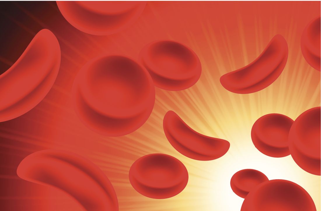
“HSCT offers a potential cure [for SCD], which may improve quantity and quality of life [for patients],” said Courtney D. Fitzhugh, MD, a Lasker Clinical Research Scholar in the Laboratory of Early Sickle Mortality Prevention at NHLBI.
Currently, HLA-matched sibling and matched unrelated donor sources provide the best outcomes for sickle cell patients undergoing allogeneic HSCT, she explained. Alternative stem cell sources include umbilical cord blood and haploidentical donors.
Over the past 2 years, the majority of novel transplant techniques have been primarily aimed at improving conditioning regimens and lowering rates of graft-versus-host disease (GVHD).
Recent evidence
A recent international survey found high survival rates in patients with SCD who underwent HLA-matched sibling HSCT during 1986-2013. At 5-years, overall- and event-free survival rates were 92.9% and 91.4%, respectively, with even higher rates (95% and 93%) seen in children aged younger than 16 years.
With respect to safety, the cumulative incidence rates of acute and chronic GVHD were 14.8% and 14.3%, Dr. Fitzhugh reported.
Much of the success seen with HLA-matched sibling donors is attributable to recent data demonstrating that complete transformation of patient’s bone marrow is unnecessary to illicit a curative effect.
With donor myeloid chimerism levels of at least 20%, the sickle disease phenotype can be reversed, and there’s a reduced risk of GVHD, she said.
In mouse models, researchers have found that inclusion of sirolimus in HLA-matched pretransplant conditioning regimens leads to higher levels of donor cell engraftment. As a result, some conditioning regimens now administer sirolimus (target 10-15 ng/dL) one-day prior to transplantation.
In 55 patients transplanted using this technique, overall- and event-free survival rates of 93% and 87% have been reported, with no transplant-related mortality or evidence of GVHD. Other institutions have also begun to adopt this technique, and have reported similar findings, Dr. Fitzhugh reported.
“When you [administer high-dose] chemotherapy, you don’t expect that patients are able to have children, but we are excited to report that 8 of our patients have had 13 healthy babies post transplant,” Dr. Fitzhugh said.
As a whole, several recent studies have emphasized the importance of the conditioning regimen in successful transplantation for patients with SCD.
With HLA-matched sibling donors, myeloablative regimens that include antithymocyte globulin have demonstrated greater efficacy, she said.
In patients receiving a transplant from a matched unrelated donor, early use of alemtuzumab is linked to higher rates of GVHD, while ongoing studies are exploring whether abatacept reduces the risk of GVHD, she further explained.
With respect to haploidentical and unrelated umbilical cord donors, T-cell depletion and higher-intensity conditioning have been shown to reduce graft rejection rates, she said.
Dr. Fitzhugh acknowledged that long-term efficacy and safety of these novel conditioning regimens is largely unknown. Thus, ongoing follow-up is essential to monitor for potential late effects.
NHLBI-funded trials
Nancy L. DiFronzo, PhD, program director at NHLBI, explained that the agency has funded specific clinical studies evaluating allogeneic HSCT in patients with severe SCD.
“[Surprisingly], this treatment modality is [actually] quite rare, with [only] approximately 9,000 allogeneic transplants occurring in the United States each year,” she said.
One of the primary barriers to HSCT for SCD is a lack of compatible donors. Currently, fewer than 20% of sickle cell patients have a matched unrelated donor or HLA-matched sibling donor, she reported.
Another common barrier are the risks associated with the procedure, including treatment-related toxicities and death. Active participation in a clinical trial is one strategy that can mitigate these risks, she said.
The Blood and Marrow Transplant Clinical Trials Network (BMT CTN) is a group of transplant centers that are recognized experts in HSCT. Dr. DiFronzo explained that the consortium is cosponsored by the National Cancer Institute and NHLBI, with the goal of improving outcomes for both pediatric and adult patients with SCD undergoing HSCT.
At present, the BMT CTN has directly funded three multicenter clinical studies for SCD, including the SCURT study, which has now been completed, as well as the STRIDE2 and Haploidentical HCT trials, both of which are currently enrolling patients.
“The goal of these new approaches [being studied in these 3 trials] is cure, where individuals can live longer with a better quality of life,” Dr. DiFronzo said. “We’ve [specifically] adjusted regimens with [this goal] in mind.”
Dr. Fitzhugh and Dr. DiFronzo did not provide information on financial disclosures.
Researchers are leading several studies designed to improve hematopoietic stem cell transplantation (HSCT) for patients with sickle cell disease (SCD), experts at the National Heart, Lung, and Blood Institute reported during a recent webinar.
“HSCT offers a potential cure [for SCD], which may improve quantity and quality of life [for patients],” said Courtney D. Fitzhugh, MD, a Lasker Clinical Research Scholar in the Laboratory of Early Sickle Mortality Prevention at NHLBI.
Currently, HLA-matched sibling and matched unrelated donor sources provide the best outcomes for sickle cell patients undergoing allogeneic HSCT, she explained. Alternative stem cell sources include umbilical cord blood and haploidentical donors.
Over the past 2 years, the majority of novel transplant techniques have been primarily aimed at improving conditioning regimens and lowering rates of graft-versus-host disease (GVHD).
Recent evidence
A recent international survey found high survival rates in patients with SCD who underwent HLA-matched sibling HSCT during 1986-2013. At 5-years, overall- and event-free survival rates were 92.9% and 91.4%, respectively, with even higher rates (95% and 93%) seen in children aged younger than 16 years.
With respect to safety, the cumulative incidence rates of acute and chronic GVHD were 14.8% and 14.3%, Dr. Fitzhugh reported.
Much of the success seen with HLA-matched sibling donors is attributable to recent data demonstrating that complete transformation of patient’s bone marrow is unnecessary to illicit a curative effect.
With donor myeloid chimerism levels of at least 20%, the sickle disease phenotype can be reversed, and there’s a reduced risk of GVHD, she said.
In mouse models, researchers have found that inclusion of sirolimus in HLA-matched pretransplant conditioning regimens leads to higher levels of donor cell engraftment. As a result, some conditioning regimens now administer sirolimus (target 10-15 ng/dL) one-day prior to transplantation.
In 55 patients transplanted using this technique, overall- and event-free survival rates of 93% and 87% have been reported, with no transplant-related mortality or evidence of GVHD. Other institutions have also begun to adopt this technique, and have reported similar findings, Dr. Fitzhugh reported.
“When you [administer high-dose] chemotherapy, you don’t expect that patients are able to have children, but we are excited to report that 8 of our patients have had 13 healthy babies post transplant,” Dr. Fitzhugh said.
As a whole, several recent studies have emphasized the importance of the conditioning regimen in successful transplantation for patients with SCD.
With HLA-matched sibling donors, myeloablative regimens that include antithymocyte globulin have demonstrated greater efficacy, she said.
In patients receiving a transplant from a matched unrelated donor, early use of alemtuzumab is linked to higher rates of GVHD, while ongoing studies are exploring whether abatacept reduces the risk of GVHD, she further explained.
With respect to haploidentical and unrelated umbilical cord donors, T-cell depletion and higher-intensity conditioning have been shown to reduce graft rejection rates, she said.
Dr. Fitzhugh acknowledged that long-term efficacy and safety of these novel conditioning regimens is largely unknown. Thus, ongoing follow-up is essential to monitor for potential late effects.
NHLBI-funded trials
Nancy L. DiFronzo, PhD, program director at NHLBI, explained that the agency has funded specific clinical studies evaluating allogeneic HSCT in patients with severe SCD.
“[Surprisingly], this treatment modality is [actually] quite rare, with [only] approximately 9,000 allogeneic transplants occurring in the United States each year,” she said.
One of the primary barriers to HSCT for SCD is a lack of compatible donors. Currently, fewer than 20% of sickle cell patients have a matched unrelated donor or HLA-matched sibling donor, she reported.
Another common barrier are the risks associated with the procedure, including treatment-related toxicities and death. Active participation in a clinical trial is one strategy that can mitigate these risks, she said.
The Blood and Marrow Transplant Clinical Trials Network (BMT CTN) is a group of transplant centers that are recognized experts in HSCT. Dr. DiFronzo explained that the consortium is cosponsored by the National Cancer Institute and NHLBI, with the goal of improving outcomes for both pediatric and adult patients with SCD undergoing HSCT.
At present, the BMT CTN has directly funded three multicenter clinical studies for SCD, including the SCURT study, which has now been completed, as well as the STRIDE2 and Haploidentical HCT trials, both of which are currently enrolling patients.
“The goal of these new approaches [being studied in these 3 trials] is cure, where individuals can live longer with a better quality of life,” Dr. DiFronzo said. “We’ve [specifically] adjusted regimens with [this goal] in mind.”
Dr. Fitzhugh and Dr. DiFronzo did not provide information on financial disclosures.
Tranexamic acid does not increase complications in high-risk joint replacement surgery patients
A study has found that administering tranexamic acid (TXA) to high-risk patients undergoing total joint arthroplasty (TJA) does not increase their odds of adverse outcomes.
“The inclusion of high-risk patients in our study increases the generalizability of our findings and is consistent with the previous studies that showed no increase in complications when TXA is administered to TJA patients,” wrote Steven B. Porter, MD, of the Mayo Clinic in Jacksonville, Fla., and coauthors. The study was published in the Journal of Arthroplasty.
To determine the safety of TXA in patients at risk for thrombotic complications, the researchers investigated 38,220 patients who underwent total knee or total hip arthroplasty between 2011 and 2017 at the Mayo Clinic. Of those patients, 20,501 (54%) patients received TXA during their operation and 17,719 (46%) did not. Overall, 8,877 were classified as “high-risk” cases, which meant they had one or more cardiovascular disease or thromboembolic event before surgery.
After multivariable analysis, high risk-patients who received TXA had no significant difference in adverse outcome odds, compared with high-risk patients who did not receive TXA (odds ratio, 1.00; 95% confidence interval, 0.85-1.18). After 90 days, high-risk patients who did not receive TXA were more likely than those who received TXA to experience deep vein thrombosis (2.3% vs 0.8%, P less than .001), pulmonary embolism (1.7% vs 1.0%, P less than .001), cerebrovascular accident (0.8% vs. 0.4%, P less than .001), or death (0.5% vs. 0.4%, P less than .001).
The authors noted their study’s limitations, including a higher baseline incidence of risk factors in high-risk patients who did not receive TXA, compared with high-risk patients who did, which could have led to that group being “self-selected” to not receive TXA. In addition, all medical histories and rates of complications were based on ICD codes, which may have been inaccurate and therefore led to mischaracterized risk or miscoded postoperative complications.
The study was funded by the Mayo Clinic’s Robert D. and Patricia E. Kern Center for the Science of Health Care Delivery. No conflicts of interest were reported.
SOURCE: Porter SB et al. J Arthroplasty. 2019 Aug 17. doi: 10.1016/j.arth.2019.08.015.
FROM THE JOURNAL OF ARTHROPLASTY
Key clinical point: Administering tranexamic acid to high-risk patients undergoing joint replacement surgery does not increase the odds of adverse outcomes.
Major finding: After multivariable analysis, high-risk patients who received tranexamic acid had no significant difference in adverse outcome odds, compared with high-risk patients who did not receive tranexamic acid (odd ratio, 1.00; 95% confidence interval, 0.85-1.18).
Study details: A retrospective case-control study of 38,220 patients who underwent primary total knee or total hip arthroplasty between 2011 and 2017.
Disclosures: The study was funded by the Mayo Clinic’s Robert D. and Patricia E. Kern Center for the Science of Health Care Delivery. No conflicts of interest were reported.
Source: Porter SB et al. J Arthroplasty. 2019 Aug 17. doi: 10.1016/j.arth.2019.08.015.
Quadruplet prolongs progression-free survival in newly diagnosed myeloma
BOSTON – A carfilzomib-based quadruplet can improve outcomes in transplant-eligible patients with newly diagnosed multiple myeloma, a phase 3 trial suggests.
In the Myeloma XI trial, carfilzomib plus cyclophosphamide, lenalidomide, and dexamethasone (KCRD) significantly prolonged progression-free survival (PFS), compared with cyclophosphamide-lenalidomide-dexamethasone (CRD) or cyclophosphamide-thalidomide-dexamethasone (CTD).
“KCRD was associated with a very high response rate and a high MRD [minimal residual disease]-negative rate at the end of induction, and it significantly improved progression-free survival compared to the triplet combinations,” said Charlotte Pawlyn, PhD, of The Institute of Cancer Research in London.
Dr. Pawlyn reported these findings at the International Myeloma Workshop held by the International Myeloma Society.
The phase 3 Myeloma XI trial enrolled 1,056 patients with newly diagnosed myeloma who were eligible for transplant. The patients were randomized to receive KCRD (n = 526), CRD (n = 265), or CTD (n = 265) as induction.
Baseline characteristics were well balanced between the treatment arms. The median age was 61 years in the KCRD and CTD arms and 62 years in the CRD arm (overall range, 33-75 years). Roughly 60% of patients in each arm were men.
About 50%-60% of patients in each arm had standard-risk cytogenetics, which was defined as the absence of any cytogenetic lesions. About 30%-40% of patients in each arm had high-risk cytogenetics, meaning they had one of the following lesions: t(4;14), t(14;16), t(14;20), del (17p), or gain(1q). About 10% of patients in each arm had ultra-high-risk cytogenetics, which was defined as having more than one lesion.
Treatment
For induction, patients were randomized to KCRD, CRD, or CTD. All patients in the KCRD arm and patients in the CRD/CTD arms who achieved a partial response or better went straight to autologous transplant after induction. Nonresponders in the CTD and CRD arms received intensification with cyclophosphamide, bortezomib, and dexamethasone before transplant.
After transplant, all eligible patients were randomized to lenalidomide maintenance or observation. Patients were eligible for this randomization if they didn’t respond to induction, had progressive disease, or had previous or concurrent active malignancies.
The median follow-up was 34.5 months. The median number of induction cycles completed was 4 (range, 1-12) in the KCRD arm, 5 (range, 1-15) in the CRD arm, and 6 (range, 1-13) in the CTD arm.
Response
At the end of induction, the rate of very good partial response or better was 82.3% in the KCRD arm, 64.9% in the CRD arm, 52.8% in the CTD arm, and 58.9% in the CTD-CRD arms combined. The odds ratio for the KCRD group compared to the triplets combined was 4.35 (P less than .0001).
At 100 days after transplant, the rate of very good partial response or better was 91.9% in the KCRD arm, 82.1% in the CRD arm, 76.1% in the CTD arm, and 79.3% in the CTD-CRD arms combined. The odds ratio for the KCRD group compared to the triplets combined was 3.01 (P less than .0001).
KCRD produced a higher proportion of MRD-negative responses both before and after transplant. After induction, the rate of MRD-negative response was 11% in the CTD arm, 21% in the CRD arm, and 55% in the KCRD arm. After transplant, the rates were 51%, 49%, and 77%, respectively.
Survival
KCRD improved PFS. The 3-year PFS rate was 64.5% in the KCRD arm and 50.3% in the CTD-CRD arms combined. The hazard ratio (HR) was 0.63 (P less than .0001).
The PFS benefit with KCRD was present in all patient subgroups. For example, KCRD improved PFS, compared with CTD-CRD, in patients with standard-risk (HR = 0.62), high-risk (HR = 0.68), and ultra-high-risk (HR = 0.50) cytogenetics.
Patients who achieved an MRD-negative response had better PFS, and early achievement of MRD negativity was associated with improved PFS, Dr. Pawlyn noted.
“But what’s also notable ... is that those patients who received KCRD and achieved MRD negativity ... had better outcomes than patients who achieved MRD negativity whilst receiving a triplet combination,” Dr. Pawlyn said. “So this suggests that the induction regimen delivered is important, not just the achievement of MRD negativity at a defined cutoff.”
Dr. Pawlyn added that overall survival data from this study are not yet mature, but the researchers did assess PFS2. PFS2 was defined as the time from randomization to second disease progression. The 3-year PFS2 was 81.8% in the KCRD arm and 75.1% in the CTD-CRD arms combined. The HR was 0.75 (P = .0451).
Myeloma XI is sponsored by University of Leeds in collaboration with Celgene, Merck Sharp & Dohme, and Amgen. Dr. Pawlyn reported relationships with Amgen, Celgene, and other companies.
SOURCE: Pawlyn C et al. IMW 2019, Abstract OAB-002.
REPORTING FROM IMW 2019
CDC awards $1.2 million to learn about people with sickle cell disease
The Centers for Disease Control and Prevention will be awarding $1.2 million in funding to help states collect data on issues faced by people with sickle cell disease.
![]()
Currently, only Georgia and California work with the CDC on the Sickle Cell Data Collection program to gather population-based, comprehensive health information about people with sickle cell disease. The new funding will expand that base to nine states. The money will go toward a 1-year project that will build infrastructure for recipient sites to gather unique data and conduct in-depth analyses in people with sickle cell disease, the CDC noted.
The sites that were awarded funding are Duke University, Durham, N.C.; Georgia State University, Atlanta; the Indiana Hemophilia and Thrombosis Center in Indianapolis; the Michigan Department of Health & Human Services; the Minnesota Department of Health; the Public Health Institute in Oakland, Calif.; the University of Alabama at Birmingham; the University of Tennessee Health Science Center in Memphis; and the Virginia Department of Health.
“Data is vital to informing new treatments and clinical care that will improve the lives of people affected by sickle cell disease. This new funding expands CDC’s partner network across the country which will accelerate efforts to ensure sickle cell patients live longer and healthier lives,” said CDC Director Robert R. Redfield, MD.
Find the full press release on the CDC website.
Several factors may affect immune suppression discontinuation after HCT
New research suggests several factors are associated with failure to discontinue immune suppression after allogeneic hematopoietic cell transplant (HCT).
Patients older than 50 years, those with advanced stage disease, patients with a mismatched unrelated donor, and those who received peripheral blood stem cells from an unrelated donor were less likely to discontinue immune suppression successfully, Joseph Pidala, MD, PhD, of Moffitt Cancer Center in Tampa, Fla., and colleagues reported in JAMA Oncology.
The researchers analyzed data from 827 patients in two national Blood and Marrow Transplant Clinical Trial Network studies (NCT00075816 and NCT00406393). These randomized, phase 3 trials enrolled patients with hematologic malignancies who received myeloablative conditioning before allogeneic HCT.
The patients’ median age at HCT was 44 years (range, less than 1 to 67 years), and 55.1% were male. The median follow-up was 72 months (range, 11-124 months).
At 5 years, 20% of patients (n = 168) had successfully discontinued immune suppression and were still alive. A total of 342 patients (41.4%) were able to stop immune suppression, but 127 of them had to resume it after developing graft-versus-host disease (GVHD). There were an additional 47 patients who died or relapsed after stopping immune suppression.
The researchers identified several factors that were significantly associated with lower odds of discontinuing immune suppression and being free of GVHD, including:
- Being older than 50 years versus younger than 30 years (adjusted odds ratio [aOR], 0.27; 99% confidence interval [CI], 0.14-0.50; P less than .001).
- Having a mismatched unrelated donor versus having a matched sibling (aOR, 0.37; 99% CI, 0.14-0.97; P = .008).
- Receiving peripheral blood stem cells versus bone marrow, from unrelated donors only (aOR, 0.46; 99% CI, 0.26-0.82; P less than .001).
- Having advanced stage disease versus early disease (aOR, 0.45; 99%CI, 0.23-0.86; P = .002).
The researchers also found that discontinuing immune suppression was not significantly associated with a decreased risk of relapse, with a hazard ratio (HR) of 1.95 (99% CI, 0.88-4.31; P = .03).
There was no significant association between acute GVHD–related variables and discontinuation of immune suppression. However, there were a few factors significantly associated with a lower likelihood of discontinuation after chronic GVHD, including:
- Current skin involvement (HR, 0.33; 99% CI, 0.14-0.80; P = .001).
- Unrelated well-matched donor versus matched sibling donor (HR, 0.29; 99% CI, 0.10-0.79; P = .001).
- Unrelated mismatched donor versus matched sibling donor (HR, 0.17; 99% CI, 0.03-0.95; P = .008).
In total, 127 patients had to resume immune suppression because of GVHD. Such failed attempts at discontinuing immune suppression were associated with receiving peripheral blood stem cells from an unrelated donor versus bone marrow from an unrelated donor, with an HR of 2.62 (99% CI, 1.30-5.29; P less than .001).
A history of acute or chronic GVHD was associated with failure to discontinue immune suppression, the researchers noted.
Lastly, the researchers developed dynamic prediction models for the probability of freedom from immune suppression and GVHD at 1, 3, and 5 years in the future. The team found that graft type, donor type, age, state history, and timing of immune suppression discontinuation were all associated with the likelihood of being free from immune suppression and GVHD at all three time points. Disease risk was only associated with freedom from immune suppression and GVHD at the 1-year mark.
The researchers said their findings must be validated in an independent cohort of patients and, after that, should be tested in a prospective trial.
The current study was funded by the National Heart Lung and Blood Institute. Two of the researchers reported relationships with more than 30 pharmaceutical companies.
SOURCE: Pidala J et al. JAMA Oncol. 2019 Sep 26. doi: 10.1001/jamaoncol.2019.2974.
FROM JAMA ONCOLOGY
POCUS for hospitalists: The SHM position statement
Background: POCUS is becoming more prevalent in the daily practice of hospitalists; however, there are currently no established standards or guidelines for the use of POCUS for hospitalists.

Study design: Position statement.
Setting: SHM Executive Committee and Multi-Institutional POCUS faculty meeting through the Society of Hospital Medicine 2018 Annual Conference reviewed and approved this statement.
Synopsis: In contrast to the comprehensive ultrasound exam, POCUS is used by hospitalists to answer focused questions, by the same clinician who is generating the clinical question, to evaluate multiple body systems, or to serially investigate changes clinical status or evaluate responses to therapy.
This position statement provides guidance on the use of POCUS by hospitalists and the administrators who oversee it by outlining POCUS in terms of common diagnostic and procedural applications; training; assessments by the categories of basic knowledge, image acquisition, interpretation, clinical integration, and certification and maintenance of skills; and program management.
Bottom line: This position statement by the SHM provides guidance for hospitalists and administrators on the use and oversight of POCUS.
Citation: Soni NJ et al. Point-of-care ultrasound for hospitalists: A position statement of the Society of Hospital Medicine. J Hosp Med. 2019 Jan 2;14:E1-E6.
Dr. Wang is an associate professor of medicine in the division of general and hospital medicine at UT Health San Antonio and a hospitalist at South Texas Veterans Health Care System.
Background: POCUS is becoming more prevalent in the daily practice of hospitalists; however, there are currently no established standards or guidelines for the use of POCUS for hospitalists.
Study design: Position statement.
Setting: SHM Executive Committee and Multi-Institutional POCUS faculty meeting through the Society of Hospital Medicine 2018 Annual Conference reviewed and approved this statement.
Synopsis: In contrast to the comprehensive ultrasound exam, POCUS is used by hospitalists to answer focused questions, by the same clinician who is generating the clinical question, to evaluate multiple body systems, or to serially investigate changes clinical status or evaluate responses to therapy.
This position statement provides guidance on the use of POCUS by hospitalists and the administrators who oversee it by outlining POCUS in terms of common diagnostic and procedural applications; training; assessments by the categories of basic knowledge, image acquisition, interpretation, clinical integration, and certification and maintenance of skills; and program management.
Bottom line: This position statement by the SHM provides guidance for hospitalists and administrators on the use and oversight of POCUS.
Citation: Soni NJ et al. Point-of-care ultrasound for hospitalists: A position statement of the Society of Hospital Medicine. J Hosp Med. 2019 Jan 2;14:E1-E6.
Dr. Wang is an associate professor of medicine in the division of general and hospital medicine at UT Health San Antonio and a hospitalist at South Texas Veterans Health Care System.
COMISAIR: CGM ‘makes the difference’ in type 1 diabetes
BARCELONA – Real-time continuous glucose monitoring (rtCGM) was better than self-monitoring of blood glucose (SMBG) in reducing hemoglobin A1c (HbA1c) and other glycemic endpoints in people with type 1 diabetes, regardless of the type of insulin delivery method used in a 3-year follow-up of a prospective, real-world clinical trial.
Long-term results from the COMISAIR study showed that the end-of-study HbA1c values were significantly lower, compared with baseline values, in people with type 1 diabetes who used rtCGM with multiple daily injections (MDI) of insulin (7.0% [53 mmol/mol], P = .0002) or an insulin pump (6.9% [52 mmol/mol], P less than .0001). There was no significant difference between the two rtCGM delivery-method groups.
Final HbA1c values for those who used SMBG with multiple daily injections or an insulin pump were 8.0% (64 mmol/mol) and 7.7% (61 mmol/mol), respectively, but were not significantly different from baseline (P = .3574 and P = .1, respectively).
These findings could help guide physicians when discussing treatment and monitoring options with their patients, suggested study investigator Jan Šoupal, MD, PhD, of Charles University in Prague, when he presented the findings at the annual meeting of the European Association for the Study of Diabetes.
Dr. Šoupal and associates have previously reported data from the COMISAIR (Comparison of Different Treatment Modalities for Type 1 Diabetes Including Sensor-Augmented Insulin Regimens) study at 1 year of follow-up for 65 patients (Diabetes Technol Ther. 2016;18:532-8). The findings he presented at the EASD meeting, simultaneously published online Diabetes Care, were for the full cohort of 94 patients and, with 3 years of follow-up, makes it “the longest CGM trial ever,” he said.
At the time the COMISAIR study was initiated, in 2013, “we knew that insulin pump therapy, especially in combination with real-time CGM, can improve several outcomes of patients with type 1 diabetes,” Dr. Šoupal observed. However, the effectiveness of CGM in patients with MDI was not widely described, and comparisons between continuous subcutaneous insulin infusion (CSII) and insulin MDI with rtCGM were lacking. “Moreover, we didn’t have any comparison between insulin pump therapy alone, without CGM, and MDI with CGM, and there were no long-term trials with real-time [continuous glucose monitoring].”
The aim of the COMISAIR study was therefore to compare four different treatment strategies in people with type 1 diabetes who had an HbA1c of 7%-10% (53-86 mmol/mol), despite MDI treatment with insulin analogues and SMBG. The treatment strategies tested were CSII plus rtCGM (n = 26), MDI plus rtCGM (n = 22), CSII plus SMBG (n = 25), and MDI plus SMBG (n = 21). Patients were not randomized to these treatment arms but exposed to all of them during a 4-day DAFNE-like training session and then allowed to choose which they would like to use according to their individual needs and preferences, reflecting real-life practice.
Dr. Šoupal pointed out that two different continuous glucose monitoring devices had been used in the trial, and that 100% of the CGM groups wore a sensor for more than 70% of the time, which was one of the prerequisites for inclusion in the trial. Good adherence was observed, with 93% of patients completing all study visits, and CGM users wearing their sensors on average 88% of the time. “This nice adherence may be connected to the pretty good results,” he observed.
In discussing the HbA1c results, Dr. Šoupal noted that “improvement observed in patients with [continuous glucose monitoring] is stable throughout 3 years, which is not always a reality for different types of treatment for diabetes.” In addition, “it is not so important how insulin is delivered, what is more important is how patients with type 1 diabetes monitor their glucose.”
Another key endpoint of the trial was time in range (70-180 mg/dL [3.9-10 mmol/L]). Results showed significantly more patients achieving this with rtCGM than with SMBG, regardless of whether they were using pump therapy or MDI. Comparing 3-year with baseline values, time in range was 72.3% versus 50.9% for rtCGM with CSII and 69% versus 48.7% for rtCGM with MDI (P less than .0001 for both). Results with SMBG with CSII or MDI were a respective 57.8% versus 50.6% (P = .0114) and 54.7% versus 51.8% (P = 1.0).
Glycemic variability was reduced in patients using insulin pumps with SBMG, and “not surprisingly, there was a reduction in both CGM-augmented groups,” Dr. Šoupal stated.
There was a reduction in the time spent in hypoglycemia from baseline to year 3 in all four groups, but that was significant only for the two rtCGM groups. Overall, there were seven severe hypoglycemia episodes, five in the SMBG groups (two in the CSII group, three in the MDI group) and two in the rtCGM groups (one each in the CSII and MDI groups), with one episode only occurring when the CGM sensor was not being worn.
Three episodes of ketoacidosis were reported – one each in the SMBG-pump, SMBG-MDI, and rtCGM-pump groups.
In summing up, Dr. Šoupal said that “real-time CGM, both with insulin pumps and with [multiple daily injections], provided significant, comparable, and stable improvement of glycemic outcomes.” He added that “treatment with CGM and MDI was more effective than treatment with insulin pump therapy alone, and that CGM and MDI can even be considered as a suitable alternative to treatment with insulin pumps and CMG for some patients.”
With many treatment options available, some will suit patients better than others, he suggested, but although “individualization of our treatment is important”, the COMISAIR data show that “it is CGM that makes the difference”.
The study was supported by the Agency for Healthcare Research and the Ministry of Health of the Czech Republic. Dr. Šoupal reported receiving honoraria from Abbott, AstraZeneca, Boehringer Ingelheim, Dexcom, Eli Lilly, Medtronic, Novo Nordisk, and Roche. Dexcom also paid for the development of the manuscript published in Diabetes Care.
SOURCES: J et al. EASD 2019, Abstract 40; J et al. Diabetes Care. 2019 Sep 17. doi: 10.2337/dc19-0888.
REPORTING FROM EASD 2019
Older black women have worse outcomes after fragility fracture
ORLANDO – Older black women with postmenopausal osteoporosis had significantly higher mortality rates, were more likely to be placed in a long-term nursing facility, and were more likely to become newly eligible for Medicaid after a major fragility fracture event, compared with their white counterparts, according to a findings presented at the annual meeting of the American Society for Bone and Mineral Research.
Previous studies have examined racial differences in mortality and outcomes after fracture, but the data from those studies are older or limited to a certain region or health system, Nicole C. Wright, PhD, MPH, of the department of epidemiology at the University of Alabama at Birmingham, said in her presentation.
“To our knowledge, this is the first comprehensive evaluation of fractures and outcomes post fracture by race, particularly in black women,” she said.
Using Medicare data from between 2010 and 2016, Dr. Wright and colleagues performed a cohort-based, descriptive study of 400,479 white women and 11,563 black women with postmenopausal osteoporosis, who were covered by Medicare Parts A, B, C and D and had a hip, pelvis, femur, radius/ulna, humerus, or clinical vertebral fractures. Fractures were identified by way of a validated algorithm that used inpatient and outpatient claims, outpatient physical evaluations, and management claims, together with fracture repair codes (positive predictive value range, 90.9%-98.6%; from Wright N et al. J Bone Min Res. 2019 Jun 6. doi: 10.1002/jbmr.3807).
The groups had similar proportions of patients in each age group (65-75 years, 75-84 years, 85 years and older), with a slightly higher percentage of younger black patients than younger white patients (25.0% vs. 22.4%, respectively). Black patients were more likely than white patients to be from the South (58.6% vs. 39.7%) and have a Charlson Comorbidity Index score of 2 or higher (62.9% vs. 45.4%). White patients were more likely than black patients to have a Charlson score of 0 (42.0% vs. 24.1%).
The three identifying outcomes were: death/mortality, which was determined using the date in the Medicare vital status; debility, a term used for patients newly placed in a long-term nursing facility; and destitution, used to describe patients who became newly eligible for Medicaid after a major fragility fracture.
The results showed that the most common fracture types were hip and clinical vertebral fractures, with black women having a significantly lower rate of clinical vertebral fractures (29.0% vs. 34.1%, respectively) but a significantly higher rate of femur fractures (9.1% vs. 3.8%). Black women also had a significantly higher mortality rate after a fracture (19.6% vs. 15.4%), and a significantly higher composite outcome of all three identifying outcome measurements (24.6% vs. 20.2%). However, rates of debility and destitution were similar between the groups.
When measured by fracture type, black women had significantly different 1-year postfracture outcomes, compared with white women, with a 38.0% higher incidence of mortality, 40.2% higher rate of debility, 185.0% higher rate of destitution, and 35.4% higher composite outcome for hip fracture. For radius/ulna fractures, black women also had a 59.7% higher rate of death, 8.5% higher rate of debility, 164.7% higher rate of destitution, and 43.0% higher composite outcomes; and for clinical vertebral fractures, they had 11.4% higher rate of death, 10.8% higher rate of debility, 130.6% higher rate of destitution, and 13.6% higher composite outcome, compared with white women.
Overall, black women had higher incidence risk ratios for death (IRR, 1.24; 95% confidence interval, 1.15-1.33), debility (IRR, 1.19; 95% CI, 1.06-1.33), and destitution (IRR, 2.45; 95% CI, 2.20-2.73) for fractures of the hip; higher IRRs for death (IRR, 1.48; 95% CI, 1.33-1.66), debility (IRR, 1.02; 95% CI, 0.87-1.20), and destitution (IRR, 2.70; 95% CI, 2.33-3.13) for fractures of the radius or ulna; and higher IRRs for death (IRR, 1.07; 95% CI, 0.98-1.17) and destitution (IRR, 2.40; 95% CI, 2.15-2.67) for clinical vertebral fractures.
“These data show that we need to develop interventions and/or programs to mitigate and reduce disparities in fracture outcomes,” said Dr. Wright.
She noted that the study results were limited because of its observational nature, and results cannot be generalized beyond older women with postmenopausal osteoporosis with Medicare coverage. In addition, the algorithm used to determine fracture status also had a potentially low sensitivity, which may have affected the study results, she said.
The study was funded by the National Institute of Arthritis and Musculoskeletal and Skin Diseases. Dr. Wright reported receiving grants from Amgen and serving as an expert witness for the law firm Norton Rose Fulbright and Pfizer. Dr. Chen reported receiving grants from Amgen. Dr. Curtis reported receiving grants from, and is a consultant for, Amgen, Eli Lilly, and Radius. Dr. Saag reported receiving grants from Amgen and is a consultant for Gilead and Radius.
SOURCE: Wright NC et al. ASBMR 2019, Abstract 1125.
REPORTING FROM ASBMR 2019
Meeting the obstetrical needs of trans and gender nonconforming patients
Like their cisgender counterparts, transgender and gender nonconforming patients (trans patients) may reach a point in their lives where they want to build their own families. This may be achieved through adoption, alternative insemination with donor sperm, or assisted reproductive treatment with donor sperm or egg, cryopreserved sperm or egg, or surrogacy.1 Obstetricians can provide more equitable care to trans individuals by acknowledging these needs and providing gender-inclusive counseling and guidance.

The American Society for Reproductive Medicine recommends that medical providers counsel patients about the potential effects of medical transitioning on their fertility prior to the initiation of hormonal or surgical therapies.2 Patients should be educated about options for fertility preservation and reproduction since exogenous hormones and gonadectomy impact fertility.3 A referral to a fertility specialist should be placed for patients interested in oocyte or sperm cryopreservation, embryo cryopreservation, or ovarian tissue cryopreservation.2
If a trans patient presents to the obstetrician/gynecologist for preconception counseling after undergoing medical gender transition, they should be offered evidence-based guidance based on an organ inventory (surgical history with documentation of natal sex organs still in situ). A biologic pregnancy may be a fertility option for a patient who has a vagina, uterus, fallopian tubes, and ovaries and is not currently using testosterone. Gender-affirming testosterone therapy suppresses ovulation and causes amenorrhea in most patients, although this is often reversible once the exogenous hormone is discontinued.2 When the patient is ovulating on their own or undergoes ovulation induction, conception may be achieved via the same methods used with cisgender couples: Sperm is obtained from a partner or donor, followed by intercourse if the patient is comfortable with this, intrauterine insemination (IUI), or in vitro fertilization (IVF).
Conversely, a trans patient with a penis and testicles who has already undergone medical gender affirmation with estrogen should be counseled that prior exposure to estrogen may have caused irreversible testicular damage, making assisted reproductive treatment more challenging if sperm had not been cryopreserved prior to starting gender-affirming hormone therapy.2 If spermatogenesis is successful or sperm was previously cryopreserved, the next step in reproductive counseling for these patients centers on finding gestational carriers and egg donors if the patient does not already have a partner who is willing or able to carry the child. At this point in time, uterine transplantation has not been attempted in a trans patient and therefore is not considered a viable fertility option.
The trans patient who becomes pregnant will encounter physical changes that may trigger underlying gender dysphoria. One study found that transgender men who experience pregnancy exhibited varying degrees of gender dysphoria.4 Obstetrician/gynecologists should have an awareness about the possibility of heightened gender dysphoria and sensitively approach prenatal visits by avoiding triggering language or using inappropriate pronouns. Simply asking a trans patient about preferred pronouns and terminology for body parts can be the difference between a negative and positive pregnancy experience. For example, a transman may prefer a different term for vagina/vulva/cervix. This is especially important at the time of delivery, when exams may become more frequent for the patient. However, inclusive prenatal care starts from the first prenatal visit when the patient checks in and continues all the way through the doctor/patient experience. All office staff should be trained to use preferred names and pronouns and gender-neutral restrooms should be easily accessible. Likewise, waiting rooms should include visible support for the LGBTQ (lesbian, gay, bisexual, transgender and queer or questioning) patient population.
The anatomy ultrasound and “gender reveal” during the pregnancy and at the time of delivery can understandably also be a sensitive subject for a pregnant trans patient. Previous cultural practice has been to describe the gender of the fetus at the anatomy ultrasound, when in fact, gender can only be self-determined by an individual many years after birth. What the anatomy ultrasound does convey is the appearance of external genitalia to help predict the assigned sex. As obstetrician/gynecologists who practice evidence-based medicine, we are encouraged to challenge the cultural norm of announcing the gender of the baby at time of ultrasound and at time of birth. We should focus instead on conveying what objective information we do know. After the infant is born, we know the sex they are assigned based on the what external reproductive organs are seen.
In the postpartum period, trans patients who successfully carried a pregnancy may choose to feed their infant with their own human milk. For some trans patients, breastfeeding may be referred to as chestfeeding, since this terminology is more gender neutral. Having prior chest masculinization surgery does not exclude a transmasculine patient from lactating, although milk production may vary. Patients should be counseled that there is limited data on the safety of testosterone use while lactating.1 We found only one case report of induced lactation in a nonpuerperal transfeminine patient.5 In addition to addressing infant feeding concerns, obstetrician/gynecologists should counsel postpartum trans patients about contraceptive options and screen for perinatal mood disorders, especially those patients with a history of mood disorders before pregnancy.
Ultimately, trans patients seeking fertility options and obstetrical care have a right to obtain reliable information and access gender-inclusive treatment from their obstetrician/gynecologists. Each family makeup is unique and should be respected by all health care professionals taking care of the patient. As obstetrician/gynecologists, it is our duty to coordinate and advocate for the equitable care of our trans patients who want to grow their families.
Dr. Joyner is an assistant professor at Emory University, Atlanta, and is the director of gynecologic services in the Gender Center at Grady Memorial Hospital in Atlanta. Dr. Joyner identifies as a cisgender female and uses she/hers/her as her personal pronouns. Dr. Katie Riddle is an ob.gyn. in Connecticut who is passionate about LGBTQ health care. She recently completed her residency in Ann Arbor, Mich. Dr. Riddle identifies as a cisgender female and uses she/hers/her as her personal pronouns. Dr. Joyner and Dr. Riddle said they had no financial disclosures. Email them at obnews@mdedge.com.
References
1. Viloria RP. “Reproductive Health and Obstetric Care in Transgender Patients.” Fenway Health.
2. Amato P. “Fertility options for transgender persons.” University of California, San Francisco Transgender Care and Treatment Guidelines. 2016 Jun 17.
3. Fertil Steril. 2015 Nov. doi: 10.1016/j.fertnstert.2015.08.021.
4. Obstet Gynecol. 2014. doi: 10.1097/AOG.0000000000000540.
5. Transgend Health. 2018 Jan 1. doi: 10.1089/trgh.2017.0044.
Like their cisgender counterparts, transgender and gender nonconforming patients (trans patients) may reach a point in their lives where they want to build their own families. This may be achieved through adoption, alternative insemination with donor sperm, or assisted reproductive treatment with donor sperm or egg, cryopreserved sperm or egg, or surrogacy.1 Obstetricians can provide more equitable care to trans individuals by acknowledging these needs and providing gender-inclusive counseling and guidance.
The American Society for Reproductive Medicine recommends that medical providers counsel patients about the potential effects of medical transitioning on their fertility prior to the initiation of hormonal or surgical therapies.2 Patients should be educated about options for fertility preservation and reproduction since exogenous hormones and gonadectomy impact fertility.3 A referral to a fertility specialist should be placed for patients interested in oocyte or sperm cryopreservation, embryo cryopreservation, or ovarian tissue cryopreservation.2
If a trans patient presents to the obstetrician/gynecologist for preconception counseling after undergoing medical gender transition, they should be offered evidence-based guidance based on an organ inventory (surgical history with documentation of natal sex organs still in situ). A biologic pregnancy may be a fertility option for a patient who has a vagina, uterus, fallopian tubes, and ovaries and is not currently using testosterone. Gender-affirming testosterone therapy suppresses ovulation and causes amenorrhea in most patients, although this is often reversible once the exogenous hormone is discontinued.2 When the patient is ovulating on their own or undergoes ovulation induction, conception may be achieved via the same methods used with cisgender couples: Sperm is obtained from a partner or donor, followed by intercourse if the patient is comfortable with this, intrauterine insemination (IUI), or in vitro fertilization (IVF).
Conversely, a trans patient with a penis and testicles who has already undergone medical gender affirmation with estrogen should be counseled that prior exposure to estrogen may have caused irreversible testicular damage, making assisted reproductive treatment more challenging if sperm had not been cryopreserved prior to starting gender-affirming hormone therapy.2 If spermatogenesis is successful or sperm was previously cryopreserved, the next step in reproductive counseling for these patients centers on finding gestational carriers and egg donors if the patient does not already have a partner who is willing or able to carry the child. At this point in time, uterine transplantation has not been attempted in a trans patient and therefore is not considered a viable fertility option.
The trans patient who becomes pregnant will encounter physical changes that may trigger underlying gender dysphoria. One study found that transgender men who experience pregnancy exhibited varying degrees of gender dysphoria.4 Obstetrician/gynecologists should have an awareness about the possibility of heightened gender dysphoria and sensitively approach prenatal visits by avoiding triggering language or using inappropriate pronouns. Simply asking a trans patient about preferred pronouns and terminology for body parts can be the difference between a negative and positive pregnancy experience. For example, a transman may prefer a different term for vagina/vulva/cervix. This is especially important at the time of delivery, when exams may become more frequent for the patient. However, inclusive prenatal care starts from the first prenatal visit when the patient checks in and continues all the way through the doctor/patient experience. All office staff should be trained to use preferred names and pronouns and gender-neutral restrooms should be easily accessible. Likewise, waiting rooms should include visible support for the LGBTQ (lesbian, gay, bisexual, transgender and queer or questioning) patient population.
The anatomy ultrasound and “gender reveal” during the pregnancy and at the time of delivery can understandably also be a sensitive subject for a pregnant trans patient. Previous cultural practice has been to describe the gender of the fetus at the anatomy ultrasound, when in fact, gender can only be self-determined by an individual many years after birth. What the anatomy ultrasound does convey is the appearance of external genitalia to help predict the assigned sex. As obstetrician/gynecologists who practice evidence-based medicine, we are encouraged to challenge the cultural norm of announcing the gender of the baby at time of ultrasound and at time of birth. We should focus instead on conveying what objective information we do know. After the infant is born, we know the sex they are assigned based on the what external reproductive organs are seen.
In the postpartum period, trans patients who successfully carried a pregnancy may choose to feed their infant with their own human milk. For some trans patients, breastfeeding may be referred to as chestfeeding, since this terminology is more gender neutral. Having prior chest masculinization surgery does not exclude a transmasculine patient from lactating, although milk production may vary. Patients should be counseled that there is limited data on the safety of testosterone use while lactating.1 We found only one case report of induced lactation in a nonpuerperal transfeminine patient.5 In addition to addressing infant feeding concerns, obstetrician/gynecologists should counsel postpartum trans patients about contraceptive options and screen for perinatal mood disorders, especially those patients with a history of mood disorders before pregnancy.
Ultimately, trans patients seeking fertility options and obstetrical care have a right to obtain reliable information and access gender-inclusive treatment from their obstetrician/gynecologists. Each family makeup is unique and should be respected by all health care professionals taking care of the patient. As obstetrician/gynecologists, it is our duty to coordinate and advocate for the equitable care of our trans patients who want to grow their families.
Dr. Joyner is an assistant professor at Emory University, Atlanta, and is the director of gynecologic services in the Gender Center at Grady Memorial Hospital in Atlanta. Dr. Joyner identifies as a cisgender female and uses she/hers/her as her personal pronouns. Dr. Katie Riddle is an ob.gyn. in Connecticut who is passionate about LGBTQ health care. She recently completed her residency in Ann Arbor, Mich. Dr. Riddle identifies as a cisgender female and uses she/hers/her as her personal pronouns. Dr. Joyner and Dr. Riddle said they had no financial disclosures. Email them at obnews@mdedge.com.
References
1. Viloria RP. “Reproductive Health and Obstetric Care in Transgender Patients.” Fenway Health.
2. Amato P. “Fertility options for transgender persons.” University of California, San Francisco Transgender Care and Treatment Guidelines. 2016 Jun 17.
3. Fertil Steril. 2015 Nov. doi: 10.1016/j.fertnstert.2015.08.021.
4. Obstet Gynecol. 2014. doi: 10.1097/AOG.0000000000000540.
5. Transgend Health. 2018 Jan 1. doi: 10.1089/trgh.2017.0044.
Delaying revision knee replacement increases the odds of infection
According to a study on patients undergoing revision knee replacement, a delay of more than 24 hours between hospital admission and total knee arthroplasty (TKA) for periprosthetic fracture (PPF) led to increased odds of complications such as surgical site and urinary tract infections.
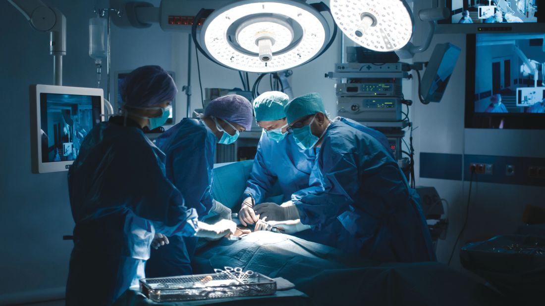
“Although this association is an important finding, the confounding factors that cause delay to surgery must be elucidated in non-database studies,” wrote Venkat Boddapati, MD, of Columbia University Medical Center, New York, and coauthors. The study was published in Arthroplasty Today.
To assess the best time for revision TKA after PPF of the knee, the researchers analyzed data from 484 patients who underwent another TKA from 2005 to 2016. Of those patients, 377 (78%) had expedited surgery – defined as less than or equal to 24 hours from hospital admission – and 107 (22%) had non-expedited surgery. Non-expedited patients averaged 3.2 days from admission to surgery.
After multivariate analysis, non-expedited patients had more complications overall, compared with expedited patients (odds ratio 2.35, P = .037). They also had comparative increases in surgical site infections (OR 12.87, P = .029), urinary tract infections (OR 10.46, P = .048), non-home discharge (OR 4.27, P less than .001), and blood transfusions (OR 4.53, P less than .001). The two groups saw no statistical difference in mortality.
The authors noted their study’s limitations, including an inability to assess complications beyond 30 days after surgery, which may affect tracking longer-term outcomes such as mortality. In addition, they were only able to classify surgery as expedited or non-expedited based on when the patient was admitted to the hospital, not the time since their injury. Finally, they lacked “relevant variables that may have contributed to this analysis,” including the type of fracture and the revision implants used.
Three authors reported being paid consultants for, and receiving research support from, several medical companies. The others reported no conflicts of interest.
SOURCE: Boddapati V et al. Arthroplast Today. 2019 Sep 1. doi: 10.1016/j.artd.2019.05.002.
According to a study on patients undergoing revision knee replacement, a delay of more than 24 hours between hospital admission and total knee arthroplasty (TKA) for periprosthetic fracture (PPF) led to increased odds of complications such as surgical site and urinary tract infections.
“Although this association is an important finding, the confounding factors that cause delay to surgery must be elucidated in non-database studies,” wrote Venkat Boddapati, MD, of Columbia University Medical Center, New York, and coauthors. The study was published in Arthroplasty Today.
To assess the best time for revision TKA after PPF of the knee, the researchers analyzed data from 484 patients who underwent another TKA from 2005 to 2016. Of those patients, 377 (78%) had expedited surgery – defined as less than or equal to 24 hours from hospital admission – and 107 (22%) had non-expedited surgery. Non-expedited patients averaged 3.2 days from admission to surgery.
After multivariate analysis, non-expedited patients had more complications overall, compared with expedited patients (odds ratio 2.35, P = .037). They also had comparative increases in surgical site infections (OR 12.87, P = .029), urinary tract infections (OR 10.46, P = .048), non-home discharge (OR 4.27, P less than .001), and blood transfusions (OR 4.53, P less than .001). The two groups saw no statistical difference in mortality.
The authors noted their study’s limitations, including an inability to assess complications beyond 30 days after surgery, which may affect tracking longer-term outcomes such as mortality. In addition, they were only able to classify surgery as expedited or non-expedited based on when the patient was admitted to the hospital, not the time since their injury. Finally, they lacked “relevant variables that may have contributed to this analysis,” including the type of fracture and the revision implants used.
Three authors reported being paid consultants for, and receiving research support from, several medical companies. The others reported no conflicts of interest.
SOURCE: Boddapati V et al. Arthroplast Today. 2019 Sep 1. doi: 10.1016/j.artd.2019.05.002.
FROM ARTHROPLASTY TODAY