User login
Heavy menstrual bleeding in teens often linked to bleeding disorders
Over one-third of adolescents presenting with heavy menstrual bleeding were diagnosed with a bleeding disorder after screening, according to results of a retrospective study.

The high incidence of bleeding disorders detected argues for routine screening of adolescents with heavy menstrual bleeding (HMB), Brooke O’Brien, MD, of the University of Queensland, Brisbane, Australia, and her colleagues wrote in the Journal of Pediatric & Adolescent Gynecology.
“These findings support comprehensive and systematic hemostatic evaluation in adolescents with HMB,” Dr. O’Brien and her colleagues wrote. “A higher level of awareness of bleeding disorders as a cause for HMB in adolescence, especially [von Willebrand disease] and platelet function disorders, is needed and close multidisciplinary collaboration between the pediatric and adolescent gynecologist and hematologist in a specialized tertiary center should be established in the management of these patients.”
In their study, Dr. O’Brien and her colleagues retrospectively evaluated 124 adolescents with HMB at a pediatric and adolescent gynecology tertiary care center between July 2007 and July 2017. Of these, 77 patients (62.1%) underwent screening for blood disorders.
The researchers found 27 adolescents overall were diagnosed with a blood disorder, which consisted of 35.0% of patients screened and 21.7% of all patients studied. Specifically, 14 of 27 patients (51.6%) screened were diagnosed with von Willebrand disease, 9 of 27 patients (33.3%) screened were found to have inherited platelet function disorders, 3 of 27 patients (11.1%) had inherited or acquired thrombocytopenia, and 1 of 27 patients (3.7%) had factor IX deficiency. The researchers also screened for iron deficiency and/or anemia and found 53 of 107 patients (49.5%) who were screened received a diagnosis, and 19 of 27 patients (70.3%) who were diagnosed with a bleeding disorder also had iron deficiency and/or anemia.
“In adolescents who are already known to have a bleeding disorder, consultation with a pediatric gynecologist and/or hematologist prior to menarche may be helpful to outline abnormal patterns of menstrual bleeding and to discuss options of treatment in the event of heavy menstrual bleeding,” Dr. O’Brien and her colleagues wrote.
Potential limitations in the study include the refractory nature of referrals at a tertiary care center potentially overestimating the prevalence of HMB in this population as well as the study’s retrospective design when investigating and measuring heavy menstrual bleeding, but researchers noted patients were reviewed and classified by a specialist pediatric hematologist.
The authors reported no relevant conflicts of interest.
SOURCE: O’Brien B et al. J Pediatr Adolesc Gynecol. 2018 Nov 22. doi: 10.1016/j.jpag.2018.11.005.
Over one-third of adolescents presenting with heavy menstrual bleeding were diagnosed with a bleeding disorder after screening, according to results of a retrospective study.
The high incidence of bleeding disorders detected argues for routine screening of adolescents with heavy menstrual bleeding (HMB), Brooke O’Brien, MD, of the University of Queensland, Brisbane, Australia, and her colleagues wrote in the Journal of Pediatric & Adolescent Gynecology.
“These findings support comprehensive and systematic hemostatic evaluation in adolescents with HMB,” Dr. O’Brien and her colleagues wrote. “A higher level of awareness of bleeding disorders as a cause for HMB in adolescence, especially [von Willebrand disease] and platelet function disorders, is needed and close multidisciplinary collaboration between the pediatric and adolescent gynecologist and hematologist in a specialized tertiary center should be established in the management of these patients.”
In their study, Dr. O’Brien and her colleagues retrospectively evaluated 124 adolescents with HMB at a pediatric and adolescent gynecology tertiary care center between July 2007 and July 2017. Of these, 77 patients (62.1%) underwent screening for blood disorders.
The researchers found 27 adolescents overall were diagnosed with a blood disorder, which consisted of 35.0% of patients screened and 21.7% of all patients studied. Specifically, 14 of 27 patients (51.6%) screened were diagnosed with von Willebrand disease, 9 of 27 patients (33.3%) screened were found to have inherited platelet function disorders, 3 of 27 patients (11.1%) had inherited or acquired thrombocytopenia, and 1 of 27 patients (3.7%) had factor IX deficiency. The researchers also screened for iron deficiency and/or anemia and found 53 of 107 patients (49.5%) who were screened received a diagnosis, and 19 of 27 patients (70.3%) who were diagnosed with a bleeding disorder also had iron deficiency and/or anemia.
“In adolescents who are already known to have a bleeding disorder, consultation with a pediatric gynecologist and/or hematologist prior to menarche may be helpful to outline abnormal patterns of menstrual bleeding and to discuss options of treatment in the event of heavy menstrual bleeding,” Dr. O’Brien and her colleagues wrote.
Potential limitations in the study include the refractory nature of referrals at a tertiary care center potentially overestimating the prevalence of HMB in this population as well as the study’s retrospective design when investigating and measuring heavy menstrual bleeding, but researchers noted patients were reviewed and classified by a specialist pediatric hematologist.
The authors reported no relevant conflicts of interest.
SOURCE: O’Brien B et al. J Pediatr Adolesc Gynecol. 2018 Nov 22. doi: 10.1016/j.jpag.2018.11.005.
FROM THE JOURNAL OF PEDIATRIC & ADOLESCENT GYNECOLOGY
Key clinical point: More than one-third of adolescents with heavy menstrual bleeding were diagnosed with a bleeding disorder.
Major finding: After screening, 35% of women with heavy menstrual bleeding had a bleeding disorder; over half of those screened had von Willebrand disease.
Study details: A retrospective study of 124 adolescents at the Queensland Paediatric and Adolescent Gynaecology Service between July 2007 and July 2017.
Disclosures: The authors reported no relevant conflicts of interest.
Source: O’Brien B et al. J Pediatr Adolesc Gynecol. 2018 Nov 22 . doi: 10.1016/j.jpag.2018.11.005.
FLYER: R-CHOP 4 safer, as effective for low-risk DLBCL patients under 60
SAN DIEGO – Patients aged younger than 60 years with favorable-prognosis diffuse large B-cell lymphoma who were randomly assigned to therapy with four cycles of R-CHOP (rituximab, cyclophosphamide, doxorubicin, vincristine, and prednisone) had progression-free, event-free, and overall survival rates comparable with those of patients assigned to six cycles, investigators in the FLYER trial reported.
The four-cycle regimen was associated with a marked reduction in adverse events, with an overall drop in nonhematologic malignancies of approximately one-third compared with the six-cycle regimen.
For younger patients with favorable-prognosis DLBCL – defined as an age-adjusted International Prognostic Index score of 0 and low tumor burden (less than 7.5 cm) – four cycles of R-CHOP can be a new standard of care.
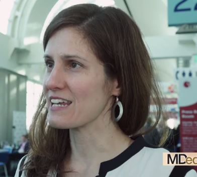
In this video interview at the annual meeting of the American Society of Hematology, Viola Poeschel, MD, of Saarland University in Homburg, Germany, describes the patient population who may benefit from shorter duration therapy.
SAN DIEGO – Patients aged younger than 60 years with favorable-prognosis diffuse large B-cell lymphoma who were randomly assigned to therapy with four cycles of R-CHOP (rituximab, cyclophosphamide, doxorubicin, vincristine, and prednisone) had progression-free, event-free, and overall survival rates comparable with those of patients assigned to six cycles, investigators in the FLYER trial reported.
The four-cycle regimen was associated with a marked reduction in adverse events, with an overall drop in nonhematologic malignancies of approximately one-third compared with the six-cycle regimen.
For younger patients with favorable-prognosis DLBCL – defined as an age-adjusted International Prognostic Index score of 0 and low tumor burden (less than 7.5 cm) – four cycles of R-CHOP can be a new standard of care.
In this video interview at the annual meeting of the American Society of Hematology, Viola Poeschel, MD, of Saarland University in Homburg, Germany, describes the patient population who may benefit from shorter duration therapy.
REPORTING FROM ASH 2018

Sickle cell disease phenotype reversed by gene therapy
SAN DIEGO – An adult with sickle cell disease has had significant remissions in symptoms and a near elimination of transfusion requirements after receiving an infusion of autologous stem cells genetically modified to simultaneously induce the fetal form of hemoglobin and decrease sickle hemoglobin.
In a first-in-human, proof-of-concept study, transduction of hematopoietic stem cells with a lentiviral vector targeted against the gamma globin repressor BCL11A in erythroid cells led to rapid induction of fetal hemoglobin and a reversal of the sickle cell disease (SCD) phenotype in the early phase of stem cell reconstitution, reported Erica B. Esrick, MD, from the Dana-Farber/Boston Children’s Cancer and Blood Disorders Center in Boston.
“The potential advantage of this approach over the gene-addition strategy of gene therapy is that we can harness the physiologic switch machinery that exists in the cell to simultaneously increase fetal hemoglobin and decrease sickle hemoglobin,” she said at a briefing prior to her presentation at the annual meeting of the American Society of Hematology.
Several research groups are developing autologous gene therapy for beta-hemoglobinopathies, including the use of CRISPR-Cas9 technology to mimic a rare, naturally occurring mutation that causes the fetal type of hemoglobin to persist into adulthood in some patients with SCD and beta-thalassemia.
Dr. Esrick and her colleagues are trying a different approach: Using gene therapy to knock down BCL11A expression to induce gamma globin expression.
For the treatment, autologous hematopoietic stem cells are collected from patients following mobilization with plerixafor. The cells are then transduced with a lentiviral vector consisting of a novel short hairpin RNA embedded in an endogenous micro-RNA. The investigators refer to the construct as a shmiR (“schmeer”). The construct is designed to be erythroid specific, with BCL11A knocked down only in the red cell lineage, to avoid potential off-target effects of the therapy.
Following stem cell collection and transduction, patients undergo conditioning with busulfan prior to infusion of the modified stem cells.
In three patients treated thus far, the process has been shown to be highly efficient, with approximately 96% of treated cells transduced.
In the patient mentioned before, neutrophil engraftment was confirmed on day 22 after transfusion of the modified cells. He experienced adverse events that were consistent with myeloablative conditioning, but no adverse events associated with the modified cells.
During 6 months of follow-up the patient did not experience SCD-related pain, respiratory events, or neurologic events, and did not have anemia, with a total hemoglobin of 11 g/dL at 6 months. He has not required any transfusions since engraftment.
Patients in the trial will be followed for 2 years, and then will be enrolled in a 15-year follow-up study designed to evaluate the safety and the durability of therapy.
Dr. Esrick reported receiving honoraria from Bluebird Bio, maker of the short hairpin RNA construct used in the trial.
SOURCE: Esrick EB et al. ASH 2018, Abstract 1023.
REPORTING FROM ASH 2018
Key clinical point: Gene therapy to induce the fetal form of hemoglobin reversed the symptoms of sickle cell disease in an adult patient.
Major finding: During 6 months of follow-up the patient did not experience sickle cell disease–related pain, respiratory events, or neurologic events, and did not have anemia.
Study details: A first-in-human study in seven adults with sickle cell disease.
Disclosures: Dr. Esrick reported receiving honoraria from Bluebird Bio, maker of the short hairpin RNA construct used in the trial.
Source: Esrick EB et al. ASH 2018, Abstract 1023.
Daily hydroxyurea effective, safe for African children
SAN DIEGO – Daily hydroxyurea treatment for sickle cell disease is feasible, safe, and effective for children in sub-Saharan Africa, according to the results of a large open-label, phase 1-2, international trial.

Hydroxyurea was associated with reduced rates of malaria and other infections, resulting in improved survival, according to Léon Tshilolo, MD, PhD, of Centre Hospitalier Monkole in Kinshasa, the Democratic Republic of the Congo.
“Based on that data, we believe that wider access to hydroxyurea for sickle cell anemia has the potential to save millions of lives in Africa,” Dr. Tshilolo said in a press conference at the annual meeting of the American Society of Hematology.
Use of hydroxyurea has been limited in Africa because of cost, access issues, and challenges associated with laboratory monitoring, according to the researchers.
Moreover, most of the data on the efficacy of hydroxyurea come from studies conducted in the United States, Europe, and other high-income settings, said the study’s senior author Russell E. Ware, MD, PhD, of Cincinnati Children’s Hospital Center.
“Now that there’s data in an African setting, I think this will go a long way to advancing [hydroxyurea therapy] and encouraging governments, organizations, and pharmaceutical companies to bring it in,” Dr. Ware said in an interview.
In the study by Dr. Ware, Dr. Tshilolo, and their colleagues, 606 children in four sub-Saharan African countries completed a 2-month pretreatment screening phase designed to capture baseline clinical and laboratory data. The children, who were aged 1-10 years, were started at 15-20 mg/kg of hydroxyurea for 6 months, followed by escalation to the maximum tolerated dose.
With a median of 2.5 years of treatment, treated children experienced less pain and anemia, fewer cases of malaria and other infections, and lower rates of transfusions and death versus rates observed in the pretreatment screening phase of the trial.
The rate of vasoocclusive pain during hydroxyurea treatment was 44.6 events per 100 patient-years, compared with 98.3 events per 100 patient-years in the pretreatment period (incidence rate ratio, 0.45; 95% confidence interval, 0.37-0.56), according to data simultaneously published in the New England Journal of Medicine.
Malaria infection rates were 22.9 events per 100 patient-years in the hydroxyurea treatment period versus 46.9 events in the pretreatment period (IRR, 0.49; 95% CI, 0.37-0.66). Rates of nonmalaria infections were 90.0 events per 100 patient-years in the hydroxyurea treatment period versus 142.5 events per 100 patient-years in the pretreatment period (IRR, 0.62; 95% CI, 0.53-0.72).
Dr. Tshilolo said investigators were “encouraged” by the reduced infection rates, particularly in light of previous concerns that hydroxyurea could suppress the immune system and put children at risk for malaria.
Death rates were 1.1 per 100 patient-years in the hydroxyurea group and 3.6 per 100 patient-years in the pretreatment period (IR, 0.30; 95% CI, 0.10-0.88). Dose-limiting toxic events occurred in 5.1% of the children, which was below the protocol-specified threshold for safety, Dr. Tshilolo added.
Dr. Tshilolo reported grants from the National Institutes of Health/National Heart, Lung, and Blood Institute and Cincinnati Children’s Research Foundation, along with nonfinancial support from Bristol-Myers Squibb. Dr. Ware reported grants from the NIH/NHLBI and Bristol-Myers Squibb.
SOURCE: Tshilolo L et al. ASH 2018, Abstract 3.
SAN DIEGO – Daily hydroxyurea treatment for sickle cell disease is feasible, safe, and effective for children in sub-Saharan Africa, according to the results of a large open-label, phase 1-2, international trial.
Hydroxyurea was associated with reduced rates of malaria and other infections, resulting in improved survival, according to Léon Tshilolo, MD, PhD, of Centre Hospitalier Monkole in Kinshasa, the Democratic Republic of the Congo.
“Based on that data, we believe that wider access to hydroxyurea for sickle cell anemia has the potential to save millions of lives in Africa,” Dr. Tshilolo said in a press conference at the annual meeting of the American Society of Hematology.
Use of hydroxyurea has been limited in Africa because of cost, access issues, and challenges associated with laboratory monitoring, according to the researchers.
Moreover, most of the data on the efficacy of hydroxyurea come from studies conducted in the United States, Europe, and other high-income settings, said the study’s senior author Russell E. Ware, MD, PhD, of Cincinnati Children’s Hospital Center.
“Now that there’s data in an African setting, I think this will go a long way to advancing [hydroxyurea therapy] and encouraging governments, organizations, and pharmaceutical companies to bring it in,” Dr. Ware said in an interview.
In the study by Dr. Ware, Dr. Tshilolo, and their colleagues, 606 children in four sub-Saharan African countries completed a 2-month pretreatment screening phase designed to capture baseline clinical and laboratory data. The children, who were aged 1-10 years, were started at 15-20 mg/kg of hydroxyurea for 6 months, followed by escalation to the maximum tolerated dose.
With a median of 2.5 years of treatment, treated children experienced less pain and anemia, fewer cases of malaria and other infections, and lower rates of transfusions and death versus rates observed in the pretreatment screening phase of the trial.
The rate of vasoocclusive pain during hydroxyurea treatment was 44.6 events per 100 patient-years, compared with 98.3 events per 100 patient-years in the pretreatment period (incidence rate ratio, 0.45; 95% confidence interval, 0.37-0.56), according to data simultaneously published in the New England Journal of Medicine.
Malaria infection rates were 22.9 events per 100 patient-years in the hydroxyurea treatment period versus 46.9 events in the pretreatment period (IRR, 0.49; 95% CI, 0.37-0.66). Rates of nonmalaria infections were 90.0 events per 100 patient-years in the hydroxyurea treatment period versus 142.5 events per 100 patient-years in the pretreatment period (IRR, 0.62; 95% CI, 0.53-0.72).
Dr. Tshilolo said investigators were “encouraged” by the reduced infection rates, particularly in light of previous concerns that hydroxyurea could suppress the immune system and put children at risk for malaria.
Death rates were 1.1 per 100 patient-years in the hydroxyurea group and 3.6 per 100 patient-years in the pretreatment period (IR, 0.30; 95% CI, 0.10-0.88). Dose-limiting toxic events occurred in 5.1% of the children, which was below the protocol-specified threshold for safety, Dr. Tshilolo added.
Dr. Tshilolo reported grants from the National Institutes of Health/National Heart, Lung, and Blood Institute and Cincinnati Children’s Research Foundation, along with nonfinancial support from Bristol-Myers Squibb. Dr. Ware reported grants from the NIH/NHLBI and Bristol-Myers Squibb.
SOURCE: Tshilolo L et al. ASH 2018, Abstract 3.
REPORTING FROM ASH 2018
Key clinical point: Daily hydroxyurea treatment in sub-Saharan African children with sickle cell disease is feasible, safe, and effective, and has the additional benefit of reducing their rates of malaria and nonmalaria infections.
Major finding: Malaria infection rates were 22.9 versus 46.9 events per 100 patient-years in the hydroxyurea treatment period and pretreatment period, respectively (incidence rate ratio, 0.49; 95% CI, 0.37-0.66).
Study details: A phase 1-2, international, open-label trial including 606 children in four sub-Saharan African countries who completed a 2-month pretreatment screening phase and went on to receive hydroxyurea.
Disclosures: Dr. Tshilolo reported grants from the National Institutes of Health/National Heart, Lung, and Blood Institute and Cincinnati Children’s Research Foundation, along with nonfinancial support from Bristol-Myers Squibb. Dr. Ware reported grants from the NIH/NHLBI and Bristol-Myers Squibb.
Source: Tshilolo L et al. ASH 2018, Abstract 3.
Flipping the fetal hemoglobin switch reverses sickle cell symptoms
SAN DIEGO – Researchers were able to “flip the switch” from the adult to fetal form of hemoglobin using autologous stem cells genetically modified to simultaneously induce the fetal form of hemoglobin and decrease sickle hemoglobin.
The advance was announced by investigators at the Dana-Farber Cancer Institute and Boston Children’s Hospital at the annual meeting of the American Society of Hematology. At 6 months of follow-up, one adult patient in the proof-of-concept study has experienced a reversal of the sickle cell phenotype, with no pain episodes or respiratory or neurologic events.
The fetal form of hemoglobin is known to be protective against the signs and symptoms of sickle cell disease, but apart from a few rare exceptions, people with the disorder begin to experience debilitating symptoms as levels of the fetal form begin to decline in early childhood and levels of the adult form of hemoglobin steadily rise.
In this video interview, Erica B. Esrick, MD, from the Dana-Farber/Boston Children’s Cancer and Blood Disorders Center, describes the novel approach of using RNA interference to knock down a repressor that suppresses expression of gamma globin in sickle cell disease.
REPORTING FROM ASH 2018
RELIEF: In Behçet’s, apremilast improves oral ulcers for up to 28 weeks
CHICAGO – Apremilast was effective and well tolerated for up to 28 weeks for the treatment of oral ulcers in patients with active Behçet’s disease, based on findings from the randomized, placebo-controlled, phase 3 RELIEF trial.
At baseline, mean oral ulcer counts were 4.2 in 104 patients randomized to receive the oral phosphodiesterase-4 inhibitor and 3.9 in 103 patients in the placebo group. Mean visual analog scale (VAS) pain scores were 61.2 and 60.8 in the two groups, respectively.
The primary study endpoint of area under the curve for total number of oral ulcers over a 12-week period (AUCWk0-12) – a measure that reflects the number of oral ulcers that occur over time and also accounts for the recurring-remitting course of oral ulcers – was achieved. AUCWk0-12 was significantly lower in the apremilast group than in the placebo group (129.54 vs. 222.14, respectively; P less than .0001), Gulen Hatemi, MD, reported at the annual meeting of the American College of Rheumatology.
From baseline to week 12, apremilast treatment also resulted in a significantly lower number of oral ulcers (mean of 1.1 vs. 2.0 for placebo at 12 weeks) and significantly reduced pain from oral ulcers at every visit from week 1 through week 12 of the study, compared with placebo (mean VAS score change from baseline, –40.7 vs. –15.9), said Dr. Hatemi, a professor of medicine at Istanbul University.
“The [12-week] complete response rate ... was 53% in the apremilast group and 22.3% in the placebo group. The [12-week] partial response rate ...was 76% in the apremilast group and 48% in the placebo group,” she said, adding that the efficacy of apremilast was sustained with continued treatment through 28 weeks.
Study participants were adults (mean age, 40 years) with active Behçet’s disease and three or more oral ulcers at randomization or two or more at screening and at randomization. All had been previously treated with at least one nonbiologic medication for oral ulcers and were allowed to have received previous biologic therapies for other disease manifestations. Those with active major organ involvement were excluded.
Treatment included a 30-mg dose of apremilast twice daily for 12 weeks or placebo. After 12 weeks, all patients received apremilast through at least 28 weeks of the 64-week study.
At the 28-week analysis, patients who were initially randomized to placebo and who switched to apremilast after week 12 had benefits comparable with those seen in those randomized to apremilast at the start of the study. A complete response was seen in 59% and 62% of patients in the groups, respectively, and a partial response was seen in 90% and 85%, respectively. Additionally, the mean change in the VAS score for oral ulcer pain in the groups at that time was –40.6 and –41.9, Dr. Hatemi said.
Apremilast was well tolerated in this study; the incidence of adverse events was comparable in the treatment and placebo groups during the 12-week placebo-controlled phase of the study – 78.8% and 71.8%, respectively. The most common events were diarrhea, nausea, headache, and upper respiratory tract infection, she said.
“These were generally mild to moderate, and only two patients had to discontinue the study due to gastrointestinal adverse events,” she said, noting that no new safety signals were observed.
Behçet’s disease is a chronic, relapsing, multisystem inflammatory disorder characterized by recurrent oral ulcers that can be disabling and have a substantial effect on quality of life. These findings, which include efficacy data up to 28 weeks and safety data for at least 100 patients exposed to apremilast for at least 1 year, demonstrate the efficacy of apremilast for the treatment oral ulcers in patients with Behçet’s disease, she said, noting that “the safety findings were consistent with the known safety profile of apremilast.”
The RELIEF study was supported by Celgene. Dr. Hatemi reported receiving grant/research support from Celgene and serving as a speaker for AbbVie, Mustafa Nevzet Pharmaceuticals, and UCB.
SOURCE: Hatemi G et al. Arthritis Rheumatol. 2018;70(Suppl 10), Abstract 2789.
REPORTING FROM THE ACR ANNUAL MEETING
Key clinical point: Apremilast is safe and effective for treating oral ulcers in patients with Behçet’s disease.
Major finding: The AUCWk0-12 was significantly lower with apremilast (129.54) versus placebo (222.14).
Study details: A randomized, placebo-controlled, phase 3 study of 207 patients.
Disclosures: The RELIEF study was supported by Celgene. Dr. Hatemi reported receiving grant/research support from Celgene and serving as a speaker for AbbVie, Mustafa Nevzet Pharmaceuticals, and UCB.
Source: Hatemi G et al. Arthritis Rheumatol. 2018;70(Suppl 10), Abstract 2789
Depression is linked to seizure frequency in patients with epilepsy
NEW ORLEANS –
The conclusion comes from a study of 120 people with epilepsy, 62 of whom had at least moderate depression based on the Patient Health Questionnaire-9 (PHQ-9). The Rapid Estimate of Adult Literacy in Medicine (REALM-R), Quality of Life in Epilepsy (QOLIE-10) and Charlson Comorbidity Index were used to assess patients’ health literacy, quality of life, and medical comorbidity, respectively
Among demographic characteristics, only inability to work was significantly associated with depression severity. Higher 30-day seizure frequency, panic disorder, and obsessive-compulsive disorder were correlated with more severe depression severity. Medical comorbidity was not associated with increased risk of depression.
Identifying and treating psychiatric comorbidities should be part of the management of patients with epilepsy, said Martha X. Sajatovic, MD, director of the Neurological and Behavioral Outcomes Center at Case Western Reserve University in Cleveland, who presented the data. “Following up to ensure they receive treatment is vital, because it can truly change patient outcomes and help them achieve their best quality of life.”
The study findings are consistent with those of previous research indicating that people with symptoms of depression are more likely to have more frequent seizures and decreased quality of life, said Dr. Sajatovic.
“Health care providers should screen their epilepsy patients for depression, but they shouldn’t stop there,” she advised. “A person may have depressive symptoms that don’t reach the level of depression but should be assessed for other types of mental health issues that could easily be overlooked.”
Patients with epilepsy should respond to the PHQ-9 annually, or more frequently, if warranted, she added.
“It’s important that people with epilepsy who have depression or other mental health issues get treatment such as cognitive behavioral therapy and medication,” said Dr. Sajatovic. “Even being in a self-management program helps, because the better they are at self management, the less likely they are to suffer negative health effects.”
This study was supported by a grant from the Centers for Disease Control and Prevention SIP 14-007 1U48DP005030.
SOURCE: Kumar N et al. AES 2018, Abstract 1.371.
REPORTING FROM AES 2018
Key clinical point: Identification and treatment of psychiatric comorbidities are appropriate components of epilepsy management.
Major finding: Half of participants in a randomized, controlled trial had depression of at least moderate severity.
Study details: Researchers analyzed data from a trial of 120 people with epilepsy.
Disclosures: This study was supported by a grant from the CDC SIP 14-007 1U48DP005030.
Source: Kumar N et al. Abstract 1.371.
Enzyme-inducing AEDs may raise vitamin D dose requirements
NEW ORLEANS – Patients taking enzyme-inducing antiepileptic drugs (AEDs) may require a clinically meaningful increase in their vitamin D doses to achieve the same 25-hydroxyvitamin D (25[OH]D) plasma levels as patients taking nonenzyme-inducing AEDs, based on a retrospective chart review presented at the annual meeting of the American Epilepsy Society.
While patients receiving either type of AED had similar average 25(OH)D levels in the study (32.0 ng/mL in the enzyme-inducing AED group and 33.2 ng/mL in the noninducing AED group), those in the enzyme-inducing group required 1,587 U/day to meet the goal – a 409-unit increase in dose, compared with the 1,108 U/day dose taken by patients in the nonenzyme-inducing group.
“Patients taking enzyme-inducing AEDs may benefit from more intensive monitoring of their vitamin D supplementation, and clinicians should anticipate this likely pharmacokinetic interaction,” said Barry E. Gidal, PharmD, professor of pharmacy and neurology at the University of Wisconsin–Madison, and his colleagues.
Researchers have suggested that enzyme-inducing AEDs may affect CYP450 isoenzymes, increase vitamin D metabolism, and reduce 25(OH)D plasma levels. “It follows … that a potential pharmacokinetic interaction could exist between enzyme-inducing AEDs and oral formulations of vitamin D used for supplementation,” the investigators said.
To test the hypothesis, Dr. Gidal and his colleagues reviewed the charts of patients with epilepsy who were on any AED regimen and were prescribed vitamin D at William S. Middleton Memorial Veterans Hospital in Madison, Wisconsin, between January 2013 and September 2017.
The researchers grouped patients by those using enzyme-inducing AEDs and those taking noninducing AEDs. Patients who were taking AEDs in both categories were placed in the enzyme-inducing AED group. Patients with malabsorptive conditions and patients using calcitriol were excluded from the analysis.
Data included AEDs used, prescription and over-the-counter vitamin D use, 25(OH)D plasma concentration, renal function, age, gender, and ethnicity. Patients’ 25(OH)D levels were measured using a chemiluminescence immunoassay, and a minimum 25(OH)D plasma level of 30 ng/mL was the therapeutic goal.
The multivariant analysis was adjusted for potentially confounding variables including 25(OH)D concentration, over-the-counter vitamin D use, chronic kidney disease, age, gender, and ethnicity.
The analysis included 1,113 observations from 315 patients, and 263 of the observations (23.6%) were in the enzyme-inducing AED group. The enzyme-inducing group and noninducing groups were mostly male (90.5% and 91.8%, respectively) and similar in average age (65.9 and 61.4 years, respectively). Variables were evenly distributed between the groups, with the exceptions of chronic kidney disease, which was less common in the enzyme-inducing group (6.1% vs. 13.8%), and ethnicity (78.7% Caucasian in the enzyme-inducing group vs. 87.7% Caucasian in the noninducing group). The most common enzyme-inducing AED was phenytoin (50.6%), followed by carbamazepine (31.9%), phenobarbital (14.1%), oxcarbazepine (6.8%), primidone (1.9%), and eslicarbazepine (0.8%).
Dr. Gidal reported honoraria from Eisai, Sunovion, Lundbeck, and GW Pharmaceuticals.
SOURCE: Gidal BE et al. AES 2018, Abstract 1.315.
REPORTING FROM AES 2018
Key clinical point: Enzyme-inducing antiepileptic drugs affect vitamin D dose requirements.
Major finding: Patients taking enzyme-inducing antiepileptic drugs require a higher daily dose of vitamin D, compared with patients taking noninducing antiepileptic drugs (1,587 U/day vs. 1,108 U/day).
Study details: A retrospective chart review of data from 315 patients treated at a Veterans Affairs hospital.
Disclosures: Dr. Gidal reported honoraria from Eisai, Sunovion, Lundbeck, and GW Pharmaceuticals..
Source: Gidal BE et al. AES 2018, Abstract 1.315.
Teenagers with epilepsy may benefit from depression screening
NEW ORLEANS – Referral to a mental health provider is adequate for most patients with moderately severe symptoms of depression, but some patients may require active intervention during the clinical visit, said the researchers.

“We know that depression is more common in people with epilepsy, compared to the general population, but there is less information about depression in children and teens than adults, and little is known about the factors that increase the likelihood of depressive symptoms,” said Hillary Thomas, PhD, a pediatric psychologist at Children’s Medical Center in Dallas. “Depression screening should be routine at epilepsy treatment centers and can identify children and teens who would benefit from intervention.”
Following 2015 guidelines from the American Academy of Neurology, the Comprehensive Epilepsy Center at Children’s Health System in Dallas developed a behavioral health screening protocol for teens with epilepsy. The center aims to identify patients with depressive symptoms and ensure that they are referred to appropriate behavioral health practitioners. Clinicians also review the screening data and seizure variables for their potential implications for clinical care. Researchers at the center also seek to elucidate the relationship between depressive symptoms and seizure diagnosis and treatment.
As part of the protocol, Dr. Thomas and her colleagues administer the Patient Health Questionnaire-9 (adolescent version) to all patients aged 15-18 years during their visit to the epilepsy clinic. Patients with intellectual disability or other factors that prevent them from providing valid responses are excluded. If a patient’s PHQ-9 score indicates at least moderately severe depressive symptoms, or if he or she reports suicidal ideation, clinicians follow a specific response protocol that includes providing referrals, encouraging follow-up with the patient’s current mental health provider, and obtaining a suicide risk assessment from a psychologist or social worker. After the screener is completed, clinicians retrieve demographic and clinical data (e.g., seizure diagnosis, medication, number of clinic or emergency department visits) from the patient’s medical record and include them in a database for subsequent analysis.
Dr. Thomas and her colleagues presented data from 394 youth with epilepsy whom they had screened. Patients’ mean age was 16 years, and half of the population was female. The study population had rates of depression similar to those identified in previous studies, said Dr. Thomas. Approximately 87% of patients had minimal or mild depressive symptoms, and 8% had moderately severe depressive symptoms. Furthermore, 5% of the patients reported suicidal ideation or previous suicide attempt. Several of the patients with suicidal ideation had a current mental health provider, and the others required an in-clinic risk assessment. Overall, 13% of the population required behavioral health referral or intervention. When the researchers conducted chi-squared analysis, they found no significant association between seizure type and depression severity.
“Our results don’t mean that only 13% of the teens with epilepsy had depressive symptoms,” said Susan Arnold, MD, director of the Comprehensive Epilepsy Center and a coauthor of the study. “They indicate the significant percentage of teens whose level of depressive symptoms warranted behavioral health referrals or further evaluation or even intervention during a clinic visit. Health care providers need to be vigilant about continually screening children and teens for depression.” As part of each patient’s comprehensive care, epilepsy treatment centers should provide psychosocial teams that include social workers or psychologists, she added.
The investigators plan to continue analyzing the data for specific depression symptoms that are most common in teens. These symptoms could be the basis for developing additional resources for families, such as lists of warning signs and guides to symptom management, as well as group therapy and support groups.
SOURCE: Thomas HM et al. Abstract 1.388.
NEW ORLEANS – Referral to a mental health provider is adequate for most patients with moderately severe symptoms of depression, but some patients may require active intervention during the clinical visit, said the researchers.
“We know that depression is more common in people with epilepsy, compared to the general population, but there is less information about depression in children and teens than adults, and little is known about the factors that increase the likelihood of depressive symptoms,” said Hillary Thomas, PhD, a pediatric psychologist at Children’s Medical Center in Dallas. “Depression screening should be routine at epilepsy treatment centers and can identify children and teens who would benefit from intervention.”
Following 2015 guidelines from the American Academy of Neurology, the Comprehensive Epilepsy Center at Children’s Health System in Dallas developed a behavioral health screening protocol for teens with epilepsy. The center aims to identify patients with depressive symptoms and ensure that they are referred to appropriate behavioral health practitioners. Clinicians also review the screening data and seizure variables for their potential implications for clinical care. Researchers at the center also seek to elucidate the relationship between depressive symptoms and seizure diagnosis and treatment.
As part of the protocol, Dr. Thomas and her colleagues administer the Patient Health Questionnaire-9 (adolescent version) to all patients aged 15-18 years during their visit to the epilepsy clinic. Patients with intellectual disability or other factors that prevent them from providing valid responses are excluded. If a patient’s PHQ-9 score indicates at least moderately severe depressive symptoms, or if he or she reports suicidal ideation, clinicians follow a specific response protocol that includes providing referrals, encouraging follow-up with the patient’s current mental health provider, and obtaining a suicide risk assessment from a psychologist or social worker. After the screener is completed, clinicians retrieve demographic and clinical data (e.g., seizure diagnosis, medication, number of clinic or emergency department visits) from the patient’s medical record and include them in a database for subsequent analysis.
Dr. Thomas and her colleagues presented data from 394 youth with epilepsy whom they had screened. Patients’ mean age was 16 years, and half of the population was female. The study population had rates of depression similar to those identified in previous studies, said Dr. Thomas. Approximately 87% of patients had minimal or mild depressive symptoms, and 8% had moderately severe depressive symptoms. Furthermore, 5% of the patients reported suicidal ideation or previous suicide attempt. Several of the patients with suicidal ideation had a current mental health provider, and the others required an in-clinic risk assessment. Overall, 13% of the population required behavioral health referral or intervention. When the researchers conducted chi-squared analysis, they found no significant association between seizure type and depression severity.
“Our results don’t mean that only 13% of the teens with epilepsy had depressive symptoms,” said Susan Arnold, MD, director of the Comprehensive Epilepsy Center and a coauthor of the study. “They indicate the significant percentage of teens whose level of depressive symptoms warranted behavioral health referrals or further evaluation or even intervention during a clinic visit. Health care providers need to be vigilant about continually screening children and teens for depression.” As part of each patient’s comprehensive care, epilepsy treatment centers should provide psychosocial teams that include social workers or psychologists, she added.
The investigators plan to continue analyzing the data for specific depression symptoms that are most common in teens. These symptoms could be the basis for developing additional resources for families, such as lists of warning signs and guides to symptom management, as well as group therapy and support groups.
SOURCE: Thomas HM et al. Abstract 1.388.
REPORTING FROM AES 2018
Key clinical point: Screening children with epilepsy regularly for depression may be advisable.
Major finding: About 13% of patients screened required referral or intervention.
Study details: Prospective study of 394 patients with epilepsy.
Disclosures: The investigators have no disclosures and received no funding for this study.
Source: Thomas HM et al. Abstract 1.388.
Gatekeeper
One evening as the oncology fellow on call, I received a phone call from the ICU fellow.

“Can you meet me in the emergency room?” he asked. “I want to make sure we’re on the same page.”
A patient we had discharged from the hospital 2 days before was back. He had metastatic stomach cancer that had spread into his lungs and the lymph nodes in his chest. While he was in the hospital, he had required several liters of oxygen to maintain a normal work of breathing.
But now, he was in the emergency room, he was requiring a full face mask to help him breathe – and his oxygen levels were still dropping.
The ICU had been called. The next step along the algorithm of worsening breathing would be intubation. They would have to sedate him, put a breathing tube down his throat, and connect him to a ventilator to keep him alive.
But they didn’t want to do that if he was dying from his cancer.
Hence the call to me. My job, as the oncologist on call, was to answer the question: Is he dying?
Specifically, that meant weigh in on his cancer prognosis. Put his disease into context. Does he have any more options, chemotherapy or otherwise?
As an oncology fellow, I’ve found this to be one of the most common calls I get. Someone is critically ill and they need something to survive – maybe it’s intubation; maybe it’s surgery. The patient also happens to have metastatic cancer. The question posed to me is: Should we proceed?
It’s also one of the most difficult calls. Because doctors are historically bad at prognosticating. Because often I’m meeting the patient for the first time. Because the decision is huge and often final, and because both options are bad.
Suppose I say he has a good year or 2 ahead of him, and we intubate him – and then he never comes off the ventilator. We are eventually forced to withdraw care, and to the family it’s as though they are killing their father. It’s traumatic; it’s painful; and it deprives someone of a comfortable passing. Suppose I say he is dying from his cancer and we decide against a breathing tube. If I am wrong in that direction, a person’s life is cut short. It’s a perfect storm of high risk and low certainty.
Many people with metastatic cancer say they wouldn’t want invasive treatment near the end of life. But how do we know when it’s the end? There is still a moment when you must determine: Is this it? The truth is it’s not always clear.
Whenever I can, I reach out to the primary oncologist who knows the patient best. Then, I do a quick search for something reversible. Did the patient take too much morphine at home, and should we trial a dose of Narcan? Does he have a pneumonia that could be cured with antibiotics, a blood clot that could improve with blood thinners, or some extra fluid that can be diuresed? But usually it’s a mix, and even if there is a reversible injury, it can tip the very ill person over to the irreversible. This is how passing away from an aggressive cancer plays out.
Down in the emergency room, my patient’s breathing is rapid. His chest is heaving. The nurse shows me his blood gas with a carbon dioxide level more than twice the upper limit of normal. Now fading in and out of consciousness, he is a different man from the one who had walked out of the hospital 2 days earlier.
His daughter stands next to him. “He always said he wanted to do everything. I think we should give the breathing tube a try,” she says.
I tell her my concerns. I am afraid if we do it the likelihood of ever coming off is slim. And if we place a breathing tube he would have to be sedated so as not to be uncomfortable, and you won’t be able to communicate with him. You can’t say good bye, or I love you. If we keep the mask, he may wake up enough to interact.
The daughter – whom I knew well from prior visits, who was always articulate and poised and the spokesperson for the family – had held it together this entire time. Now, she breaks down. We all wait as I hand her a box of tissues. I look down, channeling all of my energy into not crying in front of her.
He’s waking up, one of us notes.
She goes over. “I need to ask him,” she says.
“Papa.”
At first he doesn’t answer.
“Papa, do you want the breathing tube?”
“No,” he says.
“Without it you can die. You know that, Papa?”
“No breathing tube,” he says.
“OK,” she turns to us, with tears of sadness but also what seems like relief.
Forty-eight hours later, he passed away. His family had time to come in, and he had periods of alertness where he could speak with them. They were able to say good-bye. He was able to say I love you.
Another patient’s wife once told me he had given her the “gift of clarity” when he plainly stated before he passed that he didn’t want to be saved. She didn’t have to make the decision for him, and neither did the doctors. I liked that term, and I thought about it then.
I am grateful my patient’s wishes were clear. But we aren’t always so lucky. It’s a chilling part of the job description, being a gatekeeper to the question: Is this the end?
Dr. Yurkiewicz is a fellow in hematology and oncology at Stanford (Calif.) University. Follow her on Twitter @ilanayurkiewicz.
One evening as the oncology fellow on call, I received a phone call from the ICU fellow.
“Can you meet me in the emergency room?” he asked. “I want to make sure we’re on the same page.”
A patient we had discharged from the hospital 2 days before was back. He had metastatic stomach cancer that had spread into his lungs and the lymph nodes in his chest. While he was in the hospital, he had required several liters of oxygen to maintain a normal work of breathing.
But now, he was in the emergency room, he was requiring a full face mask to help him breathe – and his oxygen levels were still dropping.
The ICU had been called. The next step along the algorithm of worsening breathing would be intubation. They would have to sedate him, put a breathing tube down his throat, and connect him to a ventilator to keep him alive.
But they didn’t want to do that if he was dying from his cancer.
Hence the call to me. My job, as the oncologist on call, was to answer the question: Is he dying?
Specifically, that meant weigh in on his cancer prognosis. Put his disease into context. Does he have any more options, chemotherapy or otherwise?
As an oncology fellow, I’ve found this to be one of the most common calls I get. Someone is critically ill and they need something to survive – maybe it’s intubation; maybe it’s surgery. The patient also happens to have metastatic cancer. The question posed to me is: Should we proceed?
It’s also one of the most difficult calls. Because doctors are historically bad at prognosticating. Because often I’m meeting the patient for the first time. Because the decision is huge and often final, and because both options are bad.
Suppose I say he has a good year or 2 ahead of him, and we intubate him – and then he never comes off the ventilator. We are eventually forced to withdraw care, and to the family it’s as though they are killing their father. It’s traumatic; it’s painful; and it deprives someone of a comfortable passing. Suppose I say he is dying from his cancer and we decide against a breathing tube. If I am wrong in that direction, a person’s life is cut short. It’s a perfect storm of high risk and low certainty.
Many people with metastatic cancer say they wouldn’t want invasive treatment near the end of life. But how do we know when it’s the end? There is still a moment when you must determine: Is this it? The truth is it’s not always clear.
Whenever I can, I reach out to the primary oncologist who knows the patient best. Then, I do a quick search for something reversible. Did the patient take too much morphine at home, and should we trial a dose of Narcan? Does he have a pneumonia that could be cured with antibiotics, a blood clot that could improve with blood thinners, or some extra fluid that can be diuresed? But usually it’s a mix, and even if there is a reversible injury, it can tip the very ill person over to the irreversible. This is how passing away from an aggressive cancer plays out.
Down in the emergency room, my patient’s breathing is rapid. His chest is heaving. The nurse shows me his blood gas with a carbon dioxide level more than twice the upper limit of normal. Now fading in and out of consciousness, he is a different man from the one who had walked out of the hospital 2 days earlier.
His daughter stands next to him. “He always said he wanted to do everything. I think we should give the breathing tube a try,” she says.
I tell her my concerns. I am afraid if we do it the likelihood of ever coming off is slim. And if we place a breathing tube he would have to be sedated so as not to be uncomfortable, and you won’t be able to communicate with him. You can’t say good bye, or I love you. If we keep the mask, he may wake up enough to interact.
The daughter – whom I knew well from prior visits, who was always articulate and poised and the spokesperson for the family – had held it together this entire time. Now, she breaks down. We all wait as I hand her a box of tissues. I look down, channeling all of my energy into not crying in front of her.
He’s waking up, one of us notes.
She goes over. “I need to ask him,” she says.
“Papa.”
At first he doesn’t answer.
“Papa, do you want the breathing tube?”
“No,” he says.
“Without it you can die. You know that, Papa?”
“No breathing tube,” he says.
“OK,” she turns to us, with tears of sadness but also what seems like relief.
Forty-eight hours later, he passed away. His family had time to come in, and he had periods of alertness where he could speak with them. They were able to say good-bye. He was able to say I love you.
Another patient’s wife once told me he had given her the “gift of clarity” when he plainly stated before he passed that he didn’t want to be saved. She didn’t have to make the decision for him, and neither did the doctors. I liked that term, and I thought about it then.
I am grateful my patient’s wishes were clear. But we aren’t always so lucky. It’s a chilling part of the job description, being a gatekeeper to the question: Is this the end?
Dr. Yurkiewicz is a fellow in hematology and oncology at Stanford (Calif.) University. Follow her on Twitter @ilanayurkiewicz.