User login
Exercise PH poised for comeback as new definition takes hold
Patients with a pulmonary artery pressure/cardiac output slope greater than 3 mm Hg/L/min on cardiopulmonary exercise tests have more than double the risk of cardiovascular hospitalization and all-cause mortality, according to a prospective study of 714 subjects with exertional dyspnea but preserved ejection fractions.

The findings “suggest that across a wide range of individuals with chronic dyspnea, exercise can unmask abnormal pulmonary vascular responses that in turn bear significant clinical implications. These findings, coupled with a growing body of work ... suggest that reintroduction of an exercise based definition of [pulmonary hypertension (PH)] in PH guidelines” – using the pulmonary artery pressure/cardiac output slope – “merits consideration,” wrote Jennifer Ho, MD, a heart failure and transplantation cardiologist at Massachusetts General Hospital, Boston, and colleagues (J Am Coll Cardiol. 2020 Jan 7;75[1]:17-26. doi: 10.1016/j.jacc.2019.10.048).
A new definition takes hold
The slope captures the steepness of pulmonary artery pressure increase as cardiac output goes up, giving a measure of overall pulmonary resistance. A value above 3 mm Hg/L/min means that pulmonary artery pressure (PAP) is too high for a given cardiac output (CO). The slope “is preferable to using a single absolute cut point value for exercise PAP” to define exercise pulmonary hypertension.“ Indeed, we confirm that in the absence of elevated PAP/CO, an absolute exercise PAP [above] 30 mm Hg” – the definition of exercise-induced pulmonary hypertension in years past – “does not portend worse outcomes,” Dr. Ho and her team noted.
In an accompanying editorial titled, “Exercise Pulmonary Hypertension Is Back,” Marius Hoeper, MD, a senior physician in the department of respiratory medicine at Hannover (Germany) Medical School, explained that the findings likely signal the revival of exercise pulmonary hypertension as a useful clinical concept (J Am Coll Cardiol. 2020 Jan 7;75[1]:27-8. doi: 10.1016/j.jacc.2019.11.010).
The standalone 30 mm Hg cut point was largely abandoned about a decade ago when it was realized that pressures above that mark were “not necessarily abnormal in certain subjects, for instance in athletes or elderly individuals,” he said.
But it’s become clear in recent years, and now confirmed by Dr. Ho and her team, that what matters is not the stand-alone measurement, but it’s relationship to cardiac output. “There is now sufficient evidence to define exercise PH by an abnormal [mean]PAP/CO slope [above] 3 mm Hg/L/min,” Dr. Hoeper said.
Abnormal slopes in over 40%
Each subject in the Massachusetts General study had an average of 10 paired PAP and CO measurements taken by invasive hemodynamic monitoring, including pulmonary artery catheterization via the internal jugular vein, while they road a stationary bicycle. The measurements were used to calculate the PAP/CO slope. A slope greater than 3 mm Hg/L/min was defined as abnormal based on previous research.
Results of the one-time assessment were correlated with the study’s primary outcome – cardiovascular hospitalization or all-cause death – over a mean follow up of 3.7 years. Subjects were 57 years old, on average, and 59% were women; just 2% had a previous diagnosis of pulmonary hypertension. Overall, 41% of the subjects had abnormal PAP/CO slopes, 26% had abnormal slopes without resting pulmonary hypertension, and 208 subjects (29%) met the primary outcome.
After adjustments for age, sex, and cardiopulmonary comorbidities, abnormal slopes more than doubled the risk of the primary outcome (hazard ratio [HR] 2.03; 95% confidence interval [CI]: 1.48-2.78; P less than .001). The risk remained elevated even in the absence of resting pulmonary hypertension (HR 1.75, 95% CI 1.21-2.54, P = .003), and in people with only mildly elevated resting PAPs of 21-29 mm Hg.
Older people were more likely to have abnormally elevated slopes, as well as were those with cardiopulmonary comorbidities, lower exercise tolerance, lower peak oxygen uptake, and more severely impaired right ventricular function. Diabetes, prior heart failure, chronic obstructive pulmonary disease, and interstitial lung disease were more prevalent in the elevated slope group, and their median N-terminal pro–B type natriuretic peptide level was 154 pg/mL, versus 52 pg/mL among people with normal slopes.
A simpler test is needed
In his editorial, Dr. Hoeper noted that diagnosing exercise PH by elevated slope “will occasionally help physicians and patients to better understand exertional dyspnea and to detect early pulmonary vascular disease in patients at risk,” but for the most part, the new definition “will have little immediate [effect] on clinical practice, as evidence-based treatments for this condition are not yet available.”
Even so, “having a globally accepted gold standard” for exercise PH based on the PAP/CO slope might well spur development of “simpler, noninvasive” ways to measure it so it can be used outside of specialty settings.
Dr. Ho and her team agreed. “These findings should prompt additional work using less invasive measurement modalities such as exercise echocardiography to evaluate” exercise PAP/CO slopes, they said.
The work was funded by the National Institutes of Health, Gilead Sciences, the American Heart Association, and the Massachusetts General Hospital Heart Failure Research Innovation Fund. The investigators had no relevant disclosures. Dr. Hoeper reported lecture and consultation fees from Actelion, Bayer, Merck Sharp and Dohme, and Pfizer.
SOURCE: Ho JE et al., J Am Coll Cardiol. 2020 Jan 7;75(1):17-26. doi: 10.1016/j.jacc.2019.10.048.
Patients with a pulmonary artery pressure/cardiac output slope greater than 3 mm Hg/L/min on cardiopulmonary exercise tests have more than double the risk of cardiovascular hospitalization and all-cause mortality, according to a prospective study of 714 subjects with exertional dyspnea but preserved ejection fractions.
The findings “suggest that across a wide range of individuals with chronic dyspnea, exercise can unmask abnormal pulmonary vascular responses that in turn bear significant clinical implications. These findings, coupled with a growing body of work ... suggest that reintroduction of an exercise based definition of [pulmonary hypertension (PH)] in PH guidelines” – using the pulmonary artery pressure/cardiac output slope – “merits consideration,” wrote Jennifer Ho, MD, a heart failure and transplantation cardiologist at Massachusetts General Hospital, Boston, and colleagues (J Am Coll Cardiol. 2020 Jan 7;75[1]:17-26. doi: 10.1016/j.jacc.2019.10.048).
A new definition takes hold
The slope captures the steepness of pulmonary artery pressure increase as cardiac output goes up, giving a measure of overall pulmonary resistance. A value above 3 mm Hg/L/min means that pulmonary artery pressure (PAP) is too high for a given cardiac output (CO). The slope “is preferable to using a single absolute cut point value for exercise PAP” to define exercise pulmonary hypertension.“ Indeed, we confirm that in the absence of elevated PAP/CO, an absolute exercise PAP [above] 30 mm Hg” – the definition of exercise-induced pulmonary hypertension in years past – “does not portend worse outcomes,” Dr. Ho and her team noted.
In an accompanying editorial titled, “Exercise Pulmonary Hypertension Is Back,” Marius Hoeper, MD, a senior physician in the department of respiratory medicine at Hannover (Germany) Medical School, explained that the findings likely signal the revival of exercise pulmonary hypertension as a useful clinical concept (J Am Coll Cardiol. 2020 Jan 7;75[1]:27-8. doi: 10.1016/j.jacc.2019.11.010).
The standalone 30 mm Hg cut point was largely abandoned about a decade ago when it was realized that pressures above that mark were “not necessarily abnormal in certain subjects, for instance in athletes or elderly individuals,” he said.
But it’s become clear in recent years, and now confirmed by Dr. Ho and her team, that what matters is not the stand-alone measurement, but it’s relationship to cardiac output. “There is now sufficient evidence to define exercise PH by an abnormal [mean]PAP/CO slope [above] 3 mm Hg/L/min,” Dr. Hoeper said.
Abnormal slopes in over 40%
Each subject in the Massachusetts General study had an average of 10 paired PAP and CO measurements taken by invasive hemodynamic monitoring, including pulmonary artery catheterization via the internal jugular vein, while they road a stationary bicycle. The measurements were used to calculate the PAP/CO slope. A slope greater than 3 mm Hg/L/min was defined as abnormal based on previous research.
Results of the one-time assessment were correlated with the study’s primary outcome – cardiovascular hospitalization or all-cause death – over a mean follow up of 3.7 years. Subjects were 57 years old, on average, and 59% were women; just 2% had a previous diagnosis of pulmonary hypertension. Overall, 41% of the subjects had abnormal PAP/CO slopes, 26% had abnormal slopes without resting pulmonary hypertension, and 208 subjects (29%) met the primary outcome.
After adjustments for age, sex, and cardiopulmonary comorbidities, abnormal slopes more than doubled the risk of the primary outcome (hazard ratio [HR] 2.03; 95% confidence interval [CI]: 1.48-2.78; P less than .001). The risk remained elevated even in the absence of resting pulmonary hypertension (HR 1.75, 95% CI 1.21-2.54, P = .003), and in people with only mildly elevated resting PAPs of 21-29 mm Hg.
Older people were more likely to have abnormally elevated slopes, as well as were those with cardiopulmonary comorbidities, lower exercise tolerance, lower peak oxygen uptake, and more severely impaired right ventricular function. Diabetes, prior heart failure, chronic obstructive pulmonary disease, and interstitial lung disease were more prevalent in the elevated slope group, and their median N-terminal pro–B type natriuretic peptide level was 154 pg/mL, versus 52 pg/mL among people with normal slopes.
A simpler test is needed
In his editorial, Dr. Hoeper noted that diagnosing exercise PH by elevated slope “will occasionally help physicians and patients to better understand exertional dyspnea and to detect early pulmonary vascular disease in patients at risk,” but for the most part, the new definition “will have little immediate [effect] on clinical practice, as evidence-based treatments for this condition are not yet available.”
Even so, “having a globally accepted gold standard” for exercise PH based on the PAP/CO slope might well spur development of “simpler, noninvasive” ways to measure it so it can be used outside of specialty settings.
Dr. Ho and her team agreed. “These findings should prompt additional work using less invasive measurement modalities such as exercise echocardiography to evaluate” exercise PAP/CO slopes, they said.
The work was funded by the National Institutes of Health, Gilead Sciences, the American Heart Association, and the Massachusetts General Hospital Heart Failure Research Innovation Fund. The investigators had no relevant disclosures. Dr. Hoeper reported lecture and consultation fees from Actelion, Bayer, Merck Sharp and Dohme, and Pfizer.
SOURCE: Ho JE et al., J Am Coll Cardiol. 2020 Jan 7;75(1):17-26. doi: 10.1016/j.jacc.2019.10.048.
FROM THE JOURNAL OF THE AMERICAN COLLEGE OF CARDIOLOGY
Oncologists are average in terms of happiness, survey suggests
When it comes to physician happiness both in and outside the workplace, oncologists are about average, according to Medscape’s 2020 Lifestyle, Happiness, and Burnout Report.
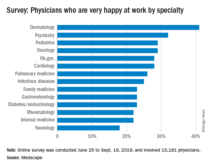
Oncologists landed in the middle of the pack among all physicians surveyed for happiness. Rheumatologists were most likely to report being very or extremely happy outside of work (60%) and neurologists were least likely to do so (44%), but about half of oncologists (51%) reported being very/extremely happy outside of work. For happiness at work, dermatologists topped the list (41%), neurologists came in last (18%), and oncologists remained in the middle (29%).
Oncologists were average when it came to burnout as well, matching the rate of overall physicians. Specifically, 32% of oncologists were burned out, 4% were depressed, and 9% were both burned out and depressed.
The most commonly reported factors contributing to burnout among oncologists were an overabundance of bureaucratic tasks (74%), spending too many hours at work (42%), and a lack of respect from colleagues in the workplace (36%).
Exercise was the most commonly reported way oncologists dealt with burnout (51%), followed by talking with family and friends (49%), and isolating themselves from others (38%). In addition, 57% of oncologists took 3-4 weeks’ vacation, compared with 44% of physicians overall; 29% of oncologists took less than 3 weeks’ vacation.
About 18% of oncologists said they had contemplated suicide, and 1% said they’d attempted it; 72% said they’d never had thoughts of suicide. Just under one-quarter of oncologists said they were currently seeking professional help or were planning to seek help for symptoms of depression and/or burnout.
“The survey results are concerning on several levels,” Maurie Markman, MD, of Cancer Treatment Centers of America, Philadelphia, said in an interview.
“First, the data suggest a considerable number of oncologists are simply burned out from the day-to-day bureaucracy (paperwork, etc.) of medical practice, which has absolutely nothing to do with the actual care delivered. This likely impacts the willingness to continue in this role. Second, one must be concerned for the future recruitment of physicians to become clinical oncologists. And finally, one must wonder about the impact of these concerning figures on the quality of care being provided to cancer patients.”
This survey was conducted from June 25 to Sept. 19, 2019, and involved 15,181 physicians. Oncologists made up 1% of the survey pool.
When it comes to physician happiness both in and outside the workplace, oncologists are about average, according to Medscape’s 2020 Lifestyle, Happiness, and Burnout Report.
Oncologists landed in the middle of the pack among all physicians surveyed for happiness. Rheumatologists were most likely to report being very or extremely happy outside of work (60%) and neurologists were least likely to do so (44%), but about half of oncologists (51%) reported being very/extremely happy outside of work. For happiness at work, dermatologists topped the list (41%), neurologists came in last (18%), and oncologists remained in the middle (29%).
Oncologists were average when it came to burnout as well, matching the rate of overall physicians. Specifically, 32% of oncologists were burned out, 4% were depressed, and 9% were both burned out and depressed.
The most commonly reported factors contributing to burnout among oncologists were an overabundance of bureaucratic tasks (74%), spending too many hours at work (42%), and a lack of respect from colleagues in the workplace (36%).
Exercise was the most commonly reported way oncologists dealt with burnout (51%), followed by talking with family and friends (49%), and isolating themselves from others (38%). In addition, 57% of oncologists took 3-4 weeks’ vacation, compared with 44% of physicians overall; 29% of oncologists took less than 3 weeks’ vacation.
About 18% of oncologists said they had contemplated suicide, and 1% said they’d attempted it; 72% said they’d never had thoughts of suicide. Just under one-quarter of oncologists said they were currently seeking professional help or were planning to seek help for symptoms of depression and/or burnout.
“The survey results are concerning on several levels,” Maurie Markman, MD, of Cancer Treatment Centers of America, Philadelphia, said in an interview.
“First, the data suggest a considerable number of oncologists are simply burned out from the day-to-day bureaucracy (paperwork, etc.) of medical practice, which has absolutely nothing to do with the actual care delivered. This likely impacts the willingness to continue in this role. Second, one must be concerned for the future recruitment of physicians to become clinical oncologists. And finally, one must wonder about the impact of these concerning figures on the quality of care being provided to cancer patients.”
This survey was conducted from June 25 to Sept. 19, 2019, and involved 15,181 physicians. Oncologists made up 1% of the survey pool.
Another round of research shows ketamine may help alcoholism
More research suggests that a single infusion of ketamine combined with counseling may help alcohol-dependent patients curb their drinking.
In a pilot study of 40 participants, those who were randomly assigned to receive intravenous ketamine plus outpatient motivational enhancement therapy (MET) showed greater abstinence rates, longer time to relapse, and fewer heavy drinking days than did those who received MET plus midazolam.
The findings support a U.K. study published late last year showing that a single dose of intravenous ketamine plus therapy that focused on reactivating drinking-related “maladaptive reward memories” reduced drinking urges and alcohol intake more than just ketamine or a placebo infusion alone (Nat Commun. 2019 Nov 26;10[1]:5187).
of the New York State Psychiatric Institute, Columbia University, New York, said in an interview.
“It’s an important area of research to understand in order to make behavioral treatments more effective, and ketamine appears to have the properties to address those vulnerabilities,” Dr. Dakwar said.
The study was published in the American Journal of Psychiatry (2019 Dec 2. doi: 10.1176/appi.ajp.2019.19070684).
Real-world approach
Pathologic alcohol use is responsible for an estimated 3.8% of all deaths globally, yet current interventions for alcohol use disorder have limited efficacy, the researchers noted.
New treatments with innovative mechanisms would be valuable, they added.
Ketamine is a high-affinity N-methyl-d-aspartate receptor (NMDAR) antagonist.
Previously, research offered “promising results” with the use of ketamine for cocaine use disorder, including increased motivation to quit and decreased craving, Dr. Dakwar noted.
“Those results led us to think about how ketamine might be helpful for other substance use disorders, especially given the overlap in clinical vulnerabilities and epidemiology,” he said.
The study from the U.K. researchers was conducted in 90 patients with harmful drinking behavior but who had not been diagnosed with alcohol use disorder.
Dr. Dakwar noted that this was “a nontreatment study. None of the people there had alcohol use disorder; they were heavy drinkers. Also, the effects there were fairly modest.
“My interest was how to integrate ketamine into a clinical, real-world framework that could be helpful for people,” he added.
The study included 40 participants (52.5% women; 70.3% white; mean age, 53 years) with alcohol dependence whose average consumption was five drinks per day.
All entered a 5-week outpatient program of MET, which involved engaging in strategies to promote motivation and self-directed change.
During the program’s second week, the participants were randomly assigned to received a 52-minute IV infusion of ketamine 0.71 mg/kg (n = 17) or the benzodiazepine midazolam 0.025 mg/kg (n = 23).
This ketamine dose was selected “because it was the highest dose tolerated by participants in preliminary studies,” the researchers reported.
“Midazolam was chosen as the active control because it alters consciousness without any known persistent ... effect on alcohol dependence,” they added.
The “timeline follow back method” was used to assess alcohol use after treatment. Abstinence was confirmed by measuring urine ethyl glucuronide levels with urine toxicology tests.
Other measures included use of a visual analogue scale, the Clinical Institute Withdrawal Assessment, and the modified Perceived Stress Scale.
Primary outcome met
Results showed that 47.1% of the ketamine group and 59.1% of the midazolam group used alcohol during the 21 days after treatment infusion; 17.6% and 40.9%, respectively, had a heavy drinking day.
For the primary outcome measure of alcohol abstinence, the “quadratic effect of time was significant” (P = .004), as was time-by-treatment interaction (P less than .001).
Although the model-estimated proportions of alcohol abstinence remained stable for the ketamine group for 21 days post infusion, the proportions decreased significantly for the control group.
The odds of having a heavy drinking day did not change significantly after treatment for the ketamine group (odds ratio, 0.98; P = .74) but increased significantly with each postinfusion day for the midazolam group (OR, 1.19; P less than .001).
For the ketamine group, time to relapse was also significantly longer (P = .04).
No significant differences were found between the groups in rates of withdrawal, craving, or stress sensitivity.
A new direction?
The most common adverse events after treatment were sedation, seen in 12 members of the midazolam group and in 8 members of the ketamine group, and headache, seen in four and six members, respectively.
Although two ketamine-group members experienced mild agitation for up to 1 hour post infusion, no incidents of persistent psychoactive effects were reported in either group.
No participants who received ketamine dropped out during the study period; among those who received midazolam, six dropped out.
“These preliminary data suggest new directions in integrated pharmacotherapy-behavioral treatments for alcohol use disorder,” the investigators wrote.
However, a larger patient population will be needed in future research in order to “replicate these promising results,” they added.
Dr. Dakwar noted that the time to first drink after treatment was comparable between the groups.
“But what was different in the ketamine group was that they didn’t continue drinking after that first drink. They didn’t initiate heavy drinking, they didn’t relapse, they were able to bounce back and stay with the program,” he said.
“It was surprising but still consistent with the central hypothesis that ketamine provides this opportunity for setting the foundation for the requisite commitment so that, once things become difficult, they’re still able to maintain recovery,” Dr. Dakwar said.
‘Provocative findings’
In an accompanying editorial, Sanjay J. Mathew, MD, of the department of psychiatry and behavioral sciences at Baylor College of Medicine in Houston, and Rebecca B. Price, PhD, of the department of psychiatry at the University of Pittsburgh, noted that ketamine’s effects on abstinence “were robust” in this trial.
“It is also noteworthy that, in spite of recruiting from a population of patients with active and significant substance use history (a group that has routinely been excluded from ketamine trials in depression), no participant showed evidence of new drug-seeking behaviors,” Dr. Mathew and Dr. Price wrote.
“Overall, these findings are provocative and hypothesis generating but certainly not definitive because of the small sample size,” they add.
Other limitations cited include the short follow-up period and the fact that only half of the participants were available for a 6-month follow-up telephone interview. In addition, generalizability was limited because the population did not have additional medical or psychiatric illnesses or additional substance use disorders, the editorialists wrote.
Because of the limitations, the investigators “are appropriately circumspect about the immediate clinical implications of this small pilot study.”
Still, the results “affirm the potential of rational combinatorial approaches for a vexing medical and public health problem,” Dr. Mathew and Dr. Price concluded.
The study was funded by grants from the National Institute on Alcohol Abuse and Alcoholism, the National Institute on Drug Abuse, and the New York State Psychiatric Institute. The study authors and Dr. Price reported no relevant financial relationships. Dr Mathew reported serving as a consultant to or having received research support from several companies, including Alkermes, Allergan, Clexio Biosciences, and Janssen. The original article includes a full list of his disclosures.
A version of this article first appeared on Medscape.com.
Tramadol use for noncancer pain linked with increased hip fracture risk
The risk of hip fracture was higher among patients treated with tramadol for chronic noncancer pain than among those treated with other commonly used NSAIDs in a large population-based cohort in the United Kingdom.

The incidence of hip fracture over a 12-month period among 293,912 propensity score-matched tramadol and codeine recipients in The Health Improvement Network (THIN) database during 2000-2017 was 3.7 vs. 2.9 per 1,000 person-years, respectively (hazard ratio for hip fracture, 1.28), Jie Wei, PhD, of Xiangya Hospital, Central South University, Changsha, China, and colleagues reported in the Journal of Bone and Mineral Research.
Hip fracture incidence per 1,000 person-years was also higher in propensity score–matched cohorts of patients receiving tramadol vs. naproxen (2.9 vs. 1.7; HR, 1.69), ibuprofen (3.4 vs. 2.0; HR, 1.65), celecoxib (3.4 vs. 1.8; HR, 1.85), or etoricoxib (2.9 vs. 1.5; HR, 1.96), the investigators found.
Tramadol is considered a weak opioid and is commonly used for the treatment of pain based on a lower perceived risk of serious cardiovascular and gastrointestinal effects versus NSAIDs, and of addiction and respiratory depression versus traditional opioids, they explained. Several professional organizations also have “strongly or conditionally recommended tramadol” as a first- or second-line treatment for conditions such as osteoarthritis, fibromyalgia, and chronic low back pain.
The potential mechanisms for the association between tramadol and hip fracture require further study, but “[c]onsidering the significant impact of hip fracture on morbidity, mortality, and health care costs, our results point to the need to consider tramadol’s associated risk of fracture in clinical practice and treatment guidelines,” they concluded.
This study was supported by the National Institutes of Health, the National Natural Science Foundation of China, and the Postdoctoral Science Foundation of Central South University. The authors reported having no conflicts of interest.
SOURCE: Wei J et al. J Bone Miner Res. 2019 Feb 5. doi: 10.1002/jbmr.3935.
The risk of hip fracture was higher among patients treated with tramadol for chronic noncancer pain than among those treated with other commonly used NSAIDs in a large population-based cohort in the United Kingdom.
The incidence of hip fracture over a 12-month period among 293,912 propensity score-matched tramadol and codeine recipients in The Health Improvement Network (THIN) database during 2000-2017 was 3.7 vs. 2.9 per 1,000 person-years, respectively (hazard ratio for hip fracture, 1.28), Jie Wei, PhD, of Xiangya Hospital, Central South University, Changsha, China, and colleagues reported in the Journal of Bone and Mineral Research.
Hip fracture incidence per 1,000 person-years was also higher in propensity score–matched cohorts of patients receiving tramadol vs. naproxen (2.9 vs. 1.7; HR, 1.69), ibuprofen (3.4 vs. 2.0; HR, 1.65), celecoxib (3.4 vs. 1.8; HR, 1.85), or etoricoxib (2.9 vs. 1.5; HR, 1.96), the investigators found.
Tramadol is considered a weak opioid and is commonly used for the treatment of pain based on a lower perceived risk of serious cardiovascular and gastrointestinal effects versus NSAIDs, and of addiction and respiratory depression versus traditional opioids, they explained. Several professional organizations also have “strongly or conditionally recommended tramadol” as a first- or second-line treatment for conditions such as osteoarthritis, fibromyalgia, and chronic low back pain.
The potential mechanisms for the association between tramadol and hip fracture require further study, but “[c]onsidering the significant impact of hip fracture on morbidity, mortality, and health care costs, our results point to the need to consider tramadol’s associated risk of fracture in clinical practice and treatment guidelines,” they concluded.
This study was supported by the National Institutes of Health, the National Natural Science Foundation of China, and the Postdoctoral Science Foundation of Central South University. The authors reported having no conflicts of interest.
SOURCE: Wei J et al. J Bone Miner Res. 2019 Feb 5. doi: 10.1002/jbmr.3935.
FROM THE JOURNAL OF BONE AND MINERAL RESEARCH
Cardiologists’ happiness average both in and outside the office
Compared with their colleagues, cardiologists are in the middle when it comes to happiness both in and outside the workplace, according to Medscape’s 2020 Lifestyle, Happiness, and Burnout Report.
About 28% of cardiologists reported that they were very happy in the workplace, according to the Medscape report, with dermatologists taking the top spot at 41%; 51% of cardiologists said that they were very happy outside of work, 9 percentage points behind rheumatologists at 60%.
The burnout rate for cardiologists was 29%, well below the 41% average for all physicians. About 15% of cardiologists reported that they were both burned out and depressed. An overabundance of bureaucratic tasks was the most commonly reported contributing factor to burnout at 64%, followed by spending too many hours at work at 39% and increased usage of EHRs at 33%.
Cardiologists most commonly dealt with burnout by exercising (45%), talking with friends/family (43%), and isolating themselves from others (42%). In addition, 47% of cardiologists reported taking 3-4 weeks of vacation, slightly more than the 44% average for all physicians; only 29% said they had taken less than 3 weeks’ vacation.
About 10% of cardiologists said that they’d contemplated suicide and 1% reported that they’d attempted it; 83% reported that they’d never thought about suicide. Only 10% of cardiologists reported that they were either currently seeking help or were planning to seek professional help for symptoms of burnout and/or depression, while 76% said they had no plans to consult help and had not done so in the past.
The Medscape survey was conducted from June 25 to Sept. 19, 2019, and involved 15,181 physicians.
An epidemic of fear and misinformation
As I write this, the 2019 novel coronavirus* continues to spread, exceeding 59,000 cases and 1,300 deaths worldwide. With it spreads fear. In the modern world of social media, misinformation spreads even faster than disease.

The news about a novel and deadly illness crowds out more substantial worries. Humans are not particularly good at assessing risk or responding rationally and consistently to it. Risk is hard to fully define. If you look up “risk” in Merriam Webster’s online dictionary, you get the simple definition of “possibility of loss or injury; peril.” If you look up risk in Wikipedia, you get 12 pages of explanation and 8 more pages of links and references.
People handle risk differently. Some people are more risk adverse than others. Some get a pleasurable thrill from risk, whether a slot machine or a parachute jump. Most people really don’t comprehend small probabilities, with tens of billions of dollars spent annually on U.S. lotteries.
Because 98% of people who get COVID-19 are recovering, this is not an extinction-level event or the zombie apocalypse. It is a major health hazard, and one where morbidity and mortality might be assuaged by an early and effective public health response, including the population’s adoption of good habits such as hand washing, cough etiquette, and staying home when ill.
Three key factors may help reduce the fear factor.
One key factor is accurate communication of health information to the public. This has been severely harmed in the last few years by the promotion of gossip on social media, such as Facebook, within newsfeeds without any vetting, along with a smaller component of deliberate misinformation from untraceable sources. Compare this situation with the decision in May 1988 when Surgeon General C. Everett Koop chose to snail mail a brochure on AIDS to every household in America. It was unprecedented. One element of this communication is the public’s belief that government and health care officials will responsibly and timely convey the information. There are accusations that the Chinese government initially impeded early warnings about COVID-19. Dr. Koop, to his great credit and lifesaving leadership, overcame queasiness within the Reagan administration about issues of morality and taste in discussing some of the HIV information. Alas, no similar leadership occurred in the decade of the 2010s when deaths from the opioid epidemic in the United States skyrocketed to claim more lives annually than car accidents or suicide.
A second factor is the credibility of the scientists. Antivaxxers, climate change deniers, and mercenary scientists have severely damaged that credibility of science, compared with the trust in scientists 50 years ago during the Apollo moon shot.
A third factor is perspective. Poor journalism and clickbait can focus excessively on the rare events as news. Airline crashes make the front page while fatal car accidents, claiming a hundred times more lives annually, don’t even merit a story in local media. Someone wins the lottery weekly but few pay attention to those suffering from gambling debts.
Influenza is killing many times more people than the 2019 novel coronavirus, but the news is focused on cruise ships. In the United States, influenza annually will strike tens of millions, with about 10 per 1,000 hospitalized and 0.5 per 1,000 dying. The novel coronavirus is more lethal. SARS (a coronavirus epidemic in 2003) had 8,000 cases with a mortality rate of 96 per 1,000 while the novel 2019 strain so far is killing about 20 per 1,000. That value may be an overestimate, because there may be a significant fraction of COVID-19 patients with symptoms mild enough that they do not seek medical care and do not get tested and counted.
For perspective, in 1952 the United States reported 50,000 cases of polio (meningitis or paralytic) annually with 3,000 deaths. As many as 95% of cases of poliovirus infection have no or mild symptoms and would not have been reported, so the case fatality rate estimate is skewed. In the 1950s, the United States averaged about 500,000 cases of measles per year, with about 500 deaths annually for a case fatality rate of about 1 per 1,000 in a population that was well nourished with good medical care. In malnourished children without access to modern health care, the case fatality rate can be as high as 100 per 1,000, which is why globally measles killed 142,000 people in 2018, a substantial improvement from 536,000 deaths globally in 2000, but still a leading killer of children worldwide. Vaccines had reduced the annual death toll of polio and measles in the U.S. to zero.
In comparison, in this country the annual incidences are about 70,000 overdose deaths, 50,000 suicides, and 40,000 traffic deaths.
Reassurance is the most common product sold by pediatricians. We look for low-probability, high-impact bad things. Usually we don’t find them and can reassure parents that the child will be okay. Sometimes we spot a higher-risk situation and intervene. My job is to worry professionally so that parents can worry less.
COVID-19 worries me, but irrational people worry me more. The real enemies are fear, disinformation, discrimination, and economic warfare.
Dr. Powell is a pediatric hospitalist and clinical ethics consultant living in St. Louis. Email him at pdnews@mdedge.com.
*This article was updated 2/21/2020.
As I write this, the 2019 novel coronavirus* continues to spread, exceeding 59,000 cases and 1,300 deaths worldwide. With it spreads fear. In the modern world of social media, misinformation spreads even faster than disease.
The news about a novel and deadly illness crowds out more substantial worries. Humans are not particularly good at assessing risk or responding rationally and consistently to it. Risk is hard to fully define. If you look up “risk” in Merriam Webster’s online dictionary, you get the simple definition of “possibility of loss or injury; peril.” If you look up risk in Wikipedia, you get 12 pages of explanation and 8 more pages of links and references.
People handle risk differently. Some people are more risk adverse than others. Some get a pleasurable thrill from risk, whether a slot machine or a parachute jump. Most people really don’t comprehend small probabilities, with tens of billions of dollars spent annually on U.S. lotteries.
Because 98% of people who get COVID-19 are recovering, this is not an extinction-level event or the zombie apocalypse. It is a major health hazard, and one where morbidity and mortality might be assuaged by an early and effective public health response, including the population’s adoption of good habits such as hand washing, cough etiquette, and staying home when ill.
Three key factors may help reduce the fear factor.
One key factor is accurate communication of health information to the public. This has been severely harmed in the last few years by the promotion of gossip on social media, such as Facebook, within newsfeeds without any vetting, along with a smaller component of deliberate misinformation from untraceable sources. Compare this situation with the decision in May 1988 when Surgeon General C. Everett Koop chose to snail mail a brochure on AIDS to every household in America. It was unprecedented. One element of this communication is the public’s belief that government and health care officials will responsibly and timely convey the information. There are accusations that the Chinese government initially impeded early warnings about COVID-19. Dr. Koop, to his great credit and lifesaving leadership, overcame queasiness within the Reagan administration about issues of morality and taste in discussing some of the HIV information. Alas, no similar leadership occurred in the decade of the 2010s when deaths from the opioid epidemic in the United States skyrocketed to claim more lives annually than car accidents or suicide.
A second factor is the credibility of the scientists. Antivaxxers, climate change deniers, and mercenary scientists have severely damaged that credibility of science, compared with the trust in scientists 50 years ago during the Apollo moon shot.
A third factor is perspective. Poor journalism and clickbait can focus excessively on the rare events as news. Airline crashes make the front page while fatal car accidents, claiming a hundred times more lives annually, don’t even merit a story in local media. Someone wins the lottery weekly but few pay attention to those suffering from gambling debts.
Influenza is killing many times more people than the 2019 novel coronavirus, but the news is focused on cruise ships. In the United States, influenza annually will strike tens of millions, with about 10 per 1,000 hospitalized and 0.5 per 1,000 dying. The novel coronavirus is more lethal. SARS (a coronavirus epidemic in 2003) had 8,000 cases with a mortality rate of 96 per 1,000 while the novel 2019 strain so far is killing about 20 per 1,000. That value may be an overestimate, because there may be a significant fraction of COVID-19 patients with symptoms mild enough that they do not seek medical care and do not get tested and counted.
For perspective, in 1952 the United States reported 50,000 cases of polio (meningitis or paralytic) annually with 3,000 deaths. As many as 95% of cases of poliovirus infection have no or mild symptoms and would not have been reported, so the case fatality rate estimate is skewed. In the 1950s, the United States averaged about 500,000 cases of measles per year, with about 500 deaths annually for a case fatality rate of about 1 per 1,000 in a population that was well nourished with good medical care. In malnourished children without access to modern health care, the case fatality rate can be as high as 100 per 1,000, which is why globally measles killed 142,000 people in 2018, a substantial improvement from 536,000 deaths globally in 2000, but still a leading killer of children worldwide. Vaccines had reduced the annual death toll of polio and measles in the U.S. to zero.
In comparison, in this country the annual incidences are about 70,000 overdose deaths, 50,000 suicides, and 40,000 traffic deaths.
Reassurance is the most common product sold by pediatricians. We look for low-probability, high-impact bad things. Usually we don’t find them and can reassure parents that the child will be okay. Sometimes we spot a higher-risk situation and intervene. My job is to worry professionally so that parents can worry less.
COVID-19 worries me, but irrational people worry me more. The real enemies are fear, disinformation, discrimination, and economic warfare.
Dr. Powell is a pediatric hospitalist and clinical ethics consultant living in St. Louis. Email him at pdnews@mdedge.com.
*This article was updated 2/21/2020.
Gene therapy effective in hemophilia B patients with neutralizing antibodies
The gene therapy etranacogene dezaparvovec (AMT-061) continues to demonstrate safety and efficacy in patients with hemophilia B, according to a 1-year update of a phase 2b trial.

All three patients in this trial experienced sustained increases in factor IX (FIX) activity and were able to stop prophylaxis without suffering any bleeds. Adverse events related to treatment were mild and transient.
These favorable results are particularly noteworthy because all three patients had anti-AAV5 neutralizing antibodies at baseline, according to Steven W. Pipe, MD, of the University of Michigan, Ann Arbor. He noted that studies of etranacogene dezaparvovec and its predecessor, AMT-060, are the only studies that have not excluded hemophilia patients based on preexisting immunity.
Dr. Pipe presented the latest phase 2b results with etranacogene dezaparvovec at the annual congress of the European Association for Haemophilia and Allied Disorders.
Etranacogene dezaparvovec uses an AAV5 serotype with a transgene expression cassette that codes for the hyperactive Padua FIX variant, Dr. Pipe explained. Etranacogene dezaparvovec has a structure that is nearly identical to that of AMT-060, except for two nucleotide substitutions in the coding sequence for FIX.
AMT-060 enabled stable expression of FIX that has persisted for up to 4 years without any late-emergent safety signals (Blood 2019. 134 Supplement 1: 2059). Dr. Pipe said the “enhanced version” of AMT-060, etranacogene dezaparvovec, has produced even higher levels of FIX activity in the phase 2b study (NCT03489291).
The ongoing study enrolled three men with moderate to severe FIX deficiency at baseline. The patients were 43, 50, and 47 years of age, respectively. Two patients are HIV positive, and all had hepatitis C that resolved.
All three patients were receiving FIX prophylaxis and on-demand treatment at baseline. In the year prior to screening, patients had one, three, and five bleeds, respectively. All three patients had anti-AAV5 neutralizing antibodies.
Efficacy
Patients received a single dose of etranacogene dezaparvovec at 2 x 10¹³ genome copies/kg. All three patients achieved the primary endpoint, which was FIX activity of at least 5% at 6 weeks.
At 52 weeks, the mean FIX activity was 41%. Patients 1 and 3 have maintained FIX activity of 40% or greater, which is in the nonhemophilic range. Patient 2 has maintained FIX activity in the mild range. At 52 weeks, FIX activity levels were 50.2%, 40.8%, and 31.3%, respectively.
All patients remain free of prophylaxis and bleeds. Patient 3 received a single FIX infusion as a precaution in the perioperative setting. There was no evidence of a bleed in this patient.
Safety
Etranacogene dezaparvovec was generally well tolerated, Dr. Pipe said. One patient had two adverse events that were possibly related to etranacogene dezaparvovec. Both events – transient, self-limiting headache and slightly elevated C-reactive protein – resolved without intervention.
There was one serious adverse event, but it was considered unrelated to treatment. Patient 3 required hip surgery for preexisting avascular necrosis.
Dr. Pipe said there was no evidence of transaminitis. There were modest, transient elevations in liver enzymes, but this was not enough to trigger protocol-specified immunosuppression.
Specifically, one patient had ALT elevations at weeks 22 and 44, and one patient had AST elevations at weeks 2, 4, and 31. All of these resolved quickly without treatment or an impact on FIX activity, Dr. Pipe noted.
Next steps
This study is ongoing, and patients will be followed for 5 years. Dr. Pipe said the main focus of follow-up will be to determine if patients maintain durable expression of FIX.
A phase 3 trial of etranacogene dezaparvovec is ongoing as well. The trial, HOPE-B (NCT03569891), is fully enrolled, and dosing is planned for 55 patients.
“We’re looking forward to data analysis later this year,” Dr. Pipe said. “This will be the only phase 3 study, and really the only platform so far, that is not planning to exclude patients based on preexisting immunity.”
If all goes well in the phase 3 study, etranacogene dezaparvovec could be approved by the Food and Drug Administration very soon, Dr. Pipe added.
UniQure, the company developing etranacogene dezaparvovec, is planning to submit the biologics license application to the FDA next year. Etranacogene dezaparvovec was granted breakthrough designation from the FDA and is therefore eligible for priority review, so the gene therapy could be approved as early as 2021.
The phase 2b trial of etranacogene dezaparvovec is sponsored by uniQure. Dr. Pipe disclosed relationships with uniQure and other companies.
SOURCE: Pipe SW et al. EAHAD 2020, Abstract OR10.
The gene therapy etranacogene dezaparvovec (AMT-061) continues to demonstrate safety and efficacy in patients with hemophilia B, according to a 1-year update of a phase 2b trial.
All three patients in this trial experienced sustained increases in factor IX (FIX) activity and were able to stop prophylaxis without suffering any bleeds. Adverse events related to treatment were mild and transient.
These favorable results are particularly noteworthy because all three patients had anti-AAV5 neutralizing antibodies at baseline, according to Steven W. Pipe, MD, of the University of Michigan, Ann Arbor. He noted that studies of etranacogene dezaparvovec and its predecessor, AMT-060, are the only studies that have not excluded hemophilia patients based on preexisting immunity.
Dr. Pipe presented the latest phase 2b results with etranacogene dezaparvovec at the annual congress of the European Association for Haemophilia and Allied Disorders.
Etranacogene dezaparvovec uses an AAV5 serotype with a transgene expression cassette that codes for the hyperactive Padua FIX variant, Dr. Pipe explained. Etranacogene dezaparvovec has a structure that is nearly identical to that of AMT-060, except for two nucleotide substitutions in the coding sequence for FIX.
AMT-060 enabled stable expression of FIX that has persisted for up to 4 years without any late-emergent safety signals (Blood 2019. 134 Supplement 1: 2059). Dr. Pipe said the “enhanced version” of AMT-060, etranacogene dezaparvovec, has produced even higher levels of FIX activity in the phase 2b study (NCT03489291).
The ongoing study enrolled three men with moderate to severe FIX deficiency at baseline. The patients were 43, 50, and 47 years of age, respectively. Two patients are HIV positive, and all had hepatitis C that resolved.
All three patients were receiving FIX prophylaxis and on-demand treatment at baseline. In the year prior to screening, patients had one, three, and five bleeds, respectively. All three patients had anti-AAV5 neutralizing antibodies.
Efficacy
Patients received a single dose of etranacogene dezaparvovec at 2 x 10¹³ genome copies/kg. All three patients achieved the primary endpoint, which was FIX activity of at least 5% at 6 weeks.
At 52 weeks, the mean FIX activity was 41%. Patients 1 and 3 have maintained FIX activity of 40% or greater, which is in the nonhemophilic range. Patient 2 has maintained FIX activity in the mild range. At 52 weeks, FIX activity levels were 50.2%, 40.8%, and 31.3%, respectively.
All patients remain free of prophylaxis and bleeds. Patient 3 received a single FIX infusion as a precaution in the perioperative setting. There was no evidence of a bleed in this patient.
Safety
Etranacogene dezaparvovec was generally well tolerated, Dr. Pipe said. One patient had two adverse events that were possibly related to etranacogene dezaparvovec. Both events – transient, self-limiting headache and slightly elevated C-reactive protein – resolved without intervention.
There was one serious adverse event, but it was considered unrelated to treatment. Patient 3 required hip surgery for preexisting avascular necrosis.
Dr. Pipe said there was no evidence of transaminitis. There were modest, transient elevations in liver enzymes, but this was not enough to trigger protocol-specified immunosuppression.
Specifically, one patient had ALT elevations at weeks 22 and 44, and one patient had AST elevations at weeks 2, 4, and 31. All of these resolved quickly without treatment or an impact on FIX activity, Dr. Pipe noted.
Next steps
This study is ongoing, and patients will be followed for 5 years. Dr. Pipe said the main focus of follow-up will be to determine if patients maintain durable expression of FIX.
A phase 3 trial of etranacogene dezaparvovec is ongoing as well. The trial, HOPE-B (NCT03569891), is fully enrolled, and dosing is planned for 55 patients.
“We’re looking forward to data analysis later this year,” Dr. Pipe said. “This will be the only phase 3 study, and really the only platform so far, that is not planning to exclude patients based on preexisting immunity.”
If all goes well in the phase 3 study, etranacogene dezaparvovec could be approved by the Food and Drug Administration very soon, Dr. Pipe added.
UniQure, the company developing etranacogene dezaparvovec, is planning to submit the biologics license application to the FDA next year. Etranacogene dezaparvovec was granted breakthrough designation from the FDA and is therefore eligible for priority review, so the gene therapy could be approved as early as 2021.
The phase 2b trial of etranacogene dezaparvovec is sponsored by uniQure. Dr. Pipe disclosed relationships with uniQure and other companies.
SOURCE: Pipe SW et al. EAHAD 2020, Abstract OR10.
REPORTING FROM EAHAD 2020
Shorter time to metastases associated with worse RCC outcomes
in an international review of over 7,000 renal cell carcinoma (RCC) patients treated with first-line tyrosine kinase inhibitors.
“Patients with synchronous disease, compared with patients with metachronous disease, have more adverse prognostic features, significantly shorter TTF [time to treatment failure], and poorer survival. This may help in patient counseling and may be taken into consideration in clinical trial designs in the future, in order to avoid an imbalance between treatment arms,” wrote investigators led by Frede Donskov, MD, a clinical professor at Aarhus (Denmark) University Hospital, in European Urology Oncology.
In the largest study to date to address the impact of timing of metastases on outcomes from tyrosine kinase inhibitor (TKI) treatment, something that’s been unclear until now, Dr. Donskov and associates turned to the International Metastatic Renal Cell Carcinoma Database Consortium (IMDC) to compare outcomes of 3,906 patients with synchronous metastases, meaning metastases within 3 months of initial RCC diagnosis, with 3,480 with metachronous disease, meaning metastases after that point.
They found that more patients with synchronous versus metachronous disease had higher T stage (T1-2, 19% vs. 34%), N1 disease (21% vs. 6%), presence of sarcomatoid differentiation (15.8% vs. 7.9%), Karnofsky performance status less than 80 (25.9% vs. 15.1%), anemia (62.5% vs. 42.3%), elevated neutrophils (18.9% vs. 10.9%), elevated platelets (21.6% vs. 11.4%), bone metastases (40.4% vs. 29.8%); and IMDC poor risk (40.6% vs.11.3%).
Synchronous versus metachronous disease by intervals of more than 3-12 months, more than 1-2 years, more than 2-7 years, and more than 7 years correlated with poor TTF (5.6 months vs. 7.3, 8.0, 10.8, and 13.3 months; P less than .0001) and poor overall survival (median, 16.7 months vs. 23.8, 30.2, 34.8, and 41.7 months; P less than .0001).
On multivariable regressions adjusting for baseline variables, metachronous disease was protective versus synchronous RCC on overall survival and TTF, with a greater protective effect the longer it took for the disease to metastasize.
“Synchronous disease may represent a distinct pathologic and molecular phenotype ... a high proportion of patients with synchronous disease have tumors with punctuated evolution, harboring aggressive disease features, consolidating in worse risk factors, requiring systemic therapy earlier, and having almost half the expected survival after the initiation of targeted therapy, compared with the latest metastatic timing, as shown in our study,” the investigators wrote.
The findings “reflect the underlying aggressive tumor biology. Whether [time to metastasis] impacts outcome to checkpoint immunotherapy is yet to be elucidated,” they added.
Patients were a median of 59 years at diagnosis, and 72.9% were men. None of the synchronous patients had a surgical nephrectomy, compared with 95.4% of metachronous patients; 67.2% of patients in both groups were treated with the TKI sunitinib (Sutent).
The work was funded by the IMDC. The lead investigator reported institutional grants from Ipsen and Pfizer, maker of sunitinib.
SOURCE: Donskov F et al. Eur Urol Oncol. 2020 Feb 6. doi: 10.1016/j.euo.2020.01.001.
FROM EUROPEAN UROLOGY ONCOLOGY
To Improve TB Vaccination, Change The Way It’s Given?
The standard intradermal route of delivery for Bacille Calmette–Guérin (BCG) does not necessarily generate a strong enough response from lung T-cells, the researchers say. They hypothesized that administering BCG by IV or aerosol might be more effective.
They gave a group of rhesus macaques the BGC vaccine by intradermal, aerosol, or IV routes, then assessed immune responses in blood and fluid drawn from the lungs over a 24-week follow-up. Six months after vaccination, the researchers injected the vaccinated animals with a virulent strain of Mycobacterium tuberculosis (M tuberculosis) and tracked infection and disease development over 3 months.
The IV vaccination resulted in the highest durable levels of T-cells in blood and lungs. Nine of 10 animals vaccinated via IV were highly protected; 6 showed no detectable infection in any tissue tested and 3 had only very low counts of M tuberculosis in lung tissue. All unvaccinated animals and those immunized via intradermal or aerosol routes showed signs of significantly greater infection.
Upping the dose did not improve protection. The IV BCG group showed 90% protection at a threshold as low as 50 colony-forming units (the standard human ID dose is 5 x 10⁵ CFUs).
The researchers say several unique quantitative and qualitative differences in the immune responses may underlie protection. Perhaps most noteworthy, they say, was the large population of T- cells in the tissue across all lung parenchyma lobes.
The study provides a “paradigm shift,” the researchers conclude, adding that the IV route may also improve the protective capacity of other vaccines.
Lorcaserin withdrawn from U.S. market due to cancer risk
The Food and Drug Administration asked Eisai to voluntary withdraw the weight-loss drug lorcaserin (Belviq and Belviq XR) on Feb. 13 after a post-marketing trial with more than 12,000 subjects revealed an increased occurrence of cancer.

In a Drug Safety Communication, the agency said “health care professionals should stop prescribing and dispensing lorcaserin to patients. Contact patients currently taking lorcaserin, inform them of the increased occurrence of cancer seen in the clinical trial, and ask them to stop taking the medicine. Discuss alternative weight-loss medicines or strategies with your patients.”
Eisai is complying with the withdrawal request.
The decision is based on the agency’s review of the 5-year trial, which was designed to evaluate cardiac risk with the drug and ended in June 2018. In total, 7.7% of patients randomized to 10 mg lorcaserin twice daily were diagnosed with 520 primary cancers, compared with 7.1% of placebo subjects diagnosed with 470 cancers, over a median follow-up of 3 years and 3 months. There was one additional cancer observed for every 470 patients treated for 1 year.
“There was no apparent difference in the incidence of cancer over the initial months of treatment, but the imbalance increased with longer duration on lorcaserin,” FDA said. Pancreatic, colorectal, and lung cancers were among those diagnosed.
In short, “we believe that the risks of lorcaserin outweigh its benefits based on our completed review of” the data, the agency said. The FDA is not recommending special cancer screenings for patients who have taken lorcaserin.
The action follows an FDA alert in January about a possible elevated cancer risk based on its preliminary analysis of the study.
Patients were also advised Feb. 13 to stop taking the drug and talk to their providers about alternative weight-loss medications and weight-management programs.
They were also told to dispose of the pills at a drug take-back location if available, but if not, to mix them with an “unappealing substance” such as dirt, cat litter, or used coffee grounds; seal them in plastic bag; and put them in the trash.
The Food and Drug Administration asked Eisai to voluntary withdraw the weight-loss drug lorcaserin (Belviq and Belviq XR) on Feb. 13 after a post-marketing trial with more than 12,000 subjects revealed an increased occurrence of cancer.
In a Drug Safety Communication, the agency said “health care professionals should stop prescribing and dispensing lorcaserin to patients. Contact patients currently taking lorcaserin, inform them of the increased occurrence of cancer seen in the clinical trial, and ask them to stop taking the medicine. Discuss alternative weight-loss medicines or strategies with your patients.”
Eisai is complying with the withdrawal request.
The decision is based on the agency’s review of the 5-year trial, which was designed to evaluate cardiac risk with the drug and ended in June 2018. In total, 7.7% of patients randomized to 10 mg lorcaserin twice daily were diagnosed with 520 primary cancers, compared with 7.1% of placebo subjects diagnosed with 470 cancers, over a median follow-up of 3 years and 3 months. There was one additional cancer observed for every 470 patients treated for 1 year.
“There was no apparent difference in the incidence of cancer over the initial months of treatment, but the imbalance increased with longer duration on lorcaserin,” FDA said. Pancreatic, colorectal, and lung cancers were among those diagnosed.
In short, “we believe that the risks of lorcaserin outweigh its benefits based on our completed review of” the data, the agency said. The FDA is not recommending special cancer screenings for patients who have taken lorcaserin.
The action follows an FDA alert in January about a possible elevated cancer risk based on its preliminary analysis of the study.
Patients were also advised Feb. 13 to stop taking the drug and talk to their providers about alternative weight-loss medications and weight-management programs.
They were also told to dispose of the pills at a drug take-back location if available, but if not, to mix them with an “unappealing substance” such as dirt, cat litter, or used coffee grounds; seal them in plastic bag; and put them in the trash.