User login
Beyond C. difficile: The future of fecal microbial transplantation
SAN DIEGO – Two leading figures in microbiome research took time during the annual Digestive Disease Week to share their perspective with members of the press. Identifying key research presented at the meeting and painting a broader picture of trends and challenges in research on the interplay with the microbiota with gut health, Colleen Kelly, MD, of Brown University, Providence, R.I., cochair and principal investigator, AGA FMT Registry Steering Committee, led off the round table with a discussion of human research on fecal microbial transplantation (FMT) and obesity.
Purna Kashyap, MBBS, of the Mayo Clinic, Rochester, Minn., member, AGA Center for Gut Microbiome Research and Education Scientific Advisory Board, delved into the potential for donor microbiota transplant to address small bowel disorders, such as small intestinal bacterial overgrowth, and provided commentary regarding the potential – and limitations – of using FMT in functional bowel disorders such as irritable bowel syndrome (IBS).
Obesity
Dr. Kelly noted that two abstracts at the conference presented data about FMT in obesity. The first study was presented by Jessica Allegretti, MD, a gastroenterologist at Brigham and Women’s Hospital, Boston; the second study was presented by Elaine Yu, MD, an endocrinologist at Massachusetts General Hospital, Boston.
The two studies shared some similarities, but had some differences, said Dr. Kelly. “They both used lean donor encapsulated FMT ... and they both were placebo controlled, using placebo capsules.” The first study looked at metabolically healthy obese patients, while the second study included patients with mild to moderate insulin resistance.
Dr. Allegretti’s work looked at the effect that FMT from lean donors had on levels of a satiety peptide, glucagonlike peptide–1 (GLP-1), while also looking at changes in weight and microbiota, as well as safety. Patients received an initial 30-capsule dose as well as two later doses of 12 capsules each. The 22-patient study, in which individuals were randomized 1:1 to FMT or placebo capsules, didn’t show statistically significant changes in GLP-1 levels or body mass index with FMT over the 12-week study period. “But they were able to show engraftment, which I think is an important thing that you do wonder about – over this period of time, the bacteria that came from the lean donor actually engrafted into the recipient and affected the diversity. The recipient became more similar to the donor,” said Dr. Kelly.
There were some clues among the findings that engraftment was effecting metabolic change in the recipient, she said, including differences in bile acid conversion among gut bacteria; also, lower levels of the primary bile acid taurocholic acid in recipients after FMT. “So it was a negative study in what she was looking for, but an example of these smaller studies kind of pushing the field along.”
In discussing the study presented by Dr. Yu that examined lean donor FMT in individuals with insulin resistance, Dr. Kelly said, “It was actually pretty sophisticated.” By using hyperinsulinemic euglycemic clamping, the investigators were able to measure insulin sensitivity based on glucose load. In this study of 24 individuals randomized 1:1 to receive FMT or placebo capsules, recipients received weekly doses over a period of 6 weeks. Here again, though Dr. Yu and colleagues again found engraftment, “they did not find the big changes in metabolic parameters that they hoped that they would,” said Dr. Kelly.
These two studies furthered previous work completed in the Netherlands examining lean donor FMT for individuals with metabolic syndrome. “Those [studies] did show both engraftment and some changes in insulin resistance,” but they were also small studies, noted Dr. Kelly. Dr. Kashyap pointed out that the earlier studies had shown in a subgroup analysis that response to FMT was limited to those patients who lacked microbial diversity pretransplant.
This makes some mechanistic sense when thinking about FMT’s greatest success to date: Treating Clostridioides difficile infection, a condition whose very hallmark is dysbiosis characterized by monospecies gut domination, noted Dr. Kashyap.
Added Dr. Kelly, “I think everyone’s hoping there’s going to be this pill that’s going to make us skinny, but I don’t think we’re going to find that with FMT and obesity. I do think these studies are important, because there’s so much animal data already, and we’re kind of like, ‘How much more can you do in mice?’ ” By translating this preclinical work into humans, the mechanisms of obesity and the role of the microbiome can be better understood.
IBS
Turning to functional bowel disorders, Dr. Kelly pointed out a new study examining FMT for IBS. “So far, the research has been pretty disappointing,” she noted. The study, presented by Prashant Singh, MBBS, a gastroenterology fellow at Beth Israel Deaconess Medical Center, Boston, examined FMT for patients with moderate to severe diarrhea-predominant IBS. The study had four arms, randomizing the 44 participants to FMT alone; pretreatment with metronidazole and ciprofloxacin or rifaximin, each followed by FMT; or placebo. “It really didn’t show any differences in their severity of IBS in any group; so no effect: a negative study,” said Dr. Kelly.
“It’s challenging. FMT is an appealing strategy because we don’t have to put a lot of thought process into it,” said Dr. Kashyap, but it’s no panacea. “We aren’t going to be able to treat everybody with FMT.
“The challenge with IBS is that if we look at all the compositional studies, the majority of them show that at least a big subset of patients with IBS already have a normal-appearing microbiome,” said Dr. Kashyap. In those patients, “It’s very hard to know what FMT is doing.” He noted that IBS subclassifications are made by pathophysiology, without regard to the intestinal microbiome, so these classifications may not be useful for determining who may benefit from FMT.
“Again, there’s always an opportunity to learn from these studies,” whether they’re positive or negative, as long as they’re well done, said Dr. Kashyap. “There’s always an opportunity to go back and see, was there a specific subgroup of patients who responded, where there might be one or more causes which might be more amenable” to FMT.
Small intestine
And most intestinal microbial research to date, noted Dr. Kelly, has focused on the colon. “Most of our knowledge is of fecal microbiota.” New techniques including double-balloon enteroscopy of the small bowel have promise to “provide completely new information about patterns of bacteria throughout the small bowel,” and of the role of small bowel bacteria in overall gut health, she said.
“The role of the small bowel has been ignored because of accessibility,” agreed Dr. Kashyap. There’s a current focus on research into enteroscopy and other techniques to sample small intestine microbiota, he said.
In a podium presentation, Eugene Chang, MD, Martin Boyer Professor in the University of Chicago’s department of medicine, gave a broad overview of how the small intestine microbiome modulates lipid regulation. This choice of topic for the Charles M. Mansbach Memorial Lecture shows that gastroenterologists are recognizing the importance of microbiome along the entire span of the gut, said Dr. Kashyap. Dr. Chang’s approach, he added, represents a departure in that “it’s not simply just looking at what’s present and what’s not, but seeing what’s functionally relevant to metabolism.”
“We always have known that the small intestine is the workhorse; that’s where everything happens” in terms of motility, absorption, and digestion, said Dr. Kashyap. “But because of our inability to reach it easily we’ve always chosen to ignore it; we always go after the low-hanging fruit.” Despite challenges, more microbiome research should be small-bowel focused. “Eventually, it’s no pain, no gain.”
Dr. Kashyap is on the advisory board of uBiome, and is an ad hoc advisory board member for Salix Pharmaceuticals. Dr. Kelly reported no conflicts of interest.
This story was updated on July 30, 2019.
EXPERT ANALYSIS FROM DDW 2019
Older adults’ interested in conversations about deprescribing
Clinical question: Among older adults, what attitudes exist toward deprescribing?
Background: Polypharmacy in older adults is common and can be associated with increased hospitalizations and reduced quality of life.
Study design: Population-based survey study.
Setting: Medicare beneficiaries in the United States.

Synopsis: The investigators used data from the National Health and Aging Trends Study (NHATS), which collects information annually on a nationally representative sample of Medicare beneficiaries ages 65 and older. Of 1,981 responses on the NHATS Medication Attitudes module, 92% of older adults expressed willingness to stop a medication if their doctor said it was possible. While 89% agreed that all their medications were necessary, 66.6% also agreed that they would like to reduce the number of their medications. Patients taking more than six medications, compared with those taking fewer than six (adjusted odds ratio, 2.9; 95% confidence interval, 1.74-4.82) and those with three or more medical conditions, compared with patients with fewer than two (aOR 2.87; 95% CI 1.53-5.37) had greater odds of willingness to stop a medication. Importantly, the study did not collect data about specific medications.
Bottom line: A vast majority of older adults would be willing to stop one or more of their medications if considered possible by their physician, and two-thirds want to reduce the number of their medications. If appropriate, hospitalists should consider having a conversation about deprescribing with their older patients.
Citation: Reeve E et al. Assessment of attitudes toward deprescribing in older Medicare beneficiaries in the United States. JAMA Intern Med. 2018;178(12):1673-180.
Dr. Stanley is assistant professor of medicine at Northwestern University Feinberg School of Medicine and a hospitalist at Northwestern Memorial Hospital, both in Chicago.
Clinical question: Among older adults, what attitudes exist toward deprescribing?
Background: Polypharmacy in older adults is common and can be associated with increased hospitalizations and reduced quality of life.
Study design: Population-based survey study.
Setting: Medicare beneficiaries in the United States.
Synopsis: The investigators used data from the National Health and Aging Trends Study (NHATS), which collects information annually on a nationally representative sample of Medicare beneficiaries ages 65 and older. Of 1,981 responses on the NHATS Medication Attitudes module, 92% of older adults expressed willingness to stop a medication if their doctor said it was possible. While 89% agreed that all their medications were necessary, 66.6% also agreed that they would like to reduce the number of their medications. Patients taking more than six medications, compared with those taking fewer than six (adjusted odds ratio, 2.9; 95% confidence interval, 1.74-4.82) and those with three or more medical conditions, compared with patients with fewer than two (aOR 2.87; 95% CI 1.53-5.37) had greater odds of willingness to stop a medication. Importantly, the study did not collect data about specific medications.
Bottom line: A vast majority of older adults would be willing to stop one or more of their medications if considered possible by their physician, and two-thirds want to reduce the number of their medications. If appropriate, hospitalists should consider having a conversation about deprescribing with their older patients.
Citation: Reeve E et al. Assessment of attitudes toward deprescribing in older Medicare beneficiaries in the United States. JAMA Intern Med. 2018;178(12):1673-180.
Dr. Stanley is assistant professor of medicine at Northwestern University Feinberg School of Medicine and a hospitalist at Northwestern Memorial Hospital, both in Chicago.
Trained interpreters essential for treating non–English-speaking patients
NEW YORK – Clinicians should resist the temptation to use untrained interpreters, such as a child, another relative, or their own limited language skills, when treating patients who cannot communicate in English, according to an expert reviewing this issue at the American Academy of Dermatology summer meeting.
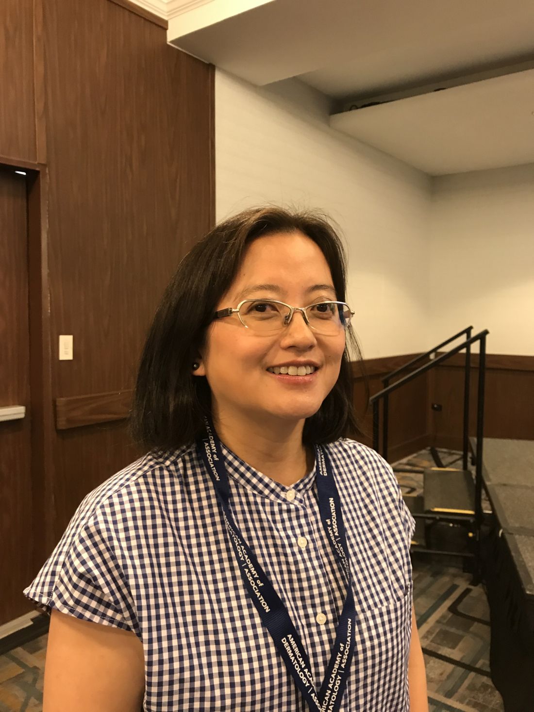
In clinical encounters with patients who have limited English proficiency, “ reported Amy Y.Y. Chen, MD, who is affiliated with Central Connecticut Dermatology in Canton.
In many situations, interpreter services are required by law. This includes a provision of the 1963 Civil Rights Act that specifies these services should be made available to any individual with limited English proficiency receiving federal financial assistance (with the exception of Medicare Part B).
In reviewing this and other laws, Dr. Chen explained that many prohibitions are explicit. For example, it is against the law for clinicians to communicate with the patient through children, whether or not they are related to the patient. A patient’s adult companions are also prohibited from interpreting unless the patient has provided express permission.
Despite the rules, some clinicians might be tempted to forgo a translator when none is readily available, opting for an improvised solution. Dr. Chen said that this is ill advised even when it is not illegal.
“There are a lot of potential problems with using nonprofessional interpreters, starting with the issue of confidentiality,” Dr. Chen warned.
As defined by the Department of Health & Human Services, a qualified interpreter establishes competency by developing familiarity with specialized terminology; by communicating accurately, effectively, and impartially; and by recognizing the ethical issues, including confidentiality, essential to their role.
By itself, language fluency might not be sufficient. Many physicians have conversational fluency in one or more languages other than English, but Dr. Chen pointed out that complex and nuanced clinical descriptions might be difficult to follow for a nonnative speaker. Moreover, many individuals who have no problem posing questions in a foreign language don’t do nearly as well in following the answers.
As interpreters, family members can be particularly problematic. In addition to the issues of confidentiality and medical terminology, a family member might have his or her own agenda that influences how questions and answers are conveyed.
Moreover, family members and others untrained in translating might edit answers based on their own sense of relevance. Many clinicians working through an interpreter will recognize the experience of receiving a yes or no answer after a lengthy discussion between a nontrained interpreter and patient. In such situations, the clinician can reasonably worry that important information was lost.
Typically, major hospitals already offer a systematic approach to providing interpreters when needed, but physicians working in private practice or other smaller practice settings might not. According to Dr. Chen, who recently collaborated on review of this issue (J Am Acad Dermatol. 2019 Mar;80:829-31), they should.
Interpreter services are available by telephone or Internet. Fees typically fall in the range of $2-$5 per minute. In offices with bilingual staff members, formal medical interpreter training might make sense. The Certification Commission for Healthcare Interpreters and the National Board of Certification for Medical Interpreters can help in this process.
When using a medical interpreter, Dr. Chen had some tips.
“Maintain eye contact and talk to the patient,” said Dr. Chen, suggesting that the interpreter, if present in the room, be seated next to or behind the patient. Whether the interpreter is in the room or participating remotely, Dr. Chen advised against speaking through the interpreter with such phases as “tell her that.” Rather, she advised speaking directly to the patient with the interpreter providing the translation.
More practically, Dr. Chen recommended speaking slowly and posing only one question at a time. She also recommended strategies to elicit reassurance that the patient has understood what was communicated. Not least, she recommended a “show me” approach in which a patient can repeat or demonstrate what he or she has learned.
Citing evidence that poor and incomplete translation contributes to medical errors and patient dissatisfaction, Dr. Chen reiterated that engaging unbiased trained translators is advisable for good clinical care even if it were not mandated by law.
NEW YORK – Clinicians should resist the temptation to use untrained interpreters, such as a child, another relative, or their own limited language skills, when treating patients who cannot communicate in English, according to an expert reviewing this issue at the American Academy of Dermatology summer meeting.
In clinical encounters with patients who have limited English proficiency, “ reported Amy Y.Y. Chen, MD, who is affiliated with Central Connecticut Dermatology in Canton.
In many situations, interpreter services are required by law. This includes a provision of the 1963 Civil Rights Act that specifies these services should be made available to any individual with limited English proficiency receiving federal financial assistance (with the exception of Medicare Part B).
In reviewing this and other laws, Dr. Chen explained that many prohibitions are explicit. For example, it is against the law for clinicians to communicate with the patient through children, whether or not they are related to the patient. A patient’s adult companions are also prohibited from interpreting unless the patient has provided express permission.
Despite the rules, some clinicians might be tempted to forgo a translator when none is readily available, opting for an improvised solution. Dr. Chen said that this is ill advised even when it is not illegal.
“There are a lot of potential problems with using nonprofessional interpreters, starting with the issue of confidentiality,” Dr. Chen warned.
As defined by the Department of Health & Human Services, a qualified interpreter establishes competency by developing familiarity with specialized terminology; by communicating accurately, effectively, and impartially; and by recognizing the ethical issues, including confidentiality, essential to their role.
By itself, language fluency might not be sufficient. Many physicians have conversational fluency in one or more languages other than English, but Dr. Chen pointed out that complex and nuanced clinical descriptions might be difficult to follow for a nonnative speaker. Moreover, many individuals who have no problem posing questions in a foreign language don’t do nearly as well in following the answers.
As interpreters, family members can be particularly problematic. In addition to the issues of confidentiality and medical terminology, a family member might have his or her own agenda that influences how questions and answers are conveyed.
Moreover, family members and others untrained in translating might edit answers based on their own sense of relevance. Many clinicians working through an interpreter will recognize the experience of receiving a yes or no answer after a lengthy discussion between a nontrained interpreter and patient. In such situations, the clinician can reasonably worry that important information was lost.
Typically, major hospitals already offer a systematic approach to providing interpreters when needed, but physicians working in private practice or other smaller practice settings might not. According to Dr. Chen, who recently collaborated on review of this issue (J Am Acad Dermatol. 2019 Mar;80:829-31), they should.
Interpreter services are available by telephone or Internet. Fees typically fall in the range of $2-$5 per minute. In offices with bilingual staff members, formal medical interpreter training might make sense. The Certification Commission for Healthcare Interpreters and the National Board of Certification for Medical Interpreters can help in this process.
When using a medical interpreter, Dr. Chen had some tips.
“Maintain eye contact and talk to the patient,” said Dr. Chen, suggesting that the interpreter, if present in the room, be seated next to or behind the patient. Whether the interpreter is in the room or participating remotely, Dr. Chen advised against speaking through the interpreter with such phases as “tell her that.” Rather, she advised speaking directly to the patient with the interpreter providing the translation.
More practically, Dr. Chen recommended speaking slowly and posing only one question at a time. She also recommended strategies to elicit reassurance that the patient has understood what was communicated. Not least, she recommended a “show me” approach in which a patient can repeat or demonstrate what he or she has learned.
Citing evidence that poor and incomplete translation contributes to medical errors and patient dissatisfaction, Dr. Chen reiterated that engaging unbiased trained translators is advisable for good clinical care even if it were not mandated by law.
EXPERT ANALYSIS FROM SUMMER AAD 2019
Lenabasum, a novel cannabinoid, shows promise in dermatomyositis
MILAN – according to Victoria Werth, MD, who presented early-stage findings at the World Congress of Dermatology.
Lenabasum – previously known as anabasum – is a synthetic cannabinoid that binds to the CB2 receptor present on a variety of cells, including lymphocytes, explained Dr. Werth, professor of medicine at the University of Pennsylvania, Philadelphia, and chief of dermatology at the Philadelphia Veterans Administration Hospital. The nonpsychoactive compound’s mechanism of action helps prevent tissue thickening and fibrosis. In addition to its potential for dermatomyositis treatment, it is also being investigated as a treatment for cystic fibrosis, scleroderma, and lupus.
Dr. Werth added that earlier in vitro work with peripheral blood mononuclear cells of patients with dermatomyositis showed that lenabasum markedly suppressed the cells’ secretion of tumor necrosis factor–alpha, interferon-alpha, and interferon-beta (J Invest Dermatol. 2017 Nov;137[11]:2445-7). A randomized, placebo-controlled trial tested lenabasum, known in the trial as JBT-101, in 22 patients with skin-predominant myositis. The study was structured so patients took a half dose of lenabasum for 1 month, followed by 2 months of taking a full dose, and finally 1 month with no lenabasum dosing. Compared with placebo, scores on the Cutaneous Dermatomyositis Disease Area and Severity Index (CDASI) were significantly improved with full dosing of lenabasum (P = .02), Dr. Werth said.
In this 16-week trial, the mean change in CDASI score dropped for participants in both the lenabasum and placebo groups for the first 4 weeks. After that, however, those taking lenabasum saw an improvement of about 8 points from baseline CDASI score at 8 weeks, while those taking placebo had a decline of 3 points (P = .05). The differences remained significant at trial’s end.
Patient-reported outcomes for pain were significantly better with lenabasum, with patients on lenabasum recording a decrease on the Patient-Reported Outcomes Measurement Information System–29 pain interference scale, while patients on placebo reported an increase in pain by the end of the study period (P = .026). Scores on two other patient-reported dermatomyositis scales were numerically improved for patients taking lenabasum, but the differences from the patients on placebo were not statistically significant, Dr. Werth said.
However, patients in the initial clinical trial were given the opportunity to continue in a long-term extension arm, and that group has seen the clinician-rated CDASI scores continue to fall, with a mean decrease of 22 points from baseline (about 12 further points from the mean for those on lenabasum in the initial trial) at the 68-week mark, she added.
Itch scores continued to drop as well, with a reduction of 3.7 points on the 5-D Itch scale by 68 weeks; at 16 weeks, the reduction for the lenabasum arm had been 1.3 points.
Skin samples taken from trial participants showed many fewer CD4 cells in those taking lenabasum, compared with placebo at week 12 of the initial study. Interferon-beta and -gamma levels also dropped in those taking lenabasum, but not in the placebo arm.
“Lenabasum has effects on cytokine signatures and inflammatory cells correlating with response to therapy,” Dr. Werth said, adding that the findings gave support for a planned global phase 3 clinical trial of lenabasum in dermatomyositis.
Dr. Werth reported receiving grants from Pfizer and Corbus, and consulting fees from Pfizer, Janssen, Neovacs, Idera Pharmaceuticals, Octapharma, CSL Behring, and Corbus Pharmaceuticals. The study was funded by Corbus, the National Institutes of Health, and the University of Pennsylvania.
Cannabinoids represent a broad class of chemical compounds originally comprised only phytocannabinoids – cannabinoids produced by the cannabis plant. The best-known and most-studied cannabinoids are tetrahydrocannabinol and cannabidiol, which are plant-derived, or “phyto”, cannabinoids. While tetrahydrocannabinol is best known for its mind-altering, munchy-causing properties, it was in fact the study of this illegal substance that led to the groundbreaking discovery of the human endocannabinoid system. (Meaning, we make our very own cannabinoids and receptors for them, which make up an extraordinary biological network that play a role in everything from sensations of pain and itch to mood, inflammation regulation, and wound healing.) Gold star for drugs?

It was this understanding that led to the development of synthetic cannabinoids, like ajulemic acid (aka lenabasum), all of which has created a flurry of investigative productivity and creativity to capitalize on these bioactive agents. That said, development, research and development, and even education in/on this area has been hindered for many years because of both regulatory limitations and negative public perceptions, given the Schedule 1 designation of every component of the cannabis plant (even nonpsychoactive actives) up until a few months ago with the passing of the Farm Bill (which made hemp legal).
However, the interest level among consumers, patients, and physicians is quickly growing regarding the effectiveness of cannabinoids in the treatment of a laundry list of skin conditions and symptoms, including psoriasis, atopic dermatitis, and wound healing. But there are just so many unanswered questions for anyone to be certain about the benefits. We recently published a study surveying 531 dermatologists to get a better idea about our community’s attitude and awareness on cannabinoids as therapeutics, and it turned out there’s a lot we all need to learn (J Drugs Dermatol. 2018 Dec 1;17[12]:1273-8). Some highlights of our findings:
- Dermatologists are being approached by their patients with questions on this subject matter, and this is more likely to occur in states where medical cannabis is legalized.
- While more than 90% of respondents agreed that this is an important area for research and development and 86% thought medical cannabinoids should be legal, more than 80% were not comfortable with their understanding or knowledge on this subject matter, which is not surprising given that 64% of respondents incorrectly responded that cannabidiol has psychoactive effects.
With the fast-tracking of lenabasum down the Food and Drug Administration approval pathway for not one but two diseases we as dermatologists manage, I am optimistic that this addition to our much needed armament will serve as further stimulus to expand the role of cannabinoids in the management of dermatologic diseases and spotlight the need for more education and research in this vastly underrecognized yet intriguing and promising space.
Adam Friedman, MD, is professor and interim chair of dermatology, director of translational research, and director of the supportive oncodermatology clinic at George Washington University, Washington, and is on the board of Dermatology News. Dr. Friedman is on the scientific advisory board for Corbus Pharmaceuticals.
Cannabinoids represent a broad class of chemical compounds originally comprised only phytocannabinoids – cannabinoids produced by the cannabis plant. The best-known and most-studied cannabinoids are tetrahydrocannabinol and cannabidiol, which are plant-derived, or “phyto”, cannabinoids. While tetrahydrocannabinol is best known for its mind-altering, munchy-causing properties, it was in fact the study of this illegal substance that led to the groundbreaking discovery of the human endocannabinoid system. (Meaning, we make our very own cannabinoids and receptors for them, which make up an extraordinary biological network that play a role in everything from sensations of pain and itch to mood, inflammation regulation, and wound healing.) Gold star for drugs?
It was this understanding that led to the development of synthetic cannabinoids, like ajulemic acid (aka lenabasum), all of which has created a flurry of investigative productivity and creativity to capitalize on these bioactive agents. That said, development, research and development, and even education in/on this area has been hindered for many years because of both regulatory limitations and negative public perceptions, given the Schedule 1 designation of every component of the cannabis plant (even nonpsychoactive actives) up until a few months ago with the passing of the Farm Bill (which made hemp legal).
However, the interest level among consumers, patients, and physicians is quickly growing regarding the effectiveness of cannabinoids in the treatment of a laundry list of skin conditions and symptoms, including psoriasis, atopic dermatitis, and wound healing. But there are just so many unanswered questions for anyone to be certain about the benefits. We recently published a study surveying 531 dermatologists to get a better idea about our community’s attitude and awareness on cannabinoids as therapeutics, and it turned out there’s a lot we all need to learn (J Drugs Dermatol. 2018 Dec 1;17[12]:1273-8). Some highlights of our findings:
- Dermatologists are being approached by their patients with questions on this subject matter, and this is more likely to occur in states where medical cannabis is legalized.
- While more than 90% of respondents agreed that this is an important area for research and development and 86% thought medical cannabinoids should be legal, more than 80% were not comfortable with their understanding or knowledge on this subject matter, which is not surprising given that 64% of respondents incorrectly responded that cannabidiol has psychoactive effects.
With the fast-tracking of lenabasum down the Food and Drug Administration approval pathway for not one but two diseases we as dermatologists manage, I am optimistic that this addition to our much needed armament will serve as further stimulus to expand the role of cannabinoids in the management of dermatologic diseases and spotlight the need for more education and research in this vastly underrecognized yet intriguing and promising space.
Adam Friedman, MD, is professor and interim chair of dermatology, director of translational research, and director of the supportive oncodermatology clinic at George Washington University, Washington, and is on the board of Dermatology News. Dr. Friedman is on the scientific advisory board for Corbus Pharmaceuticals.
MILAN – according to Victoria Werth, MD, who presented early-stage findings at the World Congress of Dermatology.
Lenabasum – previously known as anabasum – is a synthetic cannabinoid that binds to the CB2 receptor present on a variety of cells, including lymphocytes, explained Dr. Werth, professor of medicine at the University of Pennsylvania, Philadelphia, and chief of dermatology at the Philadelphia Veterans Administration Hospital. The nonpsychoactive compound’s mechanism of action helps prevent tissue thickening and fibrosis. In addition to its potential for dermatomyositis treatment, it is also being investigated as a treatment for cystic fibrosis, scleroderma, and lupus.
Dr. Werth added that earlier in vitro work with peripheral blood mononuclear cells of patients with dermatomyositis showed that lenabasum markedly suppressed the cells’ secretion of tumor necrosis factor–alpha, interferon-alpha, and interferon-beta (J Invest Dermatol. 2017 Nov;137[11]:2445-7). A randomized, placebo-controlled trial tested lenabasum, known in the trial as JBT-101, in 22 patients with skin-predominant myositis. The study was structured so patients took a half dose of lenabasum for 1 month, followed by 2 months of taking a full dose, and finally 1 month with no lenabasum dosing. Compared with placebo, scores on the Cutaneous Dermatomyositis Disease Area and Severity Index (CDASI) were significantly improved with full dosing of lenabasum (P = .02), Dr. Werth said.
In this 16-week trial, the mean change in CDASI score dropped for participants in both the lenabasum and placebo groups for the first 4 weeks. After that, however, those taking lenabasum saw an improvement of about 8 points from baseline CDASI score at 8 weeks, while those taking placebo had a decline of 3 points (P = .05). The differences remained significant at trial’s end.
Patient-reported outcomes for pain were significantly better with lenabasum, with patients on lenabasum recording a decrease on the Patient-Reported Outcomes Measurement Information System–29 pain interference scale, while patients on placebo reported an increase in pain by the end of the study period (P = .026). Scores on two other patient-reported dermatomyositis scales were numerically improved for patients taking lenabasum, but the differences from the patients on placebo were not statistically significant, Dr. Werth said.
However, patients in the initial clinical trial were given the opportunity to continue in a long-term extension arm, and that group has seen the clinician-rated CDASI scores continue to fall, with a mean decrease of 22 points from baseline (about 12 further points from the mean for those on lenabasum in the initial trial) at the 68-week mark, she added.
Itch scores continued to drop as well, with a reduction of 3.7 points on the 5-D Itch scale by 68 weeks; at 16 weeks, the reduction for the lenabasum arm had been 1.3 points.
Skin samples taken from trial participants showed many fewer CD4 cells in those taking lenabasum, compared with placebo at week 12 of the initial study. Interferon-beta and -gamma levels also dropped in those taking lenabasum, but not in the placebo arm.
“Lenabasum has effects on cytokine signatures and inflammatory cells correlating with response to therapy,” Dr. Werth said, adding that the findings gave support for a planned global phase 3 clinical trial of lenabasum in dermatomyositis.
Dr. Werth reported receiving grants from Pfizer and Corbus, and consulting fees from Pfizer, Janssen, Neovacs, Idera Pharmaceuticals, Octapharma, CSL Behring, and Corbus Pharmaceuticals. The study was funded by Corbus, the National Institutes of Health, and the University of Pennsylvania.
EXPERT ANALYSIS FROM WCD2019
What’s hot in knee OA rehab research
TORONTO – Emerging evidence indicates that patients with knee osteoarthritis who engage in high-intensity interval training obtain significantly greater improvement in physical function than with conventionally prescribed moderate-intensity exercise, Monica R. Maly, PhD, said at the OARSI 2019 World Congress.

This was one of the key conclusions she and her coworkers drew from their analysis of the past year’s published research on diet and exercise interventions to improve outcomes in patients with OA, where obesity and physical inactivity figure prominently as modifiable lifestyle factors.
Another finding: Exercise interventions are where all the action is at present in the field of lifestyle-modification research aimed at achieving better health-related quality of life and other positive outcomes in OA. Dietary interventions are simply not a hot research topic. Indeed, her review of the past year’s literature included 38 randomized, controlled trials (RCTs) and 15 meta-analyses and systematic reviews – and all 38 RCTs addressed exercise interventions.
“It’s interesting to note that we found no new RCT data on diet to modify obesity in OA in the past year,” Dr. Maly said at the meeting sponsored by the Osteoarthritis Research Society International.
Additionally, 32 of the 38 RCTs devoted to exercise interventions for OA focused specifically on knee OA, noted Dr. Maly of the department of kinesiology at the University of Waterloo (Ont.).
Aerobic exercise dosage and intensity
Australian investigators conducted a pilot randomized trial of high-intensity interval training (HIIT) versus more conventional moderate-intensity exercise to improve health-related quality of life and physical function in 27 patients with knee OA. The exercise programs involved unsupervised home-based cycling, with participants requested to do four roughly 25-minute sessions per week for 8 weeks.
The two exercise intensity groups showed similar gains in health-related quality of life as assessed by the Western Ontario and McMaster Universities Osteoarthritis Index (WOMAC). However, the HIIT group showed significantly greater improvement in physical function as measured on the Timed Up and Go test (PeerJ. 2018 May 9;6:e4738).
Dr. Maly noted that adherence to the home-based exercise programs was a challenge: Only 17 of the 27 patients completed the 8-week Australian study, for a 37% dropout rate. However, most study withdrawals were because of family-related issues, illness, or injuries unrelated to cycling, with no signal that HIIT placed knee OA patients at higher injury risk.
Other investigators performed a systematic review of 45 studies in an effort to generate evidence-based guidance about the optimal exercise dosing in order to improve outcomes in knee OA patients. They concluded that programs comprising 24 therapeutic exercise sessions over the course of 8-12 weeks resulted in the largest improvements in measures of pain and physical function. And, importantly, one exercise session per week conferred no benefits (J Orthop Sports Phys Ther. 2018 Mar;48[3]:146-61).
“Frequency probably matters,” Dr. Maly observed.
Patients and their physicians often wonder if long-term, land-based exercise might have deleterious impacts on joint structure in patients with knee OA. Reassurance on this score was provided by a recent meta-analysis of RCTs that concluded, on the basis of moderate-strength evidence, that exercise therapy of longer than 6 months duration had no adverse effect on tibiofemoral radiographic disease severity, compared with no exercise. Nor was there evidence of a long-term-exercise–associated deterioration of tibiofemoral cartilage morphology or worsening of synovitis or effusion. Plus, the meta-analysis provided limited evidence to suggest long-term exercise had a protective effect on the composition of patellar cartilage (Semin Arthritis Rheum. 2019 Jun;48[6]:941-9).
“While there was a little bit of evidence suggesting that long-term exercise could worsen bone marrow lesions, really there was no other evidence that it could change the structure of a joint,” according to Dr. Maly.
Internet-based exercise training
Using the Internet to deliver an individually tailored exercise-training program for patients with symptomatic knee OA might sound like an efficient strategy, but in fact it proved fruitless in a large randomized trial. The 350 participants were assigned to an 8-visit, 4-month program of physical therapy, a wait-list control group, or an internet-based program that delivered tailored exercises and video demonstrations with no face-to-face contact. The bottom line is that improvement in WOMAC scores didn’t differ significantly between the three groups when evaluated at 4 and 12 months (Osteoarthritis Cartilage. 2018 Mar;26[3]:383-96).
“When we deliver exercise with the use of technology, it may require some support, including face to face,” Dr. Maly concluded from the study results.
Strength training
High-intensity resistance training such as weight lifting aimed at strengthening the quadriceps and other large muscles is often eschewed in patients with knee OA because of concern about possible damage to their already damaged joints. Intriguingly, Brazilian investigators may have found a workaround. They randomized 48 women with knee OA to 12 weeks of either supervised low-intensity resistance training performed with partial blood-flow restriction using an air cuff, to low-intensity resistance training alone, or to high-intensity resistance training. The low-intensity resistance workouts involved exercises such as leg presses and knee extensions performed at 30% of maximum effort.
The low-intensity resistance training performed with blood-flow restriction and the high-intensity strength training programs proved similarly effective in improving quadriceps muscle mass, muscle strength, and physical function to a significantly greater extent than with low-intensity resistance training alone. Moreover, low-intensity resistance training with blood-flow restriction also resulted in a significant improvement in pain scores. That finding, coupled with the fact that 4 of the 16 patients in the high-intensity resistance training group dropped out of the trial because of exercise-induced knee pain, suggests that low-intensity strength training carried out with partial blood-flow restriction may have a bright future (Med Sci Sports Exerc. 2018 May;50[5]:897-905).
Exercise plus diet-induced weight loss
How does the combination of dietary weight loss plus exercise stack up against diet alone in terms of benefits on pain and physical function in obese patients with knee OA? A systematic review and meta-analysis of nine RCTs aimed at answering that question concluded that diet-alone strategies are less effective. Both the diet-plus-exercise and diet-only interventions resulted in comparably moderate improvement in physical function. However, diet-only treatments didn’t reduce pain, whereas diet-plus-exercise interventions achieved moderate pain relief (Semin Arthritis Rheum. 2019 Apr;48[5]:765-77).
Dr. Maly reported having no financial conflicts of interest regarding her presentation.
TORONTO – Emerging evidence indicates that patients with knee osteoarthritis who engage in high-intensity interval training obtain significantly greater improvement in physical function than with conventionally prescribed moderate-intensity exercise, Monica R. Maly, PhD, said at the OARSI 2019 World Congress.
This was one of the key conclusions she and her coworkers drew from their analysis of the past year’s published research on diet and exercise interventions to improve outcomes in patients with OA, where obesity and physical inactivity figure prominently as modifiable lifestyle factors.
Another finding: Exercise interventions are where all the action is at present in the field of lifestyle-modification research aimed at achieving better health-related quality of life and other positive outcomes in OA. Dietary interventions are simply not a hot research topic. Indeed, her review of the past year’s literature included 38 randomized, controlled trials (RCTs) and 15 meta-analyses and systematic reviews – and all 38 RCTs addressed exercise interventions.
“It’s interesting to note that we found no new RCT data on diet to modify obesity in OA in the past year,” Dr. Maly said at the meeting sponsored by the Osteoarthritis Research Society International.
Additionally, 32 of the 38 RCTs devoted to exercise interventions for OA focused specifically on knee OA, noted Dr. Maly of the department of kinesiology at the University of Waterloo (Ont.).
Aerobic exercise dosage and intensity
Australian investigators conducted a pilot randomized trial of high-intensity interval training (HIIT) versus more conventional moderate-intensity exercise to improve health-related quality of life and physical function in 27 patients with knee OA. The exercise programs involved unsupervised home-based cycling, with participants requested to do four roughly 25-minute sessions per week for 8 weeks.
The two exercise intensity groups showed similar gains in health-related quality of life as assessed by the Western Ontario and McMaster Universities Osteoarthritis Index (WOMAC). However, the HIIT group showed significantly greater improvement in physical function as measured on the Timed Up and Go test (PeerJ. 2018 May 9;6:e4738).
Dr. Maly noted that adherence to the home-based exercise programs was a challenge: Only 17 of the 27 patients completed the 8-week Australian study, for a 37% dropout rate. However, most study withdrawals were because of family-related issues, illness, or injuries unrelated to cycling, with no signal that HIIT placed knee OA patients at higher injury risk.
Other investigators performed a systematic review of 45 studies in an effort to generate evidence-based guidance about the optimal exercise dosing in order to improve outcomes in knee OA patients. They concluded that programs comprising 24 therapeutic exercise sessions over the course of 8-12 weeks resulted in the largest improvements in measures of pain and physical function. And, importantly, one exercise session per week conferred no benefits (J Orthop Sports Phys Ther. 2018 Mar;48[3]:146-61).
“Frequency probably matters,” Dr. Maly observed.
Patients and their physicians often wonder if long-term, land-based exercise might have deleterious impacts on joint structure in patients with knee OA. Reassurance on this score was provided by a recent meta-analysis of RCTs that concluded, on the basis of moderate-strength evidence, that exercise therapy of longer than 6 months duration had no adverse effect on tibiofemoral radiographic disease severity, compared with no exercise. Nor was there evidence of a long-term-exercise–associated deterioration of tibiofemoral cartilage morphology or worsening of synovitis or effusion. Plus, the meta-analysis provided limited evidence to suggest long-term exercise had a protective effect on the composition of patellar cartilage (Semin Arthritis Rheum. 2019 Jun;48[6]:941-9).
“While there was a little bit of evidence suggesting that long-term exercise could worsen bone marrow lesions, really there was no other evidence that it could change the structure of a joint,” according to Dr. Maly.
Internet-based exercise training
Using the Internet to deliver an individually tailored exercise-training program for patients with symptomatic knee OA might sound like an efficient strategy, but in fact it proved fruitless in a large randomized trial. The 350 participants were assigned to an 8-visit, 4-month program of physical therapy, a wait-list control group, or an internet-based program that delivered tailored exercises and video demonstrations with no face-to-face contact. The bottom line is that improvement in WOMAC scores didn’t differ significantly between the three groups when evaluated at 4 and 12 months (Osteoarthritis Cartilage. 2018 Mar;26[3]:383-96).
“When we deliver exercise with the use of technology, it may require some support, including face to face,” Dr. Maly concluded from the study results.
Strength training
High-intensity resistance training such as weight lifting aimed at strengthening the quadriceps and other large muscles is often eschewed in patients with knee OA because of concern about possible damage to their already damaged joints. Intriguingly, Brazilian investigators may have found a workaround. They randomized 48 women with knee OA to 12 weeks of either supervised low-intensity resistance training performed with partial blood-flow restriction using an air cuff, to low-intensity resistance training alone, or to high-intensity resistance training. The low-intensity resistance workouts involved exercises such as leg presses and knee extensions performed at 30% of maximum effort.
The low-intensity resistance training performed with blood-flow restriction and the high-intensity strength training programs proved similarly effective in improving quadriceps muscle mass, muscle strength, and physical function to a significantly greater extent than with low-intensity resistance training alone. Moreover, low-intensity resistance training with blood-flow restriction also resulted in a significant improvement in pain scores. That finding, coupled with the fact that 4 of the 16 patients in the high-intensity resistance training group dropped out of the trial because of exercise-induced knee pain, suggests that low-intensity strength training carried out with partial blood-flow restriction may have a bright future (Med Sci Sports Exerc. 2018 May;50[5]:897-905).
Exercise plus diet-induced weight loss
How does the combination of dietary weight loss plus exercise stack up against diet alone in terms of benefits on pain and physical function in obese patients with knee OA? A systematic review and meta-analysis of nine RCTs aimed at answering that question concluded that diet-alone strategies are less effective. Both the diet-plus-exercise and diet-only interventions resulted in comparably moderate improvement in physical function. However, diet-only treatments didn’t reduce pain, whereas diet-plus-exercise interventions achieved moderate pain relief (Semin Arthritis Rheum. 2019 Apr;48[5]:765-77).
Dr. Maly reported having no financial conflicts of interest regarding her presentation.
EXPERT ANALYSIS FROM OARSI 2019
Lichenoid dermatitis with mycosis fungoides features linked to checkpoint inhibitor therapy
A patient treated with immune checkpoint inhibitor therapy for thyroid carcinoma presented with lichenoid dermatitis that resembled mycosis fungoides and also showed with monoclonal T-cell receptor gene rearrangement.
A case report published in the Journal of Cutaneous Pathology describes the “unusual” case and highlights another form of lichenoid dermatitis that may occur with immune checkpoint inhibitor therapy.
The patient was a 66-year-old man with BRAFV600E-mutated anaplastic thyroid carcinoma, who was enrolled in a clinical trial of the checkpoint inhibitor atezolizumab, in combination with the BRAF inhibitor vemurafenib and MEK inhibitor cobimetinib.
Around 11 months after starting treatment, he presented to a dermatology department with a 1.5-cm x 1.2-cm crusted, erythematous plaque on his abdomen that had appeared 3 weeks earlier. He also had several follicular-based erythematous papules on his extremities and a single verrucous papule on his index finger, which the authors wrote were likely associated with the vemurafenib therapy.
The patient had previously had a squamous cell carcinoma removed but had no history of cutaneous lymphoma.
A punch biopsy of the abdominal lesion showed dense lichenoid, lymphohistiocytic infiltrate with papillary dermal fibrosis, and scattered multinucleated giant histiocytes. Immunohistochemical studies showed the lesion had an abnormal immunophenotype in which CD4+ cells were four to five times more common than CD8+ cells, and there was partial loss of CD7 expression.
The lesion was treated with 0.05% clobetasol cream and monitored without interrupting the cancer therapy. The lesion gradually reduced in size, but 4 months later, another lesion appeared on the patient’s right clavicle.
A skin biopsy revealed lichenoid lymphohistiocytic infiltrate with occasional giant cells in the superficial dermis, as well as atypical, hyperchromatic lymphocytes with clear halos. Immunohistochemical studies showed that the new lesion was similar to the earlier abdominal lesion.
T-cell receptor gene rearrangement studies on both lesions showed that the abdominal lesion had both monoclonal TCR-gamma and TCR-beta gene rearrangements. The clavicle lesion showed the same monoclonal TCR-gamma rearrangement as the abdominal lesion, but lacked the TCR-beta gene rearrangement.
The lesions continued to be treated with clobetasol cream (0.05%), and the patient remained on the anticancer treatment regimen.
Michael T. Tetzlaff, MD, PhD, of the University of Texas MD Anderson Cancer Center, Houston, and coauthors wrote that up to half of all patients treated with immune checkpoint inhibitors develop some kind of cutaneous immune-related adverse event, and lichenoid dermatitis is one of the most common seen in biopsies.
“Clinical and pathological recognition of monoclonal [lichenoid dermatitis associated with immune checkpoint inhibitors] in the context of [immune checkpoint inhibitor] therapy will be important for accurate diagnosis and patient care,” they wrote.
The researchers did not report financial disclosures.
SOURCE: Tetzlaff MT et al. J Cutan Pathol. 2019 Jun 29. doi: 10.1111/cup.13536.
FROM THE JOURNAL OF CUTANEOUS PATHOLOGY
Key clinical point:
Major finding: Checkpoint inhibitor therapy can trigger lichenoid dermatitis with mycosis fungoides–like features.
Study details: A case report involving a 66-year-old man with BRAFV600E-mutated anaplastic thyroid carcinoma.
Disclosures: The researchers did not report financial disclosures.
Source: Tetzlaff MT et al. J Cutan Pathol. 2019 Jun 29. doi: 10.1111/cup.13536.
Become a Social Media Ambassador
If you are a medical student, a general surgery resident and/or a trainee who’d like to get more involved within the Society for Vascular Surgery, apply to be an SVS Social Media Ambassador. Ambassadors must be members of the SVS and understand the basic concept of social media. Once a part of the program, they are encouraged to be advocates for the SVS and provide honest feedback to the staff as to what they’d like to see on our channels. If you are interested, learn more about the program here and apply today.
If you are a medical student, a general surgery resident and/or a trainee who’d like to get more involved within the Society for Vascular Surgery, apply to be an SVS Social Media Ambassador. Ambassadors must be members of the SVS and understand the basic concept of social media. Once a part of the program, they are encouraged to be advocates for the SVS and provide honest feedback to the staff as to what they’d like to see on our channels. If you are interested, learn more about the program here and apply today.
If you are a medical student, a general surgery resident and/or a trainee who’d like to get more involved within the Society for Vascular Surgery, apply to be an SVS Social Media Ambassador. Ambassadors must be members of the SVS and understand the basic concept of social media. Once a part of the program, they are encouraged to be advocates for the SVS and provide honest feedback to the staff as to what they’d like to see on our channels. If you are interested, learn more about the program here and apply today.
Interested in joining an SVS Task Force?
The Society for Vascular Surgery is establishing a new Health Information Technology Task Force. The SVS Executive Board is seeking members with a desire to explore and advance technology-based health improvement for vascular patients. Volunteers should be interested in, as well as have some experience with, technology-related health improvements, medical informatics, etc. Selected task force members will collaborate with non-member industry technology experts, as well as SVS leadership. If you’d like to be considered, please email a brief statement of interest to ehall@vascularsociety.org. Read all the details here.
The Society for Vascular Surgery is establishing a new Health Information Technology Task Force. The SVS Executive Board is seeking members with a desire to explore and advance technology-based health improvement for vascular patients. Volunteers should be interested in, as well as have some experience with, technology-related health improvements, medical informatics, etc. Selected task force members will collaborate with non-member industry technology experts, as well as SVS leadership. If you’d like to be considered, please email a brief statement of interest to ehall@vascularsociety.org. Read all the details here.
The Society for Vascular Surgery is establishing a new Health Information Technology Task Force. The SVS Executive Board is seeking members with a desire to explore and advance technology-based health improvement for vascular patients. Volunteers should be interested in, as well as have some experience with, technology-related health improvements, medical informatics, etc. Selected task force members will collaborate with non-member industry technology experts, as well as SVS leadership. If you’d like to be considered, please email a brief statement of interest to ehall@vascularsociety.org. Read all the details here.
Can sunscreens cause frontal fibrosing alopecia? The jury’s out
NEW YORK – Is there an association between sunscreen use and frontal fibrosing alopecia? Perhaps, but causation is far from well established, according to Henry Lim, MD.
“Frontal fibrosing alopecia is becoming more common – definitely,” said Dr. Lim, speaking in an exclusive interview at the American Academy of Dermatology summer meeting. “Those of us who see patients would see, on average, probably a few patients a week with new-onset frontal fibrosing alopecia.”
Dr. Lim, program director of dermatology research and the photomedicine fellowship at Henry Ford Hospital, Detroit, said in a presentation at the meeting that, in the medical literature, just four surveys have been identified that examined the potential association of UV filters in sunscreen preparations with the risk for frontal fibrosing alopecia (FFA).
The first survey-based study questioned 205 women – 105 of whom had FFA – about sunscreen use, finding increased risk for FFA when sunscreen use was reported at least twice a week (Br J Dermatol. 2016 Oct;175:762-7). The second study, by contrast, looked at men only, examining 17 patients with FFA and 73 control participants. This study found increased frequency of FFA in patients who reported use of sunscreen or sunscreen-containing moisturizers (Br J Dermatol. 2011 Jul;177:260-1).
The final two survey-based studies each also found increased FFA frequency in patients using sunscreens, said Dr. Lim. The first of these was the largest among the FFA studies, surveying 308 patients, 19 of whom were men, with FFA and 347 control participants (Clin Exp Dermatol. 2019 Jun;44:404-10). The other study, involving women only, compared 130 patients with FFA and 130 control participants (Br J Dermatol. 2019 Apr;180:943-4).
Several factors need to be taken into consideration when evaluating the available data, said Dr. Lim. “With any type of retrospective study, there are some limitations, recall bias being one. No. 2, the studies were not designed to look at which sunscreens’ ingredient is causing the frontal fibrosing alopecia, so all we can say is that there is an association of sunscreen with frontal fibrosing alopecia without knowing which active ingredient is involved.”
Also, “there is no conclusion that we can draw in terms of causation,” he said. Data are insufficient for this kind of inference.
Titanium dioxide, a mineral filter used in some sunscreen preparations, has also been implicated in FFA. Elemental titanium has been detected in the hair shafts of some patients with alopecia, said Dr. Lim, noting a case report and another small study. In the smaller study, 20 patients were included. Of them, 16 were FFA-affected women, 4 were unaffected women, and 1 was an unaffected man. Here, however, titanium was found in the hair shaft of all participants save the one unaffected man (Br J Dermatol. 2019 Jul;181:216-7).
However, he warned, “the studies are still early. The numbers are still relatively small that have been examined.” Furthermore, the controls, meaning individuals who did not have FFA in the study, also had titanium element in their hair shaft. All of this means that it’s too early to draw firm conclusions about whether there’s a causal relationship between titanium-containing preparations and FFA.
In questioning after the session, an audience member pointed out that sunscreens are not the sole source of titanium among topical preparations; many cosmetics and hair products also use titanium.
Dr. Lim reported financial relationships with Eli Lilly, Estee Lauder, Ferndale Laboratories, Incyte Corporation, ISDIN, Pierre Fabre Dermatologie, and Unigen.
EXPERT ANALYSIS FROM SUMMER AAD 2019
A personal touch can be key to state-level advocacy
WASHINGTON –
That was the message Matthew Lesser (D), deputy majority leader of the Connecticut state senate, told attendees at the National Comprehensive Cancer Network policy summit examining the impact of state policy on access to cancer care.
Mr. Lesser framed the advice around his own battle with cancer, a diagnosis that came with a need for immediate surgery within 36 hours. His diagnosis came with a recommendation for fertility preservation before he started treatment. And while the action for men is relatively inexpensive, he found after the fact that it could cost upwards of $15,000 for female cancer patients to undergo fertility preservation, and it was something insurance usually did not cover.
He made it a personal legislative priority to get a law on the books preventing insurance companies from restricting fertility coverage for those undergoing cancer treatment.
Mr. Lesser said he tried four times to get the bill passed but it failed to gain any traction. Then, in 2017, as he decided it would be his last attempt at getting a law passed, an advocate stepped up.
“This time something different happened,” he said. “We did the usual thing. We had a public hearing. Anybody can come up and speak. We don’t know in advance who that’s going to be.” A 32-year-old single mother battling advanced breast cancer, Melissa Thompson, “showed up at our public hearing and told her story.”
And it was her testimony, and persistent advocacy that followed, that won over the state legislators.
“Her story was incredibly powerful,” he recalled. “But for some of my more hard-nosed colleagues, stories aren’t enough. They want to see dollars and cents. They want to know the facts and the figures, and Melissa, a former Goldman Sachs analyst with an MBA from Columbia School of Business, was able to lay out dollars and cents argument much better than a lot of health professionals have been able to do.”
Her testimony was so powerful that Mr. Lesser said it was the only time he had seen a committee give an ovation to a witness.
Ms. Thompson followed up her appearance before a state committee with outreach to leadership on both sides of the aisle.
“Melissa changed the way we talked about the issue, and partly because this one person, this one Connecticut resident, reached out to every single member of the State senate,” he said. “She recorded a little video that she played to the 36 members of the state senate. She went and talked to Republican and Democratic leaders. She told her story. And she wouldn’t give up.”
And her work paid off, as H.B. 7124 passed with no dissenting votes in both chambers of the Connecticut state legislature, and was signed into law in 2017.
“This wasn’t something I did,” Mr. Lesser said. “It’s not something I could have done. I tried to do it and failed. But it does show the incredible power of individual advocates on the state level.”
The reason he thinks individual advocacy has a lot more impact on the state level is one of size and resources.
“Because state [governments] are so small, the power of individual advocates is enormous,” he stated. “Because we are so understaffed and so underresourced, compared to our colleagues on the federal level, that personal connection is magnified in importance.”
Mr. Lesser recalled other examples of how personal advocacy in Connecticut that have moved legislation in a bipartisan fashion in ways that would likely have otherwise failed without a voice outside the legislative body telling a personal story, including breast cancer imaging screening for women with dense breasts and raising the age for tobacco purchasing to 21.
REPORTING FROM THE NCCN POLICY SUMMIT