User login
Bispecific CAR T-cell therapy yields complete responses in relapsed/refractory non-Hodgkin lymphomas
CHICAGO – A bispecific anti-CD19, anti-CD20 chimeric antigen receptor (CAR) T cell approach is safe and produced complete responses in the majority of patients with relapsed or refractory non-Hodgkin lymphoma in a phase 1 study, an investigator reported.
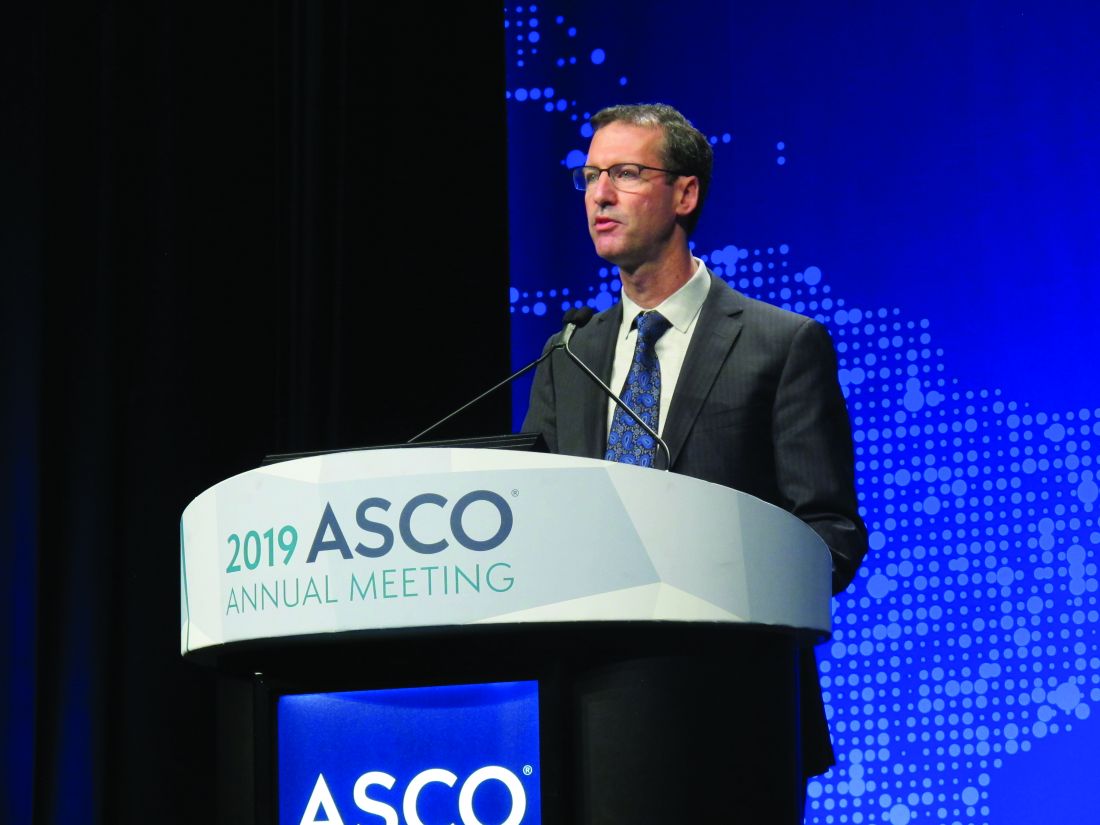
Eleven of 17 assessable patients had a response to treatment with the bispecific lentiviral CAR T cell (LV20.19CAR) at day 28, and of those 11 patients, 9 had complete responses, all of which are ongoing, said Nirav Niranjan Shah, MD, of the Medical College of Wisconsin in Milwaukee.
“To date, there’s no dose-limiting toxicity, no ICU-level care, no deaths attributed to treatment, no grade 3 to 4 cytokine release syndrome, and only two patients had reversible grade 3 neurotoxicity,” Dr. Shah said at the annual meeting of the American Society of Clinical Oncology.
Patients who did relapse or progress on treatment maintained CD19 or CD20 positivity, with no observed downregulation of target receptors, he reported in an oral abstract session.
Of note, the CAR T cells were produced locally at the point of care, with a 100% success rate and a set 14-day manufacturing time, he added.
Bispecific targeting of CD19 and CD20 is a new approach being investigated at a time when there are already two CD19-specific CAR T cell therapies approved for aggressive B-cell non-Hodgkin lymphomas, Dr. Shah told attendees.
“Despite the great promise of CD19 CAR T cell therapies, very quickly after the development of these therapies, we discovered mechanisms of resistance—specifically, the development of a CD19 negative relapse,” he said.
The hypothesis that targeting more than one B-cell antigen could potentially mitigate that effect stemmed from preclinical studies showing that targeting both CD19 and CD20 decreased downregulation of CD19 but not other B-cell antigens, he added.
In the present phase 1 study of the first-in-human, bispecific tandem CAR T cell against CD19 and CD20, patients have been treated at several dose levels, some with a split infusion over 2 days to evaluate safety, and some with a single infusion, Dr. Shah said.
A total of 17 patients have been treated with a lymphodepletion regimen followed by LV20.19CAR: 8 patients with diffuse large B-cell lymphoma, 6 with mantle cell lymphoma, 2 with chronic lymphocytic leukemia, and 1 with follicular lymphoma, according to the investigator. The median age of patients is 59 years, and patients had received at least 3 and up to 11 prior lines of therapy.
There have been no dose-limiting toxicities to date with dosing up to the target of 2.5 x 10⁶ cells/kg, Dr. Shah reported, adding that there has been no grade 3-4 cytokine release syndrome and no grade 4 neurotoxicity. Grade 1-2 cytokine release syndrome has been seen in 11 patients, while grade 3 neurotoxicity occurred in 2 patients.
Fourteen of 17 patients had a response, including 11 complete responses and 3 partial responses. Eleven patients were treated at the target dose of 2.5 x 10⁶ cells/kg, and of those, 9 had a complete response and 1 had a partial response (overall response rate, Dr. Shah said.
To date, all patients in complete response have remained in a complete response, with durations of response of 1 to 18 months.
Next, investigators plan to conduct phase 2 studies in more specific cohorts, including patients with mantle cell lymphoma, and patients who have relapsed after CD19 CAR T cell therapy, Dr. Shah said.
Dr. Shah reported disclosures related to Cidara Therapeutics, Exelixis, Geron, Oncosec, Incyte, Jazz Pharmaceuticals, Juno Therapeutics, Kite Pharma, and Miltenyi Biotec.
SOURCE: Shah NN et al. ASCO 2019. Abstract 2510.
This article was updated on 7/8/2019
CHICAGO – A bispecific anti-CD19, anti-CD20 chimeric antigen receptor (CAR) T cell approach is safe and produced complete responses in the majority of patients with relapsed or refractory non-Hodgkin lymphoma in a phase 1 study, an investigator reported.
Eleven of 17 assessable patients had a response to treatment with the bispecific lentiviral CAR T cell (LV20.19CAR) at day 28, and of those 11 patients, 9 had complete responses, all of which are ongoing, said Nirav Niranjan Shah, MD, of the Medical College of Wisconsin in Milwaukee.
“To date, there’s no dose-limiting toxicity, no ICU-level care, no deaths attributed to treatment, no grade 3 to 4 cytokine release syndrome, and only two patients had reversible grade 3 neurotoxicity,” Dr. Shah said at the annual meeting of the American Society of Clinical Oncology.
Patients who did relapse or progress on treatment maintained CD19 or CD20 positivity, with no observed downregulation of target receptors, he reported in an oral abstract session.
Of note, the CAR T cells were produced locally at the point of care, with a 100% success rate and a set 14-day manufacturing time, he added.
Bispecific targeting of CD19 and CD20 is a new approach being investigated at a time when there are already two CD19-specific CAR T cell therapies approved for aggressive B-cell non-Hodgkin lymphomas, Dr. Shah told attendees.
“Despite the great promise of CD19 CAR T cell therapies, very quickly after the development of these therapies, we discovered mechanisms of resistance—specifically, the development of a CD19 negative relapse,” he said.
The hypothesis that targeting more than one B-cell antigen could potentially mitigate that effect stemmed from preclinical studies showing that targeting both CD19 and CD20 decreased downregulation of CD19 but not other B-cell antigens, he added.
In the present phase 1 study of the first-in-human, bispecific tandem CAR T cell against CD19 and CD20, patients have been treated at several dose levels, some with a split infusion over 2 days to evaluate safety, and some with a single infusion, Dr. Shah said.
A total of 17 patients have been treated with a lymphodepletion regimen followed by LV20.19CAR: 8 patients with diffuse large B-cell lymphoma, 6 with mantle cell lymphoma, 2 with chronic lymphocytic leukemia, and 1 with follicular lymphoma, according to the investigator. The median age of patients is 59 years, and patients had received at least 3 and up to 11 prior lines of therapy.
There have been no dose-limiting toxicities to date with dosing up to the target of 2.5 x 10⁶ cells/kg, Dr. Shah reported, adding that there has been no grade 3-4 cytokine release syndrome and no grade 4 neurotoxicity. Grade 1-2 cytokine release syndrome has been seen in 11 patients, while grade 3 neurotoxicity occurred in 2 patients.
Fourteen of 17 patients had a response, including 11 complete responses and 3 partial responses. Eleven patients were treated at the target dose of 2.5 x 10⁶ cells/kg, and of those, 9 had a complete response and 1 had a partial response (overall response rate, Dr. Shah said.
To date, all patients in complete response have remained in a complete response, with durations of response of 1 to 18 months.
Next, investigators plan to conduct phase 2 studies in more specific cohorts, including patients with mantle cell lymphoma, and patients who have relapsed after CD19 CAR T cell therapy, Dr. Shah said.
Dr. Shah reported disclosures related to Cidara Therapeutics, Exelixis, Geron, Oncosec, Incyte, Jazz Pharmaceuticals, Juno Therapeutics, Kite Pharma, and Miltenyi Biotec.
SOURCE: Shah NN et al. ASCO 2019. Abstract 2510.
This article was updated on 7/8/2019
REPORTING FROM ASCO 2019
APHINITY trial: Biomarker analysis IDs predictive, prognostic factors
CHICAGO – Higher levels of several immune markers confer better response and outcomes in patients with HER2-positive breast cancer treated with trastuzumab and pertuzumab, according to a comprehensive biomarker analysis of data from the randomized, phase 3 APHINITY trial.
APHINITY randomized 4,805 patients with HER2-positive breast cancer to adjuvant chemotherapy with trastuzumab plus either pertuzumab or placebo and demonstrated a small improvement of just 1.7% in invasive disease–free survival at 4 years with the addition of adjuvant pertuzumab. The current analysis involved a nested case-control assessment of 1,023 patient samples from the trial to identify “biomarkers beyond clinical parameters” that could identify subgroups of patients who might benefit more from the addition of pertuzumab, Ian E. Krop, MD, PhD, reported at the annual meeting of the American Society of Clinical Oncology.
The genomic and immune marker-based analysis included DNA, whole transcriptome, tumor-infiltrating lymphocytes (TILs), and HER2 analyses, and after adjustment for treatment, hormone receptor status, nodal status, age, and chemotherapy type. Topoisomerase II-alpha amplification and higher messenger RNA expression of an immune signature consisting of interferon-gamma (IFNG), programmed death-ligand 1 (PD-L1), and chemokine (C-X-C motif) ligand 9 (CXCL9) were associated with better prognosis (hazard ratios, 0.68 and 0.91, respectively), said Dr. Krop, associate chief of the division of breast oncology at the Susan F. Smith Center for Women’s Cancers and clinical research director of the breast oncology center at Dana-Farber Cancer Institute, both in Boston.
TILs also suggested better outcomes (HR, 0.91), and HER2 copy number of six or greater versus lower levels of HER2 copy number was also associated with better prognosis (HR, 0.68), he noted.
Conversely, PI3K/PTEN/AKT gene alterations and MYC and ZNF703 amplification each were associated with worse outcomes (HRs, 1.35, 1.61, and 1.62, respectively).
As for predictive value, no significant association was seen between any of the amplification events and benefit of pertuzumab, nor was an interaction seen between the three-gene signature and pertuzumab benefit, Dr. Krop said.
“But if you look at the individual genes and ... the highest quartiles of expression of these genes – particularly interferon gamma and CXCL9 – it did appear that there was a statistically significant improvement in the benefit of pertuzumab if you had the highest levels of these genes, compared to lower levels of these genes,” he added.
The hazard ratios for CXCL9 of 0%-75% and greater than 75%, for example, were 0.95 and 0.49, respectively.
The interaction P values for IFNG and CXCL9 were statistically significant, but a trend toward benefit with PD-L1 did not reach statistical significance, Dr. Krop noted.
As with IFNG and CXCL9, the highest quartiles of TILs also predicted greater pertuzumab benefit (HRs for TILs at 0-75% and greater than 75%, 0.95 and 0.35, respectively), and the association was highly significant (P = .003).
HER2 copy number of six or greater was also associated with significantly greater benefit with pertuzumab (HRs for copy number of six or greater vs. less than six, 0.75 and 1.41, respectively).
A trend was seen toward decreased benefit of pertuzumab in patients with P13K/PTEN/AKT alteration, but this was not statistically significant, he noted.
However, the trend, coupled with the poor prognosis found to be associated with P13K-altered cancers, “would suggest that we need to identify new therapies – alternative approaches – to maximize treatment benefit in this cancer subtype,” he said.
“This biomarker analysis is possibly the largest and most comprehensive to date in HER2-positive breast cancer,” Dr. Krop said, noting that the findings provide support for an immune-mediated mechanism of action for pertuzumab, and suggest a need for alternative therapies for patients with low levels of TILs or immune gene markers in order to maximize their outcomes.
“We hope these data will be useful to refine future trials of early-stage HER2-positive breast cancer,” he said.
Dr. Krop reported relationships – including employment, leadership, and stock ownership – with AMAG, as well as honoraria from Genentech/Roche; consulting or advisory roles with Context Therapeutics, Daiichi Sankyo, Genentech/Roche, MacroGenics, Seattle Genetics, and Taiho Pharmaceutical; and research funding from Genentech and Pfizer to his institution.
SOURCE: Krop IE et al. ASCO 2019, Abstract 1012 .
REPORTING FROM ASCO 2019
Why we need another article on suicide contracts
Every guideline and lecture on suicide risk assessment includes the message: “Do not use suicide contracts.” Yet, as forensic psychiatrists, we continue to see medical records that rely solely on the patient verbalizing, agreeing, or signing that they will be safe, in order to justify medical decision-making. A recent case we reviewed involving a grossly psychotic male spotlighted the meaninglessness of suicide contracts. In an attempt to understand the impulse by clinicians to use suicide contracts, we decided to review the topic.

Suicide risk assessment is a confusing and poorly explained skill in our field. Suicide risk assessment tools are well-intended. They are meant to identify and stratify risk, and help guide medical decision-making. Popular tools are startlingly different. How can two scales represent adequate psychiatric knowledge yet be completely different? SADPERSONS1 is widely used and still considered standard of care yet has nothing in common with the Columbia–Suicide Severity Rating Scale (CSSRS).2
For those of us working in forensic settings, we are aghast that neither assessment is modified for use in correctional settings or accounts for essential risk factors of suicide in jails and prisons (placement in solitary, significant charges, homeless, etc.) Yet, they are widely used in jails and prisons across the country. This can be extrapolated to all of us who work with specific populations yet are asked to follow generic scales by administrators.
In reviewing the literature, we are surprised to see the lack of acknowledgment that many tools used in suicide risk assessment have little to no evidence. Despite their numerous appearances in medical records that we review, we are not aware of existing evidence for asking patients whether patients are suicidal on an hourly basis, for psychotropic treatment other than lithium and clozapine (Clozaril), and for safety plans that involve telling the patient to call 911. Of even greater concern, suicide risk assessments themselves may have limited value because of a lack of evidence as suggested by large study findings. It may surprise some to learn that the National Institute for Health and Care Excellence (NICE) in the United Kingdom includes the following statement in its guidelines: “Do not use risk assessment tools and scales to predict future suicide or repetition of self-harm.”3
In 2017, Carter et al.4 reviewed 70 studies using suicide risk scales to stratify patients in higher-risk groups for self-harm or suicide, during a follow-up period. The study reviewed biological tests such as the dexamethasone suppression test and 5-hydroxyindoleacetic acid; as well as psychological scales, including Buglass & Horton, SADPERSONS, the Beck Hopelessness Scale, the Beck’s Depression Inventory, Manchester Self Harm Rule, and the Edinburgh Risk Rating Scale. Their conclusion was clear: “No individual predictive instrument or pooled subgroups of instruments were able to classify patients as being at high risk of suicidal behavior with a level of accuracy suitable to be used to allocate treatment.”
Despite the bad reputation, one must admit that suicide contracts intuitively feel right. Just as we ask patients whether they believe they will stay sober in the future, or ask patients if they will be compliant with their psychotropics, asking them if they feel that they can maintain safety seems relevant. Reading through the literature, one can even find articles promoting this approach. In 2011, researchers simply asked 147 patients in psychiatric hospitals considered to be high risk for suicide whether they would engage in self-harm in the following weeks. They followed those patients for 15 weeks after their discharge for acts of self-harm. They concluded that “self-perceptions of risk seem to perform as well as the best [standardized assessment tools] the field has to offer” for the prediction of self-harm.5 We are unconvinced that juries would find suicide contracts irrelevant despite the lack of evidence. American society values individual autonomy and self-decision making. Patients telling their clinicians, “I will be OK” is relevant to suicide risk assessment. One can argue that the problem is not with the suicide contract itself, but with its blind use as a marker of safety.
The standard of care dictates that we try to assess suicide risk using evidence-based techniques. To the providers who see merit in asking patients whether they will be able to maintain their safety, we empathize with this impulse despite the lack of evidence. This will contribute in our shared effort to minimize suicide.
We acknowledge that the evidence of any assessment is limited and might miss a greater point in this entire discussion: Why are new iterations of suicide risk assessments not an improvement on the prior ones but a competing theory? New assessments emphasizing different facets of suicidal thinking do not include key demographic factors, while older tools do not include more recent understanding, such as the importance of hopelessness. From a provider’s perspective, the debate appears to be a battle of trends, theories, and acronyms rather than comprehensive analysis of the latest evidence. We, therefore, are concerned by “suicide experts” who advocate for any one assessment as the only gold standard and give false hopes about its efficacy.
As suicide rates continue to climb across the country, one wonders what we, as psychiatrists, are trying to achieve. Promises of zero suicides by hospitals,6 academic institutions,7 and even governments8 are well-meaning but possibly misleading to families and patients. Psychiatry should advocate within the standard of care for reasonable attempts at suicide risk assessment, including demographic factors (see SADPERSONS), as well as examination of the actual suicidality (see the CSSRS). Our professional organizations should clarify expectations for clinicians while also clarifying the limitations of our current knowledge base.
References
1. Patterson WM et al. Evaluation of suicidal patients: the SADPERSONS scale. Psychosomatics. 1983 Apr;24[4]:343-5, 348-9.
2. Posner K et al. The Columbia-Suicide Severity Rating Scale: initial validity and internal consistency findings from three multisite studies with adolescents and adults. Am J Psychiatry. 2011 Dec;168(12):1266-77.
3. Kendall T et al. Longer term management of self harm: summary of NICE guidance. BMJ. 2011;343. doi: 10.1136/bmj.d7073.
4. Carter G et al. Predicting suicidal behaviors using clinical instruments: systematic review and meta-analysis of positive predictive values for risk scales. Br J Psychiatry. 2017 Jun;210(6):387-95.
5. Peterson J et al. If you want to know, consider asking: How likely is it that patients will hurt themselves in the future? Psychol Assess. 2011 Sep;23(3):626-34.
5. Byrne JM et al. Implementation and impact of the central district of California’s suicide prevention program for crime defendants. Federal Probation. 2012 Jun;76(1):3-13.
6. “R.I.’s Butler Hospital sets ‘zero suicide’ goal for patients”/audio. Providence Journal. May 15, 2018.
7. “NIMH funds 3 ‘zero suicide’ grants.” National Institute of Mental Health. Sep 16, 2016.
8. Rothschild N. “Is it possible to eliminate suicide?” Atlantic. Jun 5, 2015.
Dr. Badre is a forensic psychiatrist in San Diego and an expert in correctional mental health. He holds teaching positions at the University of California, San Diego, and the University of San Diego. Dr. Rao is a San Diego–based board-certified psychiatrist with expertise in forensic psychiatry, correctional psychiatry, telepsychiatry, and inpatient psychiatry.
Every guideline and lecture on suicide risk assessment includes the message: “Do not use suicide contracts.” Yet, as forensic psychiatrists, we continue to see medical records that rely solely on the patient verbalizing, agreeing, or signing that they will be safe, in order to justify medical decision-making. A recent case we reviewed involving a grossly psychotic male spotlighted the meaninglessness of suicide contracts. In an attempt to understand the impulse by clinicians to use suicide contracts, we decided to review the topic.
Suicide risk assessment is a confusing and poorly explained skill in our field. Suicide risk assessment tools are well-intended. They are meant to identify and stratify risk, and help guide medical decision-making. Popular tools are startlingly different. How can two scales represent adequate psychiatric knowledge yet be completely different? SADPERSONS1 is widely used and still considered standard of care yet has nothing in common with the Columbia–Suicide Severity Rating Scale (CSSRS).2
For those of us working in forensic settings, we are aghast that neither assessment is modified for use in correctional settings or accounts for essential risk factors of suicide in jails and prisons (placement in solitary, significant charges, homeless, etc.) Yet, they are widely used in jails and prisons across the country. This can be extrapolated to all of us who work with specific populations yet are asked to follow generic scales by administrators.
In reviewing the literature, we are surprised to see the lack of acknowledgment that many tools used in suicide risk assessment have little to no evidence. Despite their numerous appearances in medical records that we review, we are not aware of existing evidence for asking patients whether patients are suicidal on an hourly basis, for psychotropic treatment other than lithium and clozapine (Clozaril), and for safety plans that involve telling the patient to call 911. Of even greater concern, suicide risk assessments themselves may have limited value because of a lack of evidence as suggested by large study findings. It may surprise some to learn that the National Institute for Health and Care Excellence (NICE) in the United Kingdom includes the following statement in its guidelines: “Do not use risk assessment tools and scales to predict future suicide or repetition of self-harm.”3
In 2017, Carter et al.4 reviewed 70 studies using suicide risk scales to stratify patients in higher-risk groups for self-harm or suicide, during a follow-up period. The study reviewed biological tests such as the dexamethasone suppression test and 5-hydroxyindoleacetic acid; as well as psychological scales, including Buglass & Horton, SADPERSONS, the Beck Hopelessness Scale, the Beck’s Depression Inventory, Manchester Self Harm Rule, and the Edinburgh Risk Rating Scale. Their conclusion was clear: “No individual predictive instrument or pooled subgroups of instruments were able to classify patients as being at high risk of suicidal behavior with a level of accuracy suitable to be used to allocate treatment.”
Despite the bad reputation, one must admit that suicide contracts intuitively feel right. Just as we ask patients whether they believe they will stay sober in the future, or ask patients if they will be compliant with their psychotropics, asking them if they feel that they can maintain safety seems relevant. Reading through the literature, one can even find articles promoting this approach. In 2011, researchers simply asked 147 patients in psychiatric hospitals considered to be high risk for suicide whether they would engage in self-harm in the following weeks. They followed those patients for 15 weeks after their discharge for acts of self-harm. They concluded that “self-perceptions of risk seem to perform as well as the best [standardized assessment tools] the field has to offer” for the prediction of self-harm.5 We are unconvinced that juries would find suicide contracts irrelevant despite the lack of evidence. American society values individual autonomy and self-decision making. Patients telling their clinicians, “I will be OK” is relevant to suicide risk assessment. One can argue that the problem is not with the suicide contract itself, but with its blind use as a marker of safety.
The standard of care dictates that we try to assess suicide risk using evidence-based techniques. To the providers who see merit in asking patients whether they will be able to maintain their safety, we empathize with this impulse despite the lack of evidence. This will contribute in our shared effort to minimize suicide.
We acknowledge that the evidence of any assessment is limited and might miss a greater point in this entire discussion: Why are new iterations of suicide risk assessments not an improvement on the prior ones but a competing theory? New assessments emphasizing different facets of suicidal thinking do not include key demographic factors, while older tools do not include more recent understanding, such as the importance of hopelessness. From a provider’s perspective, the debate appears to be a battle of trends, theories, and acronyms rather than comprehensive analysis of the latest evidence. We, therefore, are concerned by “suicide experts” who advocate for any one assessment as the only gold standard and give false hopes about its efficacy.
As suicide rates continue to climb across the country, one wonders what we, as psychiatrists, are trying to achieve. Promises of zero suicides by hospitals,6 academic institutions,7 and even governments8 are well-meaning but possibly misleading to families and patients. Psychiatry should advocate within the standard of care for reasonable attempts at suicide risk assessment, including demographic factors (see SADPERSONS), as well as examination of the actual suicidality (see the CSSRS). Our professional organizations should clarify expectations for clinicians while also clarifying the limitations of our current knowledge base.
References
1. Patterson WM et al. Evaluation of suicidal patients: the SADPERSONS scale. Psychosomatics. 1983 Apr;24[4]:343-5, 348-9.
2. Posner K et al. The Columbia-Suicide Severity Rating Scale: initial validity and internal consistency findings from three multisite studies with adolescents and adults. Am J Psychiatry. 2011 Dec;168(12):1266-77.
3. Kendall T et al. Longer term management of self harm: summary of NICE guidance. BMJ. 2011;343. doi: 10.1136/bmj.d7073.
4. Carter G et al. Predicting suicidal behaviors using clinical instruments: systematic review and meta-analysis of positive predictive values for risk scales. Br J Psychiatry. 2017 Jun;210(6):387-95.
5. Peterson J et al. If you want to know, consider asking: How likely is it that patients will hurt themselves in the future? Psychol Assess. 2011 Sep;23(3):626-34.
5. Byrne JM et al. Implementation and impact of the central district of California’s suicide prevention program for crime defendants. Federal Probation. 2012 Jun;76(1):3-13.
6. “R.I.’s Butler Hospital sets ‘zero suicide’ goal for patients”/audio. Providence Journal. May 15, 2018.
7. “NIMH funds 3 ‘zero suicide’ grants.” National Institute of Mental Health. Sep 16, 2016.
8. Rothschild N. “Is it possible to eliminate suicide?” Atlantic. Jun 5, 2015.
Dr. Badre is a forensic psychiatrist in San Diego and an expert in correctional mental health. He holds teaching positions at the University of California, San Diego, and the University of San Diego. Dr. Rao is a San Diego–based board-certified psychiatrist with expertise in forensic psychiatry, correctional psychiatry, telepsychiatry, and inpatient psychiatry.
FDA panel: Continue using paclitaxel-eluting PAD devices, with caveats
GAITHERSBURG, MD. – There was sufficient evidence of a late mortality signal seen at 2-5 years post procedurally for paclitaxel-eluting stents and coated balloons to warrant a label change for the devices, the Food and Drug Administration’s Circulatory System Devices Panel unanimously agreed after 2 days of deliberation.

That signal was brought to light in a meta-analysis published last December by Konstantinos Katsanos, MD, of Patras University Hospital, Rion, Greece, and colleagues (J Am Heart Assoc. 2018;7:e011245). Although there were concerns about the quality of the industry data used in the study, the caliber of the analysis itself and the subsequent data presented by the FDA to the panel were deemed sufficient to recommend a warning of concern to patients and providers.
Much of the new data from industry and large database registries presented to the panel, which was chaired by Richard A. Lange, MD, indicated a lessening to no evidence of the mortality effect. But this evidence was deemed insufficient to counter the evidence of the randomized controlled trials individually and collectively as presented in the Katsanos meta-analysis and subsequent information presented by the FDA that examined various parameters in a variety of sensitivity analyses that confirmed the late mortality signal. There was also concern that the industry and the registry analyses presented were not peer reviewed.
However, the panel also determined that it would be inappropriate to pull the devices from the market and from general use for several reasons.
One key reason was that, according to the panel, there was no mechanistic cause apparent for the late mortality. In addition, no convincing dose-response data could be teased from the preclinical and clinical trials studied because of their variability of devices, application methods, and lack of appropriate tissue analysis across studies.
Finally, the industry data used to create the meta-analysis were considered to be fundamentally flawed: in blinding, in the relatively small numbers of patients, and in the large percentage of patients lost to follow-up. The latter could have dramatically influenced the perceived results, especially as the studies were not powered or designed to follow mortality over such a period of time, according to the panel.
These limitations to the signal were especially important to the panel because of the obvious benefits with regard to quality of life provided to patients from these devices, which were attested to during the 2-day meeting by numerous presenters from industry, medical organizations – including societies and nonprofits – and providers.
In responding to FDA requests on a variety of concerns, the panel reiterated that there was a credible mortality signal, but that they could not be confident about the magnitude and whether it was caused by the paclitaxel treatment or some factor in the design or conduct of the studies. In addition, the panel members felt that they could neither confirm nor eliminate a class effect, given the fact that the information was based on a meta-analysis and thus none of the included devices could safely be removed from consideration.
They suggested that further safety information should be obtained, potentially by assessing and perhaps altering data collection in 29 ongoing studies over the next 5 years or so in more than 10,000 patients.
In addition, several of the panel members felt that additional animal studies might be performed including the use of older rat models; and using animal models that mimicked the kind of comorbidities present in the treated population, such as diabetes and atherosclerosis. They suggested cross-company industry cooperation with the FDA on these models, including looking at drug interactions and mimicking the dose application of stents/balloons.
Both the FDA representative and the panel were especially concerned with the benefit/risk profile.
The recommendation to still market the devices with a label warning was warranted, according to many members of the panel. They pointed to the clear benefits in quality of life and the lowered need for revascularization despite the evidence of the mortality signal, which, while statistically significant, could not be pinned town with regard to mechanisms or specific causes of death.
Overall, there was a concern that there should be a dialogue between patients and their doctors to discuss clear short-term benefits with unknown long-term risk, and that the label should support this by clearly mentioning the mortality signal that was found, although there was no attempt to develop exact wording.
Panel member Joaquin E. Cigarroa, MD, head of cardiovascular medicine at Oregon Health & Science University, Portland, suggested with regard to labeling that a statement “that ‘there may be’ – not ‘there is’ – a late mortality signal, should be included.”
Panel member John C. Somberg, MD, program director of clinical research and bioinformatics at Rush University in Lake Bluff, Ill., stated: “The label should say something like, ‘when looking at a meta-analysis that combined all studies with stents and balloons that carried paclitaxel, there may be a late mortality, which must be balanced against an early and sustained benefit in terms of pain on walking and potential loss of circulation to your extremity.’ ”
Other panel members thought that the meta-analysis should not be privileged and that somehow the totality of the evidence should somehow be distilled down into the label, including the evidence against the signal.
“We’re meeting because of a signal, of a concern – an honest, well-meaning concern – of increased mortality. And my opinion is that the patients need to be informed of it,” said Dr. Lange, president, Texas Tech University, El Paso.
Some members of the panel felt that it may not be justifiable to use these devices in patients with low intrinsic risk and low recurrence risk, and the whole spectrum of patients may need to be considered in further studies to figure out the subgroups that have more benefits and more risks, and also to consider how to mitigate risks in patients who receive the device, whether through medical therapy or lifestyle modification.
In particular, Frank W. LoGerfo, MD, the William V. McDermott Distinguished Professor of Surgery at Harvard Medical School, Boston, stated: “Interventions for claudication should be extremely rare. It rarely progresses, and the pain should be worked through by exercise with low risk of limb loss.” He added that intervention with these devices “takes away options. Trading life for something that is not limb-threatening is something we should not be considering.”
There was no firm consensus on whether new randomized trials should be done, although they were of course the ideal solution. Kevin E. Kip, PhD, Distinguished USF Health Professor at the University of South Florida, Tampa, and others argued that, whether new trials were necessary or not, to deal with the safety question in a timely fashion, existing trials have to capture as much of the missing data as possible, and carry out follow-up out further.
FDA representative Bram Zuckerman, MD, director of the Office of Cardiovascular Devices at the Center for Devices and Radiological Health, indicated that those things might not be easily be accomplished due to regulatory constraints and the financial costs, and that to do so there would be need for community effort among stakeholders, including collaborative efforts with existing prospective registries such as that run by the Vascular Quality Initiative.
One overall conclusion by both the FDA and panel members was that the quality of these and other such studies going forward must improve, by standardizing definitions and data forms to make studies more uniform across the industry. They reemphasized the need to work with the registries to get common data included, and to incorporate of insurance provider and Social Security death data as much as possible to help alleviate the lost follow-up problem.
SOURCE: Webcasts of the complete 2 days of the FDA panel meeting are available online.
GAITHERSBURG, MD. – There was sufficient evidence of a late mortality signal seen at 2-5 years post procedurally for paclitaxel-eluting stents and coated balloons to warrant a label change for the devices, the Food and Drug Administration’s Circulatory System Devices Panel unanimously agreed after 2 days of deliberation.
That signal was brought to light in a meta-analysis published last December by Konstantinos Katsanos, MD, of Patras University Hospital, Rion, Greece, and colleagues (J Am Heart Assoc. 2018;7:e011245). Although there were concerns about the quality of the industry data used in the study, the caliber of the analysis itself and the subsequent data presented by the FDA to the panel were deemed sufficient to recommend a warning of concern to patients and providers.
Much of the new data from industry and large database registries presented to the panel, which was chaired by Richard A. Lange, MD, indicated a lessening to no evidence of the mortality effect. But this evidence was deemed insufficient to counter the evidence of the randomized controlled trials individually and collectively as presented in the Katsanos meta-analysis and subsequent information presented by the FDA that examined various parameters in a variety of sensitivity analyses that confirmed the late mortality signal. There was also concern that the industry and the registry analyses presented were not peer reviewed.
However, the panel also determined that it would be inappropriate to pull the devices from the market and from general use for several reasons.
One key reason was that, according to the panel, there was no mechanistic cause apparent for the late mortality. In addition, no convincing dose-response data could be teased from the preclinical and clinical trials studied because of their variability of devices, application methods, and lack of appropriate tissue analysis across studies.
Finally, the industry data used to create the meta-analysis were considered to be fundamentally flawed: in blinding, in the relatively small numbers of patients, and in the large percentage of patients lost to follow-up. The latter could have dramatically influenced the perceived results, especially as the studies were not powered or designed to follow mortality over such a period of time, according to the panel.
These limitations to the signal were especially important to the panel because of the obvious benefits with regard to quality of life provided to patients from these devices, which were attested to during the 2-day meeting by numerous presenters from industry, medical organizations – including societies and nonprofits – and providers.
In responding to FDA requests on a variety of concerns, the panel reiterated that there was a credible mortality signal, but that they could not be confident about the magnitude and whether it was caused by the paclitaxel treatment or some factor in the design or conduct of the studies. In addition, the panel members felt that they could neither confirm nor eliminate a class effect, given the fact that the information was based on a meta-analysis and thus none of the included devices could safely be removed from consideration.
They suggested that further safety information should be obtained, potentially by assessing and perhaps altering data collection in 29 ongoing studies over the next 5 years or so in more than 10,000 patients.
In addition, several of the panel members felt that additional animal studies might be performed including the use of older rat models; and using animal models that mimicked the kind of comorbidities present in the treated population, such as diabetes and atherosclerosis. They suggested cross-company industry cooperation with the FDA on these models, including looking at drug interactions and mimicking the dose application of stents/balloons.
Both the FDA representative and the panel were especially concerned with the benefit/risk profile.
The recommendation to still market the devices with a label warning was warranted, according to many members of the panel. They pointed to the clear benefits in quality of life and the lowered need for revascularization despite the evidence of the mortality signal, which, while statistically significant, could not be pinned town with regard to mechanisms or specific causes of death.
Overall, there was a concern that there should be a dialogue between patients and their doctors to discuss clear short-term benefits with unknown long-term risk, and that the label should support this by clearly mentioning the mortality signal that was found, although there was no attempt to develop exact wording.
Panel member Joaquin E. Cigarroa, MD, head of cardiovascular medicine at Oregon Health & Science University, Portland, suggested with regard to labeling that a statement “that ‘there may be’ – not ‘there is’ – a late mortality signal, should be included.”
Panel member John C. Somberg, MD, program director of clinical research and bioinformatics at Rush University in Lake Bluff, Ill., stated: “The label should say something like, ‘when looking at a meta-analysis that combined all studies with stents and balloons that carried paclitaxel, there may be a late mortality, which must be balanced against an early and sustained benefit in terms of pain on walking and potential loss of circulation to your extremity.’ ”
Other panel members thought that the meta-analysis should not be privileged and that somehow the totality of the evidence should somehow be distilled down into the label, including the evidence against the signal.
“We’re meeting because of a signal, of a concern – an honest, well-meaning concern – of increased mortality. And my opinion is that the patients need to be informed of it,” said Dr. Lange, president, Texas Tech University, El Paso.
Some members of the panel felt that it may not be justifiable to use these devices in patients with low intrinsic risk and low recurrence risk, and the whole spectrum of patients may need to be considered in further studies to figure out the subgroups that have more benefits and more risks, and also to consider how to mitigate risks in patients who receive the device, whether through medical therapy or lifestyle modification.
In particular, Frank W. LoGerfo, MD, the William V. McDermott Distinguished Professor of Surgery at Harvard Medical School, Boston, stated: “Interventions for claudication should be extremely rare. It rarely progresses, and the pain should be worked through by exercise with low risk of limb loss.” He added that intervention with these devices “takes away options. Trading life for something that is not limb-threatening is something we should not be considering.”
There was no firm consensus on whether new randomized trials should be done, although they were of course the ideal solution. Kevin E. Kip, PhD, Distinguished USF Health Professor at the University of South Florida, Tampa, and others argued that, whether new trials were necessary or not, to deal with the safety question in a timely fashion, existing trials have to capture as much of the missing data as possible, and carry out follow-up out further.
FDA representative Bram Zuckerman, MD, director of the Office of Cardiovascular Devices at the Center for Devices and Radiological Health, indicated that those things might not be easily be accomplished due to regulatory constraints and the financial costs, and that to do so there would be need for community effort among stakeholders, including collaborative efforts with existing prospective registries such as that run by the Vascular Quality Initiative.
One overall conclusion by both the FDA and panel members was that the quality of these and other such studies going forward must improve, by standardizing definitions and data forms to make studies more uniform across the industry. They reemphasized the need to work with the registries to get common data included, and to incorporate of insurance provider and Social Security death data as much as possible to help alleviate the lost follow-up problem.
SOURCE: Webcasts of the complete 2 days of the FDA panel meeting are available online.
REPORTING FROM AN FDA PANEL MEETING
EULAR revises its RA management recommendations
MADRID – No change to designating methotrexate the first disease-modifying drug to prescribe, before any biologic drug, and no adoption of imaging criteria to determine whether a patient is in remission.

“Imaging with ultrasound or MRI is out” as a remission criterion. “It’s high risk and a waste of resources,” declared Josef S. Smolen, MD, head of the EULAR writing panel, in the most forceful declaration he made while presenting the pending recommendation revision at the European Congress of Rheumatology.
Dr. Smolen’s strong warning against an imaging parameter when treating RA patients toward a remission target was no surprise, as he had already voiced this opinion in an editorial he coauthored earlier this year (JAMA. 2019 Feb 5;321[5]:457-8). The editorial cited data from three independent studies that compared an RA treatment strategy that used an imaging measure of joint inflammation as a treatment target along with clinical assessment against clinical assessment alone. All three studies found no benefit from ultrasound or MRI for defining a treatment goal, and two of the studies showed evidence for harm. “Using imaging to guide therapy led to prescription of potentially harmful medicines without differences in the primary outcomes, but at high costs and potential burden of unnecessary treatment changes and risks for patients,” noted Dr. Smolen and his coauthor in the editorial.
The report that this editorial addressed (JAMA. 2019 Feb 5;321[5]:461-72) also provided some of the most recent evidence for the second omission from the new revision that Dr. Smolen called out: No change to the recommendation to use methotrexate as initial treatment for any RA patient. “We continue to say that methotrexate is the first treatment strategy. There is no new evidence that any biological treatment is better than methotrexate, so there is no change,” said Dr. Smolen, professor of medicine at the Medical University of Vienna, who also led the EULAR writing panel for the immediately preceding set of RA treatment recommendations first unveiled 3 years before (Ann Rheum Dis. 2017 Jun;76[6]:960-77).
Perhaps the most notable changes to the recommendations are the way they handle targeted-synthetic disease-modifying antirheumatic drugs (tsDMARDs), a class that currently is synonymous with the Janus kinase (JAK) inhibitors. “Because of new evidence we have lifted up the tsDMARDs” so that no preference is given to biologic DMARDs over the ts class as happened in the 2016 version, Dr. Smolen said. Another revision to this recommendation was to change the addition of either a biologic or tsDMARD to a patient not fully responsive to a conventional-synthetic (cs) DMARD and with poor prognostic factors from a “should be considered” to a “should be added” recommendation.
Another way in which the pending revision uplifted tsDMARDs was in the wording for the recommendation that deals with patients who do not respond to a first tumor necrosis factor (TNF) inhibitor plus methotrexate or another csDMARD, and now lists as the first option switching to a biologic or tsDMARD with a different mode of action followed by a different TNF inhibitor, a reversal of order from before when a different TNF inhibitor got first mention. This order change was a modest revision that reflected observational evidence that was modestly persuasive that switching to an agent with a different mechanism of action is often the most effective approach, Dr. Smolen said.
The new recommendations also reaffirmed the eleventh recommendation from the 2016 version, which called for tapering of the biologic or tsDMARD from a patient in remission while retaining the csDMARD, usually methotrexate. Dr. Smolen cited new evidence in favor of this approach (Ann Rheum Dis. 2019 Jun;78[6]:746-53), which allowed the writing panel to upgrade the evidence supporting this recommendation to the A level. The concept of tapering down the biologic or tsDMARD for a patient in sustained remission while maintaining the csDMARD was “fully confirmed” in a recent report, he added. The writing panel also upticked its rating of the evidence in favor of cautiously tapering the csDMARD in patients who maintain remission on just a csDMARD.
One final element in the pending revision called out a newly identified safety signal, an increased risk for venous thromboembolism among patients on certain high dosages of JAK inhibitors, especially in patients with increased risk for venous thromboembolism. This new safety concern adds to the already-described increased risk for herpes zoster from JAK inhibitors, especially in Japanese and Korean populations, Dr. Smolen said. In general, more long-term safety data for JAK inhibitors are needed.
The draft update also added one new overarching principle: “Patients require access to multiple drugs with different modes of action to address the heterogeneity of RA, and patients may require multiple, successive treatments throughout life.” Overall, pending changes to the RA recommendations were limited because “the EULAR recommendations have achieved a steady state of the art” for defining whom to treat, treatment targets, and appropriate treatment strategies, Dr. Smolen said.
Dr. Smolen had been a consultant to or a speaker on behalf of several drug companies.
MADRID – No change to designating methotrexate the first disease-modifying drug to prescribe, before any biologic drug, and no adoption of imaging criteria to determine whether a patient is in remission.
“Imaging with ultrasound or MRI is out” as a remission criterion. “It’s high risk and a waste of resources,” declared Josef S. Smolen, MD, head of the EULAR writing panel, in the most forceful declaration he made while presenting the pending recommendation revision at the European Congress of Rheumatology.
Dr. Smolen’s strong warning against an imaging parameter when treating RA patients toward a remission target was no surprise, as he had already voiced this opinion in an editorial he coauthored earlier this year (JAMA. 2019 Feb 5;321[5]:457-8). The editorial cited data from three independent studies that compared an RA treatment strategy that used an imaging measure of joint inflammation as a treatment target along with clinical assessment against clinical assessment alone. All three studies found no benefit from ultrasound or MRI for defining a treatment goal, and two of the studies showed evidence for harm. “Using imaging to guide therapy led to prescription of potentially harmful medicines without differences in the primary outcomes, but at high costs and potential burden of unnecessary treatment changes and risks for patients,” noted Dr. Smolen and his coauthor in the editorial.
The report that this editorial addressed (JAMA. 2019 Feb 5;321[5]:461-72) also provided some of the most recent evidence for the second omission from the new revision that Dr. Smolen called out: No change to the recommendation to use methotrexate as initial treatment for any RA patient. “We continue to say that methotrexate is the first treatment strategy. There is no new evidence that any biological treatment is better than methotrexate, so there is no change,” said Dr. Smolen, professor of medicine at the Medical University of Vienna, who also led the EULAR writing panel for the immediately preceding set of RA treatment recommendations first unveiled 3 years before (Ann Rheum Dis. 2017 Jun;76[6]:960-77).
Perhaps the most notable changes to the recommendations are the way they handle targeted-synthetic disease-modifying antirheumatic drugs (tsDMARDs), a class that currently is synonymous with the Janus kinase (JAK) inhibitors. “Because of new evidence we have lifted up the tsDMARDs” so that no preference is given to biologic DMARDs over the ts class as happened in the 2016 version, Dr. Smolen said. Another revision to this recommendation was to change the addition of either a biologic or tsDMARD to a patient not fully responsive to a conventional-synthetic (cs) DMARD and with poor prognostic factors from a “should be considered” to a “should be added” recommendation.
Another way in which the pending revision uplifted tsDMARDs was in the wording for the recommendation that deals with patients who do not respond to a first tumor necrosis factor (TNF) inhibitor plus methotrexate or another csDMARD, and now lists as the first option switching to a biologic or tsDMARD with a different mode of action followed by a different TNF inhibitor, a reversal of order from before when a different TNF inhibitor got first mention. This order change was a modest revision that reflected observational evidence that was modestly persuasive that switching to an agent with a different mechanism of action is often the most effective approach, Dr. Smolen said.
The new recommendations also reaffirmed the eleventh recommendation from the 2016 version, which called for tapering of the biologic or tsDMARD from a patient in remission while retaining the csDMARD, usually methotrexate. Dr. Smolen cited new evidence in favor of this approach (Ann Rheum Dis. 2019 Jun;78[6]:746-53), which allowed the writing panel to upgrade the evidence supporting this recommendation to the A level. The concept of tapering down the biologic or tsDMARD for a patient in sustained remission while maintaining the csDMARD was “fully confirmed” in a recent report, he added. The writing panel also upticked its rating of the evidence in favor of cautiously tapering the csDMARD in patients who maintain remission on just a csDMARD.
One final element in the pending revision called out a newly identified safety signal, an increased risk for venous thromboembolism among patients on certain high dosages of JAK inhibitors, especially in patients with increased risk for venous thromboembolism. This new safety concern adds to the already-described increased risk for herpes zoster from JAK inhibitors, especially in Japanese and Korean populations, Dr. Smolen said. In general, more long-term safety data for JAK inhibitors are needed.
The draft update also added one new overarching principle: “Patients require access to multiple drugs with different modes of action to address the heterogeneity of RA, and patients may require multiple, successive treatments throughout life.” Overall, pending changes to the RA recommendations were limited because “the EULAR recommendations have achieved a steady state of the art” for defining whom to treat, treatment targets, and appropriate treatment strategies, Dr. Smolen said.
Dr. Smolen had been a consultant to or a speaker on behalf of several drug companies.
EXPERT ANALYSIS FROM EULAR 2019 CONGRESS
Chemo-free neoadjuvant approaches emerge in NSCLC and breast cancer
In this edition of “How I will treat my next patient,” I take a look at two “chemo-free” neoadjuvant studies reported at the annual meeting of the American Society of Clinical Oncology. One summarizes the potential utility of immune checkpoint inhibitors (ICIs) in non–small cell lung cancer (NSCLC) patients who are – or can become – candidates for curative resection and the other highlights the potential utility of neoadjuvant trastuzumab emtansine (T-DM1) for HER2-positive breast cancer patients.
NEOSTAR in NSCLC

With the accumulation of comorbid conditions in an aging population, we all see NSCLC patients who are potential candidates for curative surgery, but for whom we have concerns about standard preoperative chemotherapy plus or minus radiation. At ASCO 2019, abstracts 8503 (atezolizumab, the LCMC3 trial) and 8504 (nivolumab plus or minus ipilimumab, NEOSTAR) addressed the neoadjuvant use of ICIs. I will focus on NEOSTAR, because the major pathologic response (mPR) rate – reduction in viable tumor cells to 10% or less – was higher with the combination of nivolumab plus ipilimumab in NEOSTAR than with single agent nivolumab or atezolizumab in the NEOSTAR or LCMC3 trials, respectively.
Briefly, 44 patients with stage I-IIIA NSCLC were randomized to nivolumab plus or minus ipilimumab. In total, 93% completed 6 weeks of neoadjuvant therapy and 89% were resected. The mPR rate was 33% with nivolumab plus ipilimumab (about twice as high as with nivolumab alone in NEOSTAR or atezolizumab in LCMC3).
Among resected patients, nivolumab plus ipilimumab had a 44% mPR rate and a pathologic complete response rate of 38%. Although RECIST (Response Evaluation Criteria in Solid Tumors) responses were more likely in patients who had an mPR, 11% of patients had radiographic “nodal immune flare” because of noncaseating granulomas in regional (or nonregional) nodes. Elevated baseline programmed death-ligand 1 was associated with a higher rate of mPR. Surgical complications seemed similar to expectations – 1 bronchopleural fistula and 8 air leaks among the 39 resected patients.
What this means in practice
Although the mPR endpoint has no validated association with survival and the studies were relatively small, neoadjuvant use of ICIs in patients for whom tolerance to standard chemotherapy plus or minus radiation might be problematic is attractive – especially in view of the reality of an approximately 50% relapse rate after surgical resection with standard therapy.
If I had a potential candidate for neoadjuvant ICI therapy – especially one with a high proportion of cells with PD-L1 or someone with an equivocal distant metastasis on a preoperative PET-CT – I would consider using an ICI as given in LCMC3 or NEOSTAR.
PREDIX in HER2-positive breast cancer
As Mark Pegram, MD, of Stanford (Calif.) University suggested in his discussion at the local/regional/adjuvant breast cancer session at ASCO 2019, the goal of HER2-targeted therapy was originally that it could replace – not supplement – the use of cytotoxic chemotherapy.
Abstracts 500 (the KRISTINE trial: neoadjuvant T-DM1 plus pertuzumab vs. docetaxel, carboplatin, and trastuzumab plus pertuzumab); 501 (the PREDIX study: T-DM1 vs. docetaxel plus trastuzumab plus pertuzumab [DTP] for six cycles); and 502 (HER2 heterogeneity as a predictor of response) addressed the potential for the antibody-drug conjugate to replace standard preoperative cytotoxic chemotherapy plus HER2-targeting.
In PREDIX, it was anticipated that toxicity would be lower with T-DM1 than with DTP – and it was, with better quality of life scores. The authors found a pathologic complete response rate of 45% among 98 participants with stage IIA-IIIA HER2-positive breast cancer, with higher rates among hormone receptor–negative than hormone receptor–positive patients, as expected.
PREDIX patients were allowed to switch from T-DM1 to DTP for progression, lack of clinical/radiographic response, or toxicity. More than twice as many patients switched from DTP to T-DM1 than vice versa for progression or lack of response.
What this means in practice
Although neither DTP nor T-DM1 are National Comprehensive Cancer Network guideline-endorsed neoadjuvant regimens at present, the KRISTINE and PREDIX trials and Abstract 502 advance the discussion about further personalizing therapy for HER2-amplified breast cancer with high HER2 copy number and lack of intratumor heterogeneity for HER2. They also raise questions about de-escalating therapy for patients with good prognosis and HER2-positive cancers, and the creative use of T-DM1 in the neoadjuvant setting.
Neoadjuvant T-DM1 may not be standard of care yet, but watch this space.
Dr. Lyss has been a community-based medical oncologist and clinical researcher for more than 35 years, practicing in St. Louis. His clinical and research interests are in the prevention, diagnosis, and treatment of breast and lung cancers and in expanding access to clinical trials to medically underserved populations.
In this edition of “How I will treat my next patient,” I take a look at two “chemo-free” neoadjuvant studies reported at the annual meeting of the American Society of Clinical Oncology. One summarizes the potential utility of immune checkpoint inhibitors (ICIs) in non–small cell lung cancer (NSCLC) patients who are – or can become – candidates for curative resection and the other highlights the potential utility of neoadjuvant trastuzumab emtansine (T-DM1) for HER2-positive breast cancer patients.
NEOSTAR in NSCLC
With the accumulation of comorbid conditions in an aging population, we all see NSCLC patients who are potential candidates for curative surgery, but for whom we have concerns about standard preoperative chemotherapy plus or minus radiation. At ASCO 2019, abstracts 8503 (atezolizumab, the LCMC3 trial) and 8504 (nivolumab plus or minus ipilimumab, NEOSTAR) addressed the neoadjuvant use of ICIs. I will focus on NEOSTAR, because the major pathologic response (mPR) rate – reduction in viable tumor cells to 10% or less – was higher with the combination of nivolumab plus ipilimumab in NEOSTAR than with single agent nivolumab or atezolizumab in the NEOSTAR or LCMC3 trials, respectively.
Briefly, 44 patients with stage I-IIIA NSCLC were randomized to nivolumab plus or minus ipilimumab. In total, 93% completed 6 weeks of neoadjuvant therapy and 89% were resected. The mPR rate was 33% with nivolumab plus ipilimumab (about twice as high as with nivolumab alone in NEOSTAR or atezolizumab in LCMC3).
Among resected patients, nivolumab plus ipilimumab had a 44% mPR rate and a pathologic complete response rate of 38%. Although RECIST (Response Evaluation Criteria in Solid Tumors) responses were more likely in patients who had an mPR, 11% of patients had radiographic “nodal immune flare” because of noncaseating granulomas in regional (or nonregional) nodes. Elevated baseline programmed death-ligand 1 was associated with a higher rate of mPR. Surgical complications seemed similar to expectations – 1 bronchopleural fistula and 8 air leaks among the 39 resected patients.
What this means in practice
Although the mPR endpoint has no validated association with survival and the studies were relatively small, neoadjuvant use of ICIs in patients for whom tolerance to standard chemotherapy plus or minus radiation might be problematic is attractive – especially in view of the reality of an approximately 50% relapse rate after surgical resection with standard therapy.
If I had a potential candidate for neoadjuvant ICI therapy – especially one with a high proportion of cells with PD-L1 or someone with an equivocal distant metastasis on a preoperative PET-CT – I would consider using an ICI as given in LCMC3 or NEOSTAR.
PREDIX in HER2-positive breast cancer
As Mark Pegram, MD, of Stanford (Calif.) University suggested in his discussion at the local/regional/adjuvant breast cancer session at ASCO 2019, the goal of HER2-targeted therapy was originally that it could replace – not supplement – the use of cytotoxic chemotherapy.
Abstracts 500 (the KRISTINE trial: neoadjuvant T-DM1 plus pertuzumab vs. docetaxel, carboplatin, and trastuzumab plus pertuzumab); 501 (the PREDIX study: T-DM1 vs. docetaxel plus trastuzumab plus pertuzumab [DTP] for six cycles); and 502 (HER2 heterogeneity as a predictor of response) addressed the potential for the antibody-drug conjugate to replace standard preoperative cytotoxic chemotherapy plus HER2-targeting.
In PREDIX, it was anticipated that toxicity would be lower with T-DM1 than with DTP – and it was, with better quality of life scores. The authors found a pathologic complete response rate of 45% among 98 participants with stage IIA-IIIA HER2-positive breast cancer, with higher rates among hormone receptor–negative than hormone receptor–positive patients, as expected.
PREDIX patients were allowed to switch from T-DM1 to DTP for progression, lack of clinical/radiographic response, or toxicity. More than twice as many patients switched from DTP to T-DM1 than vice versa for progression or lack of response.
What this means in practice
Although neither DTP nor T-DM1 are National Comprehensive Cancer Network guideline-endorsed neoadjuvant regimens at present, the KRISTINE and PREDIX trials and Abstract 502 advance the discussion about further personalizing therapy for HER2-amplified breast cancer with high HER2 copy number and lack of intratumor heterogeneity for HER2. They also raise questions about de-escalating therapy for patients with good prognosis and HER2-positive cancers, and the creative use of T-DM1 in the neoadjuvant setting.
Neoadjuvant T-DM1 may not be standard of care yet, but watch this space.
Dr. Lyss has been a community-based medical oncologist and clinical researcher for more than 35 years, practicing in St. Louis. His clinical and research interests are in the prevention, diagnosis, and treatment of breast and lung cancers and in expanding access to clinical trials to medically underserved populations.
Efforts toward producing CNO/CRMO classification criteria show first results
MADRID – according to recent findings from international surveys of pediatric rheumatologists that were presented at the European Congress of Rheumatology.
Melissa Oliver, MD, a pediatric rheumatologist at Riley Hospital for Children, Indianapolis, and colleagues recently undertook the multiphase study as part of an international collaborative effort led by the Childhood Arthritis and Rheumatology Research Alliance to establish consensus-based diagnostic and classification criteria for CNO, an autoinflammatory bone disease of unknown cause that primarily affects children and adolescents. CNO is also known as chronic recurrent multifocal osteomyelitis (CRMO). If this disease is not diagnosed and treated appropriately in a timely fashion, damage and long-term disability is possible. In the absence of widely accepted, consensus-driven criteria, treatment is based largely on expert opinion, Dr. Oliver explained in an interview.
“There is an urgent need for a new and more robust set of classification criteria for CRMO, based on large expert consensus and the analysis of a large sample of patients and controls,” she said.
There are two proposed diagnostic criteria, the 2007 classification of nonbacterial osteitis and the 2016 Bristol diagnostic criteria for CRMO, but both are derived from single-center cohort studies and have not been validated, Dr. Oliver explained.
The list of candidate items that have come out of the study is moving clinicians a step closer toward the design of a practical patient data collection form that appropriately weighs each item included in the classification criteria.
The study employed anonymous survey and nominal group techniques with the goal of developing a set of classification criteria sensitive and specific enough to identify CRMO/CNO patients. In phase 1, a Delphi survey was administered among international rheumatologists to generate candidate criteria items. Phase 2 sought to reduce candidate criteria items through consensus processes via input from physicians managing CNO and patients or caregivers of children with CNO.
Altogether, 259 of 865 pediatric rheumatologists (30%) completed an online questionnaire addressing features key to the classification of CNO, including 77 who practice in Europe (30%), 132 in North America (51%), and 50 on other continents (19%). Of these, 138 (53%) had greater than 10 years of clinical practice experience, and 108 (42%) had managed more than 10 CNO patients.
Initially, Dr. Oliver and colleagues identified 33 candidate criteria items that fell into six domains: clinical presentation, physical exam, laboratory findings, imaging findings, bone biopsy, and treatment response. The top eight weighted items that increased the likelihood of CNO/CRMO were exclusion of malignancy by bone biopsy; multifocal bone lesions; presence of bone pain, swelling, and/or warmth; signs of fibrosis and/or inflammation on bone biopsy; typical location of CNO/CRMO lesion, such as the clavicle, metaphysis of long bones, the mandible, and vertebrae; presence of CNO/CRMO–related comorbidities; normal C-reactive protein (CRP) or erythrocyte sedimentation rate (ESR); and typical MRI findings of CNO/CRMO.
By phase 2, candidate items, which were presented to 39 rheumatologists and 7 parents, were refined or eliminated using item-reduction techniques. A second survey was issued to 77 of 82 members of a work group so that the remaining items could be ranked by their power of distinguishing CNO from conditions that merely mimicked the disease. The greatest mean discriminatory scores were identified with multifocal lesions (ruling out malignancy and infection) and typical location on imaging. Normal C-reactive protein and/or an erythrocyte sedimentation rate more than three times the upper limit of normal had the greatest negative mean discriminatory scores.
The next steps will be to form an expert panel who will use 1000minds software to determine the final criteria and identify a threshold for disease. The investigators hope to build a large multinational case repository of at least 500 patients with CNO/CRMO and 500 patients with mimicking conditions from which to derive a development cohort and an external validation cohort. So far, 10 sites, including 4 in Europe, have obtained approval from an institutional review board. The group has also submitted a proposal for classification criteria to the American College of Rheumatology and the European League Against Rheumatism, Dr. Oliver said.
Dr. Oliver had no disclosures to report, but several coauthors reported financial ties to industry.
SOURCE: Oliver M et al. Ann Rheum Dis. Jun 2019;78(Suppl 2):254-5, Abstract OP0342. doi: 10.1136/annrheumdis-2019-eular.1539.
REPORTING FROM EULAR 2019 CONGRESS

Medical cannabis laws appear no longer tied to drop in opioid overdose mortality
Correlations do not hold when analysis is expanded to 2017
Contrary to previous research indicating that medical cannabis laws reduced opioid overdose mortality, the association between these two has reversed, with opioid overdose mortality increased in states with comprehensive medical cannabis laws, according to Chelsea L. Shover, PhD, and associates.

The original research by Marcus A. Bachhuber, MD, and associates showed that the introduction of state medical cannabis laws was associated with a 24.8% reduction in opioid overdose deaths per 100,000 population between 1999 and 2010. In contrast, the new research – which looked at a longer time period than the original research did – found that the association between state medical cannabis laws and opioid overdose mortality reversed direction, from –21% to +23%.
“We find it unlikely that medical cannabis – used by about 2.5% of the U.S. population – has exerted large conflicting effects on opioid overdose mortality,” wrote Dr. Shover, of the department of psychiatry and behavioral sciences at Stanford (Calif.) University, and associates. “A more plausible interpretation is that this association is spurious.” Their study was published in the Proceedings of the National Academy of Sciences.
To conduct their analysis, Dr. Shover and associates extended the timeline reviewed by Dr. Bachhuber and associates to 2017. During 2010-2017, 32 states enacted medical cannabis laws, including 17 allowing only medical cannabis with low levels of tetrahydrocannabinol (THC), and 8 legalized recreational marijuana. In the expanded timeline during 1999-2017, states possessing a comprehensive medical marijuana law saw an increase in opioid overdose mortality of 28.2%. Meanwhile, states with recreational marijuana laws saw a decrease of 14.7% in opioid overdose mortality, and states with low-THC medical cannabis laws saw a decrease of 7.1%. However, the investigators noted that those values had wide confidence intervals, which indicates “compatibility with large range of true associations.”
Corporate actors with deep pockets have substantial ability to promote congenial results, and suffering people are desperate for effective solutions. Cannabinoids have demonstrated therapeutic benefits, but reducing population-level opioid overdose mortality does not appear to be among them,” Dr. Shover and associates noted.
Dr. Shover reported receiving support from National Institute on Drug Abuse and the Wu Tsai Neurosciences Institute. Another coauthor received support from the Veterans Health Administration, Wu Tsai Neurosciences Institute, and the Esther Ting Memorial Professorship at Stanford.
SOURCE: Shover CL et al. Proc Natl Acad Sci U S A. 2019 Jun 10. doi: 10.1073/pnas.1903434116.
Correlations do not hold when analysis is expanded to 2017
Correlations do not hold when analysis is expanded to 2017
Contrary to previous research indicating that medical cannabis laws reduced opioid overdose mortality, the association between these two has reversed, with opioid overdose mortality increased in states with comprehensive medical cannabis laws, according to Chelsea L. Shover, PhD, and associates.
The original research by Marcus A. Bachhuber, MD, and associates showed that the introduction of state medical cannabis laws was associated with a 24.8% reduction in opioid overdose deaths per 100,000 population between 1999 and 2010. In contrast, the new research – which looked at a longer time period than the original research did – found that the association between state medical cannabis laws and opioid overdose mortality reversed direction, from –21% to +23%.
“We find it unlikely that medical cannabis – used by about 2.5% of the U.S. population – has exerted large conflicting effects on opioid overdose mortality,” wrote Dr. Shover, of the department of psychiatry and behavioral sciences at Stanford (Calif.) University, and associates. “A more plausible interpretation is that this association is spurious.” Their study was published in the Proceedings of the National Academy of Sciences.
To conduct their analysis, Dr. Shover and associates extended the timeline reviewed by Dr. Bachhuber and associates to 2017. During 2010-2017, 32 states enacted medical cannabis laws, including 17 allowing only medical cannabis with low levels of tetrahydrocannabinol (THC), and 8 legalized recreational marijuana. In the expanded timeline during 1999-2017, states possessing a comprehensive medical marijuana law saw an increase in opioid overdose mortality of 28.2%. Meanwhile, states with recreational marijuana laws saw a decrease of 14.7% in opioid overdose mortality, and states with low-THC medical cannabis laws saw a decrease of 7.1%. However, the investigators noted that those values had wide confidence intervals, which indicates “compatibility with large range of true associations.”
Corporate actors with deep pockets have substantial ability to promote congenial results, and suffering people are desperate for effective solutions. Cannabinoids have demonstrated therapeutic benefits, but reducing population-level opioid overdose mortality does not appear to be among them,” Dr. Shover and associates noted.
Dr. Shover reported receiving support from National Institute on Drug Abuse and the Wu Tsai Neurosciences Institute. Another coauthor received support from the Veterans Health Administration, Wu Tsai Neurosciences Institute, and the Esther Ting Memorial Professorship at Stanford.
SOURCE: Shover CL et al. Proc Natl Acad Sci U S A. 2019 Jun 10. doi: 10.1073/pnas.1903434116.
FROM PROCEEDINGS OF THE NATIONAL ACADEMY OF SCIENCES
Bisphosphonates before denosumab may prevent postdenosumab BMD rebound effect
MADRID – Results from an ongoing study of postmenopausal women who discontinue osteoporosis treatment with denosumab (Prolia) so far support the use of denosumab as a second-line therapy after a bisphosphonate, unless otherwise indicated, in order to reduce the loss of bone mineral density (BMD) after its discontinuation and also to support treatment to reduce bone turnover biomarkers as much as possible after stopping denosumab.
“We saw in our study that, even if you give bisphosphonates after denosumab discontinuation, [patients] could lose bone, and the group that controlled the loss of bone had very high control of bone turnover markers,” study author and presenter Bérengère Rozier Aubry, MD, said in an interview at the European Congress of Rheumatology.
She and her colleagues at the Center of Bone Diseases at Lausanne (Switzerland) University Hospital are conducting the ReoLaus (Rebound Effect Observatory in Lausanne) Bone Project to determine whether giving a bisphosphonate to postmenopausal women with osteoporosis after they have discontinued denosumab can stop the loss of bone mineral density (BMD) observed in many patients up to 2 years after stopping denosumab. This postdenosumab BMD loss has also been observed to occur with multiple spontaneous vertebral fractures.
Nearly half of patients who start denosumab discontinue it within 1 year, and 64% by 2 years, according to U.S. administrative claims data (Osteoporos Int. 2017 Apr. doi: 10.1007/s00198-016-3886-y), even though it can be taken for up to 10 years. The discontinuation is either because the patient wishes to do so or there’s a medical indication such as stopping aromatase inhibitor treatment, resolution of osteoporosis, or side effects, Dr. Rozier Aubry said in a press conference at the European Congress of Rheumatology.
Upon discontinuing denosumab, there’s a marked rebound effect in which levels of bone turnover markers rise for 2 years, and some or all of the BMD that was gained is lost (J Clin Endocrinol Metab. 2011 Apr. doi: 10.1210/jc.2010-1502). Multiple spontaneous vertebral fractures also have been reported in 5%-7%, as Dr. Rozier Aubry and colleagues first described in 2016 (Osteoporos Int. 2016 May. doi: 10.1007/s00198-015-3380-y) and others have reported subsequently.
Recommendations from the Endocrine Society in March 2019, a 2017 position statement from the European Calcified Tissue Society, and guidelines from other groups advise giving antiresorptive treatment (bisphosphonates, hormone therapy, or selective estrogen-receptor modulators) but do not say which one, in what dose, when, or for how long, Dr. Rozier Aubry noted.
Treatment with zoledronate 6 months after the last denosumab injection achieves partial preservation of BMD, but multiple vertebral fractures have still been reported when raloxifene, ibandronate, or alendronate have been given after stopping denosumab, she said.
In the ReoLaus Bone Project, Dr. Rozier Aubry and associates are following 170 postmenopausal women with osteoporosis at Lausanne University Hospital who are taking denosumab therapy. At the congress, she reported on the first 71 women in the cohort with 1 year of follow-up. They had a mean age of 64 years, had fewer than one prevalent fracture before starting denosumab, and stopped denosumab after a mean of 7.7 injections. Overall, 8% took glucocorticoids, and 22% took aromatase inhibitors.
The investigators collected data on what treatment was used after denosumab, how bone turnover markers changed 1-3 months after the last denosumab injection and then regularly afterward, how bone mineral density changed after 1 year, and any new osteoporotic fractures.
At the time of denosumab discontinuation, 59% received zoledronate, 24% alendronate, 3% other drugs, and 14% nothing. At a mean of about 17 months after the last denosumab injection, the investigators classified 30 patients as BMD losers (losing at least 3.96%), and 41 had stable BMD. The researchers found that BMD losers were younger (61.4 years vs. 65.5 years), were less likely to use zoledronate before starting denosumab (0% vs. 12%), and had greater serum CTX (C-telopeptide cross-linked type 1 collagen) levels at denosumab initiation (644 ng/mL vs. 474 ng/mL) and 12.8 months after stopping denosumab (592 ng/mL vs. 336 ng/mL) than did those with stable BMD. All differences were statistically significant.
“Our results support the use of denosumab in second line after bisphosphonate therapy to restrain the BMD loss at its discontinuation ... and a strategy to maintain the bone turnover marker serum CTX as low as possible after denosumab discontinuation,” she concluded.
“Our proposition is to start with 1 or 2 years of bisphosphonates, and if the osteoporosis is severe, to switch to denosumab treatment for 4, 6 years. … We can use denosumab for 10 years without side effects, and after that we give bisphosphonates to consolidate the treatment,” she said.
Dr. Rozier Aubry and her associates plan to follow patients in their study for 2 years.
Dr. Rozier Aubry disclosed serving on speakers bureaus for Eli Lilly, Pfizer, Amgen, and Novartis.
SOURCE: Rozier Aubry B et al. Ann Rheum Dis. Jun 2019;78(Suppl 2):115; Abstract OP0085. doi: 10.1136/annrheumdis-2019-eular.4175.
REPORTING FROM EULAR 2019 CONGRESS

Good health goes beyond having a doctor and insurance, says AMA’s equity chief
Part of Dr. Aletha Maybank’s medical training left a sour taste in her mouth.
Her superiors told her not to worry about nonmedical issues affecting her patients’ quality of life, she said, because social workers would handle it. But she didn’t understand how physicians could divorce medical advice from the context of patients’ lives.
“How can you offer advice as recommendations that’s not even relevant to how their day to day plays out?” Dr. Maybank asked.
Today, Dr. Maybank is continuing to question that medical school philosophy. She was recently named the first chief health equity officer for the American Medical Association. In that job, she is responsible for implementing practices among doctors across the country to help end disparities in care. She has a full agenda, including launching the group’s Center for Health Equity and helping the Chicago-based doctors association reach out to people in poor neighborhoods in the city.
A pediatrician, Dr. Maybank previously worked for the New York City government as deputy commissioner for the health department and founding director of the city’s health equity center.
Carmen Heredia Rodriguez of Kaiser Health News recently spoke with Dr. Maybank about her new role and how health inequities affect Americans. This transcript has been edited for length and clarity.
Q: Can you tell me what health equity means to you, and what are some of the main drivers that are keeping health inequitable in this country?
The AMA policy around health equity is optimal health for all people.
But it’s not just an outcome; there’s a process to get there. How do we engage with people? How do we look at and collect our data to make sure our practices and processes are equitable? How do we hire differently to ensure diversity? All these things are processes to achieve health equity.
In order to understand what produces inequities, we have to understand what creates health. Health is created outside of the walls of the doctor’s office and at the hospital. What are patients’ jobs and employment like? The kind of education they have. Income. Their ability to build wealth. All of these are conditions that impact health.
Q: Is there anything along your career path that really surprised you about the state of health care in the United States?
There’s the perception that all of our health is really determined by whether you have a doctor or not, or if you have insurance. What creates health is much beyond that.
So if we really want to work on health and equity, we have to partner with people who are in the education space and the economic space and the housing spaces, because that’s where health inequities are produced. You could have insurance coverage. You could have a primary care doctor. But it doesn’t mean that you’re not going to experience health inequities.
Q: Discrimination based on racial lines is one obvious driver of health inequities. What are some of the other populations that are affected by health inequity?
I think structural racism is a system that affects us all.
It’s not just the black-white issue. So, whether it’s discrimination or inequities that exist among LGBT youth and transgender [or] nonconforming people, or if it’s folks who are immigrants or women, a lot of that is contextualized under the umbrella of white supremacy within the country.
Q: And what are some of your priorities?
A large part of my work will be how I build the organizational capacity to better understand health equity. The reality in this country is folks aren’t comfortable talking about those issues. So, we have to destigmatize talking about all of this.
Q: Are there any particular populations or relationships that you plan to focus on?
The AMA excluded black physicians until the 1960s. So one question is: How do we work to heal relationships as well as understand the impact of our past actions? The AMA definitely issued an apology in the early 2000s, and my new role is also a step in the right direction. However, there is more that we can and should do.
Another priority now is: How do we work, and who do we work with, in our own backyard of Chicago? What can we do to work directly with people experiencing the greatest burden of disease? How do we ensure that we acknowledge the power, assets and expertise of communities so that we have the process and solutions driven and led by communities? To that end, we’ve begun working with West Side United via a relationship at Rush Medical Center. West Side United is a community-driven, collective neighborhood planning, implementation and investment effort geared toward optimizing economic well-being and improved health outcomes.
Q: Is there anything else you feel is important to understand about health equity?
Health equity and social determinants of health have become jargon. But we are talking about people’s lives. We were all born equal. We are clearly not all treated equal, but we all deserve equity. I don’t live outside of it, and none of us really do. I am one of those women who were three to four times more likely to die at childbirth because I’m black. So I don’t live outside of this experience. I’m talking about my own life.
Kaiser Health News is a nonprofit national health policy news service. It is an editorially independent program of the Henry J. Kaiser Family Foundation that is not affiliated with Kaiser Permanente.