User login
Increased risk of atrial fibrillation with migraine aura
The presence of visual aura during migraine is associated with an increased risk of atrial fibrillation, a study in Neurology has found.

Researchers reported an analysis of data from the longitudinal, community-based Atherosclerosis Risk in Communities (ARIC) Study, which included 11,939 individuals with no history of atrial fibrillation or stroke. Of these, 426 experienced migraines with visual aura, 1,090 experienced migraines without aura, 1,018 experienced nonmigraine headache, and 9,405 experienced no headache.
After adjustment for age and sex, individuals who had migraine with visual aura showed a significant 46% increase in the risk of incident atrial fibrillation when compared with those who experienced migraine without aura and a 39% increased risk when compared with individuals who did not experience headache (P = .004). After adjustment for risk factors such as hypertension, smoking, coronary artery disease, and congestive heart failure, the hazard ratio of incident atrial fibrillation was 1.30 for migraineurs with aura, compared with people without headache. In addition, the hazard ratio of incident atrial fibrillation was 1.39 for migraineurs with aura, compared with migraineurs without aura.
In contrast, individuals who experienced migraines without aura did not show a significantly increased risk of atrial fibrillation.
“This finding has important clinical implications and may help us better understand the atrial fibrillation mediation of the migraine-stroke link,” wrote Souvik Sen, MD, MPH, a professor in the department of neurology at the University of South Carolina, Columbia, and his coauthors. “A randomized clinical trial may help ascertain whether patients with migraine with visual aura may benefit from atrial fibrillation detection and subsequent anticoagulation or antiplatelet therapy as a primary stroke prevention strategy.”
The study also showed a significant interaction with age and sex. While men who experienced migraine with aura had an 89% higher risk of atrial fibrillation, women with aura showed no increase in risk, compared with individuals who experienced no headache. Similarly, only individuals aged 60 years or older who experienced migraine with aura showed an increased risk of atrial fibrillation, while those younger than 60 years did not.
The authors noted that previous case reports have recorded the incidence of atrial fibrillation during a migraine attack. Autonomic dysfunction influences the pathophysiology of atrial fibrillation and migraine.
“Cardiac arrhythmia recordings have been shown to be present in ECGs of patients while experiencing migraine headaches as compared with migraine-free phases,” they wrote. “This hypothesis is further supported by atrial fibrillation ablation procedures that have shown tendencies to reduce migraine symptoms and frequencies.”
In regard to the role that migraine aura played in this, they speculated as to whether migraine aura could be the result of cardioembolic stroke that might have occurred because of the atrial fibrillation.
Overall, 167 patients had incident cardioembolic strokes, and researchers suggested strokes in 87% of these cases could be attributed to the atrial fibrillation that came before the stroke.
The stroke incidence rate also was around twice as high in individuals who experienced migraine with aura, compared with those who experienced migraine without aura (4.1 per 1,000 person-years vs. 2.07 per 1,000 person-years).
The study authors acknowledged that patent foramen ovale, which was not assessed in ARIC, is a possible confounder. Previous studies have showed that patent foramen ovale is more common in younger individuals with migraine and particularly in patients who experience migraine with aura.
However, they also noted that trials of patent foramen ovale closures as a treatment for migraine have not shown success in reducing migraine frequency and, therefore, argued against patent foramen ovale as being a major confounder.
The study was supported by the National Heart, Lung, and Blood Institute and the American Heart Association. One author declared grants from the National Institutes of health, one declared research support from Tian Medical, and one author is an associate editor for Neurology. No other conflicts of interest were declared.
SOURCE: Sen S et al. Neurology. 2018;91:1-9.
This article was updated 12/12/18.
The presence of visual aura during migraine is associated with an increased risk of atrial fibrillation, a study in Neurology has found.
Researchers reported an analysis of data from the longitudinal, community-based Atherosclerosis Risk in Communities (ARIC) Study, which included 11,939 individuals with no history of atrial fibrillation or stroke. Of these, 426 experienced migraines with visual aura, 1,090 experienced migraines without aura, 1,018 experienced nonmigraine headache, and 9,405 experienced no headache.
After adjustment for age and sex, individuals who had migraine with visual aura showed a significant 46% increase in the risk of incident atrial fibrillation when compared with those who experienced migraine without aura and a 39% increased risk when compared with individuals who did not experience headache (P = .004). After adjustment for risk factors such as hypertension, smoking, coronary artery disease, and congestive heart failure, the hazard ratio of incident atrial fibrillation was 1.30 for migraineurs with aura, compared with people without headache. In addition, the hazard ratio of incident atrial fibrillation was 1.39 for migraineurs with aura, compared with migraineurs without aura.
In contrast, individuals who experienced migraines without aura did not show a significantly increased risk of atrial fibrillation.
“This finding has important clinical implications and may help us better understand the atrial fibrillation mediation of the migraine-stroke link,” wrote Souvik Sen, MD, MPH, a professor in the department of neurology at the University of South Carolina, Columbia, and his coauthors. “A randomized clinical trial may help ascertain whether patients with migraine with visual aura may benefit from atrial fibrillation detection and subsequent anticoagulation or antiplatelet therapy as a primary stroke prevention strategy.”
The study also showed a significant interaction with age and sex. While men who experienced migraine with aura had an 89% higher risk of atrial fibrillation, women with aura showed no increase in risk, compared with individuals who experienced no headache. Similarly, only individuals aged 60 years or older who experienced migraine with aura showed an increased risk of atrial fibrillation, while those younger than 60 years did not.
The authors noted that previous case reports have recorded the incidence of atrial fibrillation during a migraine attack. Autonomic dysfunction influences the pathophysiology of atrial fibrillation and migraine.
“Cardiac arrhythmia recordings have been shown to be present in ECGs of patients while experiencing migraine headaches as compared with migraine-free phases,” they wrote. “This hypothesis is further supported by atrial fibrillation ablation procedures that have shown tendencies to reduce migraine symptoms and frequencies.”
In regard to the role that migraine aura played in this, they speculated as to whether migraine aura could be the result of cardioembolic stroke that might have occurred because of the atrial fibrillation.
Overall, 167 patients had incident cardioembolic strokes, and researchers suggested strokes in 87% of these cases could be attributed to the atrial fibrillation that came before the stroke.
The stroke incidence rate also was around twice as high in individuals who experienced migraine with aura, compared with those who experienced migraine without aura (4.1 per 1,000 person-years vs. 2.07 per 1,000 person-years).
The study authors acknowledged that patent foramen ovale, which was not assessed in ARIC, is a possible confounder. Previous studies have showed that patent foramen ovale is more common in younger individuals with migraine and particularly in patients who experience migraine with aura.
However, they also noted that trials of patent foramen ovale closures as a treatment for migraine have not shown success in reducing migraine frequency and, therefore, argued against patent foramen ovale as being a major confounder.
The study was supported by the National Heart, Lung, and Blood Institute and the American Heart Association. One author declared grants from the National Institutes of health, one declared research support from Tian Medical, and one author is an associate editor for Neurology. No other conflicts of interest were declared.
SOURCE: Sen S et al. Neurology. 2018;91:1-9.
This article was updated 12/12/18.
FROM NEUROLOGY
Key clinical point: Aura in migraine is associated with an increased risk of atrial fibrillation.
Major finding: Individuals who experience migraine with aura have a 39% higher risk of atrial fibrillation than do those without aura or without migraine.
Study details: The longitudinal, community-based Atherosclerosis Risk in Communities Study in 11,939 individuals.
Disclosures: The study was supported by the National Heart, Lung, and Blood Institute and the American Heart Association. One author declared grants from the National Institutes of health, one declared research support from Tian Medical, and one author is an associate editor for Neurology. No other conflicts of interest were declared.
Source: Sen S et al. Neurology. 2018;91:1-9.
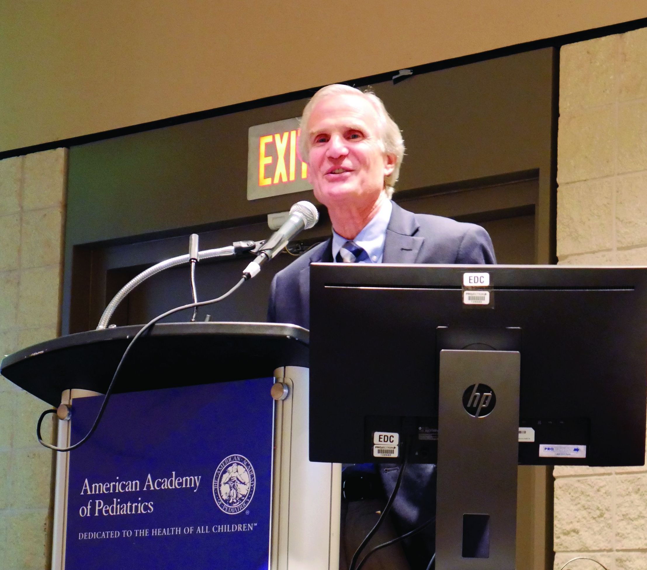
New pediatric therapies show promise for influenza, multidrug-resistant pathogens
ORLANDO – John S. Bradley, MD, said at the annual meeting of the American Academy of Pediatrics.
Dr. Bradley, director of the division of infectious diseases at Rady Children’s Hospital–San Diego, discussed a therapy for influenza, baloxavir, which was recently approved as a fast-acting single-dose medication and currently is under study in children. Also, a recent double-blind, phase 3 trial in the New England Journal of Medicine recruited patients as young as 12 years old. In the study, patients in the intervention group resolved their fever in median 25 hours, compared with 42 hours in the placebo group. Baloxavir better reduced viral load at day 2, compared with oseltamivir and placebo, but there was a similar alleviation of symptoms between both groups. There was a greater incidence of nausea and vomiting among the oseltamivir group, while the baloxavir group had a higher rate of diarrhea (N Engl J Med 2018;379:913-23).
However, Dr. Bradley noted baloxavir is much more expensive than oseltamivir, which may not justify the better tolerance of the drug for influenza treatment.
You don’t get better with it faster, so I’m not going to be recommending you all run to baloxavir this flu season for kids 12 years of age and older,” Dr. Bradley said. “I think oseltamivir is still fine, unless we end up with oseltamivir resistance.”
Solithromycin, an intravenous and oral fluoroketolide, has shown promising results against gram-positive and gram-negative pathogens for community-acquired pneumonia and other infections. During the drug’s study period, Cempra sold solithromycin to Melinta. However, one trial showed elevated liver functions in a higher number of patients than expected, and the Food and Drug Administration asked Melinta to conduct additional studies. Investigations on solithromycin have currently stopped until Melinta secures funding. “Until they get better resources, this particular drug is on hold, but you’ll see it again, I’m sure,” said Dr. Bradley, who also is professor and chief of the division of infectious diseases at the University of California, San Diego.
Dr. Bradley also discussed the efficacy of tedizolid, a protein synthesis inhibitor similar to linezolid approved in adults for the treatment of skin infections. He noted tedizolid is more active than linezolid, but the treatment course is a shorter dose for a shorter amount of time. Compared with linezolid, which can cause thrombocytopenia or neutropenia if taken for more than 10 days to 14 days, there also are fewer side effects.
“The tedizolid is much, much safer,” Dr. Bradley said, who added that trials for efficacy of tedizolid are currently underway in pediatric patients. “We’re hoping that will end up being the pediatric oxazolidinone.”
Other investigative therapies approved for adults and under study for use in children include ceftazidime/avibactam for treatment of urinary tract and complicated intra-abdominal infections, which is effective against meropenem-resistant Enterobacteriaceae and resistant Escherichia coli with extended-spectrum beta-lactamases (ESBL); ceftolozane/tazobactam has also been approved for adults, is pending approval in pediatric patients, and is active against ESBLs such as Pseudomonas; and meropenem/vaborbactam, which is active against Klebsiella pneumoniae carbapenemase (KPC)–producing isolates. Plazomicin, an aminoglycoside similar to gentamicin used to treat KPC-producing isolates, is stable against enzymes that degrade gentamicin and tobramycin.
Therapies currently under study for adults and being considered for children include imipenem/relebactam for treatment against E. coli, Enterobacter species, and KPC-producing isolates, and cefiderocol, a siderophore cephalosporin antibiotic – commonly described as a “Trojan horse” antibiotic because it binds to iron and is actively transported into the organism – is effective against Pseudomonas and has finished phase 2 trials in adults, with researchers looking to do single-dose trials in children, Dr. Bradley noted.
More experimentally, phage therapy for multidrug-resistant Acinetobacter baumannii proved effective in a 68-year-old patient with necrotizing pancreatitis who continued to deteriorate over a 4-month period despite multiple courses of antibiotics and attempted drainage of a pancreatic pseudocyst. Researchers selected a phage-specific bacterium with specificity for A. baumannii and cured him. “This is like science fiction,” Dr. Bradley said.
Dr. Bradley reported no relevant conflicts of interest.
EXPERT ANALYSIS FROM AAP 18
Team approach needed to tackle complex care for fostered, adopted children
ORLANDO – Children in foster care or who are adopted are at a higher risk of developing medical conditions, and a team approach is needed when caring for these children with a focus on medical, developmental, and mental health, Judith K. Eckerle, MD, said at the annual meeting of the American Academy of Pediatrics.
“One of the things that I’m advocating more these days is [that] we don’t operate as physicians or medical people in a vacuum. We certainly have to operate within a team approach, and that really is the best approach for these kids,” said Dr. Eckerle, a pediatrician who specializes in neurobehavioral development and is director of the Adoption Medicine Clinic at the University of Minnesota, Minneapolis.
Recognizing how early adversity is associated with conditions such as growth, brain development, motor skills, cognition, mental health, attachment, stress sensitivity, sensory processing, language, and chromosome structure can help in the medical assessment of these patients. Dr. Eckerle noted that these issues can even present in children with no early adversity who are adopted or placed into healthy foster environments.
“What we’ve learned is, we’ve just seen dozens if not hundreds of kids now in the exact same scenario who are having exactly the same types of bonding, or attachment or stress issues, as kids who did spend those few years in a really neglectful or orphanage scenarios,” Dr. Eckerle said.
“The majority of adopted and foster care kids do great in the end,” she emphasized. “As a group, they can do beautifully, but we just have to be cognizant of the things that we should be looking for and not just gloss over them so we’re not missing things along the way.”
At her institution, Dr. Eckerle said a medical assessment for new foster care or adoption patients begins with explaining the exam process with the parent and child before the child is screened by an occupational therapist. During the screening, the occupational therapist will screen the child for signs of developmental delay such as delay in gross or fine motor skills, speech, cognition, musculoskeletal, psychosocial, and sensory processing, as well as in school-based, self-care, and functional skills.
While the child is out of the room, Dr. Eckerle will ask whether the parent has any concerns about the child. After that, she will check the child’s eyes, heart, and ears to make sure they are healthy; during the end of the exam, she may order lab tests based on any concerns about prenatal or foster care trauma. Adolescents and children in whom sexual abuse is suspected should be screened for hepatitis B, hepatitis C, syphilis, and HIV.
International adoptees should undergo ova and parasite screening and the Giardia antigen test. International adoptees as well as those who had contact with the homeless or prison systems should undergo QuantiFERON-TB Gold testing for tuberculosis, and children aged under 5 years should receive targeted tuberculin skin testing. She said her center also screens children for intellectual disability, fetal alcohol spectrum disorder, short stature, and precocious puberty.
Lastly, the child is seen by a pediatric psychologist for additional screening. Dr. Eckerle noted the most common diagnoses seen at her center are reactive attachment disorder, oppositional defiant disorder, conduct disorder, pervasive development delay, autism, learning disabilities, emotional or behavioral problems, and ADHD. With regard to mental health, doctors should help a caregiver understand a child with a mental health conditions and consider pairing with a pediatric psychologist to offer evidence-based interventions for children aged under 5 years, such as child-parent psychotherapy, circle of security, and attachment biobehavioral catch-up, and offer trauma-focused cognitive behavioral therapy in children aged between 4 and 5 years.
Bad behavior often is misinterpreted as dishonesty and willful misconduct on the part of the child, but instead could be a neurodevelopmental or mental health problem, Dr. Eckerle noted. For example, a parent might interpret a child who takes a sparkling watch off of a teacher’s desk as stealing, but the child may not yet understand the concept of ownership. “Reframing these things will lead to the goal of helping that parent understand the child.”
Above all, Dr. Eckerle said she emphasizes what will happen after the exam with children she sees to establish a routine, as many children she sees have gone through multiple foster environments. It also is important to let parents know the exam is just a first step and they likely will not receive a diagnosis that day, particularly with out-of-state or international families. “I want them to have appropriate expectations that we’re not going to cure or solve or fix everything the second that they leave that room, but that we are starting the process and we are on the right track.”
Dr. Eckerle reported no conflicts of interest.
EXPERT ANALYSIS FROM AAP 18
Children with headache disorders may benefit from anti-CGRP mAb treatment
Use of anti–calcitonin gene-related peptide (CGRP) monoclonal antibodies (mAbs) may benefit children with more than 8 headache days each month, a high Pediatric Migraine Disability Assessment (PedMIDAS) score, and failure of other treatments; however, researchers cautioned that long-term safety outcomes for the treatment are not yet known, according to a recent set of recommendations published in the journal Headache.

Christina L. Szperka, MD, MSCE, of the division of neurology at the Children’s Hospital of Philadelphia and members of the Pediatric and Adolescent Headache special interest group of the American Headache Society discussed the topic of anti-CGRP mAbs at the 2018 Annual Scientific Meeting of the American Headache Society. They noted clinical outcomes for anti-CGRP mAbs in pediatric patients will likely not be available for several years and created a set of recommendations based on expert opinion of anti-CGRP mAb use in children and adolescents.
Their recommendations support using anti-CGRP mAbs for children with migraine if patients meet the following criteria: headache frequency exceeding 8 headache days per month; a PedMIDAS score of 30 or greater; failure of two or more therapies, such as pharmacologic, nonpharmacologic, or nutraceutical ones; and in patients who are past puberty.
The special interest group recommended against use of anti-CGRP mAbs in children and adolescents with recent meningitis, recent peripheral nerve injury, neurosurgery, or a central nervous system injury caused by a potentially compromised blood-brain barrier. Children and adolescents with immunodeficiency, receiving immunosuppressive medications, with structural heart defects, with pulmonary hypertension, with coronary artery disease, with cardiomyopathy, or at risk for stroke should also avoid use of anti-CGRP mAbs. Anti-CGRP mAbs are also potentially teratogenic and should not be used by adolescents or women who are pregnant, breastfeeding, or have a pregnancy wish.
Pediatric patients with significant osteoporosis or bone disease should be monitored when prescribed anti-CGRP mAbs, and the recommendations specified monitoring height and linear growth or waiting until after puberty to prescribe anti-CGRP mAbs. Although there is currently no evidence that use of anti-CGRP mAb requires pituitary hormone monitoring, the recommendations noted that weight and body mass index should also be observed.
“Pediatric and adolescent trials of anti-CGRP mAbs should be designed to maximize the chances of determining efficacy in these age groups and should focus on those who have not been successful with current multidisciplinary care,” Dr. Szperka and her colleagues wrote in the recommendations. “In the interim, the use of anti-CGRP mAbs for the treatment of headache disorders in children and adolescents may be considered in appropriate cases but should be done with close follow-up and attention to patient characteristics such as age, pubertal state, and medical comorbidities.”
Dr. Szperka receives grant support from Pfizer and Amgen and research funding from NIH. Other authors have reported grants, consulting fees, speaking fees, royalties, advisory board memberships, speaker’s bureau memberships and travel funds from Alder, Allergan, American Academy of Neurology, Amgen, Aralez, Avanir, Autonomic Technologies Inc., Biohaven, Cambridge University Press, Curelator, Depomed, Dr. Reddy’s Laboratories, Electrocore, eNeura, Genentech, Healint, Impax, JAMA Neurology, Journal Watch, Lilly, Massachusetts Medical Society, MedDay, MedicoLegal, Merck, NIH, Novartis, Oxford University Press, Quest Diagnostics, Scion, Supernus, Teva, Trigemina Inc., Upsher-Smith, UpToDate, Wolters Kluwer and Zosano.
SOURCE: Szperka CL et al. Headache. 2018. doi: 10.1111/head.13414.
Use of anti–calcitonin gene-related peptide (CGRP) monoclonal antibodies (mAbs) may benefit children with more than 8 headache days each month, a high Pediatric Migraine Disability Assessment (PedMIDAS) score, and failure of other treatments; however, researchers cautioned that long-term safety outcomes for the treatment are not yet known, according to a recent set of recommendations published in the journal Headache.
Christina L. Szperka, MD, MSCE, of the division of neurology at the Children’s Hospital of Philadelphia and members of the Pediatric and Adolescent Headache special interest group of the American Headache Society discussed the topic of anti-CGRP mAbs at the 2018 Annual Scientific Meeting of the American Headache Society. They noted clinical outcomes for anti-CGRP mAbs in pediatric patients will likely not be available for several years and created a set of recommendations based on expert opinion of anti-CGRP mAb use in children and adolescents.
Their recommendations support using anti-CGRP mAbs for children with migraine if patients meet the following criteria: headache frequency exceeding 8 headache days per month; a PedMIDAS score of 30 or greater; failure of two or more therapies, such as pharmacologic, nonpharmacologic, or nutraceutical ones; and in patients who are past puberty.
The special interest group recommended against use of anti-CGRP mAbs in children and adolescents with recent meningitis, recent peripheral nerve injury, neurosurgery, or a central nervous system injury caused by a potentially compromised blood-brain barrier. Children and adolescents with immunodeficiency, receiving immunosuppressive medications, with structural heart defects, with pulmonary hypertension, with coronary artery disease, with cardiomyopathy, or at risk for stroke should also avoid use of anti-CGRP mAbs. Anti-CGRP mAbs are also potentially teratogenic and should not be used by adolescents or women who are pregnant, breastfeeding, or have a pregnancy wish.
Pediatric patients with significant osteoporosis or bone disease should be monitored when prescribed anti-CGRP mAbs, and the recommendations specified monitoring height and linear growth or waiting until after puberty to prescribe anti-CGRP mAbs. Although there is currently no evidence that use of anti-CGRP mAb requires pituitary hormone monitoring, the recommendations noted that weight and body mass index should also be observed.
“Pediatric and adolescent trials of anti-CGRP mAbs should be designed to maximize the chances of determining efficacy in these age groups and should focus on those who have not been successful with current multidisciplinary care,” Dr. Szperka and her colleagues wrote in the recommendations. “In the interim, the use of anti-CGRP mAbs for the treatment of headache disorders in children and adolescents may be considered in appropriate cases but should be done with close follow-up and attention to patient characteristics such as age, pubertal state, and medical comorbidities.”
Dr. Szperka receives grant support from Pfizer and Amgen and research funding from NIH. Other authors have reported grants, consulting fees, speaking fees, royalties, advisory board memberships, speaker’s bureau memberships and travel funds from Alder, Allergan, American Academy of Neurology, Amgen, Aralez, Avanir, Autonomic Technologies Inc., Biohaven, Cambridge University Press, Curelator, Depomed, Dr. Reddy’s Laboratories, Electrocore, eNeura, Genentech, Healint, Impax, JAMA Neurology, Journal Watch, Lilly, Massachusetts Medical Society, MedDay, MedicoLegal, Merck, NIH, Novartis, Oxford University Press, Quest Diagnostics, Scion, Supernus, Teva, Trigemina Inc., Upsher-Smith, UpToDate, Wolters Kluwer and Zosano.
SOURCE: Szperka CL et al. Headache. 2018. doi: 10.1111/head.13414.
FROM HEADACHE
Key clinical point: Treatment of headache disorders with anti–calcitonin gene-related peptide (CGRP) monoclonal antibodies (mAbs) in children and adolescents may be indicated in some cases.
Major finding: Pediatric patients with more than eight headache days per month, PedMIDAS score of 30 or higher, and failure of other pharmacological, nonpharmacological, and nutraceutical treatments may benefit from anti-CGRP mAb treatment.
Study details: Expert opinion from the members of the Pediatric and Adolescent Headache special interest group based on recommendations made at the 2018 Annual Scientific Meeting of the American Headache Society.
Disclosures: Dr. Szperka receives grant support from Pfizer and Amgen and research funding from NIH. Other authors have reported grants, consulting fees, speaking fees, royalties, advisory board memberships, speaker’s bureau memberships, and travel funds from Alder, Allergan, American Academy of Neurology, Amgen, Aralez, Avanir, Autonomic Technologies, Biohaven, Cambridge University Press, Curelator, Depomed, Dr. Reddy’s Laboratories, Electrocore, eNeura, Genentech, Healint, Impax, JAMA Neurology, Journal Watch, Lilly, Massachusetts Medical Society, MedDay, MedicoLegal, Merck, NIH, Novartis, Oxford University Press, Quest Diagnostics, Scion, Supernus, Teva, Trigemina, Upsher-Smith, UpToDate, Wolters Kluwer, and Zosano.
Source: Szperka CL et al. Headache. 2018. doi: 10.1111/head.13414.
Risk of cancer in NAFLD is 91% higher than in control subjects
SAN FRANCISCO –

Those are key findings from a community cohort study with up to 21 years of follow-up, which one of the authors, Alina M. Allen, MD, discussed during a press briefing at the annual meeting of the American Association for the Study of Liver Diseases.
“NAFLD is the most common chronic liver disease in the Western world,” said Dr. Allen, a gastroenterologist at the Mayo Clinic, Rochester, Minn. “It affects one in four adults in the U.S. It is related to fat accumulation in the liver in people who are overweight or obese and can lead to cirrhosis and liver-related mortality. However, the most common cause of death in this population is not liver disease but malignancy and cardiovascular disease. There is a paucity of epidemiologic studies of extrahepatic cancer in NAFLD. It is not clear what types of cancers and how much higher their cancer risk is in reference to the general population.”
In an effort to determine the incidence of cancer diagnoses in NALFD, compared with controls, in a U.S. community, Dr. Allen and her colleagues drew from the Rochester Epidemiology Project to evaluate 4,791 adults diagnosed with NAFLD and 14,432 age- and sex-matched control subjects in Olmstead County, Minn., during 1997-2017. The researchers obtained corresponding Surveillance, Epidemiology, and End Results Program (SEER) rates as a quality check and used a regression model to assess the malignancy risk in NAFLD overall and by cancer type, age, sex, and body mass index. They recorded all new diagnoses of cancers that developed in both groups until 2018, for a total possible follow-up of 21 years, and they reported results in incidence rate ratios, a risk estimate similar to hazard ratios.
The mean age of the study population was 53 years, 53% were female, and the mean follow-up was 8 years with a range from 1 to 21 years. New cancers were identified in 16% of subjects with NALFD and in 12% of control subjects. The overall risk of malignancy was 91% higher in NAFLD subjects, compared with control subjects; there were higher rates in the NAFLD subjects for most types of cancers, but the largest increases were in GI cancers. The greatest malignancy risk was for cancer of the liver (RR, 3.24), followed by cancer of the uterus (RR, 2.39), stomach (RR, 2.34), pancreas (RR, 2.09), and colon (RR, 1.75). “Interestingly, the risk of colon cancer increased only in men but not in women,” Dr. Allen said. “These data provide an important hierarchical overview of the top most important malignancy risks associated with NAFLD that the medical community should be aware of.”
When the researchers looked for differences in age at cancer diagnosis between NAFLD and controls, they found that pancreas cancer occurred at a younger age among subjects with NAFLD. They also observed that colon cancer occurred at a younger age in men with NAFLD, but not in women with the disease. “What was most interesting to us was the assessment of cancer risk in NAFLD versus obesity alone,” Dr. Allen said. “Previous studies from the general population have linked obesity to a higher risk of cancer. Whether the presence of fatty liver disease would impact that risk has not been assessed. We showed that obesity is associated with a higher risk of cancer only in those with NAFLD, not in those without. If validated in independent cohorts, these findings could change our understanding of the relationship between obesity and cancer and the importance of screening for NAFLD – not only to risk-stratify liver disease but also for the risk of extrahepatic malignancy.”
Dr. Allen concluded her presentation by noting that findings from large population-based studies such as the Rochester Epidemiology Project “can offer important epidemiologic data regarding the biggest threats to the health of a community,” she said. “Such data increase awareness, enable appropriate counseling, and could inform screening policies. There is a signal in the fact that the GI cancers are increased [in NAFLD]. It’s an interesting signal that needs to be studied further.”
Dr. Allen reported having no financial conflicts.
Source: Allen AM. Hepatol. 2018;68[S1]: Abstract 31.
SAN FRANCISCO –
Those are key findings from a community cohort study with up to 21 years of follow-up, which one of the authors, Alina M. Allen, MD, discussed during a press briefing at the annual meeting of the American Association for the Study of Liver Diseases.
“NAFLD is the most common chronic liver disease in the Western world,” said Dr. Allen, a gastroenterologist at the Mayo Clinic, Rochester, Minn. “It affects one in four adults in the U.S. It is related to fat accumulation in the liver in people who are overweight or obese and can lead to cirrhosis and liver-related mortality. However, the most common cause of death in this population is not liver disease but malignancy and cardiovascular disease. There is a paucity of epidemiologic studies of extrahepatic cancer in NAFLD. It is not clear what types of cancers and how much higher their cancer risk is in reference to the general population.”
In an effort to determine the incidence of cancer diagnoses in NALFD, compared with controls, in a U.S. community, Dr. Allen and her colleagues drew from the Rochester Epidemiology Project to evaluate 4,791 adults diagnosed with NAFLD and 14,432 age- and sex-matched control subjects in Olmstead County, Minn., during 1997-2017. The researchers obtained corresponding Surveillance, Epidemiology, and End Results Program (SEER) rates as a quality check and used a regression model to assess the malignancy risk in NAFLD overall and by cancer type, age, sex, and body mass index. They recorded all new diagnoses of cancers that developed in both groups until 2018, for a total possible follow-up of 21 years, and they reported results in incidence rate ratios, a risk estimate similar to hazard ratios.
The mean age of the study population was 53 years, 53% were female, and the mean follow-up was 8 years with a range from 1 to 21 years. New cancers were identified in 16% of subjects with NALFD and in 12% of control subjects. The overall risk of malignancy was 91% higher in NAFLD subjects, compared with control subjects; there were higher rates in the NAFLD subjects for most types of cancers, but the largest increases were in GI cancers. The greatest malignancy risk was for cancer of the liver (RR, 3.24), followed by cancer of the uterus (RR, 2.39), stomach (RR, 2.34), pancreas (RR, 2.09), and colon (RR, 1.75). “Interestingly, the risk of colon cancer increased only in men but not in women,” Dr. Allen said. “These data provide an important hierarchical overview of the top most important malignancy risks associated with NAFLD that the medical community should be aware of.”
When the researchers looked for differences in age at cancer diagnosis between NAFLD and controls, they found that pancreas cancer occurred at a younger age among subjects with NAFLD. They also observed that colon cancer occurred at a younger age in men with NAFLD, but not in women with the disease. “What was most interesting to us was the assessment of cancer risk in NAFLD versus obesity alone,” Dr. Allen said. “Previous studies from the general population have linked obesity to a higher risk of cancer. Whether the presence of fatty liver disease would impact that risk has not been assessed. We showed that obesity is associated with a higher risk of cancer only in those with NAFLD, not in those without. If validated in independent cohorts, these findings could change our understanding of the relationship between obesity and cancer and the importance of screening for NAFLD – not only to risk-stratify liver disease but also for the risk of extrahepatic malignancy.”
Dr. Allen concluded her presentation by noting that findings from large population-based studies such as the Rochester Epidemiology Project “can offer important epidemiologic data regarding the biggest threats to the health of a community,” she said. “Such data increase awareness, enable appropriate counseling, and could inform screening policies. There is a signal in the fact that the GI cancers are increased [in NAFLD]. It’s an interesting signal that needs to be studied further.”
Dr. Allen reported having no financial conflicts.
Source: Allen AM. Hepatol. 2018;68[S1]: Abstract 31.
REPORTING FROM THE LIVER MEETING 2018
Key clinical point: The risk of cancer in patients with nonalcoholic fatty liver disease is 91% higher than in control subjects.
Major finding: Among subjects with NAFLD, the greatest malignancy risk was for cancer of the liver (risk ratio, 3.24).
Study details: A cohort study of 4,791 adults diagnosed with NAFLD and 14,432 age- and sex-matched control subjects from the Rochester Epidemiology Project.
Disclosures: Dr. Allen reported having no financial conflicts.
Source: Allen AM. Hepatol. 2018;68[S1]:Abstract 31
Alemtuzumab switch linked to good MS outcomes
BERLIN – Switching to alemtuzumab from fingolimod is associated with improved disease activity in patients with relapsing-remitting multiple sclerosis (RRMS), according to the results of a real-world study reported at the annual congress of the European Committee for Treatment and Research in Multiple Sclerosis.
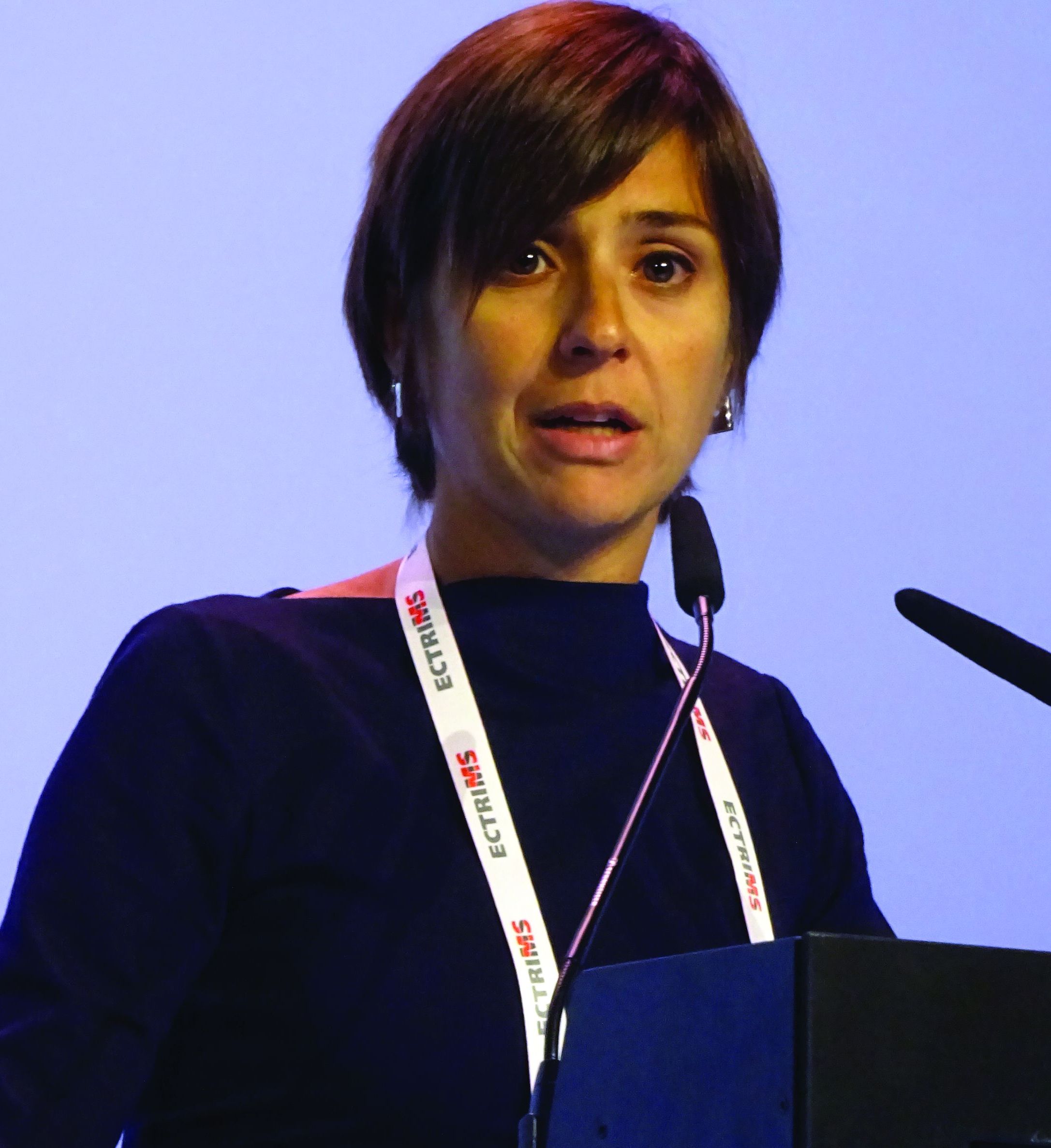
Jessica Frau, MD, of the University of Cagliari (Italy) reported that a switch from fingolimod (Gilenya) to alemtuzumab (Lemtrada) in 77 patients treated at 11 Italian centers was able to “reduce dramatically disease activity in patients who did not respond to fingolimod.”
Dr. Frau reported: “When we compared in our cohort the last year of fingolimod with the first year after the first course of alemtuzumab, we found a significant decrease in the annualized relapse rate [ARR].” The ARRs were 0.60 for fingolimod and 0.20 after 1 year of alemtuzumab treatment.
“We found also a trend towards an improvement in the EDSS [Expanded Disability Status Scale] score (P = .23), and less evidence of disease activity on MRI, both in terms of new T2 lesions and gadolinium-enhancing (Gd+) lesions.”
The last MRI during fingolimod treatment showed new T2 and Gd+ enhancing lesions in 69.2% and 58.6% of patients, respectively. Corresponding figures for the first MRI during alemtuzumab treatment were 10.4% and 2.2% of patients.
The beneficial effects of switching to fingolimod in the Italian study “was not influenced by a shorter washout [period] or a low lymphocyte count when alemtuzumab was started,” Dr. Frau said. A shorter washout period has been hypothesized to account for recent accounts of disease flares seen when switching from fingolimod to alemtuzumab, she explained.
Indeed, Dr. Frau noted that there had been a few studies that reported MS disease reactivation soon after the switch to alemtuzumab was made, which could be because lymphocytes remain in the lymph nodes when alemtuzumab is administered, this means that potentially they could repopulate the central nervous system and reactivate the disease.
However, “when alemtuzumab is started after fingolimod it is not a risk factor for reactivation of the disease,” Dr. Frau said, based on the current study’s findings.
As expected, the frequency of relapses increased during the washout period after stopping fingolimod, going from 12.7% of patients with relapse in the first month, 18.2% at 2 months, and 22.2% at 3 months. The time to first relapse from the start of alemtuzumab treatment was 6 months for 2.9% of patients, 9 months for 10.5% of patients, and 1 year for 20.7% of patients.
Asked to comment on when the optimal time to switch from fingolimod to alemtuzumab might be, Dr. Frau said: “The optimal time could be 1 month when the lymphocyte count is not too low.” However, lymphocyte counts were not measured in the entire cohort, so “these data perhaps need to have more strength.”
The switch from natalizumab to alemtuzumab
Other data on switching to alemtuzumab, this time from natalizumab (Tysabri), in the ANSWERS MS study were presented by Paul Gallagher, MBChB, of Queen Elizabeth University Hospital, London, and the University of Glasgow (Scotland).
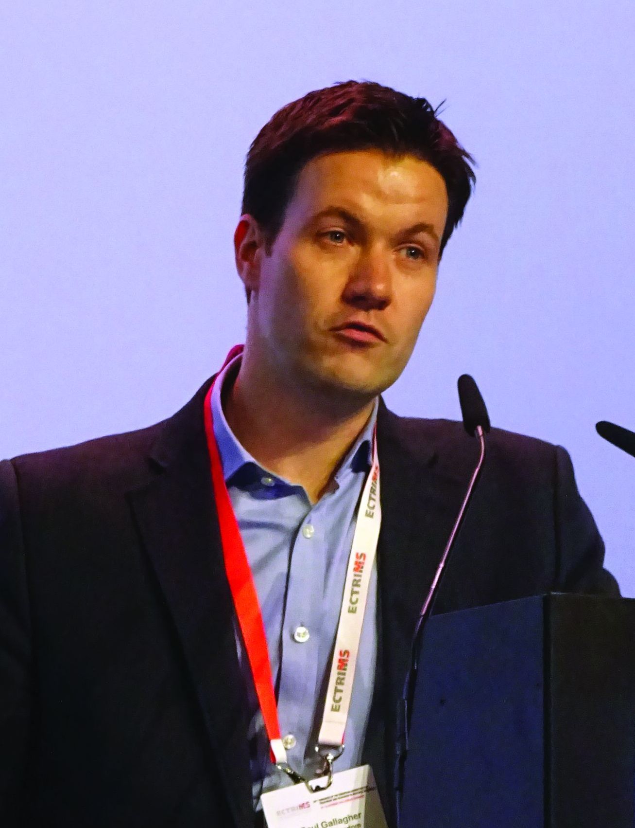
ANSWERS MS (Alemtuzumab after Natalizumab Switch in Evolving Rapidly Severe MS) is a retrospective, observational analysis of routinely collected data on the use of alemtuzumab by 13 centers the United Kingdom and Ireland. These centers have been collecting data since before alemtuzumab was licensed in 2014 for MS, Dr. Gallagher observed, with some centers having experience of making the switch for more than a decade.
ANSWERS MS addresses a common clinical question: “Is it safe and effective to switch to alemtuzumab if natalizumab fails in highly active MS?” Dr. Gallagher said. “The truth is we don’t really know the answer to this, although it’s becoming an increasingly used switch.”
Alemtuzumab was developed in Cambridge, England, in 1983, originally as an anticancer agent, and first started being used in MS patients in the 1990s. Natalizumab was first licensed in the United Kingdom in 2007.
The aim of the study was mainly to look at safety, but also examine efficacy, and to offer advice on how to best manage the switch. A total of 79 patients formed the safety cohort; 51 of these patients had more than 2 years of follow-up after their first infusion of alemtuzumab and formed the efficacy cohort.
Data were examined in five phases: before natalizumab, during natalizumab, during the switchover period, during alemtuzumab treatment, and after alemtuzumab treatment, with the latter starting 2 years after the first alemtuzumab infusion.
Dr. Gallagher noted that 43% started natalizumab as a first-line therapy, and almost half (49%) of patients stopped taking natalizumab because of breakthrough disease, making this a bit of an unusual cohort with highly active disease, although other cohort characteristics were pretty typical of an MS population.
“The headline is that there are no new safety concerns identified from this cohort,” Dr. Gallagher reported. “Most [61%] patients had infusion reactions with alemtuzumab as expected, but this gradually reduced with subsequent courses.”
Fewer than 20% of patients developed autoimmune thyroid disease, he added, and there were no cases of idiopathic thrombocytopenic purpura.
Infections were seen in nine patients, including three cases of shingles, two urinary tract infections – one of which was classed as a severe adverse event – and one case each of oral thrush, fungal skin infection, tonsillitis, and norovirus.
There was also one cytomegalovirus infection and one death from sepsis unrelated to alemtuzumab; both of these were classed as serious adverse events.
In terms of efficacy, mean ARRs were 2.3 before and 0.8 during natalizumab treatment, decreasing to 0.4 during alemtuzumab treatment and 0.5 post alemtuzumab. A “spike” in relapses was seen, however, during the switch period.
“There was a similar story with MRI imaging,” Dr. Gallagher said. “The profile suggests high disease activity during the switch phase in comparison to everything else.” The mean number of new or worsened MRI lesions was 4.32 per scan per year during the switch period. This fell, however, during alemtuzumab treatment to 0.006 per MRI scan per year and remained low after the end of alemtuzumab treatment at 0.017 per scan per year.
There was no real benefit to switching on the EDSS, with scores increasing from 3.4 in the pre-natalizumab period to 4.7 during the switch period, but then plateauing out to 4.4. and 4.3 after the initiation of alemtuzumab and in the post-alemtuzumab phase.
“These data were based on medical records, often incomplete, and so not all patients had an EDSS in every phase, for example,” Dr. Gallagher noted. He said an analysis was done to try to account for the missing information. This showed that there was an improvement in EDSS while on alemtuzumab, but the effect was not maintained.
It was evident in looking at the switch period that a shorter time between natalizumab and alemtuzumab was associated with the best outcomes, with the optimum time being around 2-4 months. Bridging therapy with fingolimod did not reduce disease activity during the switch, Dr. Gallagher said.
ANSWERS MS was funded by Sanofi-Genzyme. Paul Gallagher disclosed that he had received salary payment and travel funding for educational events from Sanofi-Genzyme and travel funding from Novartis and Biogen.
Dr. Frau disclosed that she serves on scientific advisory boards for Biogen, Merck, and Genzyme and that she has received honoraria for speaking from Merck Serono, Genzyme, Biogen, and Teva.
SOURCE: Frau J et al. Mult Scler. 2018;24(S2):100-1, Abstract 265; Gallagher P et al. Mult Scler. 2018;24(S2):99-100, Abstract 264.
BERLIN – Switching to alemtuzumab from fingolimod is associated with improved disease activity in patients with relapsing-remitting multiple sclerosis (RRMS), according to the results of a real-world study reported at the annual congress of the European Committee for Treatment and Research in Multiple Sclerosis.
Jessica Frau, MD, of the University of Cagliari (Italy) reported that a switch from fingolimod (Gilenya) to alemtuzumab (Lemtrada) in 77 patients treated at 11 Italian centers was able to “reduce dramatically disease activity in patients who did not respond to fingolimod.”
Dr. Frau reported: “When we compared in our cohort the last year of fingolimod with the first year after the first course of alemtuzumab, we found a significant decrease in the annualized relapse rate [ARR].” The ARRs were 0.60 for fingolimod and 0.20 after 1 year of alemtuzumab treatment.
“We found also a trend towards an improvement in the EDSS [Expanded Disability Status Scale] score (P = .23), and less evidence of disease activity on MRI, both in terms of new T2 lesions and gadolinium-enhancing (Gd+) lesions.”
The last MRI during fingolimod treatment showed new T2 and Gd+ enhancing lesions in 69.2% and 58.6% of patients, respectively. Corresponding figures for the first MRI during alemtuzumab treatment were 10.4% and 2.2% of patients.
The beneficial effects of switching to fingolimod in the Italian study “was not influenced by a shorter washout [period] or a low lymphocyte count when alemtuzumab was started,” Dr. Frau said. A shorter washout period has been hypothesized to account for recent accounts of disease flares seen when switching from fingolimod to alemtuzumab, she explained.
Indeed, Dr. Frau noted that there had been a few studies that reported MS disease reactivation soon after the switch to alemtuzumab was made, which could be because lymphocytes remain in the lymph nodes when alemtuzumab is administered, this means that potentially they could repopulate the central nervous system and reactivate the disease.
However, “when alemtuzumab is started after fingolimod it is not a risk factor for reactivation of the disease,” Dr. Frau said, based on the current study’s findings.
As expected, the frequency of relapses increased during the washout period after stopping fingolimod, going from 12.7% of patients with relapse in the first month, 18.2% at 2 months, and 22.2% at 3 months. The time to first relapse from the start of alemtuzumab treatment was 6 months for 2.9% of patients, 9 months for 10.5% of patients, and 1 year for 20.7% of patients.
Asked to comment on when the optimal time to switch from fingolimod to alemtuzumab might be, Dr. Frau said: “The optimal time could be 1 month when the lymphocyte count is not too low.” However, lymphocyte counts were not measured in the entire cohort, so “these data perhaps need to have more strength.”
The switch from natalizumab to alemtuzumab
Other data on switching to alemtuzumab, this time from natalizumab (Tysabri), in the ANSWERS MS study were presented by Paul Gallagher, MBChB, of Queen Elizabeth University Hospital, London, and the University of Glasgow (Scotland).
ANSWERS MS (Alemtuzumab after Natalizumab Switch in Evolving Rapidly Severe MS) is a retrospective, observational analysis of routinely collected data on the use of alemtuzumab by 13 centers the United Kingdom and Ireland. These centers have been collecting data since before alemtuzumab was licensed in 2014 for MS, Dr. Gallagher observed, with some centers having experience of making the switch for more than a decade.
ANSWERS MS addresses a common clinical question: “Is it safe and effective to switch to alemtuzumab if natalizumab fails in highly active MS?” Dr. Gallagher said. “The truth is we don’t really know the answer to this, although it’s becoming an increasingly used switch.”
Alemtuzumab was developed in Cambridge, England, in 1983, originally as an anticancer agent, and first started being used in MS patients in the 1990s. Natalizumab was first licensed in the United Kingdom in 2007.
The aim of the study was mainly to look at safety, but also examine efficacy, and to offer advice on how to best manage the switch. A total of 79 patients formed the safety cohort; 51 of these patients had more than 2 years of follow-up after their first infusion of alemtuzumab and formed the efficacy cohort.
Data were examined in five phases: before natalizumab, during natalizumab, during the switchover period, during alemtuzumab treatment, and after alemtuzumab treatment, with the latter starting 2 years after the first alemtuzumab infusion.
Dr. Gallagher noted that 43% started natalizumab as a first-line therapy, and almost half (49%) of patients stopped taking natalizumab because of breakthrough disease, making this a bit of an unusual cohort with highly active disease, although other cohort characteristics were pretty typical of an MS population.
“The headline is that there are no new safety concerns identified from this cohort,” Dr. Gallagher reported. “Most [61%] patients had infusion reactions with alemtuzumab as expected, but this gradually reduced with subsequent courses.”
Fewer than 20% of patients developed autoimmune thyroid disease, he added, and there were no cases of idiopathic thrombocytopenic purpura.
Infections were seen in nine patients, including three cases of shingles, two urinary tract infections – one of which was classed as a severe adverse event – and one case each of oral thrush, fungal skin infection, tonsillitis, and norovirus.
There was also one cytomegalovirus infection and one death from sepsis unrelated to alemtuzumab; both of these were classed as serious adverse events.
In terms of efficacy, mean ARRs were 2.3 before and 0.8 during natalizumab treatment, decreasing to 0.4 during alemtuzumab treatment and 0.5 post alemtuzumab. A “spike” in relapses was seen, however, during the switch period.
“There was a similar story with MRI imaging,” Dr. Gallagher said. “The profile suggests high disease activity during the switch phase in comparison to everything else.” The mean number of new or worsened MRI lesions was 4.32 per scan per year during the switch period. This fell, however, during alemtuzumab treatment to 0.006 per MRI scan per year and remained low after the end of alemtuzumab treatment at 0.017 per scan per year.
There was no real benefit to switching on the EDSS, with scores increasing from 3.4 in the pre-natalizumab period to 4.7 during the switch period, but then plateauing out to 4.4. and 4.3 after the initiation of alemtuzumab and in the post-alemtuzumab phase.
“These data were based on medical records, often incomplete, and so not all patients had an EDSS in every phase, for example,” Dr. Gallagher noted. He said an analysis was done to try to account for the missing information. This showed that there was an improvement in EDSS while on alemtuzumab, but the effect was not maintained.
It was evident in looking at the switch period that a shorter time between natalizumab and alemtuzumab was associated with the best outcomes, with the optimum time being around 2-4 months. Bridging therapy with fingolimod did not reduce disease activity during the switch, Dr. Gallagher said.
ANSWERS MS was funded by Sanofi-Genzyme. Paul Gallagher disclosed that he had received salary payment and travel funding for educational events from Sanofi-Genzyme and travel funding from Novartis and Biogen.
Dr. Frau disclosed that she serves on scientific advisory boards for Biogen, Merck, and Genzyme and that she has received honoraria for speaking from Merck Serono, Genzyme, Biogen, and Teva.
SOURCE: Frau J et al. Mult Scler. 2018;24(S2):100-1, Abstract 265; Gallagher P et al. Mult Scler. 2018;24(S2):99-100, Abstract 264.
REPORTING FROM ECTRIMS 2018
Key clinical point: Good results can be achieved by switching from fingolimod or natalizumab to alemtuzumab in relapsing-remitting multiple sclerosis (RRMS).
Major finding: Annualized relapse rates were 0.60 for fingolimod at the time of the switch and 0.20 after 1 year of alemtuzumab treatment in one real-world study. In another, alemtuzumab was effective in reducing inflammatory disease activity when natalizumab failed.
Study details: Two real-world, observational studies: one with 77 RRMS patients treated at 11 Italian centers and the other a retrospective analysis of routinely collected data on 79 patients.
Disclosures: Dr. Frau disclosed that she serves on scientific advisory board for Biogen, Merck, and Genzyme and that she has received honoraria for speaking from Merck Serono, Genzyme, Biogen, and Teva. ANSWERS MS was funded by Sanofi-Genzyme. Dr. Gallagher disclosed he had received salary payment and travel funding for educational events from Sanofi-Genzyme and travel funding from Novartis and Biogen.
Source: Frau J et al. Mult Scler. 2018;24(S2):100-1, Abstract 265; Gallagher P et al. Mult Scler. 2018;24(S2):99-100, Abstract 264.
Adjunctive testosterone may reduce depressive symptoms in men
Testosterone treatment has potential as an adjunct therapy for men with depressive disorders, a meta-analysis has suggested. However, more research is needed.
“This meta-analysis provides important new evidence that testosterone treatment may also be effective and efficacious for eugonadal and older men when higher testosterone dosages are administered,” Andreas Walther, PhD, and his coauthors wrote Nov. 14 in JAMA Psychiatry. However, they noted that safety monitoring in testosterone treatment trials remained important because of an absence of sufficiently powered, long-term studies to assess the increased risk of adverse events with treatment.
The link between testosterone and depression has been debated extensively because testosterone is a neuroactive steroid hormone known to influence mood and appetitive behavior, Dr. Walther and his coauthors wrote. Although testosterone treatment for various disorders in hypogonadal men has been backed by evidence, the results of randomized, placebo-controlled clinical trials for its use in depression have been inconsistent. Indeed, testosterone treatment was currently not recommended in national or international guidelines because of an “prevailing uncertainty about its efficacy, age criteria, dosage, ideal duration and method of application,” wrote Dr. Walther, of the department of biological psychology at Technische Universität Dresden (Germany).
For the current review, the researchers identified 27 randomized, controlled trials altogether including 1,890 men who were receiving testosterone treatment and had reported depressive symptoms on validated depression scales.
Results showed evidence for a moderate antidepressant association of testosterone treatment, compared with placebo (Hedges g, 0.21; 95% confidence interval, 0.10-0.32), and an efficacy odds ratio of 2.30 (95% CI, 1.30-4.06). According to the researchers, based on reference ranges for depressive symptoms, “The National Institute for Health and Care Excellence guidelines on depression suggest a reduction of 3.0 and 2.0 points on BDI scores to be clinically significant for normal depression and treatment-resistant depression, respectively,” they wrote.
Testosterone treatment also showed an efficacy OR of 2.30, a finding that the authors said suggested the “potential of testosterone treatment as adjunct therapy for men with depressive disorders.”
They said the results suggested that better treatment response might require higher doses but acknowledged that the finding required replication.
In addition, Dr. Walther and his coauthors found acceptability of testosterone treatment was high, with an OR of 0.79 for testosterone treatment–related loss to follow-up, compared with placebo. Remarkably, they added, initial testosterone status did not moderate the effects of testosterone treatment on depressive symptoms.
“Large, preregistered RCTs of good quality investigating testosterone treatment’s effect in men on depression as the primary outcome” are needed, they concluded.
Dr. Walther and his coauthors cited a few limitations, including the low number of randomized, controlled trials addressing the effects of testosterone treatment in men who were depressed but otherwise healthy.
No conflicts of interest were reported.
SOURCE: Walther A et al. JAMA Psychiatry. 2018 14 Nov. doi: 10.1001/jamapsychiatry.2018.2734.
The role of testosterone in the pathophysiology and treatment of depressive disorders in men is controversial. The meta-analysis by Walther et al. is well performed and adds to the body of evidence suggesting that testosterone treatment can lead to small improvements in men with depressive symptoms.
However, it is uncertain whether these improvements are clinically meaningful. The data should not be interpreted as testosterone treatment leads to remission or enhances response to antidepressant treatment in this population. In short, the current meta-analysis suggests testosterone replacement may enhance mood among nondepressed hypogonadal men. It is worth noting that the long-term safety of testosterone treatment remains unknown. Until more research is available, clinicians should continue to follow the Endocrine Society guideline for testosterone replacement therapy of androgen-deficient men. The data do not currently support the use of testosterone therapy, particularly in supraphysiologic doses, for the treatment of depressive disorders in men.
Shalender Bhasin, MD, is affiliated with the Brigham and Women’s Hospital in Boston, and Stuart Seidman, MD, is affiliated with Columbia University, New York. These comments are taken from an accompanying editorial (JAMA Psychiatry. 2018 Nov 14. doi: 10.1001/jamapsychiatry.2018.2661). Dr. Bhasin reported receiving research grants from several sources, and Dr. Seidman reported no disclosures.
Testosterone treatment has potential as an adjunct therapy for men with depressive disorders, a meta-analysis has suggested. However, more research is needed.
“This meta-analysis provides important new evidence that testosterone treatment may also be effective and efficacious for eugonadal and older men when higher testosterone dosages are administered,” Andreas Walther, PhD, and his coauthors wrote Nov. 14 in JAMA Psychiatry. However, they noted that safety monitoring in testosterone treatment trials remained important because of an absence of sufficiently powered, long-term studies to assess the increased risk of adverse events with treatment.
The link between testosterone and depression has been debated extensively because testosterone is a neuroactive steroid hormone known to influence mood and appetitive behavior, Dr. Walther and his coauthors wrote. Although testosterone treatment for various disorders in hypogonadal men has been backed by evidence, the results of randomized, placebo-controlled clinical trials for its use in depression have been inconsistent. Indeed, testosterone treatment was currently not recommended in national or international guidelines because of an “prevailing uncertainty about its efficacy, age criteria, dosage, ideal duration and method of application,” wrote Dr. Walther, of the department of biological psychology at Technische Universität Dresden (Germany).
For the current review, the researchers identified 27 randomized, controlled trials altogether including 1,890 men who were receiving testosterone treatment and had reported depressive symptoms on validated depression scales.
Results showed evidence for a moderate antidepressant association of testosterone treatment, compared with placebo (Hedges g, 0.21; 95% confidence interval, 0.10-0.32), and an efficacy odds ratio of 2.30 (95% CI, 1.30-4.06). According to the researchers, based on reference ranges for depressive symptoms, “The National Institute for Health and Care Excellence guidelines on depression suggest a reduction of 3.0 and 2.0 points on BDI scores to be clinically significant for normal depression and treatment-resistant depression, respectively,” they wrote.
Testosterone treatment also showed an efficacy OR of 2.30, a finding that the authors said suggested the “potential of testosterone treatment as adjunct therapy for men with depressive disorders.”
They said the results suggested that better treatment response might require higher doses but acknowledged that the finding required replication.
In addition, Dr. Walther and his coauthors found acceptability of testosterone treatment was high, with an OR of 0.79 for testosterone treatment–related loss to follow-up, compared with placebo. Remarkably, they added, initial testosterone status did not moderate the effects of testosterone treatment on depressive symptoms.
“Large, preregistered RCTs of good quality investigating testosterone treatment’s effect in men on depression as the primary outcome” are needed, they concluded.
Dr. Walther and his coauthors cited a few limitations, including the low number of randomized, controlled trials addressing the effects of testosterone treatment in men who were depressed but otherwise healthy.
No conflicts of interest were reported.
SOURCE: Walther A et al. JAMA Psychiatry. 2018 14 Nov. doi: 10.1001/jamapsychiatry.2018.2734.
FROM JAMA PSYCHIATRY
Key clinical point: Testosterone appears to be moderately effective in reducing depressive symptoms in men.
Major finding: Testosterone treatment was associated with a significant reduction in depressive symptoms, compared with placebo, with an efficacy of odds ratio of 2.30 (95% confidence interval, 1.30-4.06).
Study details: A systematic review and meta-analysis involving 27 randomized, placebo-controlled trials involving a broad range of men who were treated with testosterone and reported depressive symptoms on validated depression scales.
Disclosures: No conflicts of interest were reported.
Source: Walther A et al. JAMA Psychiatry. 2018 Nov 14. doi: 10.1001/jamapsychiatry.2018.2734.
How well are your phones being answered?
We have several new, young employees in my front office, and it had been quite awhile since I had followed my own advice of “eavesdropping” on their telephone conversations with patients. You would think that Millennials, with all the time they spend on phones, would have little to learn in that department – until you remember that Instagram and Snapchat do not require interpersonal skills.

So I. If you want to adapt it for your own use, be my guest:
• The first impression a new patient has of our office is usually made by our receptionists. Even now, in the era of texting and e-mail, the telephone remains our primary point of contact with new and long-time patients. The way we answer it determines, to a significant extent, how the community thinks of us, as people and as health care providers.
• Everyone in the office needs to know how to answer the phone professionally. If you notice that a phone is ringing and the receptionists are unable to answer it, please pick up the phone; an incoming call must never go unanswered.
• Answer all incoming calls before the third ring.• Answer warmly, enthusiastically, and professionally. Since the caller cannot see you, your voice is the only impression of our office a first-time caller will get.

• Identify yourself and our office immediately. “Good morning, Doctor Eastern’s office. This is __________, how may I help you?” (No one should ever have to ask what office they have reached, or to whom they are speaking.)
• Speak like a professional. Don’t use slang or buzzwords. Instead of “totally” or “for sure,” for example, say “certainly” or “of course.” If you tend to use fillers (“uh huh,” “um,” “like,” “you know,” etc.), train yourself not to use them in the office.
• Adopt a positive vocabulary – one that focuses on helping people. For example, rather than saying, “I don’t know,” say, “Let me find out for you,” or “I’ll make sure someone gets back to you on that.”
• Offer to take a message if the caller has a question or issue you cannot address. Assure the patient that the appropriate staffer will call back later that day. That way, office work flow is not interrupted, and the patient still receives a prompt (and correct) answer.
• All messages left overnight with the answering service must be returned as early as possible the very next business day. This is a top priority each morning. Few things annoy callers trying to reach their doctors more than unreturned calls. If the office will be closed for a holiday, or a response will be delayed for any other reason, make sure the service knows, and passes it on to patients.
• If the phone rings while you are dealing with a patient in person, that patient is your first priority. Put the caller on hold, but always ask permission before doing so, and wait for an answer. Never leave a caller on hold for more than a minute or two unless absolutely unavoidable.
• Never answer with, “Doctor’s office, please hold.” To a patient, that is even worse than not answering at all. No matter how often your hold message tells callers how important they are, they know they are being ignored. Such encounters never end well: Those who wait will be grumpy and rude when you get back to them; those who hang up will be even more grumpy and rude when they call back. Worst of all are those who don’t call back and seek care elsewhere – often leaving a nasty comment on social media besides.
• Maintaining patient confidentiality is a top priority. It makes patients feel secure about being treated in our office, and it is also the law. Be cautious about all information that is given over the phone. Don’t disclose any personal information unless you are absolutely certain you are talking to the correct patient. If the caller is not the patient, never discuss personal information without the patient’s permission.
Keep in mind that patients and others in the office may be able to overhear your phone conversations. Keep your voice down; never use the phone’s hands-free “speaker” function.
Dr. Eastern practices dermatology and dermatologic surgery in Belleville, N.J. He is the author of numerous articles and textbook chapters, and is a longtime monthly columnist for Dermatology News. Write to him at dermnews@mdedge.com.
We have several new, young employees in my front office, and it had been quite awhile since I had followed my own advice of “eavesdropping” on their telephone conversations with patients. You would think that Millennials, with all the time they spend on phones, would have little to learn in that department – until you remember that Instagram and Snapchat do not require interpersonal skills.
So I. If you want to adapt it for your own use, be my guest:
• The first impression a new patient has of our office is usually made by our receptionists. Even now, in the era of texting and e-mail, the telephone remains our primary point of contact with new and long-time patients. The way we answer it determines, to a significant extent, how the community thinks of us, as people and as health care providers.
• Everyone in the office needs to know how to answer the phone professionally. If you notice that a phone is ringing and the receptionists are unable to answer it, please pick up the phone; an incoming call must never go unanswered.
• Answer all incoming calls before the third ring.• Answer warmly, enthusiastically, and professionally. Since the caller cannot see you, your voice is the only impression of our office a first-time caller will get.
• Identify yourself and our office immediately. “Good morning, Doctor Eastern’s office. This is __________, how may I help you?” (No one should ever have to ask what office they have reached, or to whom they are speaking.)
• Speak like a professional. Don’t use slang or buzzwords. Instead of “totally” or “for sure,” for example, say “certainly” or “of course.” If you tend to use fillers (“uh huh,” “um,” “like,” “you know,” etc.), train yourself not to use them in the office.
• Adopt a positive vocabulary – one that focuses on helping people. For example, rather than saying, “I don’t know,” say, “Let me find out for you,” or “I’ll make sure someone gets back to you on that.”
• Offer to take a message if the caller has a question or issue you cannot address. Assure the patient that the appropriate staffer will call back later that day. That way, office work flow is not interrupted, and the patient still receives a prompt (and correct) answer.
• All messages left overnight with the answering service must be returned as early as possible the very next business day. This is a top priority each morning. Few things annoy callers trying to reach their doctors more than unreturned calls. If the office will be closed for a holiday, or a response will be delayed for any other reason, make sure the service knows, and passes it on to patients.
• If the phone rings while you are dealing with a patient in person, that patient is your first priority. Put the caller on hold, but always ask permission before doing so, and wait for an answer. Never leave a caller on hold for more than a minute or two unless absolutely unavoidable.
• Never answer with, “Doctor’s office, please hold.” To a patient, that is even worse than not answering at all. No matter how often your hold message tells callers how important they are, they know they are being ignored. Such encounters never end well: Those who wait will be grumpy and rude when you get back to them; those who hang up will be even more grumpy and rude when they call back. Worst of all are those who don’t call back and seek care elsewhere – often leaving a nasty comment on social media besides.
• Maintaining patient confidentiality is a top priority. It makes patients feel secure about being treated in our office, and it is also the law. Be cautious about all information that is given over the phone. Don’t disclose any personal information unless you are absolutely certain you are talking to the correct patient. If the caller is not the patient, never discuss personal information without the patient’s permission.
Keep in mind that patients and others in the office may be able to overhear your phone conversations. Keep your voice down; never use the phone’s hands-free “speaker” function.
Dr. Eastern practices dermatology and dermatologic surgery in Belleville, N.J. He is the author of numerous articles and textbook chapters, and is a longtime monthly columnist for Dermatology News. Write to him at dermnews@mdedge.com.

Preop anemia management saves blood, costs
BOSTON – A pilot anemia optimization program resulted in significant increases in day-of-surgery hemoglobin levels and reductions in RBC transfusion rates and costs in one center, but whether patient outcomes also improved is still not known.
By diagnosing anemia at the preanesthesia visit and providing anemic patients with dietary guidance and supplementation prior to cardiac surgery, blood program managers noticed a more than $360 reduction in per-patient blood-product acquisition costs, a more than $1,800 average reduction per patient in transfusion costs, and overall cost savings of more than $100,000 over 18 months, compared with historical data.
The findings were reported by Christine M. Cahill, RN, from Strong Memorial Hospital in Rochester, N.Y., and the University of Rochester (N.Y.), at AABB 2018, the annual meeting of the group formerly known as the American Association of Blood Banks.
“Anemia has been thought of as a relatively benign thing our patients live with traditionally, but what we have been finding lately is that anemia is actually more serious than we once thought, and is an independent risk factor for hospitalization, readmission, increased patient length of stay, loss of function, and diminished quality of life,” she said.
Anemia also increases the likelihood that a patient will require allogeneic transfusions and is an independent risk factor for morbidity and mortality, she added.
The pilot program, which ran from February 2016 to September 2017, was designed to test the feasibility of diagnosing anemia during a cardiology consult visit and implementing a management plan.
During the study period, 240 patients presenting for elective cardiac surgery were screened for anemia, and 58 were diagnosed as anemic, defined as a hemoglobin level of less than 12 g/dL. These patients were referred for anemia work-ups, which found that 33 patients had iron-deficient anemia and 25 had anemia from other causes. Controls were patients who underwent cardiac surgery from March to July 2015, matched by age, sex, and procedures.
Treatments for iron-deficient patients included oral iron (7 patients), intravenous iron with or without folate (20 patients), or oral folate with or without vitamin B12 (5 patients). One iron-deficient patient could not have surgery delayed for anemia management.
Of the iron-replete patients, one received oral iron and 17 received folate plus or minus vitamin B12. The remaining seven iron-replete patients were not treated for anemia.
One iron-deficient patient had a reaction to the infusion and did not receive a scheduled second dose due to the need for immediate surgery. A second patient scheduled for intravenous iron and folate broke an arm and therefore missed an intravenous infusion appointment. No other complications or reactions occurred.
Intraoperative transfusion units used in the anemia management group totaled 10, compared with 68 for controls. Postoperative transfusion units used were also significantly lower following anemia management at 13 versus 122, respectively.
The rate of RBC transfusions among patients with anemia management was 24%, compared with 60% for controls (P less than .0001). Patients in the management program also had significantly higher day-of-surgery hemoglobin, at 11.01 g/dL versus 10.16 g/dL (P less than .001), and less RBC utilization, at an average 0.40 units per patient versus 2.07 for controls (P less than .0001).
The average per patient savings in acquisition costs was $367.40, the average transfusion cost saving was $1,837, and the total cost savings over the life of the pilot program was $106,546.
The keys to success for similar programs is “to make sure you do your homework,” Ms. Cahill said. Specifically, she recommended feasibility studies, evaluation of the potential impact of infusions on the service, work flow analyses, and cost analyses. It’s also important to get high-level administrative support as well as buy-in from surgeons and patients.
Future studies should include assessment of patient outcomes, safety, and length of ICU and hospital stay, she emphasized.
The study was internally funded. Ms. Cahill reported having no conflicts of interest.
SOURCE: Cahill CM et al. AABB 2018, Abstract PBM4-ST4-22.
REPORTING FROM AABB 2018
Key clinical point:
Major finding: The total cost savings over the life of a pilot anemia management program was $106,546.
Study details: A case-control study with 58 patients scheduled for elective cardiac surgery and matched historical controls.
Disclosures: The study was internally funded. Ms. Cahill reported having no conflicts of interest.
Source: Cahill CM et al. AABB 2018, Abstract PBM4-ST4-22.
Cardiologist: Obesity has basically stopped progress on cardiovascular disease
NASHVILLE – It’s all hands on deck to fight the obesity epidemic, according to a cardiologist who made a plea for collaboration at the opening session at a meeting presented by the Obesity Society and the American Society for Metabolic and Bariatric Surgery. said Steven Nissen, MD, because when significant weight loss is achieved, “we can have an amazing effect on cardiovascular death, stroke, myocardial infarction, and these feared complications of obesity.”
From Dr. Nissen’s perspective, though, rates of death from cardiovascular disease have plateaued and are creeping up after decades of marked improvement.
“I am sorry to tell you that these rates are beginning to go up again – because of the obesity epidemic. That’s why we need to work together on this problem,” said Dr. Nissen, chair of the department of cardiovascular medicine at the Cleveland Clinic. “It has basically stopped progress on cardiovascular disease.”
Dr. Nissen drew from the broad literature intertwining obesity and cardiometabolic health to tell a story that went beyond weight loss to focus on outcomes.
For bariatric surgery, the evidence of reduction in cardiovascular risk is looking very good, said Dr. Nissen. “There are just huge changes in the metabolic risk factors. … Clearly we have evidence that if we get people to lose substantial weight, you can get people to normalize major metabolic risk factors.”
Recent data from a multisite, retrospective, matched cohort study of patients with diabetes and severe obesity show the promise of substantial weight loss in reducing risk. The study tracked 5,301 bariatric surgery patients and compared them with 14,934 “well-matched” control participants who did not have bariatric surgery but received usual care for diabetes, said Dr. Nissen.
During 7 years of follow-up, the bariatric surgery group had a hazard ratio for coronary events of 0.64 (95% confidence interval, 0.42-0.99; P less than .001). A post hoc analysis showed an HR of 0.34 for all-cause mortality among the bariatric surgery patients (95% CI, 0.15-0.74; P less than .001; JAMA. 2018;320[15]:1570-82).
“I’ve been practicing in this field for about 40 years,” said Dr. Nissen. “With statins, we get about 25% risk reduction … a 34% risk reduction is just a whopping big reduction.” Background risk was high among this population with diabetes, and this was a cohort study, not a randomized, controlled trial (RCT).
“We need RCTs,” he said. “I hope we can come together – all of us – and do a large, multicenter, global RCT on the effects of bariatric surgery on cardiovascular outcomes. But barring that, these are the best data we are going to have.”
But can these changes be achieved and sustained without surgery?
“Can diet or drug therapy favorably affect atherosclerotic cardiovascular outcomes? To me, this is the holy grail,” Dr. Nissen said.
A major cautionary note was sounded by a European Medicines Agency–mandated cardiovascular outcomes study of sibutramine, said Dr. Nissen. In clinical trials, patients taking sibutramine had seen modest weight loss, with increased HDL cholesterol and decreased triglycerides. However, blood pressure rose by 1-3 mm Hg, and heart rates also climbed by 4-5 beats per minute, changes consistent with sibutramine’s sympathomimetic effects, said Dr. Nissen. The EMA-mandated trial included over 10,000 patients and looked at a composite endpoint of major cardiovascular events, including death, MI, stroke, and resuscitated arrest. Patients were included if they were aged older than 55 years, had a body mass index of greater than 27 kg/m2, and had a history of cardiovascular disease or diabetes with an additional risk factor. Patients who had significant heart rate or blood pressure increases during the study run-in period were excluded.
In the end, patients taking sibutramine had an increased risk for the composite endpoint (11.4% vs. 10.0%; P = .02). The risk for nonfatal stroke and nonfatal MI was also significantly elevated for the sibutramine group (N Engl J Med. 2010; 363:905-17).
Phentermine is a pharmacologic relative of sibutramine, with similar effects on blood pressure and heart rate. Since it was approved prior to the current increased focus on real-world clinical outcomes in drug approvals, phentermine’s cardiovascular outcomes have never been studied by means of a RCT. “Nobody’s going to do this study unless we push for it, but it has to be done,” he said. “Although this drug reduces weight, there is considerable uncertainty whether it increases cardiovascular outcomes.”
Even looking at weight loss alone, pharmacologic treatments show marginal benefit over time, said Dr. Nissen, citing, as an example, recently published outcome data on lorcaserin. Over 40 months of treatment, there was a “complete absence of any benefit for lorcaserin,” compared with placebo, and participants saw an average weight loss of just 1.9 kg by the end of the study period, with no change in cardiovascular outcomes (N Engl J Med. 2018;379:1107-17).
To drive home the point, Dr. Nissen shared a slide entitled “Established Benefits of Weight Loss Drugs on Clinically Important Outcomes.” The slide’s text read, “This slide intentionally left blank.”
“It’s very hard for me to argue in favor of giving any of these drugs,” said Dr. Nissen. “In the absence of established outcome benefits, there are only risks and costs. I know this is not going to be popular with everyone in this audience, but I have to tell you what I really believe here: We have to do better.”
More broadly, “I think we have to demand outcome trials for obesity drugs,” said Dr. Nissen. He noted that such trials are underway for some glucagonlike peptide–1 agonists, “and I applaud them. ... I hope you will participate in those studies, because they are going to give us some answers.”
Calling for renewed efforts to improve the efficacy of lifestyle interventions, Dr. Nissen said, “What we have to do is try. .... You know as well as I that there are some outliers” who will achieve profound weight loss without surgery, and those patients are likely to reap big benefits in risk reduction.
“We’ve got a problem that affects tens of millions of people, and we’ve got to find a societal approach to this. But we share these patients; let’s work together on trying to make them better.”
Dr. Nissen did not report any relevant financial disclosures.
koakes@mdedge.com
EXPERT ANALYSIS FROM OBESITY WEEK 2018