User login
Early phase 2 data: Mocetinostat/durvalumab combo shows promise in mNSCLC
WASHINGTON, D.C. – (mNSCLC) – including patients who progressed on prior checkpoint inhibitor therapy (CIT), according to preliminary findings from a phase 2 trial.
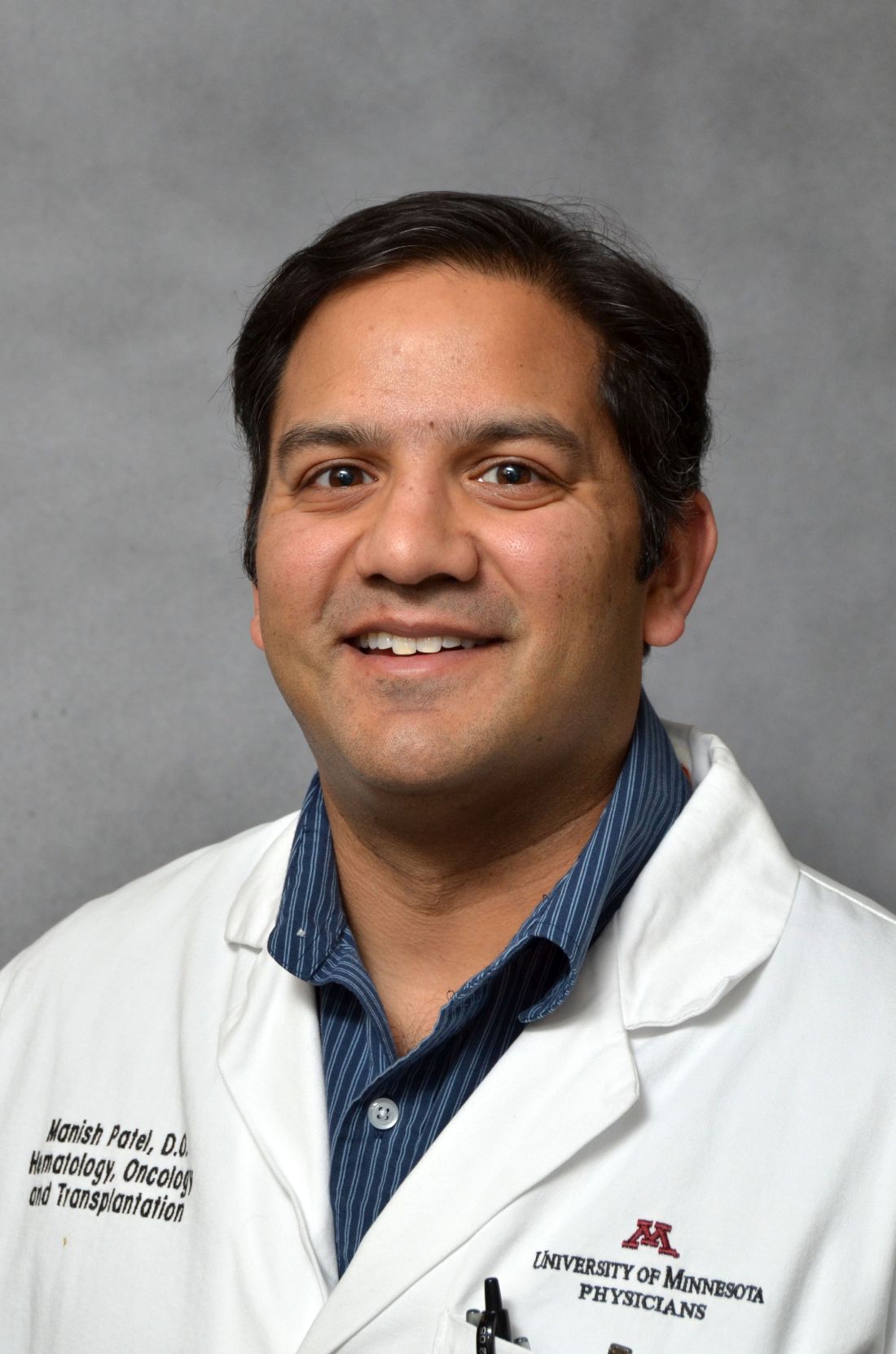
Of 29 evaluable patients who progressed on prior checkpoint blockade, 12 had “some degree of tumor regression” and 5 achieved a confirmed partial response, Manish Patel, DO, reported at the annual meeting of the Society for Immunotherapy of Cancer.
“Some of these responses were quite durable. The longest response ... was a little over 1 year,” said Dr. Patel, of the University of Minnesota Masonic Cancer Center, Minneapolis.
Several patients continue to show objective responses, and the initial estimate of response duration is a median of more than 5 months, he added.
Of note, no differences have been seen to date with respect to clinical benefit in patients who did and did not have prior clinical benefit on checkpoint blockade, Dr. Patel said.
Overall, the combination was very well tolerated. The most common adverse events were fatigue, nausea, and diarrhea, with more than 10% of patients experiencing grade 3 or higher fatigue.
“Otherwise the toxicities were relatively minor,” he said, noting, however, that 8% of patients had cardiac events during the study, including atrial fibrillation, pericardial effusion, and a few cases of pericardial tamponade.
Such effects have been described in prior mocetinostat monotherapy trials, and all patients in the current study underwent pretreatment echocardiograms and did not have evidence of pericardial effusion at the start.
“So I think this is likely to be related to mocetinostat,” Dr. Patel said.
Mocetinostat is a spectrum-selective class I and class IV histone deacetylase inhibitor with multiple potential immunomodulatory features.
For example, the agent induces major histocompatibility complex Class I and Class II expression on tumor cells, enhances the function of T effector cells, and decreases the function of immunosuppressive cell subsets, including regulatory T cells and myeloid derived suppressor cells, Dr. Patel noted.
“It was hypothesized that because of these pleiotropic immune-supportive effects, that the combination of mocetinostat and checkpoint blockade might be a successful strategy for patients with non–small cell lung cancer,” he said.
In phase 1, doses of 50 mg, 70 mg, or 90 mg given three times weekly in combination with 1,500 mg of durvalumab were studied in patients with advanced solid tumors. Based on the safety data from that phase of the study, the recommended phase 2 dose of mocetinostat was 70 mg three times weekly with 1,500 mg of durvalumab on day 1 of each 28-day cycle.
Study subjects were patients with mNSCLC who had received at least one platinum-based doublet and whose most recent treatment prior to enrollment was with a checkpoint inhibitor, or who were immunotherapy naive.
The findings show promising clinical efficacy and safety, and enrollment in the study, which began in June 2016, is currently ongoing in the United States, he said.
Dr. Patel is an advisory board member for Nektar Therapeutics and has received research funding from Merck.
SOURCE: Patel M et al. SITC 2018, Abstract 027.
REPORTING FROM SITC 2018
Key clinical point: Mocetinostat/durvalumab shows clinical activity and manageable side effects in metastatic NSCLC.
Major finding: Five patients achieved a confirmed partial response.
Study details: A phase 2 study including 29 NSCLC patients.
Disclosures: Dr. Patel is an advisory board member for Nektar Therapeutics and has received research funding from Merck.
Source: Patel M et al. SITC 2018, Abstract 027.
Azacitidine-nivolumab combo 'encouraging' in AML
The combination of azacitidine and nivolumab produced “encouraging” results in a phase 2 trial of patients with relapsed or refractory acute myeloid leukemia (AML), according to researchers.
The overall response rate was 33%, and the median overall survival (OS) was 6.3 months. However, the researchers identified factors associated with improved response and survival that could be used to select patients for this treatment.
A quarter of patients on this trial had immune-related adverse events (AEs) that were considered related to treatment, and two patients died of AEs that may have been treatment related.
Naval Daver, MD, of the University of Texas MD Anderson Cancer Center, Houston, and his colleagues reported these results in Cancer Discovery.
The trial included 70 patients with a median age of 70 years. More than half of the patients (56%) had de novo AML, and 44% had secondary AML. The median number of prior therapies was two; 64% of patients had received hypomethylating agents, 47% had received targeted therapies, and 19% had received allogeneic stem cell transplant (SCT).
For this trial, patients received azacitidine at 75 mg/m2 on days 1 to 7 and nivolumab at 3 mg/kg on days 1 and 14 of each cycle. The median number of cycles was three. Patients had a median time on study of 3.5 months and reasons for discontinuation included primary refractory disease, relapse after initial response, proceeding to SCT, patient preference, and death.
The most common treatment-related, nonhematologic AEs were constipation, diarrhea, pneumonitis, nausea, and lung infection. The rate of immune-related AEs was 25% (n = 18), with grade 2-4 immune-related AEs occurring in 16 patients (8 with grade 3-4); 14 responded to steroids and were safely rechallenged with nivolumab, according to the researchers.
Nine patients (13%) discontinued nivolumab (but continued with azacitidine) because of AEs. Two patients died of AEs that were considered possibly related to treatment. One death was caused by progressive pneumonia/pneumonitis, and one was caused by hemophagocytic lymphohistiocytosis.
The overall response rate was 33% (n = 23), with 4 patients achieving a complete response (CR) and 11 achieving a CR with incomplete count recovery (CRi). One patient had a partial response, and seven had hematologic improvement in one or more parameter maintained for more than 6 months. Six patients had stable disease lasting more than 6 months.
The researchers noted that the response rate was higher among patients who had not received prior treatment with hypomethylating agents. Additionally, a higher frequency of pretherapy CD3 and CD8 cells in the bone marrow or peripheral blood appeared to predict response.
“In particular, CD3 appeared to have a high sensitivity and specificity rate for predicting response, indicating it might serve as a reliable biomarker for selecting patients for this combination therapy,” Dr. Daver said in a statement.
At a median follow-up of 21.4 months, 81% of patients (n = 57) had died; 16 died on study treatment and 41 died after discontinuation. The median OS overall was 6.3 months, and the median event-free survival was 4.5 months.
The median OS was 16.1 months in patients with CR/CRi, partial response, hematologic improvement, or stable disease and 4.1 months in nonresponders (P less than .0001). This difference was still significant after the researchers censored the three patients who had gone on to SCT in CR/CRi (P less than .001).
The researchers also found that being in first salvage was associated with improved OS in a univariate analysis and in a comparison with historical controls.
Dr. Daver and his colleagues concluded that azacitidine and nivolumab “produced an encouraging response rate and overall survival” in patients with relapsed/refractory AML.
“We believe that implementation of clinical and immune biomarkers to select patients are likely to yield further improved outcomes with these types of therapies in AML,” Dr. Daver said.
This research was supported by Bristol-Myers Squibb, the University of Texas MD Anderson Cancer Center, and the Dick Clark Immunotherapy Research Fund. Individual researchers also reported financial relationships with Bristol-Myers Squibb.
SOURCE: Daver N et al. Cancer Discov. 2018 Nov 8. doi: 10.1158/2159-8290.CD-18-0774.
FROM CANCER DISCOVERY
Key clinical point:
Major finding: The overall response rate was 33%.
Study details: This phase 2 trial included 70 patients with relapsed/refractory acute myeloid leukemia.
Disclosures: The research was supported by Bristol-Myers Squibb, the University of Texas MD Anderson Cancer Center, and the Dick Clark Immunotherapy Research Fund. Researchers reported financial relationships with Bristol-Myers Squibb.
Source: Daver N et al. Cancer Discov. 2018 Nov 8. doi: 10.1158/2159-8290.CD-18-0774.
AAP advises moderate physical, cognitive activity after sports concussion
according to a new clinical report from the American Academy of Pediatrics.
The update to the 2010 guidelines was needed to reflect the latest research “and it was necessary to provide this new information to guide pediatricians in evaluating and treating concussions they may see in their practice,” Mark Halstead, MD, of Washington University, St. Louis, said in an interview.
The biggest changes to the guidelines involve management of concussion, noted Dr. Halstead, who was a coauthor of the AAP clinical report. “The previous recommendation called for cognitive and physical rest, which unfortunately was interpreted as complete removal from all physical activity and limiting many other things including electronic use.
“Because of research that has been conducted since the original report, it has been shown that starting some light physical activity to increase heart rate, provided it does not worsen symptoms, can be beneficial in recovery. Also, the recommendation for complete removal of electronics and computer use has unfortunately created some issues with kids getting socially isolated,” he added.
“For better or for worse, kids are connected through their electronic devices. Removing them, with no evidence that it worsens the concussion, essentially punishes kids for their injury. We also are trying to discourage prolonged removal of kids from school,” Dr. Halstead emphasized.
The new recommendations emphasize the unique nature of sports-related concussion (SRC) from one individual to another, and the need for individualized management.
Symptoms of SRC fall into five categories, according to the guidelines: somatic, vestibular, oculomotor, cognitive, and emotional/sleep. Pediatric health care providers should rule out more severe head injuries and recognize that concussion symptoms are nonspecific and may reflect preexisting conditions, such as migraine or headache disorders, learning disorders, ADHD, mental health conditions, or sleep disorders.
Use of assessments such as the Sport Concussion Management Tool (SCAT5 for 13 years and older or Child SCAT5 for 5-12 years) can help guide clinicians, but should not be used in isolation to diagnose a concussion, the guideline authors wrote.
Strategies for injury prevention are included in the guidelines as well, such as the use of appropriate headgear. As for management, computerized neurocognitive testing can play a role in decisions regarding return to play, but should not be used in isolation.
“The biggest thing we are lacking is an objective diagnostic test to determine the presence of a concussion or its resolution,” coauthor Kody A. Moffatt, MD, of Creighton University, Omaha, Nebraska, said in an interview.
“Mandatory baseline and postinjury computerized neurocognitive testing is not recommended,” he added.
Clinicians can best manage SRC with prompt recognition and diagnosis using the available tools, followed by relative rest and return to school, then noncontact physical activities, and eventually a return to sport if appropriate.
“Most concussions in children and adolescents will resolve within 4 weeks as long as there is not additional injury to the brain during that time,” Dr. Moffat said.
More research is needed in particular about concussions in elementary and middle school children, Dr. Halstead added.
In the meantime, the take-home message to pediatricians for managing SRC is one of common sense. “Extremes of removing all stimulus from a child is not likely to get them better sooner and research suggests may take them longer to get better,” Dr. Halstead noted. “That doesn’t mean they don’t have to reduce anything, as it is important to reduce physical activity and modify school workload while recovering but we should be avoiding the blanket recommendation to ‘stay home and do nothing until you are better’ approach to concussion management.”
Dr. Halstead and Dr. Moffatt reported no relevant financial conflicts to disclose; the same was true for the other report coauthors. There was no external funding for the report.
SOURCE: Halstead M et al. Pediatrics. 2018 Nov 12. doi: 10.1542/peds.2018-3074.
FROM PEDIATRICS
Update on integrated vascular surgery residencies
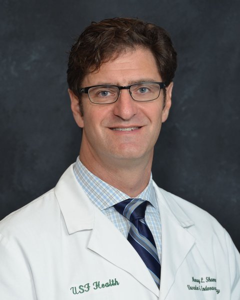
NEW YORK – In March 2005, the Accreditation Council for Graduate Medical Education approved the primary certificate in vascular surgery, and the first integrated vascular surgery residencies (0+5) were approved shortly thereafter. By 2015, there was more than a 900% increase in both the number of programs and positions, which were offered for matriculation in July 2016, according to Murray L. Shames, MD, professor of surgery and radiology at the University of South Florida, Tampa, and chief of the division of vascular surgery at Tampa General Hospital.
In 2009, Dr. Shames and his colleagues first looked at the issue to try to better understand the applicant pool, and they found that there was a 900% increase in demand for the 0+5 residency positions, compared with the traditional 5+2 vascular fellowships, and that there was a 0+5 applicant-to-position ratio of 8:1 that year. “Despite initial concerns regarding the shortened training, studies have demonstrated equivalent case volumes and job opportunities for integrated vascular residents and vascular fellows,” Dr. Shames stated at a symposium on vascular and endovascular issues sponsored by the Cleveland Clinic Foundation.
His current presentation was an update of that assessment done for the 2015 data, as integrated vascular surgery residents have begun to enter the workforce. They looked at the current supply and demand for 0+5 training programs (as well as the traditional 5+2 programs), and the quality and attributes of the 0+5 residency applicants. They obtained data for applicants for both types of programs: 2008-2015 for 0+2 and 2007-2016 for 5+2) and comparable match data were queried for 2008-2016. They looked at the number of programs, the number of positions, the total number of applicants, sex of the applicants, applications/program and applications/position, and the U.S. versus international applicant ratios.
They found that the number of integrated programs increased from 4 to 48, with an increase from 4 to 56 positions, during the study period, said Dr. Shames. Demand for integrated vascular residencies has increased nearly 300%, from 112 applicants in 2008 to 434 in 2015. The total number of U.S. medical school graduate applicants to these programs has increased from 40 in 2008 to 2,030 in 2015, with the increase in program applicants driven primarily by these U.S. medical school graduates; the number of international applicants per program decrease over this time period from 57 to 37. The percentage of women applicants has steadily increased, from 16% to 27%, and currently women constitute 41% of all integrated vascular surgery residents.
“Overall, the supply for integrated vascular surgery residency positions continues to be outnumbered by the number of applicants, with increasing applicant to position ratios at 7.8:1 in 2015, while the total number for vascular surgery fellowships has remained stable at about 1:1,” Dr. Shames concluded.
Dr. Shames reported having no relevant disclosures for his presentation.
REPORTING FROM THE VEITHSYMPOSIUM
Patient-centered primed communication and goals-of-care discussions
Clinical question: Can primed communication increase the frequency of goals-of-care conversations in the outpatient setting?
Background: Effective outpatient communication helps patients and families with serious illnesses with increased quality of life, improved quality of the dying process, and decreased resource utilization at the end of life.
Study design: Cluster-randomized trial.
Setting: Multicenter, primary care, and subspecialty clinics for adults in the Pacific Northwest.
Synopsis: Patients with advanced age and/or severe chronic medical conditions were selected to participate. The median survival for these patients was 2 years. Clinicians were eligible to participate if they had five or more of the selected patients in their panel.
In the intervention group (57 clinicians and 184 patients), all patients were surveyed to identify preferences, barriers, and facilitators for goals-of-care discussions. These data were compiled into “Jumpstart Tips” and distributed to providers before a visit with a study patient. The control group (57 clinicians and 211 patients) had patients complete these surveys but no information was provided to clinicians.
The “Jumpstart Tips” led to an increase in patient-reported (74% vs. 31%; P less than .001) and EHR-documented (62% vs. 17%; P less than .001) occurrences of goals-of-care discussions. Patients also reported increased quality of these conversations (4.6 vs. 2.1; P = .01).
However, no significant improvements were noted in goal-concordant care, depression, or anxiety scores, which may have been because of selection bias and overall difficulty in defining goal-concordant care.
Bottom line: Patient-centered primed communication can increase the frequency and quality of goals-of-care discussions in the outpatient setting.
Citation: Curtis JR et al. Effect of a patient and clinician communication-priming intervention on patient-reported goals-of-care discussions between patients with serious illness and clinicians: A randomized clinical trial. JAMA Intern Med. 2018 Jul 1;178(7):930-40.
Dr. Chowdury is an assistant professor in the division of hospital medicine, University of Colorado, Denver.
What are the barriers to solving the upcoming vascular surgeon shortage?
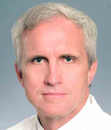
NEW YORK – Increasing the number of 0+5 integrated vascular surgery residency programs would help to alleviate a projected shortage of vascular surgeons, according to William D. Jordan, Jr., MD, professor of surgery, Emory University, Atlanta.*
“Ultimately the question is whether the workforce pipeline is large enough,” Dr. Jordan said at a symposium on vascular and endovascular issues sponsored by the Cleveland Clinic Foundation. “When you consider that there are little more than 600 vascular trainees right now, and almost 600 planned retirements over the next 5 years, the answer to the question is no. Our workforce pipeline is not big enough.”
Dr. Jordan pointed out that, in addition, if one considers the current geographic distribution of vascular surgeons across the country, and go with the new standard that 1.4 surgeons are needed per 100,000 population, there is not a single state in the country that matches up to that goal. “So we are clearly going to have a shortage,” he commented. The only way to fill that shortage is to produce more vascular surgeons. But how does the change to a 0+5 residency program model impact that need?
In a survey conducted by the Association of Program Directors in Vascular Surgery in 2016, regarding challenges as perceived by the trainees, the top two concerns expressed were regarding competing specialties and physician burnout. Statistics bear out the concern regarding competing specialties, for example, there is an increase of 85% in interventional cardiology trainees being produced and a nearly 50% increase in interventional radiology trainees. However, in vascular, it is only 18%. With regard to the goals of those vascular trainees, 90% indicated that they wanted to be attached to some academic or teaching environment. “They don’t want to be the lone wolf out there,” Dr. Jordan said, and this is from concerns regarding workload, mentorship, and camaraderie, as well as regulatory and administrative obligations that are steadily increasing and can be handled more easily in a large institution. This will not fill the need for vascular surgeons in community hospitals, creating a shortage of distribution as well as actual numbers.
One key problem with current training is the fact that the new form of student comes with almost no real surgical skills and there is a dearth of vascular surgery cases available to fully accommodate many of them throughout their training career. This is a problem exacerbated by some residency review committees, which are loathe to give vascular surgery cases to new trainees.
Integrated vascular surgery residency programs have grown and there is a substantially greater interest in them, receiving even more applicants than orthopedics or neurosurgery. U.S. interest exceeds the number of 0+5 positions available. One way to deal with the projected 31% deficit in vascular surgeons by 2025 would thus be to increase the number of these training positions. The financial accommodations to do this would be large, but perhaps the creation of an independent vascular surgery specialty board would facilitate dealing with that issue, he concluded.
Dr. Jordan reported no disclosures relevant to his talk.
Correction, 11/19/18: An earlier version of this article misidentified the speaker in the session. The speaker was William D. Jordan, Jr., MD.
REPORTING FROM THE VEITHSYMPOSIUM
December 2018
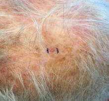
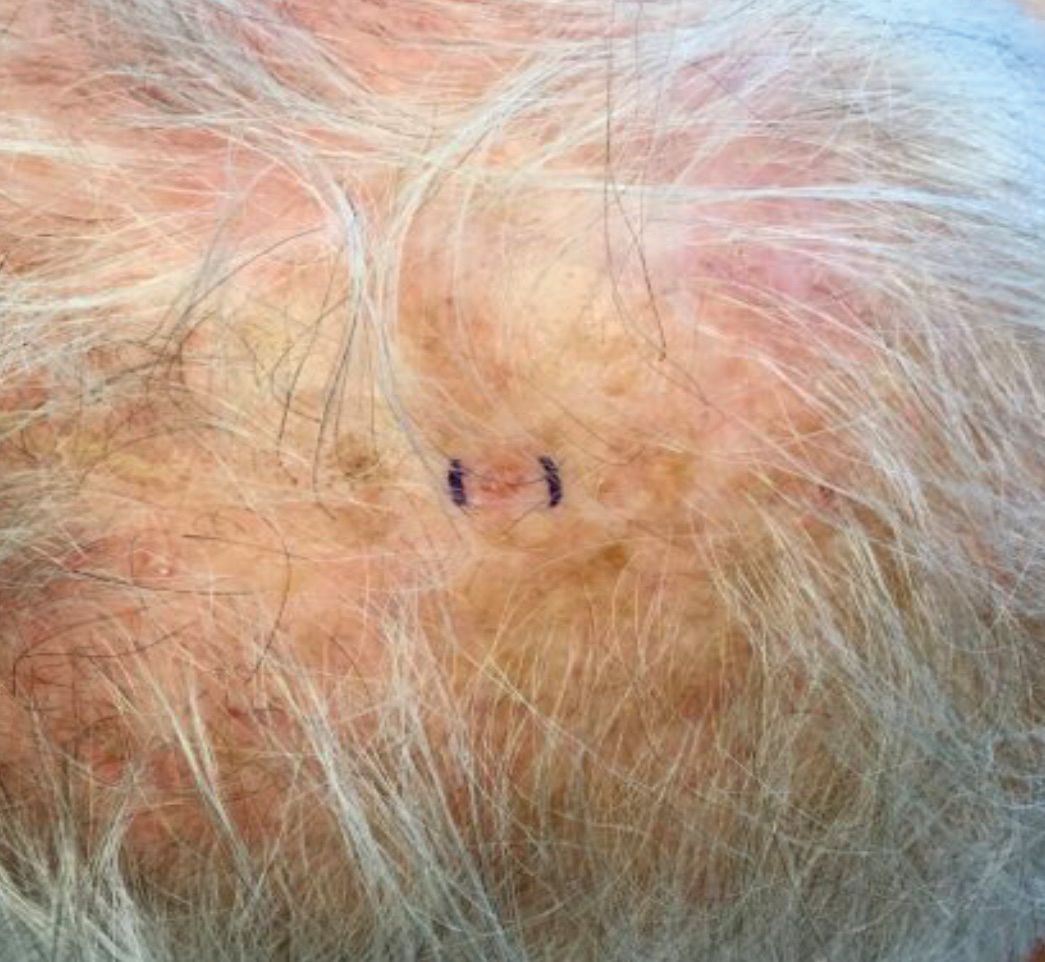
. White individuals over aged 50 years are more frequently affected. Both genders are equally affected, and 25% of cases occur on the covered areas (trunk or extremities) of younger patients. Clinically, lesions present as pink to red plaques or nodules that exhibit rapid growth. Ulceration or crusting may be present. Causes of AFX include ultraviolet radiation and ionizing radiation. AFX is considered a superficial variant of malignant fibrous histiocytoma (MFH), the most common soft tissue sarcoma of adults. Clinically, MFH involves deeper tissues than does AFX, often on the thighs or buttocks. MFH is a more aggressive malignancy that regularly metastasizes.
Histologically, the tumor occurs as a dermal proliferation of “bizarre” spindle cells, epithelioid cells, and atypical histiocytes. Vesicular changes may be present in the nucleus or cytoplasm of the spindle cells. Mitotic figures are present. Multinucleated giant cells may be present. Solar elastosis is often seen, as well. Vimentin and histiocyte stains are positive. Unlike melanoma, S-100 staining is minimal. Unlike squamous cell carcinoma, prekeratin staining is negative. CD34 is negative. AFX resembles MFH histologically.
Surgical excision by the Mohs procedure is preferred over wide excision as there is a risk of recurrence. AFX rarely metastasizes. This is more likely if inadequately excised or the patient is immunosuppressed. Sun protective practices, such as applying and reapplying sunscreen regularly, wearing sun protective clothing, and avoiding excessive UV exposure during peak hours is recommended.
This case and photo were submitted by Dr. Bilu Martin.
Dr. Bilu Martin is a board-certified dermatologist in private practice at Premier Dermatology, MD, in Aventura, Fla. More diagnostic cases are available at edermatologynews.com. To submit a case for possible publication, send an email to dermnews@mdedge.com.
A bovine arch predicts worse outcomes with type B aortic dissections
NEW YORK – The presence of a bovine arch predicts higher mortality in patients with a type B aortic dissection (TBAD), according to a study presented by Jan S. Brunkwall, MD, at a symposium on vascular and endovascular issues sponsored by the Cleveland Clinic Foundation.
The bovine arch is a congenital interruption in the evolution of the arch, and is a misnomer because it does not actually reflect the arch branching pattern found in cattle. It represents the most common variation of the aortic arch, with a prevalence of 1%-41%, depending on the literature, according to a study published by Dr. Brunkwall, chairman of the department of vascular and endovascular surgery at the University of Cologne (Germany), and his colleagues (Eur J Vasc Endovasc Surg. 2018; 55:385-391).
In order to assess the effect of the bovine arch on survival, Dr. Brunkwall and his colleagues performed a retrospective cohort analysis of patients with TBAD admitted at two centers. CT angiograms (CTAs) of patients referred because of aortic dissection were also reevaluated with regard to the presence of a bovine arch.
A total of 154 patients with TBAD and 168 with type A aortic dissection were assessed, and 110 oncologic patients who had undergone a chest CTA for disease staging during the study period acted as a control group.
There was an overall prevalence of 17.6% for bovine arch variants, with no statistical difference in prevalence between patients with a dissection and those in the control group, or between patients with a type A or type B dissection. However, mortality was 34.5% in patients with TBAD who had a bovine arch versus 16% in patients without a bovine arch. This was a significant difference (P =.04), according to Dr. Brunkwall.
Multivariate analysis showed that the presence of a bovine arch with TBAD was an independent predictor of mortality. “The reason for the high mortality cannot be explained by our data,” said Dr. Brunkwall, “but there has been a suggestion that the shear stress is different and higher in patients with a bovine arch leading to a stiffer aorta and more endothelial damage.”
Dr. Brunkwall reported that he had no disclosures.
REPORTING FROM VEITHSYMPOSIUM
Most perpetrators in school shootings brought guns from home
SEATTLE – A new analysis of all school shootings, including those with four or fewer victims, reinforces the need for prevention in the home by preventing guns from falling into unauthorized use.
The researchers examined 223 shootings in the United States that occurred during 2005-2012 and found that in more than a third of the 60 cases for which information was available, the perpetrator obtained the gun from the home and was aged 17 years or younger. Furthermore, evidence of mental illness in the shooter was rare.
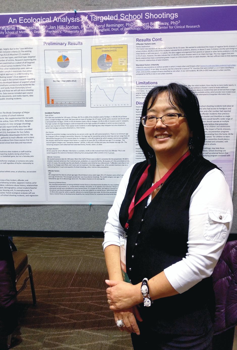
The finding complements studies of “mass shootings,” which tend to garner headlines and more research attention, according to Ayame Takahashi, MD, who presented the findings at a poster session at the annual meeting of the American Academy of Child and Adolescent Psychiatry. “What we see in the news are the big rampage shootings, with multiple victims, where the target may be anyone they can get. You hear in little bits and pieces about the single-shooter incidents, which are way more common.
“We wanted to look at as many of these cases as we could find to look at the overall variables that might be behind these smaller school shootings, which are way more common,” said Dr. Takahashi, who is an assistant professor of psychiatry at Southern Illinois University Medicine, Springfield.
The researchers identified shootings using the data from the Brady Campaign to Prevent Gun Violence during 1997-2012, and supplemented it with listings found in “The Bully Society: School shootings and the crisis of bullying in America’s schools” (New York: NYU Press, 2013). Other sources included a study on rampage shootings in U.S. high school and college settings during 2002-2008; the Virginia Tech Review Panel report, a 2014 FBI report, the Everytown for Gun Safety website, and the list of school shootings on Wikipedia.
The analysis included incidents that had occurred on a school property, including school buses, at a time when students or staff would have been at risk. The offender could have been a current or former student or employee, or anyone who came onto the school property with the intent to harm students or staff.
The sample included 223 shootings. In 60 cases, the researchers found information about how guns were obtained, and in 37% of those cases, the source was the offender’s home and the offender was 17 years old or younger. In 30% of the cases, the shooter owned the gun, but in almost all these cases, the shooter was aged 19 years or older.
Sixty-one cases had information available about the presence or absence of mental illness in the offender. In 20% of these cases, the shooter was determined to have a mental illness or, rarely, a developmental disability.
The results suggest that mental illness might be rare among school shooters, and therefore, call into question efforts to limit gun ownership among people with mental illness. “It may be barking up the wrong tree,” Dr. Takahashi said.
Instead, she advocates more messaging of gun safety in the home, to prevent unauthorized use. Psychiatrists can help by discussing these issues with patients, but she also called for more community involvement and education about the issue.
“The people who come to you, they’re kind of in your court already. so that the message gets out there beyond the folks that we see in our offices,” she said.
Dr. Takahashi has no relevant disclosures.
REPORTING FROM AACAP 2018
Key clinical point: Psychiatrists can help by discussing gun safety with patients.
Major finding: About 20% of school shooters had a confirmed mental illness or developmental disability.
Study details: Analysis of 223 school shootings in the United States between 2005 and 2012.
Disclosures: Dr. Takahashi had no relevant disclosures.
Scleroderma SCOT trial findings hold similar in lung disease
CHICAGO – Changes in quantitative lung CT scores for scleroderma-related interstitial lung disease independently validate the superiority of hematopoietic stem cell transplantation versus cyclophosphamide for severe systemic sclerosis, according to findings in a subset of patients from the SCOT (Scleroderma: Cyclophosphamide or Transplantation) trial.
The recently published findings from the SCOT trial showed that myeloablation followed by autologous hematopoietic stem cell transplant (HSCT) significantly improved event-free and overall survival of systemic sclerosis patients at 54 months, compared with 12 monthly treatments with intravenous cyclophosphamide (N Engl J Med. 2018;378:35-47).
In a subset of 75 patients from the SCOT trial, the investigators analyzed changes in lung parenchymal abnormalities on high-resolution CT scans between baseline and serial follow-up exams performed yearly for up to 5 years. Follow-up scans at 14, 26, 48, and 54 months in available patients at each time point showed that whole-lung quantitative interstitial lung disease (QILD) scores – a validated measure that combines various CT texture-based characteristics to determine disease extent – decreased significantly by 7% at 54 months in patients who underwent HSCT, compared with no change in those who received cyclophosphamide (CYC; P = .024), Keith M. Sullivan, MD, reported at the annual meeting of the American College of Rheumatology.
Additionally, whole-lung quantitative lung fibrosis (QLF) scores were stable (–1%) in the HSCT patients, but increased 3% in the CYC patients (P = .047), said Dr. Sullivan, a professor of medicine at Duke University, Durham, N.C.
Dr. Sullivan was the first author on the SCOT trial, and he reported the current study results on behalf of lead investigator Jonathan Goldin, MD, PhD, of the department of radiologic sciences at the University of California, Los Angeles.
“These are really kind of meaningful associations, especially since the worst of the [CYC] treatment group didn’t make it to month 54,” Dr. Sullivan said.
Quantitative scores of scleroderma-related interstitial lung disease were measured using computer-based quantitative image analysis of standardized, noncontrast, volumetric, thin-section, thoracic, high-resolution CT. The same CT machine was used for all time points (except for one subject) with careful attention to breath hold reproducibility and image quality. Baseline characteristics were not different between the HSCT and CYC groups, he noted, stressing the rigorous study design.
CT assessments were also compared for the most severe lobe in each patient and showed similar findings, with both QILD and QLF scores for that lobe improving in the HSCT patients relative to the CYC patients (P = .004 and P = .002, respectively), Dr. Sullivan said, adding that the direction of change in structural measures of QILD and QLF for both whole lung and most severe lobe CTs tracked with physiological pulmonary function tests, including forced vital capacity (FVC), forced expiratory volume in 1 second, and diffusing capacity of the lungs for carbon monoxide.
“The FVC improved while QILD decreased, and that’s what you would expect to see,” he said. “So for each of these ways of displaying data, there was an expected and sensible inverse correlation.”
Scleroderma-related interstitial lung disease is a major cause of morbidity and mortality in severe systemic sclerosis. In the wake of the SCOT trial findings, questions remained with respect to correlation between those findings and pulmonary function; if the improvements with HSCT are real and meaningful, they should have meaningful correlation with pulmonary function, and these findings demonstrate those correlates, he said.
“Changes in quantitative lung CT scoring of scleroderma lung disease provide an objective radiologic validation of the long-term benefits of transplant compared to cyclophosphamide in individuals with severe scleroderma and lung involvement. Improvement in imaging after transplant continues for up to 54 months after randomization, giving radiologic confirmation of a durable treatment benefit,” Dr. Sullivan concluded.
The investigators reported having no relevant disclosures.
SOURCE: Goldin J et al. Arthritis Rheumatol. 2018;70(Suppl 10): Abstract 901.
REPORTING FROM THE ACR ANNUAL MEETING
Key clinical point: Lung CT scores remain stable or improve following hematopoietic stem cell transplantation in patients with scleroderma-related interstitial lung disease when compared against monthly cyclophosphamide treatments.
Major finding: Quantitative interstitial lung disease scores decreased by 7% at 54 months in hematopoietic stem cell transplant patients versus no change in those who received cyclophosphamide (P = .024).
Study details: A study of 75 patients from the SCOT trial.
Disclosures: The investigators reported having no relevant disclosures.
Source: Goldin J et al. Arthritis Rheumatol. 2018;70(Suppl 10): Abstract 901.
