User login
Impulsivity, screen time, and sleep
If you are still struggling to understand the ADHD phenomenon and its meteoric rise to prominence over the last 3 or 4 decades, a study published in the September 2019 Pediatrics may help you make sense of why you are spending a large part of your professional day counseling parents and treating children whose lives are disrupted by their impulsivity, distractibility, and inattentiveness (“24-hour movement behaviors and impulsivity.” doi: 10.1542/peds.2019-0187). Researchers at the Children’s Hospital of Eastern Ontario (Canada) Research Institute used data collected over 10 years in 21 sites across the United States on more than 4,500 children aged 8-11 years, looking for possible associations between impulsivity and three factors – sleep duration, screen time, and physical activity.
They found that children who were exposed to fewer than 2 hours of recreational screen time each day and slept 9-11 hours nightly had significantly reduced scores on a range of impulsivity scores. While participating in at least 60 minutes of vigorous physical activity per day also was associated with less impulsivity, the effect added little to the benefit of the sleep/screen time combination. Although these nonpharmacologic strategies aimed at decreasing impulsivity may not be a cure-all for every child with symptoms that suggest ADHD, the data are compelling.
I hope that the associations these Canadian researchers have unearthed is not news to you. But their observation that 30% of the sample population met none of the recommendations for sleep, screen time, and activity and that only 5% of the sample did suggests that too few of us are delivering the message with sufficient enthusiasm and/or too many parents aren’t taking it seriously.
Over the last several years I have been encouraged to find sleep and screen time limits mentioned in articles on ADHD for both professionals and parents, but these potent contributors to impulsivity and distractibility always seem to be relegated to the oh-by-the-way category at the end of the article after a lengthy discussion of the relative values of medication and cognitive-behavioral therapy. And unfortunately, meeting these behavioral guidelines can be difficult to achieve and cannot be subcontracted out to a therapist or a pharmacist. They require parents to set and enforce limits. Saying no is difficult for all of us, particularly those without much prior experience.
How robustly have you bought into the idea that more sleep and less screen time are, if not THE answers, at least are the two we should start with? Where do your recommendations about screen time, sleep, and physical activity fit into the script when you are talking with parents about their child’s ADHD-ish behaviors? Have you put them in the oh-by-the-way category?
Do you ever say, “I know you may be expecting me to talk about medication at this visit, but I suggest you try setting and enforcing these limits on sleep and screen time for a few months and we will see how things are going”? “And I am going to give you some suggestions on how you can do this, and we will meet again as often as you feel is necessary to ease the process.”
Do you think you have the time to try this approach? Do you feel you have the skills to counsel on sleep and behavior? Do you think you can find someone with the time and experience who shares your priorities about screen time and sleep to do the parental coaching for you? It’s an approach worth considering when you step back and take the longer look at why we are living through this decades-long ADHD phenomenon.
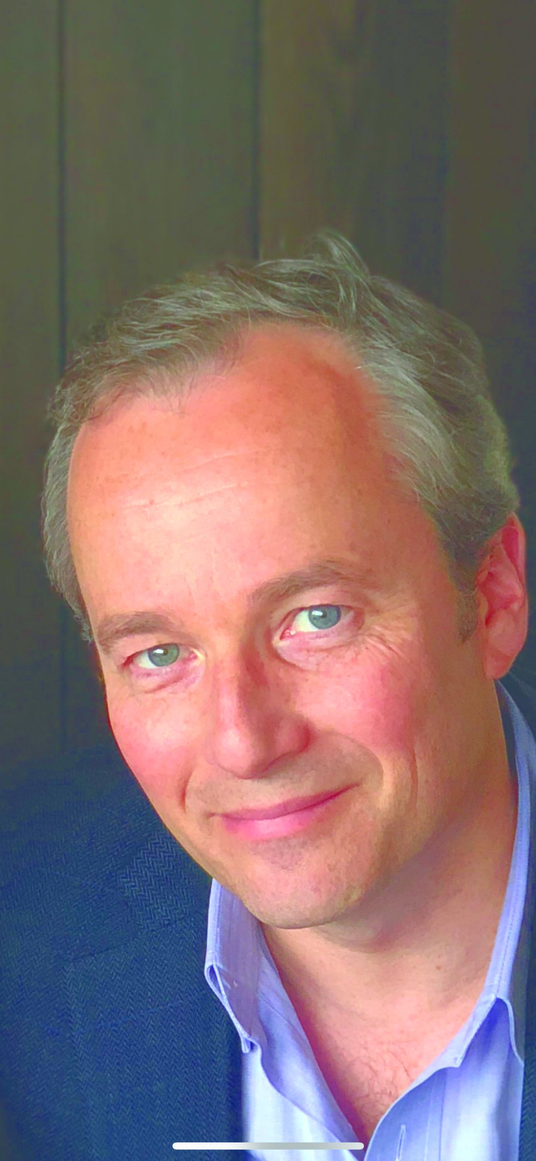
Dr. Wilkoff practiced primary care pediatrics in Brunswick, Maine, for nearly 40 years. He has authored several books on behavioral pediatrics, including “How to Say No to Your Toddler.” Email him at pdnews@mdedge.com.
PACIFIC: Patterns of lung cancer progression suggest role for local ablative therapy
Most patients with stage III non–small cell lung cancer (NSCLC) who have distant progression on standard therapy typically have one or two new lesions, often in the same organ, which suggests a role for local ablative therapy, according to investigators.
This conclusion was drawn from an exploratory analysis of the phase 3 PACIFIC trial, which previously showed that durvalumab prolonged survival among patients with NSCLC who did not progress after chemoradiotherapy, which turned the trial protocol into a new standard of care.
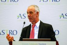
At the annual meeting of the American Society for Radiation Oncology, coauthor Andreas Rimner, MD, of the Memorial Sloan Kettering Cancer Center in New York presented findings.
“There were always questions regarding detailed patterns of failure and disease progression in [the PACIFIC] trial,” Dr. Rimner said. “This study ... focuses on these patterns of failure, including the type of first progression in the patients on the PACIFIC trial.”
During the trial, 713 patients with NSCLC were randomized in a 2:1 ratio to receive either durvalumab or placebo. After a median follow-up of 25.2 months, the superiority of durvalumab was clear, with a lower rate of progression (45.4% vs. 64.6%).
But the present analysis dug deeper into this finding by dividing patients into three groups based on site or sites of first progression: local (intrathoracic) progression only, distant (extrathoracic) progression only, or simultaneously local and distant progression. Scans were reviewed by an independent radiologist who was not involved in the original PACIFIC trial. In addition to spatial data, the investigators reported times until progression.
Regardless of site, durvalumab was associated with a longer time until progression or death. Although comparative values were not reached for distant or simultaneous spread, median time until local progression or death was reportable, at 25.2 months in the durvalumab group versus with 9.2 months in the placebo group.
These values were available, in part, because local spread was the most common type of progression: It occurred in 80.6% of patients who progressed on durvalumab and 74.5% of progressors in the placebo group.
Durvalumab reduced the rate of progression across the three spatial categories, compared with placebo, including local only (36.6% vs. 48.1%, respectively), distant only (6.9% vs. 13.1%), and simultaneously local and distant (1.9% vs. 3.4%). This means that, at first progression, new distant lesions were found in 8.8% of patients treated with durvalumab, compared with 16.5% of those treated with placebo. Of note, approximately two-thirds of patients with distant progression had only one or two distant lesions, often confined to one organ, most commonly the brain. This pattern of progression was observed in both treatment arms.
According to Dr. Rimner, this finding is clinically relevant because it suggests a potential role for local ablative therapy.
Expert perspective on the analysis was provided by Benjamin Movsas, MD, chair of radiation oncology at the Henry Ford Cancer Institute in Detroit.
“The PACIFIC trial has really transformed the standard of care for patients with locally advanced, inoperable non–small cell lung cancer by adding immunotherapy to the prior standard of care combining chemotherapy and radiation, and this has shown a dramatic improvement in survival,” Dr. Movsas said.
“By adding the immunotherapy durvalumab, you can reduce risk of local failure, you can reduce the risk of distant failure, and interestingly enough, when patients do fail distantly, and this is true in both arms, they tended to fail in only one or two spots, which is encouraging because that suggests maybe a window of opportunity to treat those one or two spots, and we have newer technologies that allow us to consider that. So we really have a new paradigm.”
The study was funded by AstraZeneca. The investigators disclosed additional relationships with Merck, Nanobiotix, Boehringer Ingelheim, and others.
SOURCE: Rimner A et al. ASTRO 2019, Abstract LBA6.
REPORTING FROM ASTRO 2019
Key clinical point: Most patients with stage 3 non–small cell lung cancer (NSCLC) who have distant progression on standard therapy typically have one or two new lesions, often in the same organ, which suggests a role for local ablative therapy.
Major finding: Approximately two-thirds of patients with distant progression had one or two new lesions.
Study details: An exploratory analysis of patterns of progression in the phase 3 PACIFIC trial, which involved 713 patients with stage III NSCLC that had not progressed after chemoradiotherapy.
Disclosures: The study was funded by AstraZeneca. The investigators disclosed additional relationships with Merck, Nanobiotix, Boehringer Ingelheim, and others.
Source: Rimner A et al. ASTRO 2019, Abstract LBA6.
SABR may put immunological brakes on oligometastatic prostate cancer
Stereotactic ablative radiation therapy (SABR) may be able to extend progression-free survival (PFS) among patients with oligometastatic prostate cancer, based on results from the phase 2 ORIOLE trial.
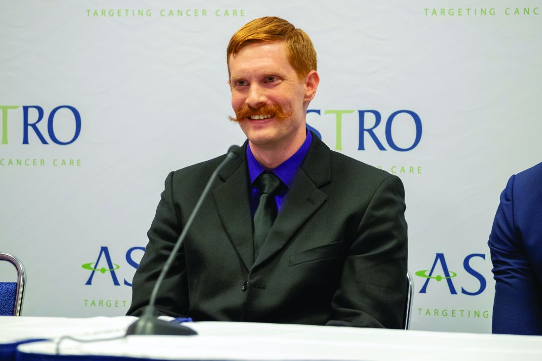
SABR appeared to control disease, in part, by triggering a systemic immune response, reported lead author Ryan Phillips, MD, PhD, chief resident of radiation oncology at Johns Hopkins Sidney Kimmel Cancer Center in Baltimore, at the annual meeting of the American Society for Radiation Oncology. This is a particularly noteworthy finding, since prostate cancer is generally considered to be immunologically cold, Dr. Phillips explained.
The ORIOLE trial also demonstrated how prostate-specific membrane antigen (PSMA)–based PET/CT scans can be used to more accurately predict metastasis by detecting lesions that would otherwise be missed by standard imaging.
“There’s a hypothesis that these first few sites of spread pave the way for additional widespread metastasis down the road,” Dr. Phillips said, “and that if we can treat all detectable disease early enough, we may be able to provide long-term control or in the best-case scenario, be able to cure these patients of early metastatic disease.”
The trial involved 54 men with recurrent, hormone-sensitive prostate cancer who had been previously treated with radiation therapy or surgery. Additional eligibility requirements included one to three lesions that were 5 cm or smaller, detectable by MRI, CT, or bone scan; a prostate-specific antigen (PSA) doubling time of less than 15 months; and an Eastern Cooperative Oncology Group performance status of 2 or less.
Patients were randomized in a 2:1 ratio to receive either SABR or observation, with follow-up every 3 months including physical exam and PSA measurement, and at 6 months, CT and bone scan. The primary endpoint was disease progression at 6 months.
In addition to this protocol, biomarker correlative analyses were performed, including PSMA-based PET/CT, T-cell clonality testing, and circulating tumor DNA (ctDNA) assessment.
Results showed that significantly fewer men treated with SABR had disease progression at 6 months (19% vs. 61%; P = .005). This benefit extended beyond the primary endpoint, as median PFS among patients treated with SABR was not yet reached after more than a year of follow-up, while those in the observation group had a median PFS of 5.8 months (P = .0023).
“Clinically, this is promising,” Dr. Phillips said, “but we also were really interested in learning more about oligometastatic prostate cancer, and something that sets ORIOLE apart are the correlative studies.”
The first of these studies involved PSMA-based PET/CT, which can identify lesions that may be missed or underappreciated with conventional imaging. Within the SABR group, 35 of 36 men had PSMA-based PET/CT performed at baseline and 6 months later. Of these, 19 had all PSMA-based PET/CT–detectable lesions treated with SABR (total consolidation), while 16 had subtotal consolidation. This difference was predictive of outcome, as only 16% of patients with total consolidation developed new lesions 6 months later, compared with 63% of those who had subtotal consolidation (P = .006).
“Not only are we treating the disease we’re detecting, but it seems to be preventing the development of new metastases outside the areas that we treated,” Dr. Phillips said. “This also held when we looked at points beyond 6 months,” he added.
Further testing showed that SABR triggered a systemic adaptive immune response involving expansion of T-cell clones. “There’s a lot more activity and changes within the immune system that were only seen within the SABR arm,” Dr. Phillips said, noting that these changes were “similar in scope to what we see after a vaccination.”
Finally, the investigators assessed ctDNA, which showed that men with at least one high-risk mutation had similar outcomes regardless of treatment group; in contrast, those without any high-risk mutations had significantly better PFS when treated with SABR.
“These are low sample size, hypothesis-generating experiments,” Dr. Phillips said, “but it is promising that there may be measurable baseline factors that will help us decide which patients are likely to benefit from this approach, and which would really be better served with an alternate treatment strategy.”
Session moderator Anthony Zietman, MD, of Massachusetts General Hospital, Boston, suggested that the ORIOLE trial could have a big future. “This is a small study, but it’s a prospective study, and the findings are really provocative,” Dr. Zietman said. “Just imagine, if there was a patient with just two or three metastatic lesions, and by irradiating those, you could liberate proteins from the destroyed metastases, generate an immune response, and suppress the development of new metastases, that will be something extraordinary. So this trial will be followed very closely as it seems to be hinting at something that’s a bit of a Holy Grail in oncology.”
The investigators disclosed relationships with RefleXion Medical, Pfizer, Genentech, and others.
SOURCE: Phillips R et al. ASTRO 2019. Abstract LBA3.
FROM ASTRO 2019
Beyond the lips: Guidance for intraoral procedures
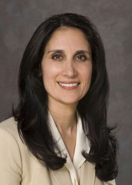
NEW YORK –The work of the dermatologist doesn’t need to stop at the lips, according to Nasim Fazel, MD, DDS, a board-certified dermatologist and dentist. she said, speaking at a session focused on oral health and procedures at the American Academy of Dermatology summer meeting.
Whether to perform an incisional or excisional biopsy on the lips or within the oral cavity is a decision driven by the clinical scenario, said Dr. Fazel, professor of clinical dermatology and director of the oral mucosal disease clinic at the University of California, Davis. Variables to consider, she said, include the size of the lesion, as well as whether the lesion is symptomatic or there’s any functional impairment. Other factors to bear in mind are whether the patient has any comorbid inflammatory conditions and whether the lesion could be malignant.
An excisional biopsy is a good procedure for small lesions that are thought to be benign, especially if they are exophytic, she noted. An example would be a traumatic fibroma, she said. This technique is also appropriate if there’s concern for dysplasia or malignancy if an office excisional biopsy is feasible given lesion size and location.
On the labial mucosa, an elliptical incision is often a good choice.
Cutaneous lip procedures
On the cutaneous lip, a punch incision can be feasible. The vermilion border should be marked to maintain orientation. “Avoid transecting the vermilion border to the extent that it’s possible,” said Dr. Fazel. Keep the vascular anatomy in mind as well, she added, since there’s a risk of severing the superior or inferior labial artery with procedures in this area. If this should happen, the branch can be identified and ligated with 4-0 fast gut or chromic gut suture material, she advised.
Another option on the cutaneous lip is shave removal. Here, a chalazion clamp can be used for both exposure and hemostasis. “Always suture parallel to the lip lines, and caution the patient that there may be significant, noticeable blanching when lip anesthesia’s used,” she said.
Gingival and tongue procedures
Moving to territory that may be less familiar for some dermatologists, Dr. Fazel walked through the process of a gingival punch biopsy. “Use local anesthetic, but a small quantity is sufficient,” she said. Using gentle pressure, the operator can work the punch instrument down to periosteal bone. At that point, just scissors can be used for undermining the specimen, with minimal disturbance of the mucosal surface.
Hemostasis after a gingival punch can be accomplished with silver nitrate or aluminum chloride or by electrocautery. A permanent defect in the gingiva can occur even with good technique, she said – a fact that should be included in the informed consent document for the procedure.
For lesions on the tongue, a shave removal can be considered, as can an incisional punch biopsy. An assistant’s hands are invaluable for stabilizing the tongue, said Dr. Fazel, illustrating that small dry gauze squares help achieve a good grip.
Considerations in biopsy technique
Dr. Fazel offered some tips and considerations when a punch biopsy is being done in the context of a chronic oral inflammatory condition, and the plan is to submit for hematoxylin and eosin (H&E) staining and direct immunofluorescence (DIF). Conditions where an intraoral punch biopsy would be considered include bullous or mucous membrane pemphigoid, pemphigus, lichen planus, and systemic lupus erythematosus, she said.
“Select a site with the most significant inflammatory changes” and aim for the most anterior site that exhibits these characteristics to maximize exposure and ensure a good specimen. The labial mucosa is a better choice in general than buccal mucosa, she said. “Maxillary and mandibular sulci are tricky sites,” especially when perilesional and lesional tissue should be included in the biopsies.
Next, Dr. Fazel walked attendees through general principles of preparing for and performing punch biopsies for H&E and DIF. In planning the biopsies, the DIF specimen should ideally be collected before the H&E specimen because the former technique will have higher yield when the specimen is taken from a less bloody field. “The yield of DIF is higher from the gingiva than from nonkeratinized surfaces.”
When infiltrating the biopsy site with local anesthesia, the needle should be centered within the lesion or general area to be biopsied, and care should be taken to maintain the orientation of the lesion to the surrounding tissue. Anesthesia can be infiltrated within the submucosal plane; the resultant ballooning will elevate the tissue to be biopsied and ease the procedure, she said.
Choose a 3-mm punch tool for gingival biopsies; 4 mm is a good size for other nonkeratinized surfaces. Unlike the procedure used for cutaneous biopsies, on delicate oral tissues, “you don’t need to hub your punch tool,” Dr. Fazel said. Similarly, the tool can be driven with firm rotation in one direction, rather than ratcheting back and forth, which may fray and deform the biopsy margins, she added. The specimen can be freed from surrounding mucosa with scissors alone and a gentle snipping motion; everting the tissue will help achieve the desired clean and gentle technique.
Because of the delicacy of the tissue, “it’s important to minimize the use of forceps, as well as any unnecessary manipulation of tissue in the process of specimen collection,” she said.
Salivary gland biopsies and intraoral suturing
Even minor salivary glands can be biopsied in a fairly straightforward way, though there’s a risk of short- and long-term loss of sensation, as well as more scarring than in other routine intraoral biopsies. Though these salivary glands lie just beneath the oral mucosa, care must be taken to avoid laceration of the orbicularis oris muscle during excision, noted Dr. Fazel. With a minor salivary gland biopsy, as with some other intraoral procedures, sutures will be needed. Consideration for the friability of the oral mucosa should drive suture material choice and closure technique.
First, “take bigger bites on each side of the incision, to minimize the risk of the suture tearing through the mucosa,” she said. Avoid forceps while suturing if possible, but be gentle if they are employed, “and be gentle tying knots: retract the mucosa as little as possible and cinch the knots down tightly with your fingers.”
Electrocautery, silver nitrate, and aluminum chloride are all reasonable options for hemostasis, although patients should be alerted that the tissue will have a charred appearance if electrocautery is used. Primary closure may be all that’s needed for hemostasis, said Dr. Fazel.
If nonabsorbable suture materials are used, nylon and prolene should be avoided since they tend to tear through the oral mucosa. Soft or braided silk are good choices, she said, and sutures can come out in 5-7 days.
Absorbable sutures have the advantage of not requiring removal, but they can be more irritating to the surrounding mucosa. Chromic or fast gut are the choices here, said Dr. Fazel. Whether absorbable or nonabsorbable sutures are placed, the size should be 4-0 or 5-0, she said.
Postoperative care, complications, and the limits of the dermatologist
Postoperatively, patients should know that swelling is to be expected, especially with procedures like minor salivary gland biopsies that involve the lip. Icing 15 minutes per hour for the first few hours will help with swelling; wound care is optimized with gentle salt water rinses. A bland diet that avoids acidic, spicy, and excessively hot foods and beverages will minimize wound irritation. Foods with sharp edges like chips, crackers, and nuts can actually catch sutures and cause pain and bleeding, so these too should be avoided.
As with dental work, care should be taken with eating or drinking until anesthesia has worn off, which may take up to 3 hours. Patients should also be cautioned that sutures are likely to come out prematurely just because of the mobility of the structures of the mouth with normal activities such as eating and talking.
Should a wound infection occur, said Dr. Fazel, it’s likely that mixed aerobic and anaerobic bacteria are to blame; accordingly, broad-spectrum beta-lactam antibiotics can be a good first-line course. More severe infections are more likely to have an anaerobic or gram-negative etiology; metronidazole is a reasonable choice for anaerobic coverage, she noted. The life-threatening complication not to miss is cellulitis of the floor of the mouth, or Ludwig angina. The swelling results in superior and posterior displacement of the tongue, obstructing the upper airway, so any patient suspected of having Ludwig angina needs emergent evaluation and treatment.
When should a dermatologist consider referral to an otolaryngologist rather than diving into a biopsy in the dermatology clinic? If the area of concern is on the posterior third of the tongue, access without special tools or higher levels of anesthesia becomes tricky, Dr. Fazel pointed out. The posterior hard palate, the soft palate, and the floor of the mouth are also regions best left to otolaryngologists, she said.
EXPERT ANALYSIS SUMMER AAD 2019
Happiness in my solo practice
I don’t want to rule the world.
Some doctors do, albeit in a non–Attila-the-Hun sort of way. They want to have offices on every street corner, in every suburb of a given city, sometimes more than one city. Like the Starbucks of medicine.
That’s not me. I’m happy in my little one-office world.
Maybe I just don’t have the ambition, or the business mindset, or whatever it takes to want to do that. I understand it’s all part of wanting to be successful, and obviously those doctors are more driven in that direction than I am. The more offices, the more patients can be seen, and the more money you make.
It’s not quite that simple, though. No one can be in more than one place at the same time, so to see more patients at more places you need more doctors. To pay more doctors requires more money, which in turn requires more patients.
There’s nothing wrong with taking over the world (or at least a suburb) if you like that sort of thing. But to me, more money brings more headaches. More offices to rent, more staff to hire, more people to handle billing, IT, HR, payroll, accounting, contracts, and so on.
You can have it. I’ve taken over all the world I want, in my case a 1,200-square-foot suite on the second floor of a small-to-medium-size medical building. To some that may sound unambitious, but to me, it’s perfect.
I know where my Keurig, Sodastream, and office supplies are. Except for my secretary and her cheerfully rambunctious young daughter, I don’t have to worry about sharing stuff here, or if anyone wants a different carpet color, or what’s going on at a satellite office halfway across town.
If other doctors want to try and take over the world, more power to them, but I’m happy with this. Enough is as good as a feast.
Dr. Block has a solo neurology practice in Scottsdale, Ariz.
Unmet Medical Needs in Triptan-Treated Migraine
Unmet medical needs are of concern to patients who experience migraine treated with triptans and there may be an undertreatment with preventive therapies whose benefit is insufficient, a new study found. The study cohort consisted of participants with ≥4 triptan dose units per month, selected from the general population. Patients were stratified into: possible Low-Frequency Episodic Migraine (pLF-EM: 4-9 triptan dose units per month), possible High-Frequency Episodic Migraine (pHF-EM: 10-14 triptan dose units per month), and possible Chronic Migraine (pCM: 14 triptan dose units per month). Researchers found:
- Of 10,270,683 adults, 8 per 1000 were triptan users and of these, 38.2% were migraineurs with unmet medical needs.
- 72.3% of patients were affected by pLF-EM, 17.4% by pHF-EM, and 10.3% by pCM.
- 19.1% of patients used oral preventive drugs.
- Triptan use reduction was found in 22.3% of patients, decreasing with the intensification of need levels.
Piccinni C, Cevoli S, Martini N. A real-world study on unmet medical needs in triptan-treated migraine: prevalence, preventive therapies and triptan use modification from a large Italian population along two years. [Published online ahead of print June 27, 2019]. J Headache Pain. doi: 10.1186/s10194-019-1027-7.
Multiple Myeloma Research Consortium, cancer centers add new leaders
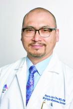
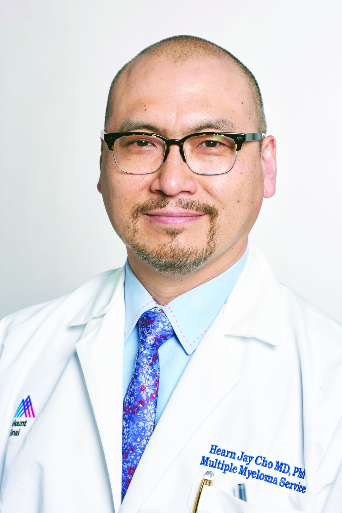
Hearn Jay Cho, MD, PhD, has been appointed chief medical officer of the Multiple Myeloma Research Foundation (MMRF). In this role, Dr. Cho will develop a clinical research strategy and accelerate drug development programs for the MMRF. He will also lead the Multiple Myeloma Research Consortium, a group of 25 research centers focused on multiple myeloma.
Dr. Cho will continue on as an associate professor of medicine at the Icahn School of Medicine at Mt. Sinai in New York, and as an attending physician with the multiple myeloma service at the Mt. Sinai Tisch Cancer Institute. He will also continue to manage his lab at Mt. Sinai.
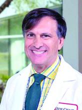
Michael Jay Styler, MD, has joined the department of hematology/oncology as an associate professor in the academic clinician track as part of the bone marrow transplant program at Fox Chase Cancer Center in Philadelphia.
Dr. Styler was previously medical director of the transplant center, apheresis center, and collection center at Hahnemann University Hospital in Philadelphia, as well as medical director of the stem cell transplant program and the clinical service chief of hematology/oncology. Dr. Styler was also an associate professor at Drexel University, Philadelphia, where he served as division director of hematology/oncology and associate fellowship director of the hematology/oncology program.
Jaspreet Chahal, MD, has joined the Oncology Institute of Hope and Innovation, which has locations in Arizona, California, and Nevada. Dr. Chahal will serve patients in Tucson and Green Valley, Ariz.
Dr. Chahal has a doctor of medicine degree from St. George’s University in West Indies, Grenada. She completed her internship in hematology and oncology at Hofstra University, Hempstead, N.Y., and her residency in internal medicine at the University of Arizona in Tucson.
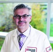
Marcin Chwistek, MD, has been appointed editor in chief of AAHPM Quarterly, which is published by the American Academy of Hospice and Palliative Medicine. Dr. Chwistek is an associate professor in the department of hematology/oncology at Fox Chase Cancer Center in Philadelphia, where he is also director of the pain and palliative care program.
As editor in chief of AAHPM Quarterly, Dr. Chwistek will work with other members of the editorial board to develop content related to hospice care and palliative medicine. Dr. Chwistek previously served as the associate editor in chief of AAHPM Quarterly and writes a column that appears in every issue.
Movers in Medicine highlights career moves and personal achievements by hematologists and oncologists. Did you switch jobs, take on a new role, climb a mountain? Tell us all about it at hematologynews@mdedge.com , and you could be featured in Movers in Medicine.
Don’t let legal considerations drive your decision making about a new job offer
SAN DIEGO – When weighing an offer to join a dermatology practice, don’t allow legal considerations to drive your decision making, Mathew M. Avram, MD, JD, advised at the annual Masters of Aesthetics Symposium.
“Determine your professional interests and follow them accordingly,” said Dr. Avram, director of laser, cosmetics, and dermatologic surgery at Massachusetts General Hospital in Boston. “Your decision as to whether to join a particular practice should not be based upon a contract. Make certain to determine the business end of your deal with any new employer first: the compensation, the work hours, etc. Decide that upfront before you get into the legal details.”
The way he sees it, your relationship with your employer will always be paramount. “This is very important,” said Dr. Avram, who practiced law before he became a dermatologist. “A good contract with a bad employer will not save you from an unhappy employment experience. Few contracts ever end up in litigation, and you cannot sue your way to a happier work experience. Trust your intuition about people; trust your underlying interest in potentially working with this person, and work from there.”
Whether you choose to work in an academic practice, a private practice, or a hybrid, the key issues to address include compensation, benefits, work hours, job responsibilities, and future partnership possibilities. “As an employee, you have the greatest leverage prior to signing an agreement,” he said. “This is the time to ask for what you want.” This may include special requests such as asking the employer to purchase lasers or other special equipment for your office, or to set aside dedicated clinical time for cosmetic procedures, so that you can build a cosmetic practice.
If you’re mulling over a job offer in academics, consider asking for an academic title, what the scope of your authority is, and about your ability to hire or prevent the hiring of others. “Let’s say you’re starting a laser center in your academic center,” said Dr. Avram, who is a past president of the American Society for Laser Medicine and Surgery. “You may not want to find out after you’ve signed your contract that they’ve hired five other people to do the same thing.”
If you’re considering a job offer from a private practice, ask for specifics about bonuses, partnership track, scope of practice, and device purchases. “You want to have that decided before you sign,” he said. “But these are business issues, not legal issues. An agreement needs to be made between you and your employer as to these issues. Once they’ve been agreed upon, it’s time to proceed with a contract. This is where legal advice becomes helpful.” The trick is to negotiate in good faith while maintaining your relationship with your new employer. “Do not destroy your relationship over legal points, but do not cave on crucial issues for fear of upsetting your new employer,” he said.
In law school, Dr. Avram learned that there is no such thing as a standard employment contract. Any contract can be amended, no matter how large or small the institution. “You can amend a mortgage agreement, so you can certainly amend a physician employment contract,” he said. “If the employer is not going to put their commitments in writing, they are probably not going to honor that commitment to you. Written agreements supersede all preceding oral agreements. Each key term needs to be stated explicitly. If not, you have lost all leverage to enforce your initial agreement.”
Key provisions are restrictive covenants and at-will employment. Dr. Avram defined restrictive covenants as contractual agreements that attempt to restrict an employee so as to limit that person’s ability to compete. “This can include noncompete clauses, nonsolicitation agreements, and confidentiality agreements,” he said. “A noncompete agreement prohibits a doctor from competing against their former practice within a specific geographic area for a period of time after the employment has ended. The nonsolicitation agreement restricts the manner and time during which a physician can solicit patients or employees from a practice after termination of employment. A confidentiality agreement is usually indefinite as to time and it restricts the employee from disclosing confidential practice information.”
State laws, he continued, govern restrictive covenants. Some states prohibit them. States that allow them limit their scope to prevent undue burden on the employee’s ability to make a living after termination. “Restrictive covenants in cities need to be narrower than those in rural areas,” he said. “Overly broad restrictive covenants may be unenforceable. Courts can ‘blue line’ covenants to make them more reasonable in scope.”
Next, Dr. Avram discussed involuntary termination in the workplace. Termination with cause means that termination can only result from violation of policy or ethics code violation or significantly poor performance. “In the absence of these circumstances, termination cannot legally proceed,” he said. “This protects the employee from an arbitrary termination.”
On the other hand, at-will employees can be terminated without cause. “It’s a much easier standard to terminate an at-will employee, as long as the reason is not illegal,” he said. “In academic centers, your employment as a physician may require a for-cause termination, whereas your administrative title may be without cause.”
One way to avoid potential legal trouble is to abide by your employer’s rules governing a physician’s interactions with industry. “It is foolish to break those rules,” Dr. Avram said. “Transparency ahead of time is always your best policy. Remember: Your day job is far more valuable to you than part-time work with industry. Also, know that pharma payments are publicly reported by the Centers for Medicare & Medicaid Services.” When it comes to establishing contracts with industry, “read them carefully and have your academic center approve them, if applicable,” he said. “Watch out for noncompete clauses, and some contracts require that you do not speak negatively about your findings. That’s something you should try to avoid.”
Dr. Avram also advised clinicians not to discuss intellectual property with industry representatives. “Do not give away your intellectual property to industry when you’re consulting with them,” he said. “It is easier to do than you might think. If you have a great idea, keep it to yourself or get it protected. This is much easier for an institution where they want you to do these things. They take ownership of a lot of it, but they make it easier to do. Otherwise, you have to go through the expensive process of getting a patent.”
Dr. Avram reported that he has received consulting fees from Allergan, Merz Pharma, Sciton, Soliton, and Zalea. He also reported having ownership and/or shareholder interest in Cytrellis Biosystems, InMode, and Zalea, and intellectual property rights with Cytrellis.
EXPERT ANALYSIS FROM MOAS 2019
Factors Associated with Headache Chronicity in Migraine Patients
Variables such as disability, depression, and lack of anger control are among the key factors associated with headache chronicity in patients who experience migraine, a new study found. The cross-sectional study included a target sample of 250 patients with acute or chronic migraine. All participants filled out questionnaires related to demographic characteristics, pain intensity, disability, depression, emotional intelligence, and anger. Researchers found:
- Patients with chronic migraine experienced higher levels of disability, depression, anger, and had lower levels of emotional intelligence vs patients with acute migraine.
- Variables that had a significant effect on headache chronicity were female gender, married status, lower level of education, headache duration, disability, depression, and anger.
Emadi F, Sharif F, Shaygan M, Sharifi N, Ashjazadeh N. Comparison of pain-related and psychological variables between acute and chronic migraine patients, and factors affecting headache chronicity. Int J Community Based Nurs Midwifery. 2019;7(3):192-200. doi: 10.30476/IJCBNM.2019.44994.
PRAGMA-CF shows disease progression of cystic fibrosis in children
reported Nynke R. Bouma, BSc, and colleagues.
“Even though bronchiectasis is present in 60% to 80% of children with CF in school age, the extent and severity of bronchiectasis in preschool children are generally lower ... however, diffuse airway abnormalities such as airway wall thickening and mucus plugging are observed in many preschool children. It is hypothesized that these preschool airway changes reflect diffuse airway disease that eventually will result in bronchiectasis in school age,” they noted.
The PRAGMA-CF image scoring system can measure airway disease and can also be used to monitor disease progression, noted Ms. Bouma of Sophia Children’s Hospital, Rotterdam, and colleagues. The study was published in Pediatric Pulmonology. PRAGMA-CF is a composite score of airway wall thickening, mucus plugging, and bronchiectasis as percent disease (%disease). “In preschool children, %disease measured by PRAGMA-CF on chest CT allows quantification of early clinically relevant morphological features of CF airway disease and it is associated with later school-age bronchiectasis,” the team wrote. “These findings support the use of %disease as a clinically relevant outcome measure in early CF lung disease.”
The team conducted a prospective cohort study of 61 children (mean age 4 years) with cystic fibrosis, following them for a mean of 5 years. A total of 122 CT scans were available from this group, in addition to spirometry data and cystic fibrosis quality of life scores.
From preschool age to school age, the %disease on PRAGMA-CF increased significantly, from a mean of 0.7% to 1.73%. Scores on another composite measuring tool (%MUPAT, a composite score of airway wall thickening and mucus plugging) went from 0.46 to 0.58 – not a significant difference.
A multivariate analysis corrected for age in each school group and the type of scanner used to acquire the images. That analysis determined that each 1% increase in %disease at preschool age resulted in an increase of 1.18% of bronchiectasis at school age.
A cross-sectional analysis of the group at school age found significant associations between the %disease and percent of forced expiratory volume and the cystic fibrosis quality of life score.
At least one pulmonary exacerbation requiring intravenous antibiotics occurred in 19 of the patients. However, the investigators didn’t find any significant interactions between the %disease in preschool and these exacerbations..
“These findings are in line with previous studies in school‐aged children that showed that mucus plugging is associated with inflammation and airway wall thickening, and that these are thought to be risk factors for later bronchiectasis,” they concluded. “On the basis of our findings, we suggest that %disease and %MUPAT could be used as a clinically relevant outcome measure in clinical studies in preschool patients with cystic fibrosis, as these measures predict later bronchiectasis. Percent disease may be preferred as it captures all the principal features of CF airways disease including bronchiectasis.”
Ms. Bouma had no financial disclosures.
SOURCE: Bouma NR et al. Pediatr Pulmonol. 2019 Sep 9 doi: 10.1002/ppul.24498; Rosenow et al. Am J Respir Crit Care Med. 2015 May 15. doi: 10.1164/rccm.201501-0061OC.
FROM PEDIATRIC PULMONOLOGY


