User login
Atopic dermatitis patients achieved freedom from itch on JAK inhibitor upadacitinib
MILAN – according to a report presented at the World Congress of Dermatology.
Compared with those in the placebo group, more patients receiving the selective Janus kinase 1 (JAK1) inhibitor achieved an itch-free state and maintained it over the 16 weeks of the phase 2b trial, said investigator Kristian Reich, MD, professor of translational research in inflammatory skin diseases at the University Medical Center Hamburg-Eppendorf (Germany).
These improvements in pruritus occurred early with upadacitinib and were pronounced at the highest dose studied, 30 mg daily, he commented. Treatment with upadacitinib also rapidly and significantly improved clinical signs of AD versus placebo, as previously reported primary endpoint data show.
“It’s a drug that works in eczema,” Dr. Reich said in an oral presentation. “We still do not fully understand what the exact relationship between itch and eczema is. Is there a neurogenic inflammation? Is there an epidermal pathology? But clearly with this drug, it does seem to reduce the itch, it does reduce the eczema, it does this early on, and the 30 mg does seem to be the right dose.”
Upadacitinib is a selective inhibitor of JAK1, a member of the signal transduction cascade for many cytokines implicated in AD, including interleukin-4, IL-13, IL-22, and others, Dr. Reich told attendees at the meeting.
In the phase 2b study, 167 patients with moderate to severe AD were randomized to placebo or upadacitinib at 7.5 mg, 15 mg, or 30 mg daily over a 16-week, double-blind period, followed by a 72-week, blinded extension. The mean age across these groups ranged from 39 to 42 years, and the mean time since onset of symptoms was 24-34 years.
Significantly improvements in Eczema Area and Severity Index (EASI) scores were seen as early as 2 weeks and were maintained throughout the 16-week, double-blind period, as previously shown. By 16 weeks, the mean percentage improvement in EASI score was 74.4% for upadacitinib 30 mg daily versus 23.0% for placebo (P less than .001).
In this more recent post hoc analysis of itch, the percentage of patients with a weekly rolling average pruritus Numerical Rating Scale (NRS) score of 0-1 was significantly higher in the upadacitinib groups, Dr. Reich said.
The placebo-adjusted difference in average pruritus NRS scores of 0-1 was highest in the 30-mg daily group, at 37.7% by week 16 (P less than .001).
Those itch scores correlated with the Patient Global Impression of Severity results, in that almost all patients rating their disease as absent or minimal by that scale also had a pruritus NRS score of 0 (81.6%) or 1 (10.5%), he said.
That link shows the important contribution of itch to the overall rating of disease severity by the patient. “Patients want to be able to say, ‘I have only minimal or absent disease,’ ” he said. “This will likely require that you really get the itch down, for example, to 0 or 1, using this pruritus numerical rating scale.”
Pruritus improvements in favor of upadacitinib were also seen when using Scoring AD itch and Patient-Oriented Eczema Measure (POEM) itch measures, Dr. Reich said. With POEM, 0% of placebo-treated patients had 0 days of itch in the past week, compared with 28.6% in the upadacitinib 30-mg daily group.
The risk-to-benefit profile of upadacitinib supports proceeding to phase 3 trials in patients with AD, according to Dr. Reich and coinvestigators.
Phase 3 trials of upadacitinib are underway in AD, psoriatic arthritis, Crohn’s disease, and ulcerative colitis, according to a recent AbbVie press release. The Food and Drug Administration accepted a New Drug Application Accepted For Priority Review for upadacitinib treatment of moderate to severe RA, based on a phase 3 program including more than 4,900 patients, the company announced in February.
Support for the study was provided by AbbVie. Dr. Reich reported disclosures related to AbbVie, Affibody, Amgen, Biogen, Boehringer Ingelheim, Celgene, Centocor, Covagen, Forward Pharma, GlaxoSmithKline, Janssen-Cilag, Leo Pharma, Eli Lilly, Medac Pharma, Merck, Novartis, Pfizer, Regeneron, Takeda, UCB, and XenoPort.
REPORTING FROM WCD2019
Survey: Patient-provider communication regarding dyspareunia disappoints
NASHVILLE, TENN. – Many women with endometriosis experience dyspareunia, but they are largely unsatisfied when it comes to discussions with health care providers about their symptoms, the results of an online survey suggest.
Of 638 women with self-reported endometriosis who responded to the survey, 81% said they always or usually experience pain during intercourse, 51% described their pain as severe, and 49% said they experience pain lasting more than 24 hours, Roberta Renzelli-Cain, DO, reported during a poster session at the annual clinical and scientific meeting of the American College of Obstetricians and Gynecologists.
“The results of our survey suggest that said Dr. Renzelli-Cain, director of the West Virginia National Center of Excellence in Women’s Health and an ob.gyn. at West Virginia University, Morgantown.
In fact, survey responses suggested that dyspareunia has a marked impact on quality of life; 69% of respondents said they find sexual intercourse unpleasant, 31% said they always or usually avoid intercourse, 44% strongly agreed that dyspareunia has affected their relationship with their spouse or partner, 63% said they worry that their spouse or partner will leave, and 63% said they feel depressed because of their dyspareunia, she and her colleagues found.
Most respondents (88%) discussed their symptoms with health care providers (HCPs), and 85% did so with their ob.gyn. Among the other HCPs who respondents spoke with about their dyspareunia were primary care physicians, nurse practitioners, emergency department doctors, fertility specialists, and pain specialists.
Among the reasons given for avoiding discussions with HCPs about painful intercourse were embarrassment (34% of respondents), thinking nothing would help (26%), the physician was a man (5%), and a feeling that the provider was not understanding (3%).
Overall, 18% of respondents said they received no advice from their HCPs regarding how to deal with their dyspareunia, and 39% found nothing that their HCPs suggested to be effective.
Advice given by HCPs included surgery, lubricant use, over-the-counter pain medication, and trying different sexual positions. The percentages of respondents receiving this advice, and the percentages who considered the advice effective, respectively, were 46%, 25% for surgery; 32%, 21% for lubricant use; 36%, 18% for OTC medication; and 21%, 14% for trying different sexual positions, the investigators said.
Importantly, 42% of respondent said they felt it would be easier to discuss dyspareunia if their HCP initiated the subject.
The findings are notable given that 6%-10% of women of childbearing age are affected by endometriosis, and about 30% of those women have related dyspareunia – a “challenging symptom associated with lower sexual functioning, as well as lower self-esteem, and body image,” the investigators wrote.
The 24-question English-language survey was conducted online among women aged 19 years or older who reported having endometriosis and dyspareunia. Participants were recruited via a social network for women with endometriosis (MyEndometriosisTeam.com) and invited by e-mail to participate.
Of the 32,865 invited participants, 361 U.S.-based women and 277 women from outside the United States completed the survey. Most (83%) were aged 19-29 years.
In this online survey, the majority of women reported suboptimal communication with HCPs when seeking help for dyspareunia, the investigators said, concluding that “these results were similar between the U.S.- and non-U.S.–based women, highlighting the need for better medical communication between patients and HCPs, and better advice for patients regarding dyspareunia.”
Dr. Renzelli-Cain reported having no relevant financial disclosures.
REPORTING FROM ACOG 2019
Study explores link between inhaled corticosteroid for COPD and reduced lung cancer risk
recent research from the European Respiratory Journal has shown.
“The appropriate use of [inhaled corticosteroids] ICS in [chronic obstructive pulmonary disease] COPD patients is often debated and not all patients might benefit from the use of ICS. The clinical benefits and risk of use in an individual patient must be weighed by the physician,” wrote Adam J.N. Raymakers, MSc, PhD, of the University of British Columbia’s Collaboration for Outcomes Research and Evaluation (CORE), Vancouver, B.C., and colleagues.
“This study, however, indicates that potential benefits may accrue from ICS use in COPD patients in terms of reduced lung cancer risk, and that sustained use may be associated with reduced risk of lung cancer.”
Dr. Raymakers and colleagues did an analysis of 39,676 patients with COPD (mean age, 70.7 years; 53% female) who received ICS between 1997 and 2007 and linked those patients to a registry of cancer patients in British Columbia. The linked databases included the Medical Services Plan (MSP) payment information file, Discharge Abstract Database (DAD), PharmaNet data file, and the British Columbia Cancer Registry (BCCR). The researchers determined a patient had COPD if he or she received three or more prescriptions related to COPD, while ICS exposure was analyzed in the context of a patient’s ICS exposure, cumulative duration, cumulative dose, and weighted cumulative duration and dose.
The analysis revealed 372,075 prescriptions for ICS were dispensed and 71.2% of the patients were “distinct users” of ICS, with patients filling a median of eight prescriptions at mean 5.2 years of follow-up. Fluticasone propionate was the most common ICS prescribed at a dose of 0.64 mg per day, and patients had median 60 days of ICS supplied per person.
Overall, there were 994 cases of lung cancer (2.5%), and exposure to ICS was linked to a 30% reduction in lung cancer risk (hazard ratio, 0.70; 95% confidence interval, 0.61-0.80), while recency-weighted duration of ICS exposure was linked to a 26% reduction in lung cancer risk (HR, 0.74; 95% CI, 0.66-0.87). There was a 43% reduced risk of lung cancer per gram of ICS when the data were measured by recency-weighted cumulative dosage.
In a multivariate analysis, ICS use was associated with a 30% reduction in risk of non–small cell lung cancer (HR, 0.70; 95% CI, 0.60-0.82), which the researchers said suggests ICS provides a protective effect for patients against lung cancer. “These results highlight the importance of properly identifying which patients might be at the highest risk of lung cancer, to enhance the therapeutic benefits of ICS in these COPD patients,” they wrote.
This study received funding from the Canadian Institutes of Health Research. The authors report no conflicts of interest.
SOURCE: Raymakers A, et al. Eur Respir J. 2019. doi: 10.1183/13993003.01257-2018.
FROM THE EUROPEAN RESPIRATORY JOURNAL
Key risk factors of pediatric cervical spinal injury identified
Risk factors that have good test accuracy in recognizing pediatric cervical spinal injury (CSI) exist, and they can be incorporated into a clinical prediction rule that has the potential to greatly lower the need for cervical spine imaging during trauma evaluation, Julie C Leonard, MD, MPH, and associates reported in Pediatrics.

Though rare, cervical spine injuries in children lead to significant morbidity and mortality, and the vast majority of these children screened radiographically have no injury at all, which makes the inherent lifetime risk of malignancy from unnecessary radiation exposure troubling to many.
In their 2014-2016 prospective observational study in which 4,091 children aged 0-17 years were evaluated for blunt trauma in one of four U.S. tertiary care children’s hospitals, 2% had CSIs.
The mean age in the cohort was 9 years; the mean age of CSI patients was 11 years. Fully 39% of patients were under 8 years of age and 23 (1%) had CSIs. Among those with CSIs, more were boys, white, and non-Hispanic. Motor vehicle crash and sports-related injuries were reported to be the most common route of injury in all children.
The main goal of the study was to “establish the infrastructure for conducting a larger cohort study,” said Dr. Leonard of Ohio State University Nationwide Children’s Hospital in Columbus, and associates. They were successful in confirming the existence of an association between CSI and head injury. Specifically, the greatest independent associations with pediatric CSI were substantial head injury, namely basilar skull fracture; signs of traumatic brain injury, such as altered mental status; respiratory failure and intubation; and head-first impacts.
The authors were careful to point out that risk factors identified in their study differed from other studies focused on adult injury, specifically with regard to neck findings. They speculated that the increased neck and spine tenderness that commonly increase following restrictive supine positioning in a cervical collar on a rigid long board could play a key role. They also speculated that ED clinicians may be more likely to “defer aspects of the neck examination” in cases where children present wearing cervical collars, which limits assessment to self reporting.
As with adult evaluation, in which adult CSI prediction rules for cervical imaging depend upon determining the extent of normal mental status after blunt trauma, successful identification of pediatric candidates will require a similar set of CSI prediction rules. “Future development of a robust pediatric CSI prediction rule should be focused on stratification based on mental status because it may be meaningful in determining which children to triage to CT scan,” the investigators advised.
Future research exploring how these risk factors can be used to build a clear, pediatric CSI prediction rule that is prospective and observational in nature is crucial to improving the timeliness and accuracy of CSI diagnosis, Dr. Leonard and associates said.
In an accompanying editorial, Mark I. Neuman, MD, MPH and Rebekah C. Mannix, MD, MPH, noted that evidence uncovered by Leonard et al. will, no doubt, provide the conceptual foundation for a future multicenter trial that can establish effective criteria needed to consider the use of imaging when evaluating CSI in children.
Previously, the National Emergency X-Ray Utilization Study (NEXUS) was the largest prospective study of CSI that also included children. The Leonard et al. study includes a much higher proportion of children, 39% of whom were younger than 8 years of age. Although the sensitivities of the models used in this latest study are lower than for those used in the NEXUS study, the specificity is much higher at 46%-50%, which has noteworthy implications for classifying children at risk of CSI. “If validated, these findings have the potential to spare imaging in over one-third of children,” said Dr. Neuman and Dr. Mannix, both of the division of emergency medicine at Boston Children’s Hospital, and the department of pediatrics at Harvard Medical School, Boston.
“The complex and varying nature of CSI in children, the result of differences in the intrinsic biomechanics of the pediatric cervical spine, mechanism of injury, and variable presentations between younger and older children pose challenges for the development of a universal, simple, and highly sensitive clinical prediction rule,” they concluded.
The National Institutes of Health funded the study, and Dr. Leonard received a grant from the Eunice Kennedy Shriver National Institute of Child Health and Human Development. The authors had no relevant disclosures. There was no external funding for the accompanying editorial and Dr. Neuman and Dr. Mannix said they had no relevant disclosures.
SOURCE: Leonard J et al. Pediatrics. 2019;144(1):e20183221; Neuman MI et al. Pediatrics. 2019;144(1):e20184052.
Risk factors that have good test accuracy in recognizing pediatric cervical spinal injury (CSI) exist, and they can be incorporated into a clinical prediction rule that has the potential to greatly lower the need for cervical spine imaging during trauma evaluation, Julie C Leonard, MD, MPH, and associates reported in Pediatrics.
Though rare, cervical spine injuries in children lead to significant morbidity and mortality, and the vast majority of these children screened radiographically have no injury at all, which makes the inherent lifetime risk of malignancy from unnecessary radiation exposure troubling to many.
In their 2014-2016 prospective observational study in which 4,091 children aged 0-17 years were evaluated for blunt trauma in one of four U.S. tertiary care children’s hospitals, 2% had CSIs.
The mean age in the cohort was 9 years; the mean age of CSI patients was 11 years. Fully 39% of patients were under 8 years of age and 23 (1%) had CSIs. Among those with CSIs, more were boys, white, and non-Hispanic. Motor vehicle crash and sports-related injuries were reported to be the most common route of injury in all children.
The main goal of the study was to “establish the infrastructure for conducting a larger cohort study,” said Dr. Leonard of Ohio State University Nationwide Children’s Hospital in Columbus, and associates. They were successful in confirming the existence of an association between CSI and head injury. Specifically, the greatest independent associations with pediatric CSI were substantial head injury, namely basilar skull fracture; signs of traumatic brain injury, such as altered mental status; respiratory failure and intubation; and head-first impacts.
The authors were careful to point out that risk factors identified in their study differed from other studies focused on adult injury, specifically with regard to neck findings. They speculated that the increased neck and spine tenderness that commonly increase following restrictive supine positioning in a cervical collar on a rigid long board could play a key role. They also speculated that ED clinicians may be more likely to “defer aspects of the neck examination” in cases where children present wearing cervical collars, which limits assessment to self reporting.
As with adult evaluation, in which adult CSI prediction rules for cervical imaging depend upon determining the extent of normal mental status after blunt trauma, successful identification of pediatric candidates will require a similar set of CSI prediction rules. “Future development of a robust pediatric CSI prediction rule should be focused on stratification based on mental status because it may be meaningful in determining which children to triage to CT scan,” the investigators advised.
Future research exploring how these risk factors can be used to build a clear, pediatric CSI prediction rule that is prospective and observational in nature is crucial to improving the timeliness and accuracy of CSI diagnosis, Dr. Leonard and associates said.
In an accompanying editorial, Mark I. Neuman, MD, MPH and Rebekah C. Mannix, MD, MPH, noted that evidence uncovered by Leonard et al. will, no doubt, provide the conceptual foundation for a future multicenter trial that can establish effective criteria needed to consider the use of imaging when evaluating CSI in children.
Previously, the National Emergency X-Ray Utilization Study (NEXUS) was the largest prospective study of CSI that also included children. The Leonard et al. study includes a much higher proportion of children, 39% of whom were younger than 8 years of age. Although the sensitivities of the models used in this latest study are lower than for those used in the NEXUS study, the specificity is much higher at 46%-50%, which has noteworthy implications for classifying children at risk of CSI. “If validated, these findings have the potential to spare imaging in over one-third of children,” said Dr. Neuman and Dr. Mannix, both of the division of emergency medicine at Boston Children’s Hospital, and the department of pediatrics at Harvard Medical School, Boston.
“The complex and varying nature of CSI in children, the result of differences in the intrinsic biomechanics of the pediatric cervical spine, mechanism of injury, and variable presentations between younger and older children pose challenges for the development of a universal, simple, and highly sensitive clinical prediction rule,” they concluded.
The National Institutes of Health funded the study, and Dr. Leonard received a grant from the Eunice Kennedy Shriver National Institute of Child Health and Human Development. The authors had no relevant disclosures. There was no external funding for the accompanying editorial and Dr. Neuman and Dr. Mannix said they had no relevant disclosures.
SOURCE: Leonard J et al. Pediatrics. 2019;144(1):e20183221; Neuman MI et al. Pediatrics. 2019;144(1):e20184052.
FROM PEDIATRICS
ADMIRAL results solidify gilteritinib as new standard for FLT3-mutated AML
AMSTERDAM – For patients with FLT3-mutated, relapsed or refractory acute myeloid leukemia (AML), gilteritinib (Xospata) offers better median overall survival than salvage chemotherapy, according to results from the phase 3 ADMIRAL trial.

Patients treated with gilteritinib also more often responded to therapy and entered remission, reported lead author Alexander Perl, MD, of the University of Pennsylvania, Philadelphia.
To overcome resistance mechanisms to existing FLT3 inhibitors, drug developers have been seeking agents with activity against both FLT3-ITD and FLT3-TKD mutations, Dr. Perl explained during his presentation at the annual congress of the European Hematology Association. “Gilteritinib is one of these agents,” he said, noting a unique mechanism of action that also may limit toxicity concerns associated with existing FLT3 inhibitors.
The international ADMIRAL trial involved 371 patients with FLT3-mutated AML who had not responded to induction therapy or were untreated after first relapse.
The population was randomized in a 2:1 ratio to receive either gilteritinib 120 mg/day or one of four salvage chemotherapy regimens: azacitidine (AZA), low-dose cytarabine (LoDAC), mitoxantrone/etoposide/cytarabine (MEC), or fludarabine/cytarabine/granulocyte colony-stimulating factor/idarubicin (FLAG-IDA).
Coprimary endpoints were overall survival and the combined rate of complete remission and complete remission with partial hematologic recovery (CR/CRh). Secondary endpoints were complete remission rate and event-free survival.
Demographic data showed that the median patient age was 62 years with a broad range (19-85 years). Most patients were positive for FLT3-ITD (88.4%), while fewer tested positive for FLT3-TKD (8.4%) or both mutations (1.9%). Relapsed AML was more common than refractory disease (60.6% vs. 39.4%).
The efficacy analysis revealed that patients treated with gilteritinib had a median overall survival of 9.3 months, significantly longer than the 5.6 months among those treated with salvage chemotherapy (hazard ratio for death = 0.637; P = .0007). The 1-year survival rate was 37.1% for the gilteritinib group, compared with 16.7% among those who received chemotherapy.
The superiority of gilteritinib was further supported by twofold higher rates of CR/CRh (34.0% vs. 15.3%) and complete remission (21.1% vs. 10.5%). Similarly, median event-free survival was significantly longer in the gilteritinib group (2.8 vs. 0.7 months). Most subgroups, such as age and sex, showed consistent benefit.
Overall, gilteritinib demonstrated a favorable safety profile. After adjusting for exposure duration, serious treatment related adverse events were more common in the chemotherapy group than the gilteritinib group (9.2% vs. 7.1%). Common grade 3 or higher adverse events related to gilteritinib were anemia (19.5%), febrile neutropenia (15.4%), thrombocytopenia (12.2%), and decreased platelet count (12.2%).
“We were able to give [gilteritinib] in an outpatient setting,” Dr. Perl said.
Although comparisons between responses based on mutation type were not possible, owing to small sample sizes, Dr. Perl highlighted that gilteritinib showed activity against both FLT3 mutation subtypes.
“This drug has been approved on the results of this study,” Dr. Perl said. “Because of this, we have a new standard of care for this population.”
The study was funded by Astellas. The investigators reported financial relationships with AbbVie, Bayer, Takeda, and other companies.
SOURCE: Perl A et al. EHA Congress, Abstract S876.
AMSTERDAM – For patients with FLT3-mutated, relapsed or refractory acute myeloid leukemia (AML), gilteritinib (Xospata) offers better median overall survival than salvage chemotherapy, according to results from the phase 3 ADMIRAL trial.
Patients treated with gilteritinib also more often responded to therapy and entered remission, reported lead author Alexander Perl, MD, of the University of Pennsylvania, Philadelphia.
To overcome resistance mechanisms to existing FLT3 inhibitors, drug developers have been seeking agents with activity against both FLT3-ITD and FLT3-TKD mutations, Dr. Perl explained during his presentation at the annual congress of the European Hematology Association. “Gilteritinib is one of these agents,” he said, noting a unique mechanism of action that also may limit toxicity concerns associated with existing FLT3 inhibitors.
The international ADMIRAL trial involved 371 patients with FLT3-mutated AML who had not responded to induction therapy or were untreated after first relapse.
The population was randomized in a 2:1 ratio to receive either gilteritinib 120 mg/day or one of four salvage chemotherapy regimens: azacitidine (AZA), low-dose cytarabine (LoDAC), mitoxantrone/etoposide/cytarabine (MEC), or fludarabine/cytarabine/granulocyte colony-stimulating factor/idarubicin (FLAG-IDA).
Coprimary endpoints were overall survival and the combined rate of complete remission and complete remission with partial hematologic recovery (CR/CRh). Secondary endpoints were complete remission rate and event-free survival.
Demographic data showed that the median patient age was 62 years with a broad range (19-85 years). Most patients were positive for FLT3-ITD (88.4%), while fewer tested positive for FLT3-TKD (8.4%) or both mutations (1.9%). Relapsed AML was more common than refractory disease (60.6% vs. 39.4%).
The efficacy analysis revealed that patients treated with gilteritinib had a median overall survival of 9.3 months, significantly longer than the 5.6 months among those treated with salvage chemotherapy (hazard ratio for death = 0.637; P = .0007). The 1-year survival rate was 37.1% for the gilteritinib group, compared with 16.7% among those who received chemotherapy.
The superiority of gilteritinib was further supported by twofold higher rates of CR/CRh (34.0% vs. 15.3%) and complete remission (21.1% vs. 10.5%). Similarly, median event-free survival was significantly longer in the gilteritinib group (2.8 vs. 0.7 months). Most subgroups, such as age and sex, showed consistent benefit.
Overall, gilteritinib demonstrated a favorable safety profile. After adjusting for exposure duration, serious treatment related adverse events were more common in the chemotherapy group than the gilteritinib group (9.2% vs. 7.1%). Common grade 3 or higher adverse events related to gilteritinib were anemia (19.5%), febrile neutropenia (15.4%), thrombocytopenia (12.2%), and decreased platelet count (12.2%).
“We were able to give [gilteritinib] in an outpatient setting,” Dr. Perl said.
Although comparisons between responses based on mutation type were not possible, owing to small sample sizes, Dr. Perl highlighted that gilteritinib showed activity against both FLT3 mutation subtypes.
“This drug has been approved on the results of this study,” Dr. Perl said. “Because of this, we have a new standard of care for this population.”
The study was funded by Astellas. The investigators reported financial relationships with AbbVie, Bayer, Takeda, and other companies.
SOURCE: Perl A et al. EHA Congress, Abstract S876.
REPORTING FROM EHA CONGRESS
Endoscopist personality linked to adenoma detection rate
Endoscopists who described themselves as “compulsive” and “thorough” had significantly higher rates of adenoma detection, according to results from a self-reported survey of 117 physician endoscopists.
Financial incentives, malpractice concerns, and perceptions of adenoma detection rate as a quality metric were not associated with endoscopists’ detection rates in the survey.
“Adenoma detection rates were higher among physicians who described themselves as more compulsive or thorough, and among those who reported feeling rushed or having difficulty accomplishing goals,” Ghideon Ezaz, MD, of Beth Israel Deaconess Medical Center in Boston and associates wrote in Clinical Gastroenterology and Hepatology.
These feelings were related to withdrawal times rather than daily procedure volume. “We hypothesize that performing a meticulous examination is mentally taxing and can cause a physician to feel rushed or perceive that it is difficult to keep pace or accomplish goals,” the researchers wrote.
Adenoma detection rates vary widely among physicians – up to threefold in some studies. Researchers have failed to attribute most of this discrepancy to seemingly obvious factors such as the type of specialty training an endoscopist completes. The traditional fee-for-service payment model is likely a culprit since physicians are paid for performing as many colonoscopies as possible rather than for procedural quality. Other potential variables include personality traits and endoscopists’ knowledge and views on the importance of adenoma detection rates.
To examine the roles of these factors in adenoma detection rates, Dr. Ezaz and coinvestigators used electronic health records data from four health systems in Boston, Pittsburgh, North Carolina, and Seattle. Detection rates were adjusted to control for differences among patient populations. Next, the researchers surveyed the physicians who performed the endoscopies about their financial motivations, knowledge and perceptions of colonoscopy quality, and personality traits.
Among 117 physicians surveyed, the median risk-adjusted adenoma detection rate was 29.3%, with an interquartile range of 24.1%-35.5%. “We found no significant association between adenoma detection rate and financial incentives, malpractice concerns, or physicians’ perceptions of adenoma detection rate as a quality metric,” the researchers wrote.
In contrast, endoscopists who described themselves as either much or somewhat more compulsive than their peers had significantly higher median adjusted rates of adenoma detection than did endoscopists who described themselves as about the same or somewhat less compulsive than others. These adenoma detection rates, in respective order, were 33.1%, 32.9%, 26.4%, and 27.3% (P = .0019). Adenoma detection rates also were significantly higher among physicians who described themselves as more thorough than their peers, who said they felt rushed during endoscopy, and who reported having difficulty pacing themselves, accomplishing goals, or managing unforeseen situations.
A secondary analysis revealed the same links between personality traits and adenomas per colonoscopy. The findings support an expert’s prior assertion (Gastrointest Endosc. 2007 Jan;65[1]:145-50) that the best endoscopists are “slow, careful, and compulsive,” the researchers noted. They recommended nurturing “meticulousness and attention to detail” during training and evaluating trainees based on these characteristics.
The National Cancer Institute provided funding. The researchers reported having no conflicts of interest.
SOURCE: Ezaz G et al. Clin Gastroenterol Hepatol. 2018 Oct 13. doi: 10.1016/j.cgh.2018.10.019.
FROM CLINICAL GASTROENTEROLOGY AND HEPATOLOGY
Key clinical point: Endoscopists’ self-reported personality traits correlated significantly with their rates of adenoma detection.
Major finding: Self-reported compulsiveness, thoroughness, feeling rushed during endoscopy, and having difficulty pacing oneself, meeting goals, or managing unforeseen situations all correlated with significantly higher rates of adenoma detection, while financial incentives, malpractice concerns, and physicians’ perception of the value of adenoma detection did not.
Study details: Surveys of 117 physician endoscopists and analyses of electronic health record from four geographically diverse health centers where they worked.
Disclosures: The National Cancer Institute provided funding. The researchers reported having no conflicts of interest.
Source: Ezaz G et al. Clin Gastroenterol Hepatol. 2018 Oct 13. doi: 10.1016/j.cgh.2018.10.019.
More than half of U.S. adults have never received HIV screening
Despite a 2006 recommendation by the Centers for Disease Control and Prevention recommending universal screening for HIV, less than half of U.S. adults have ever been tested for the disease, a new study found.
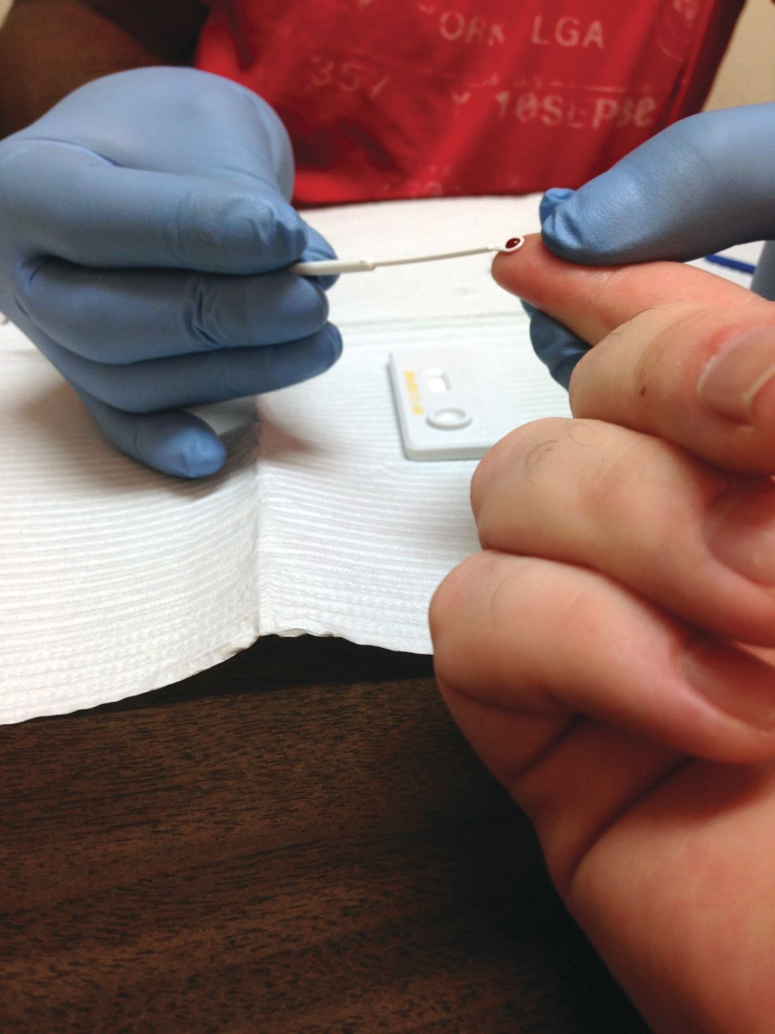
“HIV screening is a critical entry point to a range of HIV prevention and treatment options. For persons at ongoing risk for HIV infection exposure, annual screening also offers the opportunity to discuss options to reduce risk, including HIV preexposure prophylaxis,” Marc A. Pitasi, MPH, and investigators from the CDC wrote in the Morbidity and Mortality Weekly Report.
The CDC investigators analyzed data collected from the Behavioral Risk Factor Surveillance System in 2016-2017 to assess the percentage of adults screened for HIV ever and in the past year across the entirety of the United States, in 50 local jurisdictions where the majority of new U.S. HIV cases occur, and in seven states with a disproportionately high rate of HIV in rural populations.
The rate of ever testing was 38.9% across the entire country, 46.9% in the 50 local jurisdictions, and 35.5% in the seven states. The percentage of adults who had undergone HIV screening in the past year was significantly smaller at 10.1% across the entire country, 14.5% in the 50 local jurisdictions, and 9.3% in the seven states. This improved to 29.2%, 34.3%, and 26.2%, respectively, in adults who were tested in the past year who were also at risk for the disease.
The rate of ever testing varied widely among the 50 jurisdictions, ranging from 36.5% in Maricopa County, Ariz., to 70.7% in Washington, D.C. However, in addition to D.C., only Baltimore, Bronx County (N.Y.), and New York County reached or exceeded 60% coverage. Bronx County also had the highest rate of HIV screening in the past year at 31.3%, while Alameda County, Calif., had the lowest rate at 8.1%.
Of the seven states (Alabama, Arkansas, Kentucky, Mississippi, Missouri, Oklahoma, and South Carolina), Mississippi had the highest overall ever-testing rate at 40.2%, and Oklahoma had the lowest at 29.7%. People living in rural areas in those states were less likely to have ever been tested (32.1%) than those living in urban areas (37.2%). The difference between rural and urban areas increased for people at risk for HIV who had been screened in the past year, with 18.4% and 29.0%, respectively, reporting undergoing screening.
“These data provide a baseline from which to measure changes in screening in these jurisdictions and other parts of the United States over time. To achieve national goals and end the HIV epidemic in the United States, innovative and novel screening approaches might be needed to reach segments of the population that have never been tested for HIV,” the investigators concluded.
The investigators reported no conflicts of interest.
SOURCE: Pitasi MA et al. MMWR Morb Mortal Wkly Rep. 2019;68:561-7.
Despite a 2006 recommendation by the Centers for Disease Control and Prevention recommending universal screening for HIV, less than half of U.S. adults have ever been tested for the disease, a new study found.
“HIV screening is a critical entry point to a range of HIV prevention and treatment options. For persons at ongoing risk for HIV infection exposure, annual screening also offers the opportunity to discuss options to reduce risk, including HIV preexposure prophylaxis,” Marc A. Pitasi, MPH, and investigators from the CDC wrote in the Morbidity and Mortality Weekly Report.
The CDC investigators analyzed data collected from the Behavioral Risk Factor Surveillance System in 2016-2017 to assess the percentage of adults screened for HIV ever and in the past year across the entirety of the United States, in 50 local jurisdictions where the majority of new U.S. HIV cases occur, and in seven states with a disproportionately high rate of HIV in rural populations.
The rate of ever testing was 38.9% across the entire country, 46.9% in the 50 local jurisdictions, and 35.5% in the seven states. The percentage of adults who had undergone HIV screening in the past year was significantly smaller at 10.1% across the entire country, 14.5% in the 50 local jurisdictions, and 9.3% in the seven states. This improved to 29.2%, 34.3%, and 26.2%, respectively, in adults who were tested in the past year who were also at risk for the disease.
The rate of ever testing varied widely among the 50 jurisdictions, ranging from 36.5% in Maricopa County, Ariz., to 70.7% in Washington, D.C. However, in addition to D.C., only Baltimore, Bronx County (N.Y.), and New York County reached or exceeded 60% coverage. Bronx County also had the highest rate of HIV screening in the past year at 31.3%, while Alameda County, Calif., had the lowest rate at 8.1%.
Of the seven states (Alabama, Arkansas, Kentucky, Mississippi, Missouri, Oklahoma, and South Carolina), Mississippi had the highest overall ever-testing rate at 40.2%, and Oklahoma had the lowest at 29.7%. People living in rural areas in those states were less likely to have ever been tested (32.1%) than those living in urban areas (37.2%). The difference between rural and urban areas increased for people at risk for HIV who had been screened in the past year, with 18.4% and 29.0%, respectively, reporting undergoing screening.
“These data provide a baseline from which to measure changes in screening in these jurisdictions and other parts of the United States over time. To achieve national goals and end the HIV epidemic in the United States, innovative and novel screening approaches might be needed to reach segments of the population that have never been tested for HIV,” the investigators concluded.
The investigators reported no conflicts of interest.
SOURCE: Pitasi MA et al. MMWR Morb Mortal Wkly Rep. 2019;68:561-7.
FROM MMWR
Co-use of opioids, methamphetamine on rise in rural Oregon
Survey shows simultaneous use climbed from 19% to 34% between 2011 and 2017
SAN ANTONIO – A perceived low risk of using methamphetamine and a belief that methamphetamine helps with opioid addiction are both driving increasing levels of concurrent methamphetamine and opioid use in rural Oregon, according to recent qualitative research.
Use of methamphetamine by those who use opioids increased from 19% to 34% between 2011 and 2017, Gillian Leichtling, research manager at HealthInsight Oregon, said at the annual meeting of the College on Problems of Drug Dependence.
The highest prevalence of simultaneous use is in the western states, where 63% of opioid users also use methamphetamine, she said. Hospitalizations and overdoses related to methamphetamine have likewise increased, particularly in rural communities.
To better understand the motivations and implications of this trend, Ms. Leichtling and her colleagues conducted a survey from March 2018 to April 2019 of adults who had nonmedically used/injected opioids or methamphetamine in the past month. All participants lived in Lane or Douglas counties in southwestern Oregon, where half the land is controlled by the U.S. Forest Service and Bureau of Land Management, and opioid overdose rates surpass that of the state average. Additional 60-minute semistructured qualitative interviews were conducted in summer 2018.
Among the 144 surveyed, 78% had used an opioid in the past month, nearly all of whom (96%) had also used methamphetamine in the past month. The interviewees included adults fairly evenly spread across ages, but most (94%) were white.
The main themes that emerged from the interviews involved the perceived benefits and consequences of those who used both opioids and methamphetamine, and the environmental circumstances that supported methamphetamine use, Ms. Leichtling explained.
Most people interviewed had their first experience with methamphetamine early in life, typically in early or mid-adolescence, she said. Two respondents, for example, first began using at 8 and 12 years old, the former learning from a preteen neighbor.
Methamphetamine’s wide availability and low cost also increased its use. In addition, methamphetamine use carries less stigma than heroin use, participants told the researchers. One person who noted the popularity of methamphetamine added: “You get treated really badly if you’re a heroin addict.”
In addition to less stigma, many of the perceived benefits of methamphetamine use related to opioids: Participants said methamphetamine “relieves opioid withdrawal, helps reduce opioid use, enhances functioning, and combines well with opioids” for a pleasurable effect, Ms. Leichtling said. Some also perceived methamphetamine as a way to reverse opioid overdose.
“I’m getting out of [the buprenorphine] program; they’re titrating me down rapidly, and so I’ve been sick for a week,” one respondent told researchers. “I’ve been doing so much more meth just to try to deflect the pain ... they’re too hard to come down from. It’s just you can’t do it without another drug ... especially if you have a job or responsibilities or kids,” they told researchers.
Another woman said she and her mother were able to come off heroin by using methamphetamine instead, and a yet another said she and her ex-boyfriend used methamphetamine to stop using opioids.
Several respondents also mentioned using methamphetamine to help them go to work, effectively put in long days, and then care for their families when they get home.
Ms. Leichtling described three main implications of the findings for interventions in rural areas. One was the need at the community level for greater access to medication-assisted treatment (MAT) of opioid use disorder to reduce the use of methamphetamine to taper opioid use or withdrawal.
Next, clinicians need to provide tailored treatment for the co-use of opioids and methamphetamine, and educate patients on alternatives to being dropped from medication-assisted opioid use disorder treatment. Finally, individual users need education on overdose that addresses the misconceptions and risks related to methamphetamine risk, Ms. Leichtling said.
Since the survey and interviews came only from two rural Oregon counties, the findings might not be generalizable, Ms. Leichtling said, and their study did not explore social determinants of health that might be at work.
The National Institute on Drug Abuse funded the research. The authors had no conflicts of interest.
Survey shows simultaneous use climbed from 19% to 34% between 2011 and 2017
Survey shows simultaneous use climbed from 19% to 34% between 2011 and 2017
SAN ANTONIO – A perceived low risk of using methamphetamine and a belief that methamphetamine helps with opioid addiction are both driving increasing levels of concurrent methamphetamine and opioid use in rural Oregon, according to recent qualitative research.
Use of methamphetamine by those who use opioids increased from 19% to 34% between 2011 and 2017, Gillian Leichtling, research manager at HealthInsight Oregon, said at the annual meeting of the College on Problems of Drug Dependence.
The highest prevalence of simultaneous use is in the western states, where 63% of opioid users also use methamphetamine, she said. Hospitalizations and overdoses related to methamphetamine have likewise increased, particularly in rural communities.
To better understand the motivations and implications of this trend, Ms. Leichtling and her colleagues conducted a survey from March 2018 to April 2019 of adults who had nonmedically used/injected opioids or methamphetamine in the past month. All participants lived in Lane or Douglas counties in southwestern Oregon, where half the land is controlled by the U.S. Forest Service and Bureau of Land Management, and opioid overdose rates surpass that of the state average. Additional 60-minute semistructured qualitative interviews were conducted in summer 2018.
Among the 144 surveyed, 78% had used an opioid in the past month, nearly all of whom (96%) had also used methamphetamine in the past month. The interviewees included adults fairly evenly spread across ages, but most (94%) were white.
The main themes that emerged from the interviews involved the perceived benefits and consequences of those who used both opioids and methamphetamine, and the environmental circumstances that supported methamphetamine use, Ms. Leichtling explained.
Most people interviewed had their first experience with methamphetamine early in life, typically in early or mid-adolescence, she said. Two respondents, for example, first began using at 8 and 12 years old, the former learning from a preteen neighbor.
Methamphetamine’s wide availability and low cost also increased its use. In addition, methamphetamine use carries less stigma than heroin use, participants told the researchers. One person who noted the popularity of methamphetamine added: “You get treated really badly if you’re a heroin addict.”
In addition to less stigma, many of the perceived benefits of methamphetamine use related to opioids: Participants said methamphetamine “relieves opioid withdrawal, helps reduce opioid use, enhances functioning, and combines well with opioids” for a pleasurable effect, Ms. Leichtling said. Some also perceived methamphetamine as a way to reverse opioid overdose.
“I’m getting out of [the buprenorphine] program; they’re titrating me down rapidly, and so I’ve been sick for a week,” one respondent told researchers. “I’ve been doing so much more meth just to try to deflect the pain ... they’re too hard to come down from. It’s just you can’t do it without another drug ... especially if you have a job or responsibilities or kids,” they told researchers.
Another woman said she and her mother were able to come off heroin by using methamphetamine instead, and a yet another said she and her ex-boyfriend used methamphetamine to stop using opioids.
Several respondents also mentioned using methamphetamine to help them go to work, effectively put in long days, and then care for their families when they get home.
Ms. Leichtling described three main implications of the findings for interventions in rural areas. One was the need at the community level for greater access to medication-assisted treatment (MAT) of opioid use disorder to reduce the use of methamphetamine to taper opioid use or withdrawal.
Next, clinicians need to provide tailored treatment for the co-use of opioids and methamphetamine, and educate patients on alternatives to being dropped from medication-assisted opioid use disorder treatment. Finally, individual users need education on overdose that addresses the misconceptions and risks related to methamphetamine risk, Ms. Leichtling said.
Since the survey and interviews came only from two rural Oregon counties, the findings might not be generalizable, Ms. Leichtling said, and their study did not explore social determinants of health that might be at work.
The National Institute on Drug Abuse funded the research. The authors had no conflicts of interest.
REPORTING FROM CPDD 2019
Short Takes
Restrictive transfusion strategy for cardiac surgery patients remains noninferior at 6 months post op

The authors previously reported that, in moderate- to high-risk cardiac surgery patients, a restrictive transfusion strategy was noninferior to a liberal strategy based on the clinical outcomes of all-cause mortality, MI, stroke, or new renal failure with dialysis. The groups continued to show no significant difference in outcomes at 6 months post op.
Citation: Mazer CD et al. Six-month outcomes after restrictive or liberal transfusion for cardiac surgery. N Engl J Med. 2018;379:1224-33.
Restrictive transfusion strategy for cardiac surgery patients remains noninferior at 6 months post op
The authors previously reported that, in moderate- to high-risk cardiac surgery patients, a restrictive transfusion strategy was noninferior to a liberal strategy based on the clinical outcomes of all-cause mortality, MI, stroke, or new renal failure with dialysis. The groups continued to show no significant difference in outcomes at 6 months post op.
Citation: Mazer CD et al. Six-month outcomes after restrictive or liberal transfusion for cardiac surgery. N Engl J Med. 2018;379:1224-33.
Social media use linked to acceptance of cosmetic surgery
Use of social media platforms such as Tinder, Snapchat, and Instagram, particularly in conjunction with photo-editing applications, may increase an individual’s acceptance of cosmetic surgery, a new study suggests.
![]()
In JAMA Facial Plastic Surgery, researchers report the outcomes of a web-based survey study involving 252 participants, 73.0% of whom were female. The survey asked participants about their use of social media, photo-editing tools such as Photoshop, VSCO, and Snapchat filters, and answered questionnaires to assess their self-esteem, self-worth, and attitudes toward cosmetic surgery.
All participants used at least one social media platform, with a mean of seven, and used a mean of two photo-editing applications; the analysis found that those who used more social media platforms were more likely to consider cosmetic surgery.
People who used Tinder and Snapchat – with or without photo filters – showed greater acceptance of cosmetic surgery, while those who used the photography mobile app VSCO and Instagram photo filters showed greater consideration but not acceptance of cosmetic surgery, compared with nonusers.
Participants whose self-worth was more closely tied to their appearance showed greater acceptance of cosmetic surgery. When it came to self-esteem, participants who used YouTube, WhatsApp, VSCO, and Photoshop had lower self-esteem scores, compared with nonusers.
Overall, nearly two-thirds of survey participants said they used photo-editing applications to change the lighting of images, but only 5.16% said they used these applications to make changes to face or body shape. This distinction was also seen in their acceptance of cosmetic surgery scores: Those who said they made changes to face and body shape showed higher acceptance scores than nonusers, but this was not seen in those who only used it for lighting adjustments.
“The rising trend of pursuing cosmetic surgery based on social media inspiration highlights the need to better understand patients’ motivations to seek cosmetic surgery,” wrote Jonlin Chen, a medical student at Johns Hopkins University, Baltimore, and coauthors.
Commenting on the association between YouTube use, lower self-esteem, and higher acceptance of cosmetic surgery, the authors suggested that the platform may generate appearance comparisons between users by allowing them to access beauty-related videos and connect with other users interested in cosmetics.
Michael J. Reilly, MD, department of otolaryngology–head and neck surgery and Keon M. Parsa, MD, from the department of psychiatry at MedStar Georgetown University Hospital in Washington, commented in an accompanying editorial that the findings of this study illustrate an increased trend seen by facial plastic surgeons (JAMA Facial Plast Surg. 2019 June 27. doi: 10.1001/jamafacial.2019.0419). The study “shows the importance of understanding the underlying motives and characteristics of individuals seeking cosmetic surgery.” They noted that facial plastic surgeons can play a role in helping patients to improve their self-esteem, but it is also important to be aware of the clinical signs of depression, anxiety, and social isolation and refer for appropriate nonsurgical support when there are mental health concerns that go beyond the knife and needle.
The authors of the study did note that their choice of a web-based survey meant the demographic was likely to be skewed toward a younger, more social media–savvy demographic, and may not necessarily represent the broader population of individuals seeking cosmetic surgery.
No funding or conflicts of interest were declared.
SOURCE: Chen J et al. JAMA Facial Plast Surg. 2019 Jun 27. doi: 10.1001/jamafacial.2019.0328.
FROM JAMA FACIAL PLASTIC SURGERY