User login
Use of antipsychotics to treat delirium in the ICU
Background: Delirium is commonly seen in the ICU and has been associated with increased morbidity and mortality. While haloperidol, as well as atypical antipsychotics, often are used to manage ICU delirium, evidence has been mixed as to whether these medications shorten the duration of either hyperactive or hypoactive delirium.
Study design: Randomized, controlled trial.
Setting: 16 medical centers in the United States.
Synopsis: 566 adult patients with respiratory failure or shock who experienced delirium in medical or surgical ICUs in participating hospitals were randomly assigned to receive either IV haloperidol, ziprasidone, or placebo. The median exposure to the trial medication or placebo was 4 days. The median number of days without delirium was not significantly different among the three groups (P = .26) with a median length of delirium of 8.5 days in the placebo group, compared with 7.9 days in the haloperidol group and 8.7 days in the ziprasidone group. The study was powered to detect a 2-day difference.
Only 11% of patients experienced hyperactive delirium, which makes these results less generalizable to patients whose delirium presents as agitation.
Bottom line: The use of antipsychotics in ICU delirium does not affect the duration of delirium in patient with respiratory failure or shock.
Citation: Girard TD et al. Haloperidol and ziprasidone for treatment of delirium in critical illness. N Eng J Med. 2018 Dec 27;379(26):2506-16.
Dr. Defoe is an instructor of medicine at Northwestern University Feinberg School of Medicine and a hospitalist at Northwestern Memorial Hospital, both in Chicago.
When’s the right time to use dementia as a diagnosis?
Is dementia a diagnosis?
I use it myself, although I find that some neurologists consider this blasphemy.
The problem is that there aren’t many terms to cover cognitive disorders beyond mild cognitive impairment (MCI). Phrases like “cortical degeneration” and “frontotemporal disorder” are difficult for families and patients. They aren’t medically trained and want something easy to write down.
“Alzheimer’s,” or – as one patient’s family member says, “the A-word” – is often more accurate, but has stigma attached to it that many don’t want, especially at a first visit. It also immediately conjures up feared images of nursing homes, wheelchairs, and bed-bound people.
So I use a diagnosis of dementia with many families, at least initially. Since, with occasional exceptions, we tend to perform a work-up of all cognitive disorders the same way, I don’t have a problem with using a more generic blanket term. As I sometimes try to simplify things, I’ll say, “It’s like squares and rectangles. Alzheimer’s disease is a dementia, but not all dementias are Alzheimer’s disease.”
I don’t do this to avoid confrontation, be dishonest, mislead patients and families, or avoid telling the truth. I still make it very clear that this is a progressive neurologic illness that will cause worsening cognitive problems over time. But many times families aren’t ready for “the A-word” early on, or there’s a concern the patient will harm themselves while they still have that capacity. Sometimes, it’s better to use a different phrase.
It may all be semantics, but on a personal level, a word can make a huge difference.
So I say dementia. In spite of some editorials I’ve seen saying we should retire the phrase, I argue that in many circumstances it’s still valid and useful.
It may not be a final, or even specific, diagnosis, but it is often the best and most socially acceptable one at the beginning of the doctor-patient-family relationship. When you’re trying to build rapport with them, that’s equally critical when you know what’s to come down the road.
Dr. Block has a solo neurology practice in Scottsdale, Ariz.
Surprise medical billing legislation advances to the House floor
Legislation to end surprise medical billing cleared the House Energy and Commerce Committee and is headed to the House floor, but it contains a somewhat controversial arbitration mechanism that allows physicians and hospitals to seek higher payments within 30 days.
The bill, which was tacked on to H.R. 2328, passed the committee by voice vote on July 17. The amendment on arbitration also passed the committee via voice vote.
“Our most important task today is to protect patients from the unreasonable and unacceptable practice of surprise billing,” Rep. Frank Pallone (D-N.J.), chairman of the Energy and Commerce Committee, said just prior to the votes being taken by the committee. “Under the [legislation], providers would no longer be able to balance bill patients for out-of-network emergency services or for scheduled services from providers the patient was not aware would be in their treatment.”
Out-of-network providers would receive a benchmark payment for the services they provided under the legislation.
Rep. Pallone described the legislation as taking patients out of the middle of disputes between payers and providers.
He went on to describe the arbitration amendment, introduced by Rep. Raul Ruiz, MD, (D.-Calif.) and Rep. Larry Bucshon, MD, (R-Ind.), as creating an independent dispute resolution process for physicians and hospitals to file a claim in the event that they don’t think they were adequately paid for their services.
“The amendment would allow providers to have 30 days within which to file an appeal of the benchmark payment with the insurer,” Rep. Pallone said. “The insurer would then have 30 days to adjudicate the appeal, after which the provider could initiate independent dispute resolution.”
The amendment limits appeals to extenuating circumstances so that only complex cases would qualify, and it limits the variables that can be considered during arbitration to the quality of care that was provided to the patient, according to Rep. Pallone.
“Most importantly to me, it bars arbitrators from considering billed charges, which are unilaterally set by providers,” Rep. Pallone said. “Provider charges are often double or triple Medicare rates, and in some cases for some large physician staffing companies, it is around 500% of Medicare rates. If Congress sends this signal to arbiters that provider charges are to be considered, we would be creating a significantly higher standard for payment, decreasing incentives for providers to be in network and putting upward pressure on health care premiums.”
Rep. Michael Burgess, MD, (R-Texas) praised the inclusion of the arbitration amendment and said the surprise billing legislation would not be able to be passed without its inclusion.
Rep. Janice Schakowsky (D-Ill.) offered a dissenting voice to the amendment. “Arbitration, in my view, which is used as the backstop, will not lower the health care costs,” she said. “Arbitration actually comes with additional administrative costs and complexities, which could then be passed on to consumers in the form of higher premiums. Even as a backstop, I think that binding arbitration leaves a public interest, public health decision, up to an unaccountable private decision maker, and I don’t think that is a very progressive way to be dealing with the issue of pricing.”
The American Medical Association praised the inclusion of an appeals process for resolving out-of-network payment disputes. “This addition represents progress,” Patrice Harris, MD, president of the AMA, said in a statement. “While we continue to have concerns with elements of the legislation, we remain committed to working with all committee members to secure further improvements to protect patients, preserve access, and foster fair payments for out-of-network services.”
America’s Health Insurance Plans, which represents health insurers, voiced its opposition to the arbitration provision. “We strongly oppose the inclusion of arbitration because it does not solve the problem of surprise medical bills,” AHIP President and CEO Matt Eyles said in a statement. “It increases the financial burden on everyone with coverage, increasing patient premiums and driving up the cost of health care. The arbitration proposal allows private-equity firms and certain providers to price gouge patients and then shifts the final decision to a ‘third party.’ This process introduces new bureaucracy and red tape into the system, with costs to hardworking taxpayers exceeding $1 billion.”
Benedic Ippolito, research fellow in economic policy studies at the American Economic Institute, further criticized the use of arbitration in this process.
“This concept that arbitration is a backstop doesn’t really make a lot of sense,” he said during a July 17 panel discussion hosted by the Bipartisan Policy Center on surprise billing. “The arbiter has to do the same thing any sort of rate setter has to do.” He noted that if they are doing “baseball-style” arbitration, they have to choose between two offers placed in front of the arbiter, based on what the arbiter believes to be closer to whatever the reasonable rate is.
This process will eventually lead to either a system that favors the payer, the provider, or ends up being exactly what the benchmark is that has been set. “If it is better for one or the other, somebody is going to have an incentive to just trigger this thing the whole time,” Mr. Ippolito said.
Loren Adler, associate director of the USC-Brookings Schaeffer Initiative for Health Policy, said during the panel discussion that “there is no policy reason to have arbitration. There is nothing it adds for policy value.”
Mr. Adler called the arbitration amendment “a provider giveaway bill,” adding that if there has to be an arbitration option, it should have a very high threshold to be triggered.
Exposure to synthetic cannabinoids is associated with neuropsychiatric morbidity in adolescents
according to data published online July 8 ahead of print in Pediatrics. The results support a distinct neuropsychiatric profile of acute synthetic cannabinoid toxicity in adolescents, wrote the investigators.
Synthetic cannabinoids have become popular and accessible and primarily are used for recreation. The adverse effects of synthetic cannabinoid toxicity reported in the literature include tachycardia, cardiac ischemia, acute kidney injury, agitation, first episode of psychosis, seizures, and death. Adolescents are the largest age group presenting to the emergency department with acute synthetic cannabinoid toxicity, and this population requires more intensive care than adults with the same presentation.
A multicenter registry analysis
To describe the neuropsychiatric presentation of adolescents to the emergency department after synthetic cannabinoid exposure, compared with that of cannabis exposure, Sarah Ann R. Anderson, MD, PhD, an adolescent medicine fellow at Columbia University Irving Medical Center in New York, and colleagues performed a multicenter registry analysis. They examined data collected from January 2010 through September 2018 from adolescent patients who presented to sites that participate in the Toxicology Investigators Consortium. For each patient, clinicians requested a consultation by a medical toxicologist to aid care. The exposures recorded in the case registry are reported by the patients or witnesses.
Eligible patients were between ages 13 and 19 years and presented to an emergency department with synthetic cannabinoid or cannabis exposure. Dr. Anderson and colleagues collected variables such as age, sex, reported exposures, death in hospital, location of toxicology encounter, and neuropsychiatric signs or symptoms. Patients whose exposure report came from a service outside of an emergency department and those with concomitant use of cannabis and synthetic cannabinoids were excluded. For the purpose of analysis, the investigators classified patients into the following four categories: exposure to synthetic cannabinoids alone, exposure to synthetic cannabinoids and other drugs, exposure to cannabis alone, and exposure to cannabis and other drugs.
Dr. Anderson and colleagues included 348 patients in their study. The sample included 107 patients in the synthetic cannabinoid–only group, 38 in the synthetic cannabinoid/polydrug group, 86 in the cannabis-only group, and 117 in the cannabis/polydrug group. Males predominated in all groups. The one death in the study occurred in the synthetic cannabinoid–only group.
Synthetic cannabinoid exposure increased risk for seizures
Compared with the cannabis-only group, the synthetic cannabinoid–only group had an increased risk of coma or CNS depression (odds ratio, 3.42) and seizures (OR, 3.89). The risk of agitation was significantly lower in the synthetic cannabinoid–only group, compared with the cannabis-only group (OR, 0.18). The two single-drug exposure groups did not differ in their associated risks of delirium or toxic psychosis, extrapyramidal signs, dystonia or rigidity, or hallucinations.
Exposure to synthetic cannabinoids plus other drugs was associated with increased risk of agitation (OR, 3.11) and seizures (OR, 4.8), compared with exposure to cannabis plus other drugs. Among patients exposed to synthetic cannabinoids plus other drugs, the most common class of other drug was sympathomimetics (such as synthetic cathinones, cocaine, and amphetamines). Sympathomimetics and ethanol were the two most common classes of drugs among patients exposed to cannabis plus other drugs.
Synthetic cannabinoids may have distinctive neuropsychiatric outcomes
“Findings from our study further confirm the previously described association between synthetic cannabinoid–specific overdose and severe neuropsychiatric outcomes,” wrote Dr. Anderson and colleagues. They underscore “the need for targeted public health messaging to adolescents about the dangers of using synthetic cannabinoids alone or combined with other substances.”
The investigators’ finding that patients exposed to synthetic cannabinoids alone had a lower risk of agitation than those exposed to cannabis alone is not consistent with contemporary literature on synthetic cannabinoid–associated agitation. This discordance may reflect differences in the populations studied, “with more severe toxicity prompting the emergency department presentations reported in this study,” wrote Dr. Anderson and colleagues. The current study also may be affected by selection bias, they added.
The researchers acknowledged several limitations of their study. For example, the registry lacked data for variables such as race or ethnicity, concurrent illness, previous drug use, and comorbid conditions. Another limitation was that substance exposure was patient- or witness-reported, and no testing to confirm exposure to synthetic cannabinoids was performed. Finally, the study had a relatively small sample size and lacked information about patients’ long-term outcomes.
Dr. Anderson and colleagues described future research that could address open questions. Analyzing urine to identify the synthetic cannabinoid used and correlating it with the presentation in the emergency department could illuminate specific toxidromes associated with particular compounds, they wrote. Longitudinal data on the long-term effects of adolescent exposure to synthetic cannabinoids would be valuable for understanding potential long-term neurocognitive impairments. “Lastly, additional investigations into the management of adolescent synthetic cannabinoid toxicity in the emergency department is warranted, given the health care cost burden of synthetic cannabinoid–related emergency department visits,” they concluded.
The study was not supported by external funding, and the authors had no relevant disclosures.
SOURCE: Anderson SAR et al. Pediatrics. 2019 Jul 8. doi: 10.1542/peds.2018-2690.
FROM PEDIATRICS
Mechanism does not matter for second-line biologic choice in JIA
MADRID – When biologic treatment is indicated after initial tumor necrosis factor (TNF) inhibitor therapy for juvenile idiopathic arthritis (JIA) has failed, the mechanism of action of the second biologic does not appear to matter, according to data presented at the European Congress of Rheumatology.
“There appears to be no difference in effectiveness outcomes or drug survival in patients starting a second TNF inhibitor versus an alternative class of biologic,” said Lianne Kearsley-Fleet, an epidemiologist at the Centre for Epidemiology Versus Arthritis at the University of Manchester (England).
Indeed, at 6 months, there were no significant differences among patients who had switched from a TNF inhibitor to another TNF inhibitor or to a biologic with an alternative mechanism of action in terms of:
- The change in Juvenile Arthritis Disease Activity Score (JADAS)-71 from baseline (mean score change, 7.3 with second TNF inhibitor vs. 8.5 with an alternative biologic class).
- The percentage of patients achieving an American College of Rheumatology Pediatric 90% response (22% vs. 15%).
- The proportion of patients achieving minimal disease activity (30% vs. 23%).
- The percentage reaching a minimal clinically important difference (MCID; 44% vs. 43%).
There was also no difference between switching to a TNF inhibitor or alternative biologic in terms of the duration of time patients remained treated with the second-line agent.
“After 1 year, 62% of patients remained on their biologic therapy, and when we looked at drug survival over the course of that year, there was no difference between the two cohorts,” Mrs. Kearsley-Fleet reported. There was no difference also in the reasons for stopping the second biologic.
“We now have a wide range of biologic therapies available; however, there is no evidence regarding which biologic should be prescribed [in JIA], and if patients switch, which order this should be,” Mrs. Kearsley-Fleet stated. Current NHS England guidelines recommend that most patients with JIA should start a TNF inhibitor (unless they are rheumatoid factor positive, in which case they should be treated with rituximab [Rituxan]), and if the first fails, to switch to a second TNF inhibitor rather than to change class. The evidence for this is limited, she noted, adding that adult guidelines for rheumatoid arthritis now recommended a change of class if not contraindicated.
Using data from two pediatric biologics registers – the British Society for Paediatric and Adolescent Rheumatology Etanercept Cohort Study (BSPAR-ETN) and Biologics for Children with Rheumatic Diseases (BCRD) – Mrs. Kearsley-Fleet and her associates looked at data on 241 children and adolescents with polyarticular JIA (or oligoarticular-extended JIA) starting a second biologic. The aim was to compare the effectiveness of starting a second TNF inhibitor versus switching to an alternative class of agent, such as a B-cell depleting agent such as rituximab, in routine clinical practice.
A majority (n = 188; 78%) of patients had etanercept (Enbrel) as their starting TNF inhibitor and those switching to a second TNF inhibitor (n = 196) were most likely to be given adalimumab (Humira; 58%). Patients starting a biologic with another mode of action (n = 45) were most likely to be given the interleukin-6 inhibitor tocilizumab (73%), followed by rituximab in 13%, and abatacept (Orencia) in 11%. The main reasons for switching to another biologic – TNF inhibitor or otherwise – were ineffectiveness (60% with a second TNF inhibitor vs. 62% with another biologic drug class) or adverse events or intolerance (19% vs. 13%, respectively).
The strength of these data are that they come from a very large cohort of children and adolescents starting biologics for JIA, with systematic follow-up and robust statistical methods, Mrs. Kearsley-Fleet said. However, she noted that JIA was rare and that only one-fifth of patients would start a biologic, and just 30% of those patients would then switch to a second biologic.
“We don’t see any reason that the guidelines should be changed,” Mrs. Kearsley-Fleet observed. “However, repeat analysis with a larger sample size is required to reinforce whether there is any advantage of switching or not.”
Versus Arthritis (formerly Arthritis Research UK) and The British Society for Rheumatology provided funding support. Mrs. Kearsley-Fleet had no financial conflicts of interest to disclose.
SOURCE: Kearsley-Fleet L et al. Ann Rheum Dis, Jun 2019;8(Suppl 2):74-5. Abstract OP0016. doi: 10.1136/annrheumdis-2019-eular.415.
REPORTING FROM EULAR 2019 Congress
Small study suggests natural HCV clearance is caused by AR3-antibody response
Individuals who spontaneously cleared their primary hepatitis C virus (HCV) infection or reinfection had significantly more antibodies that recognized multiple HCV genotypes beyond the initial infection, compared with chronically infected individuals, according to a small molecular study of immortalized cultured B cells from patient.
In a study published in the Journal of Hepatology, Sabrina J. Merat of AIMM Therapeutics and colleagues classified patients into two groups based on the outcome of their HCV infection: individuals who became chronically infected (CHRs; n = 5) either after primary infection or after HCV reinfection and individuals who cleared one or more HCV infections and were HCV RNA negative at the end of follow-up (CLs; n = 8). The researchers considered that all CLs who cleared the infection were presumably re-exposed to HCV as they continued injecting drugs for a median of 5.9 years after primary infection. The median follow-up time of individuals after primary HCV infection was 17.5 years.
Although the frequency of total antibodies did not differ between the two groups, the antibodies from CHRs were mainly genotype specific and directed against the genotype of the ongoing infection. Antibodies from CLs showed a much broader reactivity than CHR-derived antibodies, with the absolute number of antibodies recognizing at least three or more genotypes was significantly higher in CLs than in CHRs (13 vs. 0, respectively; P = .03).
In addition, in order to determine which epitopes were being targeted in the CL patients, the researchers tested the antibodies secreted in the B-cell supernatant for binding to E2 alanine mutants in the four epitopes known to be recognized by broadly neutralizing HCV antibodies. They found that the majority of the cross-genotype antibodies (82/113; 73%) were specific for AR3 because they bound to the AR3-specific mutants.
“In chronically infected individuals, AR3-specific antibody responses may be too weak and/or may develop too late to prevent chronic infection. If confirmed, this means that a strong and broadly neutralizing antibody response should be established very early after infection in order to confer protection,” the researchers concluded.
This study was supported by the Virgo consortium, funded by the Dutch government. Sabrina Merat and several coauthors are employees of AIMM Therapeutics, as well as shareholders.
SOURCE: Merat SJ et al. J Hepatol 2019;71:14-24.
FROM THE JOURNAL OF HEPATOLOGY
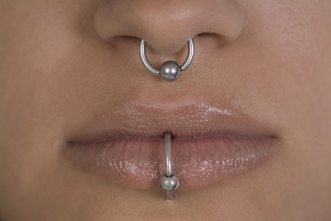
Piercing art
Body art as a form of human expression is prevalent. The most common types are skin tattoos and piercings, but also include scarification, branding, subdermal implants, and body painting. Body painting has made headlines for its artistic creativity and artistic significance at annual week long temporary communities such as the annual Burning Man art festival.
Culture and history
Culturally, however, body painting has significant historical significance, with Henna painting described in the earliest Hindu Vedic ritual books dating back 5,000 years. Henna painting, most commonly of the hands and feet, known as Mehndi in the Indian subcontinent, signifies painting of symbolic representations of the outer and the inner sun, with the idea of “awakening the inner light.” It is also a common tradition of Hindu weddings and applied in Muslim tradition in India during Eid festivals. Body painting has also been used in other cultures for ceremonial, religious reasons, as well as forms of camouflage during hunting or war. Branding and scarification were used as methods of punishment during the Middle Ages in England and commonly during slavery in the Americas. Traditionally, though, branding and scarification have been seen in darker-skinned individuals as a form of self-expression where tattoos are not as effective visually. African tribes in Ethiopia and Sudan, as well as the Maasai people in Kenya, have used scarification and branding as an ancient art that can signify everything from beauty to transition to adulthood. Some black fraternities also use it as a mark of collegiality.
While tattoos are the most recognized form of body art, body and facial piercing are far more common in the general population among cultures throughout the world. While ear piercings are the most common, historically, nostril piercing has been documented in the Middle East as far back as 4,000 years ago, and both ear and nostril piercing and jewelry are mentioned historically in the Bible (Genesis 24:22, Isaiah 3:21). Ritual tongue piercing was reportedly performed by Aztec and Mayan Indians during ceremonies to honor their deities.
Current Practice
In practice, we see different types of piercings, including but not limited to ear, nose (alar, septum, bridge), eyebrow, lip, tongue, face, nipple, umbilical, and genital piercings. Ear piercings alone may come in many forms. Not only do location, cartilage versus no cartilage involvement, and age of piercing have different implications for care and potential risks/complications, so do the size, type, and shape of jewelry used for the piercing.
Having a better understanding of piercing art is important for dermatologists and dermatologic surgeons because we sometimes treat the sequelae, including infection, allergic reactions from the jewelry, and keloid scars. Patients may intentionally create large size piercings, known as gauge piercings, and decide later they no longer want them. Or earlobe piercings can unintentionally stretch and enlarge over time from prolonged wearing of heavy earrings or trauma, sometimes resulting in a partial or complete earlobe split, requiring surgical treatment for gauge or split earlobe repair. If repiercing earlobe repair is desired, most physicians wait at least 6-8 weeks. While different earlobe surgical repair techniques (most commonly Z-plasty) and even recommendations for subdermal implant removal are described in the literature, there are no real guidelines on when to repierce in the evidenced-based literature. Healing time in general for piercings also varies by site. For example, initial earlobe piercings typically take 1-2 months to heal, whereas ear cartilage and navel piercings may take 4-12 months.
Some medical practitioners may not be aware of tips known to top piercing professionals that can help guide patients on piercing care. Cartilage piercings can sometimes present with inflammation and nodule formation, even prior to true keloid formation. In my experience, a simple solution of washing daily with a highly alkaline but gentle natural soap, such as Dr. Bronner’s mild baby soap, or compresses or soaks with warm salt water, can sometimes reduce the inflammation and resolve nodule formation before topical, intralesional corticosteroids, or surgery is needed (a situation in which surgery may lead to further cartilage inflammation and hypertrophic scar formation). Additionally, certain pressure earrings may be used to help prevent keloid formation, in addition to wearing jewelry of a metal that is nonallergenic to the user, to prevent further inflammation.
Piercing is a common form of body art and self-expression. but also develop a better understanding of and relationship with our patients by virtue of their means of artistic self-expression.
Dr. Wesley and Dr. Talakoub are cocontributors to this column. Dr. Wesley practices dermatology in Beverly Hills, Calif. Dr. Talakoub is in private practice in McLean, Va. This month’s column is by Dr. Wesley. Write to them at dermnews@mdedge.com. They had no relevant disclosures.
Dr. Lene Dreyer discusses psoriatic arthritis and cancer risk

Do patients with psoriatic arthritis face greater cancer risks? Lene Dreyer, MD, clinical professor at Aalborg (Denmark) University Hospital, talks about the mostly reassuring findings from a cancer registry analysis in four Nordic countries.

Do patients with psoriatic arthritis face greater cancer risks? Lene Dreyer, MD, clinical professor at Aalborg (Denmark) University Hospital, talks about the mostly reassuring findings from a cancer registry analysis in four Nordic countries.

Do patients with psoriatic arthritis face greater cancer risks? Lene Dreyer, MD, clinical professor at Aalborg (Denmark) University Hospital, talks about the mostly reassuring findings from a cancer registry analysis in four Nordic countries.
Statins crush early seizure risk poststroke
BANGKOK – Statin therapy, even when initiated only upon hospitalization for acute ischemic stroke, was associated with a striking reduction in the risk of early poststroke symptomatic seizure in a large observational study.
Using propensity-score matching to control for potential confounders, use of a statin during acute stroke management was associated with a “robust” 77% reduction in the risk of developing a symptomatic seizure within 7 days after hospital admission, Soichiro Matsubara, MD, reported at the International Epilepsy Congress.
This is an important finding because early symptomatic seizure (ESS) occurs in 2%-7% of patients following an acute ischemic stroke. Moreover, an Italian meta-analysis concluded that ESS was associated with a 4.4-fold increased risk of developing poststroke epilepsy (Epilepsia. 2016 Aug;57[8]:1205-14), noted Dr. Matsubara, a neurologist at the National Cerebral and Cardiovascular Center in Suita, Japan, as well as at Kumamoto (Japan) University.
He presented a study of 2,969 consecutive acute ischemic stroke patients with no history of epilepsy who were admitted to the Japanese comprehensive stroke center, of whom 2.2% experienced ESS. At physician discretion, 19% of the ESS cohort were on a statin during their acute stroke management, as were 55% of the no-ESS group. Four-fifths of patients on a statin initiated the drug only upon hospital admission.
Strokes tended to be more severe in the ESS group, with a median initial National Institutes of Health Stroke Scale score of 12.5, compared with 4 in the seizure-free patients. A cortical stroke lesion was evident upon imaging in 89% of the ESS group and 55% of no-ESS patients. Among ESS patients, 46% had a cardiometabolic stroke, compared with 34% of the no-ESS cohort. Mean C-reactive protein levels and white blood cell counts were significantly higher in the ESS cohort as well. Their median hospital length of stay was 25.5 days, versus 18 days in the no-ESS group, Dr. Matsubara said at the congress sponsored by the International League Against Epilepsy.
Of the 76 ESSs that occurred in 66 patients, 37% were focal awareness seizures, 35% were focal to bilateral tonic-clonic seizures, and 28% were focal impaired awareness seizures.
In a multivariate analysis adjusted for age, sex, body mass index, stroke subtype, and other potential confounders, statin therapy during acute management of stroke was independently associated with a 56% reduction in the relative risk of ESS. In contrast, a cortical stroke lesion was associated with a 2.83-fold increased risk.
Since this wasn’t a randomized trial of statin therapy, Dr. Matsubara and his coinvestigators felt the need to go further in analyzing the data. After extensive propensity score matching for atrial fibrillation, current smoking, systolic blood pressure, the presence or absence of a cortical stroke lesion, large vessel stenosis, and other possible confounders, they were left with two closely comparable groups: 886 statin-treated stroke patients and an equal number who were not on statin therapy during their acute stroke management. The key finding: The risk of ESS was reduced by a whopping 77% in the patients on statin therapy.
The neurologist observed that these new findings in acute ischemic stroke patients are consistent with an earlier study in a U.S. Veterans Affairs population, which demonstrated that statin therapy was associated with a significantly lower risk of new-onset geriatric epilepsy (J Am Geriatr Soc. 2009 Feb;57[2]:237-42).
As to the possible mechanism by which statins may protect against ESS, Dr. Matsubara noted that acute ischemic stroke causes toxic neuronal excitation because of blood-brain barrier disruption, ion channel dysfunction, altered gene expression, and increased release of neurotransmitters. In animal models, statins provide a neuroprotective effect by reducing glutamate levels, activating endothelial nitric oxide synthase, and inhibiting production of interleukin-6, tumor necrosis factor-alpha, and other inflammatory cytokines.
Asked about the intensity of the statin therapy, Dr. Matsubara replied that the target was typically an LDL cholesterol below 100 mg/dL.
He reported having no financial conflicts regarding the study, conducted free of commercial support.
SOURCE: Matsubara S et al. IEC 219, Abstract P002.
REPORTING FROM IEC 2019
When do I stop the code?
A hospitalist’s dilemma
I had just received my sign-out for the day. My pager beeped, and I heard it overhead “Code Blue Room X.” Hospitalist physicians lead the code team in our hospital; I quickly headed to the room.
A young man in his forties was found to be unconscious on the floor. One of the nurses had started cardiopulmonary resuscitation (CPR) as the patient was unconscious and had no palpable pulse. It was a long, drawn-out battle: CPR, cracking bones, shouting, lots of needles – an extreme roller-coaster-style situation. The patient had recently had a hip surgery and our suspicion was a massive pulmonary embolism. We ran the exhaustive code for more than an hour and then I started to debrief with my code team; discussed that treatment was getting futile and asked for opinions. Finally, I asked the team to stop and pronounced the patient dead. I felt terrible. Later that day I returned to my house, tossed my bag in the corner, and sympathized with myself – “Hello Dr. B, It was a tough one.”
Stopping resuscitation was one of the toughest decisions I had ever made, and I wondered if I would be able to make such a decision the next day. What if I had carried on? I had led code teams during my residency training and as an attending physician; but there was something different that day. This patient was a young man with no history of medical problems. Every physician knows how to initiate resuscitation for cardiopulmonary arrest (CPA); only a few know when to stop it. Did I miss this learning during my internal medicine training? I checked my red pocket leaflet with advanced cardiac life support (ACLS) algorithms, and it had no mention of it. I searched Google Scholar, PubMed, and UpToDate and surprisingly, I found no predetermined rule but only a few recommendations on when CPR should be stopped. The American Heart Association is clear that the decision to terminate resuscitative efforts rests with the treating physician in the hospital.
In my experience, the length of time to continue a code can vary widely and is mostly dependent on the physician running the code. I have seen it last 15 minutes (which is reasonable) and I have seen it last for 50 minutes when the initial rhythm was ventricular fibrillation. And if perhaps the patient regains a pulse temporarily, only to lose it again, we restart the clock. One needs to take into account various factors including time to CPR, time to defibrillation, comorbid disease, prearrest state, and initial arrest rhythm in making these decisions. It’s well understood that none of these factors alone or in combination is clearly predictive of outcome.1
Some selected patients potentially have good outcomes with prolonged, aggressive resuscitation. So when should we stop, and when should we continue resuscitation? This is always challenging. Physicians hate to stop CPR even when they know it’s time. We are guided by the Hippocratic Oath to save lives. Sometimes, even if we want to stop, we tend to continue to avoid being criticized for stopping; we are systematically biased against stopping CPR. We routinely run long codes, in part because we are not sure which patients we can bring back.
A 2012 Lancet study highlighted that the median duration of resuscitation was 12 minutes for patients achieving the return of spontaneous circulation and 20 minutes for nonsurvivors.2 The ethical guidelines issued by AHA in 2018 highlight that, in the absence of mitigating factors, prolonged resuscitative efforts for adults and children are unlikely to be successful and can be discontinued if there is no return of spontaneous circulation at any time during 30 minutes of cumulative ACLS. If the return of spontaneous circulation of any duration occurs at any time, however, it may be appropriate to consider extending the resuscitative effort.3
I believe a careful balance of the patient’s prognosis for both length of life and quality of life will determine whether continued CPR is appropriate. The responsible clinician should stop the resuscitative effort when he or she determines with a high degree of certainty that the arrest victim will not respond to further efforts. But what will help me guide my decisions next time if I ever come across this situation again?
I discussed my dilemma with one of our intensivist physicians; he expressed that in a similar scenario he would ask for opinions from other members of the code team. The role of good communication among code team members is necessary to exchange relevant knowledge in real time in a collaborative, nonhierarchical environment. The code team can provide the team leader with quick, accurate information about the patient’s clinical history that is critical to good decision making.
Family support is also an essential part of any resuscitation. Health care providers need to offer the opportunity to be present to family members during the resuscitation attempts whenever possible. One team member should be assigned to the family to answer questions, clarify information, and offer comfort, but physicians should not be asking family members to decide to stop the code. It is important to note that the decision should be made by the team leader and not the patient’s family members. Regardless of the age or condition of the patient, the loss of a loved one is difficult to deal with, even if expected. The issue becomes more difficult with changes in legal, cultural, or personal perspectives.
The AHA in 2018 stated that the treating physician is expected to understand the patient and the arrest features, and the system factors that have prognostic importance for resuscitation.3 For clinicians who work in critical care settings, the framework presented by AHA is intuitive. As a code leader, I can always give more epinephrine, try a clot-busting drug or deliver another shock. Situations vary greatly during a code, and the amount of time spent resuscitating a patient before terminating efforts is not set in stone. In many cases, it is a judgment call. The process of CPR is almost as disheartening as its bleak outcomes.
In-hospital CPAs are inevitably gruesome. Each day as an attending physician, we are faced with difficult decisions, but experiencing these incredibly difficult and life-changing events can make for good learning. A CPA situation in action is very difficult for all concerned, particularly when there is almost no chance of success. But an unsuccessful or aborted resuscitation is also a huge loss for both the family and the code team. One of the critical functions of the code team leader is to review the events of a code and exercise judgment while evaluating the length of a code. This can be an intense and emotional experience, but with these principles in mind, we can feel reassured that we are making the best decision possible, for the patient, the family, and our team.
Dr. Basnet is a hospitalist physician in the department of internal medicine at Eastern New Mexico Medical Center, Roswell.
References
1. Part 2: Ethical aspects of CPR and ECC. Circulation. 2000;102(8):I12.
2. Goldberger ZD et al. Duration of resuscitation efforts and survival after in-hospital cardiac arrest: An observational study. The Lancet. 2012;380(9852):1473-81.
3. Sirbaugh PE et al. A prospective, population-based study of the demographics, epidemiology, management, and outcome of out-of-hospital pediatric cardiopulmonary arrest. Ann Emerg Med. 1999;33(2):174-84.
A hospitalist’s dilemma
A hospitalist’s dilemma
I had just received my sign-out for the day. My pager beeped, and I heard it overhead “Code Blue Room X.” Hospitalist physicians lead the code team in our hospital; I quickly headed to the room.
A young man in his forties was found to be unconscious on the floor. One of the nurses had started cardiopulmonary resuscitation (CPR) as the patient was unconscious and had no palpable pulse. It was a long, drawn-out battle: CPR, cracking bones, shouting, lots of needles – an extreme roller-coaster-style situation. The patient had recently had a hip surgery and our suspicion was a massive pulmonary embolism. We ran the exhaustive code for more than an hour and then I started to debrief with my code team; discussed that treatment was getting futile and asked for opinions. Finally, I asked the team to stop and pronounced the patient dead. I felt terrible. Later that day I returned to my house, tossed my bag in the corner, and sympathized with myself – “Hello Dr. B, It was a tough one.”
Stopping resuscitation was one of the toughest decisions I had ever made, and I wondered if I would be able to make such a decision the next day. What if I had carried on? I had led code teams during my residency training and as an attending physician; but there was something different that day. This patient was a young man with no history of medical problems. Every physician knows how to initiate resuscitation for cardiopulmonary arrest (CPA); only a few know when to stop it. Did I miss this learning during my internal medicine training? I checked my red pocket leaflet with advanced cardiac life support (ACLS) algorithms, and it had no mention of it. I searched Google Scholar, PubMed, and UpToDate and surprisingly, I found no predetermined rule but only a few recommendations on when CPR should be stopped. The American Heart Association is clear that the decision to terminate resuscitative efforts rests with the treating physician in the hospital.
In my experience, the length of time to continue a code can vary widely and is mostly dependent on the physician running the code. I have seen it last 15 minutes (which is reasonable) and I have seen it last for 50 minutes when the initial rhythm was ventricular fibrillation. And if perhaps the patient regains a pulse temporarily, only to lose it again, we restart the clock. One needs to take into account various factors including time to CPR, time to defibrillation, comorbid disease, prearrest state, and initial arrest rhythm in making these decisions. It’s well understood that none of these factors alone or in combination is clearly predictive of outcome.1
Some selected patients potentially have good outcomes with prolonged, aggressive resuscitation. So when should we stop, and when should we continue resuscitation? This is always challenging. Physicians hate to stop CPR even when they know it’s time. We are guided by the Hippocratic Oath to save lives. Sometimes, even if we want to stop, we tend to continue to avoid being criticized for stopping; we are systematically biased against stopping CPR. We routinely run long codes, in part because we are not sure which patients we can bring back.
A 2012 Lancet study highlighted that the median duration of resuscitation was 12 minutes for patients achieving the return of spontaneous circulation and 20 minutes for nonsurvivors.2 The ethical guidelines issued by AHA in 2018 highlight that, in the absence of mitigating factors, prolonged resuscitative efforts for adults and children are unlikely to be successful and can be discontinued if there is no return of spontaneous circulation at any time during 30 minutes of cumulative ACLS. If the return of spontaneous circulation of any duration occurs at any time, however, it may be appropriate to consider extending the resuscitative effort.3
I believe a careful balance of the patient’s prognosis for both length of life and quality of life will determine whether continued CPR is appropriate. The responsible clinician should stop the resuscitative effort when he or she determines with a high degree of certainty that the arrest victim will not respond to further efforts. But what will help me guide my decisions next time if I ever come across this situation again?
I discussed my dilemma with one of our intensivist physicians; he expressed that in a similar scenario he would ask for opinions from other members of the code team. The role of good communication among code team members is necessary to exchange relevant knowledge in real time in a collaborative, nonhierarchical environment. The code team can provide the team leader with quick, accurate information about the patient’s clinical history that is critical to good decision making.
Family support is also an essential part of any resuscitation. Health care providers need to offer the opportunity to be present to family members during the resuscitation attempts whenever possible. One team member should be assigned to the family to answer questions, clarify information, and offer comfort, but physicians should not be asking family members to decide to stop the code. It is important to note that the decision should be made by the team leader and not the patient’s family members. Regardless of the age or condition of the patient, the loss of a loved one is difficult to deal with, even if expected. The issue becomes more difficult with changes in legal, cultural, or personal perspectives.
The AHA in 2018 stated that the treating physician is expected to understand the patient and the arrest features, and the system factors that have prognostic importance for resuscitation.3 For clinicians who work in critical care settings, the framework presented by AHA is intuitive. As a code leader, I can always give more epinephrine, try a clot-busting drug or deliver another shock. Situations vary greatly during a code, and the amount of time spent resuscitating a patient before terminating efforts is not set in stone. In many cases, it is a judgment call. The process of CPR is almost as disheartening as its bleak outcomes.
In-hospital CPAs are inevitably gruesome. Each day as an attending physician, we are faced with difficult decisions, but experiencing these incredibly difficult and life-changing events can make for good learning. A CPA situation in action is very difficult for all concerned, particularly when there is almost no chance of success. But an unsuccessful or aborted resuscitation is also a huge loss for both the family and the code team. One of the critical functions of the code team leader is to review the events of a code and exercise judgment while evaluating the length of a code. This can be an intense and emotional experience, but with these principles in mind, we can feel reassured that we are making the best decision possible, for the patient, the family, and our team.
Dr. Basnet is a hospitalist physician in the department of internal medicine at Eastern New Mexico Medical Center, Roswell.
References
1. Part 2: Ethical aspects of CPR and ECC. Circulation. 2000;102(8):I12.
2. Goldberger ZD et al. Duration of resuscitation efforts and survival after in-hospital cardiac arrest: An observational study. The Lancet. 2012;380(9852):1473-81.
3. Sirbaugh PE et al. A prospective, population-based study of the demographics, epidemiology, management, and outcome of out-of-hospital pediatric cardiopulmonary arrest. Ann Emerg Med. 1999;33(2):174-84.


