User login
TWILIGHT-COMPLEX: Tap ticagrelor monotherapy early after complex PCI
Patients who underwent complex PCI for acute coronary syndrome followed by 3 months of dual-antiplatelet therapy (DAPT) with ticagrelor plus aspirin fared significantly better by dropping aspirin at that point in favor of long-term ticagrelor monotherapy than with continued dual-antiplatelet therapy in the TWILIGHT-COMPLEX study.

The rate of clinically relevant bleeding was significantly lower at 12 months of follow-up in the ticagrelor monotherapy group than it was in patients randomized to continued DAPT. Moreover, this major benefit came at no cost in terms of ischemic events, which were actually numerically less frequent in the ticagrelor plus placebo group, George D. Dangas, MD, reported at the joint scientific sessions of the American College of Cardiology and the World Heart Federation. ACC organizers chose to present parts of the meeting virtually after COVID-19 concerns caused them to cancel the meeting.
“We found that the aspirin just doesn’t add that much, even in complex patients – just bleeding complications, for the most part,” explained Dr. Dangas, professor of medicine and of surgery at the Icahn School of Medicine at Mount Sinai, New York.
The TWILIGHT-COMPLEX study was a secondary post hoc analysis of outcomes in 2,342 participants in the previously reported larger parent TWILIGHT randomized trial who underwent complex PCI. The main TWILIGHT trial included 7,119 patients in 11 countries who underwent PCI for acute coronary syndrome, successfully completed 3 months of DAPT with ticagrelor plus aspirin without incident, and were then randomized double blind to 12 months of ticagrelor plus placebo or to another 12 months of ticagrelor and aspirin.
In the overall TWILIGHT trial, ticagrelor alone resulted in a significantly lower clinically relevant bleeding rate than did long-term ticagrelor plus aspirin, with no increase in the risk of death, MI, or stroke (N Engl J Med 2019; 381:2032-42). But the results left many interventional cardiologists wondering if a ticagrelor monotherapy strategy was really applicable to their more challenging patients undergoing complex PCI given that the risk of ischemic events is known to climb with PCI complexity. The TWILIGHT-COMPLEX study was specifically designed to address that concern.
To be eligible for TWILIGHT-COMPLEX, patients had to meet one or more prespecified angiographic or procedural criteria for complex PCI, such as a total stent length in excess of 60 mm, three or more treated lesions, use of an atherectomy device, or PCI of a left main lesion, a chronic total occlusion, or a bifurcation lesion with two stents. These complex PCI patients accounted for one-third of the total study population in TWILIGHT; 36% of them met more than one criteria for complex PCI.
TWILIGHT-COMPLEX findings
In the 12 months after randomization, patients who received ticagrelor plus placebo had a 4.2% incidence of clinically significant Bleeding Academic Research Consortium (BARC) type 2, 3, or 5 bleeding, which was significantly lower than the 7.7% rate in the group on long-term DAPT and represented a 46% relative risk reduction. Severe or fatal bleeding – that is, BARC type 3 or 5 – occurred in 1.1% of those on ticagrelor monotherapy and 2.6% of the DAPT group, for a significant 59% relative risk reduction.
The composite ischemic endpoint comprising cardiovascular death, MI, or ischemic stroke occurred in 3.6% of the ticagrelor monotherapy group and 4.8% of patients on long-term DAPT, a trend that didn’t achieve statistical significance. The all-cause mortality rate was 0.9% in the ticagrelor monotherapy group and 1.5% with extended DAPT, again a nonsignificant difference. Similarly, the rate of definite or probable stent thrombosis was numerically lower with ticagrelor monotherapy, by a margin of 0.4% versus 0.8%, a nonsignificant difference.
The results were consistent regardless of which specific criteria for complex PCI a patient had or how many of them.
Results are ‘reassuring’
At a press conference where Dr. Dangas presented the TWILIGHT-COMPLEX results, discussant Claire S. Duvernoy, MD, said she was “very impressed” with just how complex the PCIs were in the study participants.
“Really, these are the patients that in my own practice we’ve always been the most cautious about, the most worried about thrombotic risk, and the ones where we get down on our house staff when they drop an antiplatelet agent. So this study is very reassuring,” said Dr. Duvernoy, professor of medicine at the University of Michigan, Ann Arbor.
She identified two key differences between TWILIGHT-COMPLEX and earlier studies that showed a benefit for extended DAPT in higher-risk patients. In the earlier studies, it was the P2Y12 inhibitor that was dropped; TWILIGHT was the first major randomized trial to discontinue the aspirin instead. And patients in the TWILIGHT study received second-generation drug-eluting stents.
“That makes a huge difference,” Dr. Duvernoy said. “We have stents now that are much safer than the old ones were, and that’s what allows us to gain this incredible benefit of reduced bleeding.”
Dr. Dangas cautioned that since this was a secondary post hoc analysis, the TWILIGHT-COMPLEX study must be viewed as hypothesis-generating.
The TWILIGHT trial was funded by AstraZeneca. Dr. Dangas reported receiving institutional research grants from that company as well as Bayer and Daichi-Sankyo. He also served as a paid consultant to Abbott Vascular, Boston Scientific, and Biosensors.
Simultaneous with his presentation at ACC 2020, the TWILIGHT-COMPLEX results were published online (J Am Coll Cardiol. 2020 Mar 13. doi: 10.1016/j.jacc.2020.03.011).
SOURCE: Dangas GD. ACC 20, Abstract 410-09.
Patients who underwent complex PCI for acute coronary syndrome followed by 3 months of dual-antiplatelet therapy (DAPT) with ticagrelor plus aspirin fared significantly better by dropping aspirin at that point in favor of long-term ticagrelor monotherapy than with continued dual-antiplatelet therapy in the TWILIGHT-COMPLEX study.
The rate of clinically relevant bleeding was significantly lower at 12 months of follow-up in the ticagrelor monotherapy group than it was in patients randomized to continued DAPT. Moreover, this major benefit came at no cost in terms of ischemic events, which were actually numerically less frequent in the ticagrelor plus placebo group, George D. Dangas, MD, reported at the joint scientific sessions of the American College of Cardiology and the World Heart Federation. ACC organizers chose to present parts of the meeting virtually after COVID-19 concerns caused them to cancel the meeting.
“We found that the aspirin just doesn’t add that much, even in complex patients – just bleeding complications, for the most part,” explained Dr. Dangas, professor of medicine and of surgery at the Icahn School of Medicine at Mount Sinai, New York.
The TWILIGHT-COMPLEX study was a secondary post hoc analysis of outcomes in 2,342 participants in the previously reported larger parent TWILIGHT randomized trial who underwent complex PCI. The main TWILIGHT trial included 7,119 patients in 11 countries who underwent PCI for acute coronary syndrome, successfully completed 3 months of DAPT with ticagrelor plus aspirin without incident, and were then randomized double blind to 12 months of ticagrelor plus placebo or to another 12 months of ticagrelor and aspirin.
In the overall TWILIGHT trial, ticagrelor alone resulted in a significantly lower clinically relevant bleeding rate than did long-term ticagrelor plus aspirin, with no increase in the risk of death, MI, or stroke (N Engl J Med 2019; 381:2032-42). But the results left many interventional cardiologists wondering if a ticagrelor monotherapy strategy was really applicable to their more challenging patients undergoing complex PCI given that the risk of ischemic events is known to climb with PCI complexity. The TWILIGHT-COMPLEX study was specifically designed to address that concern.
To be eligible for TWILIGHT-COMPLEX, patients had to meet one or more prespecified angiographic or procedural criteria for complex PCI, such as a total stent length in excess of 60 mm, three or more treated lesions, use of an atherectomy device, or PCI of a left main lesion, a chronic total occlusion, or a bifurcation lesion with two stents. These complex PCI patients accounted for one-third of the total study population in TWILIGHT; 36% of them met more than one criteria for complex PCI.
TWILIGHT-COMPLEX findings
In the 12 months after randomization, patients who received ticagrelor plus placebo had a 4.2% incidence of clinically significant Bleeding Academic Research Consortium (BARC) type 2, 3, or 5 bleeding, which was significantly lower than the 7.7% rate in the group on long-term DAPT and represented a 46% relative risk reduction. Severe or fatal bleeding – that is, BARC type 3 or 5 – occurred in 1.1% of those on ticagrelor monotherapy and 2.6% of the DAPT group, for a significant 59% relative risk reduction.
The composite ischemic endpoint comprising cardiovascular death, MI, or ischemic stroke occurred in 3.6% of the ticagrelor monotherapy group and 4.8% of patients on long-term DAPT, a trend that didn’t achieve statistical significance. The all-cause mortality rate was 0.9% in the ticagrelor monotherapy group and 1.5% with extended DAPT, again a nonsignificant difference. Similarly, the rate of definite or probable stent thrombosis was numerically lower with ticagrelor monotherapy, by a margin of 0.4% versus 0.8%, a nonsignificant difference.
The results were consistent regardless of which specific criteria for complex PCI a patient had or how many of them.
Results are ‘reassuring’
At a press conference where Dr. Dangas presented the TWILIGHT-COMPLEX results, discussant Claire S. Duvernoy, MD, said she was “very impressed” with just how complex the PCIs were in the study participants.
“Really, these are the patients that in my own practice we’ve always been the most cautious about, the most worried about thrombotic risk, and the ones where we get down on our house staff when they drop an antiplatelet agent. So this study is very reassuring,” said Dr. Duvernoy, professor of medicine at the University of Michigan, Ann Arbor.
She identified two key differences between TWILIGHT-COMPLEX and earlier studies that showed a benefit for extended DAPT in higher-risk patients. In the earlier studies, it was the P2Y12 inhibitor that was dropped; TWILIGHT was the first major randomized trial to discontinue the aspirin instead. And patients in the TWILIGHT study received second-generation drug-eluting stents.
“That makes a huge difference,” Dr. Duvernoy said. “We have stents now that are much safer than the old ones were, and that’s what allows us to gain this incredible benefit of reduced bleeding.”
Dr. Dangas cautioned that since this was a secondary post hoc analysis, the TWILIGHT-COMPLEX study must be viewed as hypothesis-generating.
The TWILIGHT trial was funded by AstraZeneca. Dr. Dangas reported receiving institutional research grants from that company as well as Bayer and Daichi-Sankyo. He also served as a paid consultant to Abbott Vascular, Boston Scientific, and Biosensors.
Simultaneous with his presentation at ACC 2020, the TWILIGHT-COMPLEX results were published online (J Am Coll Cardiol. 2020 Mar 13. doi: 10.1016/j.jacc.2020.03.011).
SOURCE: Dangas GD. ACC 20, Abstract 410-09.
FROM ACC 2020

Children’s Hospitals Caring for Adults During a Pandemic: Pragmatic Considerations and Approaches
Health systems around the world have been called upon to expand acute care capacity to manage the current and projected surge of adults with COVID-19, the disease caused by severe acute respiratory syndrome coronavirus 2 (SARS-CoV-2).1 There has been mixed guidance on how pediatric facilities should consolidate and coordinate pediatric care in a way that optimizes the capacity of hospital beds, staff, and supplies, such as ventilators and medications, for both adults and children in a community.2 Furthermore, if and how these pediatric facilities should expand capacity to care for adult patients safely is uncertain.
For the last 5 years, both Boston Children’s Hospital and Cincinnati Children’s Hospital Medical Center have been caring for specific adult populations in free-standing pediatric hospitals because of the increasing prevalence of young adults with rare, complex, and historically fatal conditions (eg, chromosomal abnormalities). In the past, low life expectancies for children with such conditions contributed to the evolution of specialized care in pediatric health systems that often does not exist in adult health systems. Our teams in Boston and Cincinnati have gained insight into the multifaceted infrastructure and teams necessary to provide safe care for adults hospitalized in a pediatric setting.
In this perspective piece, we will highlight important principles that pediatric facilities and providers should prioritize if they anticipate caring for hospitalized adults during this pandemic. Designing and implementing an adult care model requires iteratively addressing the following key areas: development of a multistakeholder team, system readiness for intensive care unit (ICU) care of adults, institutional situation awareness, scope of practice, staffing considerations, patient safety, and patient populations and special considerations (eg, adults with chronic conditions of childhood onset). With these areas in mind, pediatric facilities should then consider whether they have the capacity to manage hospitalized adults.
DEVELOPMENT OF A MULTISTAKEHOLDER TEAM
Providing care for any hospitalized patient requires engagement with many health system stakeholders. By involving key stakeholders early in the planning process for our adult care model, we were able to anticipate potential obstacles when caring for a unique subset of patients and gain support of multidisciplinary partners. For instance, inclusion of bedside and support staff highlighted specific needs, such as nurses with adult training and a revised formulary to include common adult medications (eg, clopidogrel for adults with a drug-eluting stent).
Responding to the surge of hospitalized adult patients will require increasing hospital capacity.3 In pediatric settings, this will require consideration of innovative care models. These care models may include pediatric systems flexing to care for adult patients. We recommend hospital leaders from both pediatric and adult facilities have formal discussions on the best ways for pediatric facilities to respond to serve their local population. Inclusion of other key stakeholders will ensure factors imperative to the safe care of adults will not be missed.
SYSTEM READINESS FOR ICU CARE OF ADULTS
There were three levels of consideration for the use of our local pediatric ICU for these patients. First, our institutional policies allow care for adults throughout the system, which we describe in more detail later, in the “Scope of Practice” section. Second, our free-standing pediatric hospital ICUs have accreditation for the care of adults. Third, we developed clear guidelines for subspecialists regarding when adults can safely be admitted or transferred to the pediatric ICU.
Responding to a crisis still necessitates establishing a clear care-escalation plan. An initial barrier may be that some systems do not have a pediatric ICU accredited for care of patients above a certain age. During a crisis, however, as hospital volumes and mortalities rise, states may pursue executive orders, as New York State did, that ease these age restrictions.4 Otherwise, we recommend a clear transfer plan to an adult ICU or emergency credentialing and privileging of adult intensivists. Both of these options may pose challenges during a pandemic because adult ICUs will likely be full.
INSTITUTIONAL SITUATION AWARENESS
Institutional situation awareness for the identification and mitigation of risks inherent in adult care in a pediatric setting is essential for patient safety. Tracking of admitted adult patients via our electronic health record (EHR) occurs daily by an adult care–team member. Our adult care teams partner with physician safety officers and attend daily institutional multidisciplinary safety huddles to create a shared mental model for the care of adult patients. Daily huddle reports include discussion regarding the number of admitted adults, review of illness acuity, consultative advice on management, and contingency planning for potential decompensation.5,6 This integration into institutional huddles has been instrumental in proactively identifying hospitalized adults who are at risk for clinical decompensation and mitigating those risks.
Should a pediatric system admit adults to new sites or units, we recommend leveraging preexisting patient safety infrastructure similarly to identify and mitigate risks. If possible, any institutional communication about adult patients should involve adult-trained staff. Mechanisms for tracking patients will depend on local EHRs but are important to guide regular check-ins with providers caring for those patients.
SCOPE OF PRACTICE
Multiple levels of regulation affect a provider’s scope of practice. The most general of these regulations are state guidelines, followed by local institutional policy. Our institutions require consults for older adults—age varies at our specific institutions—by our adult-care team for assessment of risk and comanagement of adult-specific comorbidities. Additionally, we have agreements with our affiliated adult health facilities that allow in-person adult subspecialty consultation.
While state and institutional policies lay the foundation for pediatric systems considering new adult-care models, provider-level considerations are also needed. Often the patient’s age is a primary consideration, but comorbid conditions also affect the provider’s comfort and ability to care for these patients. We urge practitioners to exercise the full range of their capacities, but also to think critically about the ethical scope of one’s practice. As healthcare providers, it is our duty to hold each other accountable, voice concerns, and advocate to increase health system capacity equitably.7 It’s paramount that channels of communication, in-person or virtual, be arranged for supportive adult subspecialist consultation.
STAFFING CONSIDERATIONS
Med-Peds physicians and advanced practice providers are the foundation of the clinical care provided to adults at our institutions. Our Med-Peds providers practice in both the free-standing pediatric hospital and an affiliated adult health system. They offer expertise in adult clinical care and navigate between pediatric and adult systems when the need arises (eg, adult requiring urgent intervention for an acute myocardial infarction). Adult competencies of other staff must be addressed. For example, our cardiac ICUs include nurses with adult clinical care experience because critically ill adults with congenital heart disease are admitted. Advanced Care Life Support (ACLS) training is also required for staff caring for adults throughout the hospital.
There are many ways, even during a crisis, to develop an adult care model in a pediatric setting. Depending on workforce availability, internal medicine, Med-Peds, family medicine, critical care, and emergency medicine physicians could serve on either a primary service or as a consultant to support pediatrics-trained providers in caring for adults should the patient volume and acuity require staffing restructuring. Adult subspecialty access must be addressed. Telehealth may play a significant role in extending clinicians in all of these clinical roles both during the current crisis but also in underresourced settings.8 A clear process and indication for emergency or temporary credentialing and privileging necessitates understanding and addressing such challenges early. Training in adult care, or lack thereof, for other staff, such as nurses and respiratory therapists, is also crucial to consider.
PATIENT SAFETY
Adults are more likely than children to have comorbidities and clinical deterioration while hospitalized. At our institutions, when a rapid response team is called for an adult patient, an adult care–team provider responds to aid in clinical management and determines the appropriate care setting. Additionally, given that the incidence of coronary artery disease increases starting at age 35 years,9 our systems have developed procedures for managing time-sensitive conditions seen more commonly in adults, such as acute myocardial infarction, stroke, and pulmonary embolism. Despite simulation training for pediatric providers and staff, it is clear that implementing these procedures is highly dependent on involvement of the adult care team.
With the urgency of implementation, pediatric systems should consider increasing the number of providers and staff with ACLS training, especially for rapid response and code teams. Many pediatric systems may need to evaluate how their code carts are stocked and ensure they are equipped with appropriate medication dosages and sizes of supplies. Emergent and accessible adult care will be needed, especially for issues with time-to-intervention criteria like acute myocardial infarction and stroke. Hospitalized adults with COVID-19 may also have a higher incidence of arrhythmia, cardiac ischemia, and stroke.10 Consider proactively simulating common COVID-19–related scenarios to build interdisciplinary teamwork in emergency scenarios. Interhospital agreements and pathways exist for sharing medications. Outreach to pharmacies may be indicated to ensure accessibility for medications not commonly found in pediatric systems.
PATIENT POPULATIONS AND SPECIAL CONSIDERATIONS
Our children’s hospitals care for certain adult populations with chronic conditions of childhood origin because of the availability of subspecialty clinical expertise. Our adult care team aids in contingency planning to help determine place of admission (adult vs pediatric hospital) depending on patient clinical needs and system expertise. For instance, an adult with congenital heart disease may have two cardiologists—one for congenital heart disease and one for coronary artery disease. Patients with an acute issue such as new-onset arrhythmia may be admitted to our pediatric hospital; however, for a stroke they would be admitted to the adult hospital.
While important and tempting to address this issue first, creating criteria to determine which patient population to admit should be a last consideration during a pandemic. Consider if the decision to admit should be determined based on COVID-19 infection status. From there, types of conditions thought to be within the purview of pediatric practice can be considered. These include basic infectious diseases pathology (eg, skin/soft-tissue infections and pyelonephritis) and chronic conditions of childhood origin (eg, cystic fibrosis, diabetes, and inflammatory bowel disease), which have specialty providers who could work across an extended age range. Conditions potentially more challenging to safely care for in pediatric facilities include acute cardiac conditions (eg, angina, acute coronary syndrome, and arrhythmias), alcohol withdrawal, end-stage liver or kidney disease, and gastrointestinal bleeds. Considerations need to be made for research protocols and novel therapies only available at adult institutions. Through this whole process, it is especially crucial to note care equity and ensure that all patients have access to the highest attainable care possible.
CONCLUSION
Policymakers at pediatric facilities should think critically about their institution’s capacity to manage adults. In some circumstances, the decision might be to not admit adult patients based on the factors discussed in this paper or other contextual factors of the local healthcare systems. Our role in providing care for adults in pediatric hospitals involves not only ensuring age-appropriate care, but also in supporting patients and other healthcare providers to navigate a fragmented health system. Our adult-care models required building relationships between pediatric and adult health systems. Building these relationships in the setting of crisis can strengthen health systems and healthcare communities beyond the era of COVID-19. Because it’s promoted enhanced collaboration between pediatric and adult facilities, COVID-19 can be a platform to build a better system to support our already vulnerable young adults with chronic conditions of childhood origin for years to come.
1. Cavallo JJ, Donoho DA, Forman HP. Hospital capacity and operations in the coronavirus disease 2019 (COVID-19) Pandemic—planning for the Nth patient. JAMA Health Forum. 2020;1(3):e200345. https://jamanetwork.com/channels/health-forum/fullarticle/2763353. Accessed March 30, 2020.
2. Children’s Hospital Association. Consolidating Pediatric Hospital Care to Increase Capacity for Adults with COVID-19. https://www.childrenshospitals.org/Quality-and-Performance/COVID19/Resources/Consolidating-Pediatric-Hospital-Care-Increase-Capacity-Adults-COVID19. Accessed March 28, 2020.
3. Campbell J. Andrew Cuomo’s order to hospitals: expand capacity or face state takeover. Democrat & Chronicle. April 1, 2020. https://www.democratandchronicle.com/story/news/politics/albany/2020/04/01/coronavirus-cuomo-order-state-hospital-takeover/5100134002/. Accessed April 2, 2020.
4. New York State Education Department, Office of the Professions. COVID-19 Executive Orders. http://www.op.nysed.gov/COVID-19_EO.html. Accessed April 2, 2020.
5. Brady PW, Muething S, Kotagal U, et al. Improving situation awareness to reduce unrecognized clinical deterioration and serious safety events. Pediatrics. 2013;131(1):e298-e308. https://doi.org/10.1542/peds.2012-1364.
6. Conway-Habes EE, Herbst BF, Herbst LA, et al. Using quality improvement to introduce and standardize the National Early Warning Score (NEWS) for adult inpatients at a children’s hospital. Hosp Pediatr. 2017;7(3):156-163. https://doi.org/10.1542/hpeds.2016-0117.
7. Berry JG, Bloom S, Foley S, Palfrey JS. Health inequity in children and youth with chronic health conditions. Pediatrics. 2010;126(Suppl 3):S111-S119. https://doi.org/10.1542/peds.2010-1466D.
8. Smith AC, Thomas E, Snoswell CL, et al. Telehealth for global emergencies: implications for coronavirus disease 2019 (COVID-19). J Telemed Telecare. 2020:1357633X20916567. https://doi.org/10.1177/1357633X20916567.
9. Virani SS, Alonso A, Benjamin EJ, et al. Heart disease and stroke statistics—2020 update: a report from the American Heart Association. Circulation. 2020;141(9):e139-e596. https://doi.org/10.1161/CIR.0000000000000757.
10. Wu Z, McGoogan JM. Characteristics of and important lessons from the coronavirus disease 2019 (COVID-19) outbreak in China: summary of a report of 72 314 cases from the Chinese Center for Disease Control and Prevention. JAMA. 2020. https://doi.org/10.1001/jama.2020.2648.
Health systems around the world have been called upon to expand acute care capacity to manage the current and projected surge of adults with COVID-19, the disease caused by severe acute respiratory syndrome coronavirus 2 (SARS-CoV-2).1 There has been mixed guidance on how pediatric facilities should consolidate and coordinate pediatric care in a way that optimizes the capacity of hospital beds, staff, and supplies, such as ventilators and medications, for both adults and children in a community.2 Furthermore, if and how these pediatric facilities should expand capacity to care for adult patients safely is uncertain.
For the last 5 years, both Boston Children’s Hospital and Cincinnati Children’s Hospital Medical Center have been caring for specific adult populations in free-standing pediatric hospitals because of the increasing prevalence of young adults with rare, complex, and historically fatal conditions (eg, chromosomal abnormalities). In the past, low life expectancies for children with such conditions contributed to the evolution of specialized care in pediatric health systems that often does not exist in adult health systems. Our teams in Boston and Cincinnati have gained insight into the multifaceted infrastructure and teams necessary to provide safe care for adults hospitalized in a pediatric setting.
In this perspective piece, we will highlight important principles that pediatric facilities and providers should prioritize if they anticipate caring for hospitalized adults during this pandemic. Designing and implementing an adult care model requires iteratively addressing the following key areas: development of a multistakeholder team, system readiness for intensive care unit (ICU) care of adults, institutional situation awareness, scope of practice, staffing considerations, patient safety, and patient populations and special considerations (eg, adults with chronic conditions of childhood onset). With these areas in mind, pediatric facilities should then consider whether they have the capacity to manage hospitalized adults.
DEVELOPMENT OF A MULTISTAKEHOLDER TEAM
Providing care for any hospitalized patient requires engagement with many health system stakeholders. By involving key stakeholders early in the planning process for our adult care model, we were able to anticipate potential obstacles when caring for a unique subset of patients and gain support of multidisciplinary partners. For instance, inclusion of bedside and support staff highlighted specific needs, such as nurses with adult training and a revised formulary to include common adult medications (eg, clopidogrel for adults with a drug-eluting stent).
Responding to the surge of hospitalized adult patients will require increasing hospital capacity.3 In pediatric settings, this will require consideration of innovative care models. These care models may include pediatric systems flexing to care for adult patients. We recommend hospital leaders from both pediatric and adult facilities have formal discussions on the best ways for pediatric facilities to respond to serve their local population. Inclusion of other key stakeholders will ensure factors imperative to the safe care of adults will not be missed.
SYSTEM READINESS FOR ICU CARE OF ADULTS
There were three levels of consideration for the use of our local pediatric ICU for these patients. First, our institutional policies allow care for adults throughout the system, which we describe in more detail later, in the “Scope of Practice” section. Second, our free-standing pediatric hospital ICUs have accreditation for the care of adults. Third, we developed clear guidelines for subspecialists regarding when adults can safely be admitted or transferred to the pediatric ICU.
Responding to a crisis still necessitates establishing a clear care-escalation plan. An initial barrier may be that some systems do not have a pediatric ICU accredited for care of patients above a certain age. During a crisis, however, as hospital volumes and mortalities rise, states may pursue executive orders, as New York State did, that ease these age restrictions.4 Otherwise, we recommend a clear transfer plan to an adult ICU or emergency credentialing and privileging of adult intensivists. Both of these options may pose challenges during a pandemic because adult ICUs will likely be full.
INSTITUTIONAL SITUATION AWARENESS
Institutional situation awareness for the identification and mitigation of risks inherent in adult care in a pediatric setting is essential for patient safety. Tracking of admitted adult patients via our electronic health record (EHR) occurs daily by an adult care–team member. Our adult care teams partner with physician safety officers and attend daily institutional multidisciplinary safety huddles to create a shared mental model for the care of adult patients. Daily huddle reports include discussion regarding the number of admitted adults, review of illness acuity, consultative advice on management, and contingency planning for potential decompensation.5,6 This integration into institutional huddles has been instrumental in proactively identifying hospitalized adults who are at risk for clinical decompensation and mitigating those risks.
Should a pediatric system admit adults to new sites or units, we recommend leveraging preexisting patient safety infrastructure similarly to identify and mitigate risks. If possible, any institutional communication about adult patients should involve adult-trained staff. Mechanisms for tracking patients will depend on local EHRs but are important to guide regular check-ins with providers caring for those patients.
SCOPE OF PRACTICE
Multiple levels of regulation affect a provider’s scope of practice. The most general of these regulations are state guidelines, followed by local institutional policy. Our institutions require consults for older adults—age varies at our specific institutions—by our adult-care team for assessment of risk and comanagement of adult-specific comorbidities. Additionally, we have agreements with our affiliated adult health facilities that allow in-person adult subspecialty consultation.
While state and institutional policies lay the foundation for pediatric systems considering new adult-care models, provider-level considerations are also needed. Often the patient’s age is a primary consideration, but comorbid conditions also affect the provider’s comfort and ability to care for these patients. We urge practitioners to exercise the full range of their capacities, but also to think critically about the ethical scope of one’s practice. As healthcare providers, it is our duty to hold each other accountable, voice concerns, and advocate to increase health system capacity equitably.7 It’s paramount that channels of communication, in-person or virtual, be arranged for supportive adult subspecialist consultation.
STAFFING CONSIDERATIONS
Med-Peds physicians and advanced practice providers are the foundation of the clinical care provided to adults at our institutions. Our Med-Peds providers practice in both the free-standing pediatric hospital and an affiliated adult health system. They offer expertise in adult clinical care and navigate between pediatric and adult systems when the need arises (eg, adult requiring urgent intervention for an acute myocardial infarction). Adult competencies of other staff must be addressed. For example, our cardiac ICUs include nurses with adult clinical care experience because critically ill adults with congenital heart disease are admitted. Advanced Care Life Support (ACLS) training is also required for staff caring for adults throughout the hospital.
There are many ways, even during a crisis, to develop an adult care model in a pediatric setting. Depending on workforce availability, internal medicine, Med-Peds, family medicine, critical care, and emergency medicine physicians could serve on either a primary service or as a consultant to support pediatrics-trained providers in caring for adults should the patient volume and acuity require staffing restructuring. Adult subspecialty access must be addressed. Telehealth may play a significant role in extending clinicians in all of these clinical roles both during the current crisis but also in underresourced settings.8 A clear process and indication for emergency or temporary credentialing and privileging necessitates understanding and addressing such challenges early. Training in adult care, or lack thereof, for other staff, such as nurses and respiratory therapists, is also crucial to consider.
PATIENT SAFETY
Adults are more likely than children to have comorbidities and clinical deterioration while hospitalized. At our institutions, when a rapid response team is called for an adult patient, an adult care–team provider responds to aid in clinical management and determines the appropriate care setting. Additionally, given that the incidence of coronary artery disease increases starting at age 35 years,9 our systems have developed procedures for managing time-sensitive conditions seen more commonly in adults, such as acute myocardial infarction, stroke, and pulmonary embolism. Despite simulation training for pediatric providers and staff, it is clear that implementing these procedures is highly dependent on involvement of the adult care team.
With the urgency of implementation, pediatric systems should consider increasing the number of providers and staff with ACLS training, especially for rapid response and code teams. Many pediatric systems may need to evaluate how their code carts are stocked and ensure they are equipped with appropriate medication dosages and sizes of supplies. Emergent and accessible adult care will be needed, especially for issues with time-to-intervention criteria like acute myocardial infarction and stroke. Hospitalized adults with COVID-19 may also have a higher incidence of arrhythmia, cardiac ischemia, and stroke.10 Consider proactively simulating common COVID-19–related scenarios to build interdisciplinary teamwork in emergency scenarios. Interhospital agreements and pathways exist for sharing medications. Outreach to pharmacies may be indicated to ensure accessibility for medications not commonly found in pediatric systems.
PATIENT POPULATIONS AND SPECIAL CONSIDERATIONS
Our children’s hospitals care for certain adult populations with chronic conditions of childhood origin because of the availability of subspecialty clinical expertise. Our adult care team aids in contingency planning to help determine place of admission (adult vs pediatric hospital) depending on patient clinical needs and system expertise. For instance, an adult with congenital heart disease may have two cardiologists—one for congenital heart disease and one for coronary artery disease. Patients with an acute issue such as new-onset arrhythmia may be admitted to our pediatric hospital; however, for a stroke they would be admitted to the adult hospital.
While important and tempting to address this issue first, creating criteria to determine which patient population to admit should be a last consideration during a pandemic. Consider if the decision to admit should be determined based on COVID-19 infection status. From there, types of conditions thought to be within the purview of pediatric practice can be considered. These include basic infectious diseases pathology (eg, skin/soft-tissue infections and pyelonephritis) and chronic conditions of childhood origin (eg, cystic fibrosis, diabetes, and inflammatory bowel disease), which have specialty providers who could work across an extended age range. Conditions potentially more challenging to safely care for in pediatric facilities include acute cardiac conditions (eg, angina, acute coronary syndrome, and arrhythmias), alcohol withdrawal, end-stage liver or kidney disease, and gastrointestinal bleeds. Considerations need to be made for research protocols and novel therapies only available at adult institutions. Through this whole process, it is especially crucial to note care equity and ensure that all patients have access to the highest attainable care possible.
CONCLUSION
Policymakers at pediatric facilities should think critically about their institution’s capacity to manage adults. In some circumstances, the decision might be to not admit adult patients based on the factors discussed in this paper or other contextual factors of the local healthcare systems. Our role in providing care for adults in pediatric hospitals involves not only ensuring age-appropriate care, but also in supporting patients and other healthcare providers to navigate a fragmented health system. Our adult-care models required building relationships between pediatric and adult health systems. Building these relationships in the setting of crisis can strengthen health systems and healthcare communities beyond the era of COVID-19. Because it’s promoted enhanced collaboration between pediatric and adult facilities, COVID-19 can be a platform to build a better system to support our already vulnerable young adults with chronic conditions of childhood origin for years to come.
1. Cavallo JJ, Donoho DA, Forman HP. Hospital capacity and operations in the coronavirus disease 2019 (COVID-19) Pandemic—planning for the Nth patient. JAMA Health Forum. 2020;1(3):e200345. https://jamanetwork.com/channels/health-forum/fullarticle/2763353. Accessed March 30, 2020.
2. Children’s Hospital Association. Consolidating Pediatric Hospital Care to Increase Capacity for Adults with COVID-19. https://www.childrenshospitals.org/Quality-and-Performance/COVID19/Resources/Consolidating-Pediatric-Hospital-Care-Increase-Capacity-Adults-COVID19. Accessed March 28, 2020.
3. Campbell J. Andrew Cuomo’s order to hospitals: expand capacity or face state takeover. Democrat & Chronicle. April 1, 2020. https://www.democratandchronicle.com/story/news/politics/albany/2020/04/01/coronavirus-cuomo-order-state-hospital-takeover/5100134002/. Accessed April 2, 2020.
4. New York State Education Department, Office of the Professions. COVID-19 Executive Orders. http://www.op.nysed.gov/COVID-19_EO.html. Accessed April 2, 2020.
5. Brady PW, Muething S, Kotagal U, et al. Improving situation awareness to reduce unrecognized clinical deterioration and serious safety events. Pediatrics. 2013;131(1):e298-e308. https://doi.org/10.1542/peds.2012-1364.
6. Conway-Habes EE, Herbst BF, Herbst LA, et al. Using quality improvement to introduce and standardize the National Early Warning Score (NEWS) for adult inpatients at a children’s hospital. Hosp Pediatr. 2017;7(3):156-163. https://doi.org/10.1542/hpeds.2016-0117.
7. Berry JG, Bloom S, Foley S, Palfrey JS. Health inequity in children and youth with chronic health conditions. Pediatrics. 2010;126(Suppl 3):S111-S119. https://doi.org/10.1542/peds.2010-1466D.
8. Smith AC, Thomas E, Snoswell CL, et al. Telehealth for global emergencies: implications for coronavirus disease 2019 (COVID-19). J Telemed Telecare. 2020:1357633X20916567. https://doi.org/10.1177/1357633X20916567.
9. Virani SS, Alonso A, Benjamin EJ, et al. Heart disease and stroke statistics—2020 update: a report from the American Heart Association. Circulation. 2020;141(9):e139-e596. https://doi.org/10.1161/CIR.0000000000000757.
10. Wu Z, McGoogan JM. Characteristics of and important lessons from the coronavirus disease 2019 (COVID-19) outbreak in China: summary of a report of 72 314 cases from the Chinese Center for Disease Control and Prevention. JAMA. 2020. https://doi.org/10.1001/jama.2020.2648.
© 2020 Society of Hospital Medicine
COVID-19: What now?
“There are decades where nothing happens,” wrote Vladimir Lenin, “and there are weeks where decades happen.” Barely a dozen weeks ago, no one knew that the SARS-CoV-2 virus existed. Now, it has spread to almost every country on Earth, infecting over 1.8 million people whom we know about, and many more whom we do not. In so doing, it has crashed economies and health care systems, filled hospitals, emptied public spaces, and separated people from their workplaces and their friends on a scale that few of us have ever witnessed.

It has also triggered an avalanche of questions as to why our initial response was so thoroughly lethargic, rudderless, and uncoordinated; while there is plenty of blame to go around, that is for another time. The glaring question for many – including physicians trying to keep our private practices viable – is: What now?
The answer depends, of course, on how the pandemic plays out. No one yet knows exactly what will happen, but much depends on two properties of the virus, both of which are currently unknown. First: seasonality. Coronaviruses tend to thrive in winter and wane in the summer. That may also be true for SARS-CoV-2, but seasonal variations might not sufficiently slow the virus when it has so many immunologically naive hosts to infect. As I write this in mid-April, we wait anxiously to see what – if anything – summer temperatures do to its transmission in the Northern Hemisphere.
The second wild card is duration of immunity. Determining that will involve developing accurate serologic tests and administering them widely. Immune citizens, once identified, can return to work, care for the vulnerable, and anchor the economy during future outbreaks.
Even if we do get a summer hiatus, seasonal viruses typically return as winter approaches. We could conceivably still be mopping up from this outbreak when the virus – if it is seasonal – comes roaring back in October or November. Will we be ready? Or will it catch us with our pants amidships yet again?
I can envision two possibilities: Assuming we luck into a seasonal reprieve in the next few weeks, infection rates should drop, which could allow our private practices to return toward some semblance of normal – if health workers and patients alike can be convinced that our offices and clinics are safe. This might be accomplished as part of our overall preparation for a potential winter recurrence, by checking every patient’s temperature at the waiting room door. Similarly, all students should get a daily temperature check at school, as should all commuters, airline passengers, and individuals at any sizable gathering. Every fever should trigger a COVID-19 test, and every positive test should launch aggressive contact tracing and quarantines. Meanwhile, treatments and vaccines should get fast-tracked.
That’s what should happen. If it doesn’t, and COVID-19 recurs next winter, worse than before, it is anybody’s guess whether most private medical practices will be able to weather a second onslaught. Further government funding is not assured. We won’t have a vaccine by November. Chloroquine, hydroxychloroquine, and azithromycin might turn out to be helpful, but we can’t count on them.
Even if we do get lucky with seasonality, the question remains of how long it will take to restore public confidence and reboot the economy. Economies generally do not function like light switches that can be turned off for a while then simply turned back on, but act more like campfires. If you pour a bucket of water on one, it takes some time to get it cranked up again. After the “Great Recession” of 2008, it took nearly 10 years.
So now, with great reluctance, I must trot out a hoary old cliché: Hope for the best, but plan for the worst. Everyone’s situation will be different, of course, but I can make a few general suggestions. Perform a difficult mental exercise: What will you do if SARS-CoV-2 outlasts emergency funds from the Paycheck Protection and Economic Injury Disaster programs? Do the math – how long can you keep your practice afloat without floating further loans or dipping into personal savings? If you don’t know how many patients you need to see per day to break even, figure it out – now. On what day will you run out of money? When will you start putting your future at risk?
None of us thought we would ever have to face questions like these, of course – and how ironic is it that a medical emergency has forced them upon us? I sincerely hope that none of us will need to actually confront this Hobson’s choice in the coming months, but far better to address the hypothetical now than the reality later. As always, consult with your own attorney, accountant, and other business advisors before making any life-altering decisions.
Dr. Eastern practices dermatology and dermatologic surgery in Belleville, N.J. He is the author of numerous articles and textbook chapters, and is a longtime monthly columnist for Dermatology News. Write to him at dermnews@mdedge.com. He has no disclosures.
“There are decades where nothing happens,” wrote Vladimir Lenin, “and there are weeks where decades happen.” Barely a dozen weeks ago, no one knew that the SARS-CoV-2 virus existed. Now, it has spread to almost every country on Earth, infecting over 1.8 million people whom we know about, and many more whom we do not. In so doing, it has crashed economies and health care systems, filled hospitals, emptied public spaces, and separated people from their workplaces and their friends on a scale that few of us have ever witnessed.
It has also triggered an avalanche of questions as to why our initial response was so thoroughly lethargic, rudderless, and uncoordinated; while there is plenty of blame to go around, that is for another time. The glaring question for many – including physicians trying to keep our private practices viable – is: What now?
The answer depends, of course, on how the pandemic plays out. No one yet knows exactly what will happen, but much depends on two properties of the virus, both of which are currently unknown. First: seasonality. Coronaviruses tend to thrive in winter and wane in the summer. That may also be true for SARS-CoV-2, but seasonal variations might not sufficiently slow the virus when it has so many immunologically naive hosts to infect. As I write this in mid-April, we wait anxiously to see what – if anything – summer temperatures do to its transmission in the Northern Hemisphere.
The second wild card is duration of immunity. Determining that will involve developing accurate serologic tests and administering them widely. Immune citizens, once identified, can return to work, care for the vulnerable, and anchor the economy during future outbreaks.
Even if we do get a summer hiatus, seasonal viruses typically return as winter approaches. We could conceivably still be mopping up from this outbreak when the virus – if it is seasonal – comes roaring back in October or November. Will we be ready? Or will it catch us with our pants amidships yet again?
I can envision two possibilities: Assuming we luck into a seasonal reprieve in the next few weeks, infection rates should drop, which could allow our private practices to return toward some semblance of normal – if health workers and patients alike can be convinced that our offices and clinics are safe. This might be accomplished as part of our overall preparation for a potential winter recurrence, by checking every patient’s temperature at the waiting room door. Similarly, all students should get a daily temperature check at school, as should all commuters, airline passengers, and individuals at any sizable gathering. Every fever should trigger a COVID-19 test, and every positive test should launch aggressive contact tracing and quarantines. Meanwhile, treatments and vaccines should get fast-tracked.
That’s what should happen. If it doesn’t, and COVID-19 recurs next winter, worse than before, it is anybody’s guess whether most private medical practices will be able to weather a second onslaught. Further government funding is not assured. We won’t have a vaccine by November. Chloroquine, hydroxychloroquine, and azithromycin might turn out to be helpful, but we can’t count on them.
Even if we do get lucky with seasonality, the question remains of how long it will take to restore public confidence and reboot the economy. Economies generally do not function like light switches that can be turned off for a while then simply turned back on, but act more like campfires. If you pour a bucket of water on one, it takes some time to get it cranked up again. After the “Great Recession” of 2008, it took nearly 10 years.
So now, with great reluctance, I must trot out a hoary old cliché: Hope for the best, but plan for the worst. Everyone’s situation will be different, of course, but I can make a few general suggestions. Perform a difficult mental exercise: What will you do if SARS-CoV-2 outlasts emergency funds from the Paycheck Protection and Economic Injury Disaster programs? Do the math – how long can you keep your practice afloat without floating further loans or dipping into personal savings? If you don’t know how many patients you need to see per day to break even, figure it out – now. On what day will you run out of money? When will you start putting your future at risk?
None of us thought we would ever have to face questions like these, of course – and how ironic is it that a medical emergency has forced them upon us? I sincerely hope that none of us will need to actually confront this Hobson’s choice in the coming months, but far better to address the hypothetical now than the reality later. As always, consult with your own attorney, accountant, and other business advisors before making any life-altering decisions.
Dr. Eastern practices dermatology and dermatologic surgery in Belleville, N.J. He is the author of numerous articles and textbook chapters, and is a longtime monthly columnist for Dermatology News. Write to him at dermnews@mdedge.com. He has no disclosures.

Treating rectal cancer in the COVID-19 era: Expert guidance
As the COVID-19 pandemic continues, minimizing risks of infection to patients with cancer while maintaining good outcomes remains a priority. An international panel of experts has now issued recommendations for treating patients with rectal cancer, which includes using a short pre-operative course of radiotherapy (SCRT) and then delaying surgery.
Using SCRT translates to fewer hospital appointments, which will keep patients safer and allow them to maintain social distancing. The panel also found that surgery can be safely delayed by up to 12 weeks, and thus will allow procedures to be rescheduled after the pandemic peaks.
“The COVID-19 pandemic is a global emergency and we needed to work very quickly to identify changes that would benefit patients,” said David Sebag-Montefiore, MD, a professor of clinical oncology at the University of Leeds and honorary clinical oncologist with the Leeds Teaching Hospitals NHS Trust, who led the 15 member panel. “Our recommendations were published 20 days after our first meeting.”
“This process normally takes many months, if not years,” he said in a statement.
The recommendations were published online April 2 in Radiotherapy and Oncology.
The panel used the European Society for Medical Oncology (ESMO) rectal cancer guidelines as a framework to describe these new recommendations.
Recommendations by Stage
The recommendations were categorized into four subgroups based on cancer stage.
Early stage
- The ESMO guidelines recommend total mesorectal excision (TME) surgery without pre-operative radiotherapy for most cases.
- Panel recommendation also strongly supports the use of TME without pre-operative radiotherapy.
Intermediate stage
- The ESMO guidelines recommend TME alone or combined with SCRT or conventional radiotherapy (CRT) if there is uncertainty that a good quality mesorectal excision can be achieved.
- The panel strongly recommends TME alone in regions where high quality surgery is performed. The use of radiotherapy in this subgroup requires careful discussion, as the benefits of preoperative radiotherapy are likely to be small. If radiotherapy is used, then the preferred option should be SCRT.
Locally advanced
- The ESMO guideline recommends either pre-operative SCRT or CRT.
- The panel strongly recommends the use of SCRT and notes two phase 3 trials have compared SCRT and CRT and showed comparable outcomes for local recurrence, disease-free survival, overall survival, and late toxicity. In the COVID-19 setting, the panel points out that SCRT has many advantages over CRT, namely that there is less acute toxicity, fewer treatments which translate to less travel and contact with other patients and staff, and a significantly reduced risk of COVID-19 infection during treatment.
Timing of surgery after SCRT
- The ESMO guideline does not have any recommendations as they were issued before the Stockholm III trial (Lancet Oncol. 2017;18:336-46).
- The panel notes that the use of SCRT and delaying surgery has advantages that can be beneficial in both routine clinical practice and the COVID-19 setting. Several clinical trials have recommended that surgery should be performed within 3-7 days of completing radiotherapy, but the Stockholm III trial reported no difference in outcomes when surgery was delayed. It compared surgery performed within 1 week versus 4-8 weeks following SCRT and there was no difference in any survival endpoints. In addition, a longer delay to surgery was associated with a reduction in post-operative and surgical morbidity although no differences in severe complications or re-operations.
Advanced subgroup
- The ESMO guidelines recommend the use of pre-operative CRT or SCRT followed by neoadjuvant chemotherapy. CRT should be given as a fluoropyrimidine (usually capecitabine) combined with radiotherapy of 45-50.4 Gy over 5-5.5 weeks. Adjuvant chemotherapy should be considered but there is wide international variation in its use.
- The panel recommends that two options be considered based on the current evidence. The first is pre-op CRT, which is the most established standard of care, with the duration of concurrent capecitabine chemotherapy limited to 5-5.5 weeks. The second option is SCRT with or without neoadjuvant chemotherapy. In this case, the duration of radiotherapy is substantially less and has advantages versus CRT. “We consider both options to be acceptable but note the advantages of using SCRT in the COVID-19 setting,” the authors write. “The decision to use neoadjuvant chemotherapy in option 2 will reflect the attitudes to neoadjuvant and adjuvant chemotherapy in each country, the assessment of the risk-benefit ratio, considering the risk factors for COVID-19 increased mortality, and the capacity and prioritization of chemotherapy delivery.”
Organ Preservation
Organ preservation is being increasingly considered when a complete clinical response is achieved after CRT or SCRT, the panel points out. “An organ preservation approach may be considered during the COVID-19 period providing that resources for an adequate surveillance including imaging and endoscopy are available to detect local failures that require salvage surgery,” they write.
This article first appeared on Medscape.com.
Presymptomatic or asymptomatic? ID experts on shifting terminology
They also addressed racial disparities surrounding COVID-19, and announced new IDSA guidelines for diagnosis and treatment of the illness.
Regarding the shifting thinking on symptoms and transmission of the novel coronavirus, when it comes to presymptomatic or asymptomatic, “pre” is really the right terminology, Carlos del Rio, MD, professor of medicine, Division of Infectious Diseases, Emory University School of Medicine, Atlanta, Georgia, said during the briefing, because it’s not that people are asymptomatic but that they develop symptoms later and start transmitting the virus 24 to 48 hours before they develop symptoms.
“Clearly, this plays a role in transmission,” with some studies suggesting that 6% to 12% of transmissions occur during this presymptomatic stage, he explained.
Jeanne Marrazzo, MD, MPH, director of the Division of Infectious Diseases at University of Alabama at Birmingham, noted that early in the COVID-19 pandemic, the presymptomatic phase “could have been missed because we didn’t realize the wide ranging symptoms this disease has.”
This is turning out to be a “very interesting” virus with “fascinating” symptoms, she told reporters on the call.
The virus seems to have capacity to affect far more than just the respiratory tract. Initially, however, it was viewed “very much like a classic respiratory viral infection. As a result, a lot of people were refused testing because they were not showing the classic signs” of respiratory infection, Marrazzo noted.
It’s now clear that the range of symptoms is quite different, she said.
Notably, loss of smell seems to be “very characteristic and very specific to this infection. I can’t think of another common viral infection that causes loss of smell before you start to see other things,” Marrazzo said.
Data also suggest that gastrointestinal symptoms are common with COVID-19. Early data suggest that diarrhea probably occurs in about one third of patients. Some people have reported abdominal pain as the first sign, she said.
“Now that we know about the more wide range of symptoms associated [with COVID-19], we are being much more open to considering people perhaps having this infection. There is a lower index of suspicion and much lower threshold for diagnostic testing,” Marrazzo said, adding that there are still many barriers to testing and getting test results.
Stark Racial Disparities Need Greater Understanding
The second major topic of discussion at the briefing was the growing realization of racial disparities in COVID-19.
“Racial disparities in our country are not new but racial disparities in this disease are pretty stark,” del Rio said. “We live in a country where disparities have really colored a lot of what our diseases are, from HIV to diabetes to hypertension, and it’s not surprising that we are seeing this now with COVID-19.”
Marrazzo noted that, in Alabama, around 20% of the population is African American, yet almost 40% of COVID-19 deaths are occurring in this population. “The most stark statistics are coming out of Illinois and Michigan, where less than around 15% of the population is African American and yet 70% of the deaths are occurring in that group,” she said.
Both del Rio and Marrazzo agreed that understanding the racial differences in COVID-19 deaths is going to require a lot of analysis in the coming months.
Part of it likely reflects the challenge of social distancing in urban areas, Marrazzo said. “Social distancing is a luxury afforded by having a really big space, and space is money.”
The other long-standing challenge of unequal access to healthcare also likely plays a role, she said. This includes missing out on preventive health appointments and screenings, which can translate into more comorbidities, particularly hypertension.
The evolving evidence about the virus, and the stark conditions that frontline clinicians face, make this an especially challenging public health crisis, del Rio said.
“Taking care of these patients is incredibly taxing and my hat is off to physicians, residents, nurses, everybody working on this in the hospitals because they are really doing a yeoman’s work,” he said.
“These are not easy patients to take care of. Not only are [the frontline clinicians] providing care, they are caring for the patient and providing a comfort and someone to listen to when family can’t be present,” del Rio emphasized.
New Guidelines
The IDSA just released new guidelines for diagnosis and treatment of COVID-19.
“We are learning new things every day about this virus. Things are rapidly changing, and as we learn new things we have to adapt and make changes,” del Rio said.
del Rio noted that the guildelines “will evolve and change as more information comes out.”
This article first appeared on Medscape.com.
CAR T-cell therapy effective for r/r B-cell lymphoma of the GI tract
The use of anti-CD22/CD19 CAR-T sequential infusion was shown to have promising efficacy and safety for relapsed/refractory aggressive B-cell lymphoma with GI involvement, according to the results of a small study reported in Cytotherapy.
The open-label, single-center study enrolled 14 patients with relapsed/refractory aggressive B-cell lymphoma involving the GI tract between November 2017 and January 2019. The researchers examined treatment with sequential infusion of anti-CD22 and anti-CD19 CAR T cells in terms of safety and effectiveness.
An objective response was seen in 10 patients, with 7 of these having a complete response. However, 6 of the patients with partial response or stable disease went on to develop progressive disease. In terms of safety, cytokine-release syndrome and GI adverse events were generally mild and manageable, according to the authors. The most serious events were infections: Two of the patients developed bacterial infections in the GI tract, and one of these died of sepsis early after CAR T-cell infusion.
“The [CD22/CD19 CAR T sequential infusion] regimen was generally safe; however, special attention should be paid to the risk of infection in patients with lymphoma involving the GI tract,” the researchers concluded.
The study was funded by the National Science Foundation of China. The authors reported they had no conflicts of interest.
SOURCE: Zheng C et al. Cytotherapy. 2020;22:166-71.
FROM CYTOTHERAPY
History of smoking gives higher risk for ANCA-associated vasculitis
according to data from a large case-control study.

Although smokers have shown an increased risk for ANCA-associated vasculitis (AAV), compared with nonsmokers, previous studies of the association between smoking and AAV risk have been small and the results have been inconsistent, Greg McDermott, MD, and colleagues at Massachusetts General Hospital, Boston, wrote in JAMA Internal Medicine.
The researchers reviewed data from 473 adults diagnosed with AAV between 2002 and 2017 and compared them with 1,419 matched controls without AAV who had completed a smoking history questionnaire.
Overall, the odds of having a diagnosis of AAV were significantly higher among former smokers or current smokers, compared with never smokers (odds ratios, 1.58 and 2.70, respectively). In addition, the researchers found a significant dose-response relationship between pack-years of exposure and risk of AAV. The average age of the cases and controls was 59 years, 59% were women, and 84% were white.
The association between AAV risk and former or current smoking was greater among the 147 former and 29 current smokers with AAV positive for myeloperoxidase (MPO) (OR, 1.73 and 3.54, respectively). “Proteinase 3-ANCA– and MPO-ANCA–positive AAV are increasingly recognized as distinct conditions characterized by differences in genetic risk, pathogenesis, disease manifestations, and response to treatment,” the researchers said. No stronger association was noted in patients with proteinase 3-ANCA–positive AAV, they said. However, the overall associations remained strong after adjustment for demographics and disease manifestations, they noted.
The study findings were limited by several factors including the observational design, homogeneous study population at a single center, and use of self-reports, the researchers wrote. However, the results were strengthened by the large sample size and number of patients who were MPO-ANCA positive, and the data associating smoking with AAV “expand the list of potential risk factors for AAV, including genetics and silica exposure,” they said. “Further studies to confirm these results and investigate a potential pathogenic mechanism are needed,” they concluded.
The study was supported by the National Institute of Arthritis and Musculoskeletal and Skin Diseases. The researchers had no financial conflicts to disclose.
SOURCE: McDermott G et al. JAMA Intern Med. 2020 Apr 13. doi: 10.1001/jamainternmed.2020.0675.
according to data from a large case-control study.
Although smokers have shown an increased risk for ANCA-associated vasculitis (AAV), compared with nonsmokers, previous studies of the association between smoking and AAV risk have been small and the results have been inconsistent, Greg McDermott, MD, and colleagues at Massachusetts General Hospital, Boston, wrote in JAMA Internal Medicine.
The researchers reviewed data from 473 adults diagnosed with AAV between 2002 and 2017 and compared them with 1,419 matched controls without AAV who had completed a smoking history questionnaire.
Overall, the odds of having a diagnosis of AAV were significantly higher among former smokers or current smokers, compared with never smokers (odds ratios, 1.58 and 2.70, respectively). In addition, the researchers found a significant dose-response relationship between pack-years of exposure and risk of AAV. The average age of the cases and controls was 59 years, 59% were women, and 84% were white.
The association between AAV risk and former or current smoking was greater among the 147 former and 29 current smokers with AAV positive for myeloperoxidase (MPO) (OR, 1.73 and 3.54, respectively). “Proteinase 3-ANCA– and MPO-ANCA–positive AAV are increasingly recognized as distinct conditions characterized by differences in genetic risk, pathogenesis, disease manifestations, and response to treatment,” the researchers said. No stronger association was noted in patients with proteinase 3-ANCA–positive AAV, they said. However, the overall associations remained strong after adjustment for demographics and disease manifestations, they noted.
The study findings were limited by several factors including the observational design, homogeneous study population at a single center, and use of self-reports, the researchers wrote. However, the results were strengthened by the large sample size and number of patients who were MPO-ANCA positive, and the data associating smoking with AAV “expand the list of potential risk factors for AAV, including genetics and silica exposure,” they said. “Further studies to confirm these results and investigate a potential pathogenic mechanism are needed,” they concluded.
The study was supported by the National Institute of Arthritis and Musculoskeletal and Skin Diseases. The researchers had no financial conflicts to disclose.
SOURCE: McDermott G et al. JAMA Intern Med. 2020 Apr 13. doi: 10.1001/jamainternmed.2020.0675.
FROM JAMA INTERNAL MEDICINE
FDA approves Koselugo for pediatric neurofibromatosis treatment
The Food and Drug Administration has approved selumetinib (Koselugo) for the treatment of pediatric patients aged 2 years and older with type 1 neurofibromatosis (NF1) with symptomatic, inoperable plexiform neurofibromas.

FDA approval was based on results from the phase 2 SPRINT Stratum 1 trial, in which 50 patients with NF1 received selumetinib as twice-daily oral monotherapy. Of this group, 33 (66%) patients had a partial response of at least a 20% reduction in tumor volume. There were no complete responses, according to a press release.
The most common adverse events were vomiting, rash, abdominal pain, diarrhea, nausea, dry skin, fatigue, musculoskeletal pain, pyrexia, rash acneiform, stomatitis, headache, paronychia, and pruritus. Dose interruptions, dose reductions, and permanent drug discontinuation occurred in 80%, 24%, and 12% of patients, respectively.
Serious adverse reactions included cardiomyopathy, ocular toxicity, gastrointestinal toxicity, increased creatinine phosphokinase, and increased vitamin E levels and risk of bleeding, according to the press release.
“Previously, there were no medicines approved for this disease. This approval has the potential to change how symptomatic, inoperable NF1 plexiform neurofibromas are treated and provides new hope to these patients,” Roy Baynes, MD, PhD, senior vice president, head of global clinical development, and chief medical officer of Merck Research Laboratories, said in the press release.
The Food and Drug Administration has approved selumetinib (Koselugo) for the treatment of pediatric patients aged 2 years and older with type 1 neurofibromatosis (NF1) with symptomatic, inoperable plexiform neurofibromas.
FDA approval was based on results from the phase 2 SPRINT Stratum 1 trial, in which 50 patients with NF1 received selumetinib as twice-daily oral monotherapy. Of this group, 33 (66%) patients had a partial response of at least a 20% reduction in tumor volume. There were no complete responses, according to a press release.
The most common adverse events were vomiting, rash, abdominal pain, diarrhea, nausea, dry skin, fatigue, musculoskeletal pain, pyrexia, rash acneiform, stomatitis, headache, paronychia, and pruritus. Dose interruptions, dose reductions, and permanent drug discontinuation occurred in 80%, 24%, and 12% of patients, respectively.
Serious adverse reactions included cardiomyopathy, ocular toxicity, gastrointestinal toxicity, increased creatinine phosphokinase, and increased vitamin E levels and risk of bleeding, according to the press release.
“Previously, there were no medicines approved for this disease. This approval has the potential to change how symptomatic, inoperable NF1 plexiform neurofibromas are treated and provides new hope to these patients,” Roy Baynes, MD, PhD, senior vice president, head of global clinical development, and chief medical officer of Merck Research Laboratories, said in the press release.
Lymphocyte, monocyte data can predict treatment response in follicular lymphoma
Absolute lymphocyte cell (ALC) and absolute monocyte cell (AMC) counts, as well as their ratio (LMR) proved to be prognostic factors for treatment results, as shown by a database analysis of follicular lymphoma (FL) patients.
Progressive disease and stable disease after first-line therapy, as well as the mortality rate, were significantly associated with lower ALC, higher AMC, and higher LMR, according to the report published online.
Researchers analyzed the data of 100 FL variant patients admitted and treated between January 2009 and June 2018 at a single center.
Area under the curve analysis for discriminating between survival times showed 0.57 x 10⁹ cells/L was the most discriminative ALC cutoff value, 1.24 x 10⁹/L was the most discriminative AMC cutoff value, and 1.63 x 10⁹/L was the most discriminative LMR cutoff value.
Shorter overall survival (OS) was significantly associated with lower ALC, compared with those having higher ALC. Shorter OS and progression-free survival (PFS) were significantly associated with higher AMC, compared with those having lower AMC. Shorter OS and PFS were also significantly associated with lower LMR, compared with those having higher LMR.
Overall, a high-risk score in the Follicular Lymphoma International Prognostic Index (FLIPI) and having a low LMR were considered risk factors for prediction of OS in all the studied FL patients in univariate analysis and multivariate analysis, according to the researchers.
“Our results prove the effect of lymphocyte and monocyte in the tumor immune response, which gives opportunity to several therapeutic strategies that target myeloid-derived suppressor cells (MDSCs), including monocytes and their progeny and improves the T-cell function in eradication strategies,” the researchers concluded.
No study funding or disclosure details were provided.
SOURCE: Mohsen A et al. Clin Lymphoma Myeloma Leuk. 2020 Mar 20. doi: 10.1016/j.clml.2020.03.007.
FROM CLINICAL LYMPHOMA, MYELOMA AND LEUKEMIA
Edema Affecting the Penis and Scrotum
The Diagnosis: Cutaneous Crohn Disease
Crohn disease (CD) is an inflammatory bowel disease that can involve any region of the gastrointestinal (GI) tract from the mouth to the anus but most commonly presents in the terminal ileum, colon, or small bowel with transmural inflammation, fistula formation, and knife-cut fissures among the frequently described findings. Extraintestinal manifestations may be found in the liver, eyes, and joints, with cutaneous extraintestinal manifestations occurring in up to one-third of patients.1
Crohn disease can be associated with multiple cutaneous findings, including erythema nodosum, pyoderma gangrenosum, aphthous ulcers, pyodermatitis-pyostomatitis vegetans, necrotizing vasculitis, and metastatic Crohn disease (MCD).2 Typical histopathologic findings seen in MCD such as noncaseating granulomatous inflammation in the papillary and reticular dermis, possibly extending to the subcutaneous fat, are not specific to MCD. Associated genital edema is thought to be a consequence of granulomatous inflammation of lymphatics. In one study reviewing specimens from 10 cases of CD, a mean of 46% of all granulomas identified on the slides (264 granulomas in total) were located proximal to lymphatic vessels, suggesting a common pathway for development of intestinal disease and genital edema.3 The differential diagnosis for penile and scrotal swelling is broad, and the diagnosis may be missed if attention is not given to the clinical history of the patient in addition to histopathologic findings.2
Skin changes in CD also can be separated into perianal disease and true metastatic disease--the former recognized when anal lesions appear associated with segmental involvement of the GI tract and the latter as ulceration of the skin separated from the GI tract by normal tissue.1 The term sarcoidal reaction often is used to describe histopathologic findings in cutaneous CD, as it refers to the noncaseating granulomas found in approximately 60% of all cases.4 Ultimately, the location of noncaseating granulomas within the dermis of our patient's biopsy, taken in conjunction with the clinical history and the lack of defining features for other potential etiologies (eg, polarizable material, organisms on special stains), led to the diagnosis of cutaneous CD.
Cutaneous manifestations of sarcoidosis most commonly occur as papules, plaques, and subcutaneous nodules predominantly on the face, upper back, arms, and legs. Although the histologic features of sarcoidosis are characterized by lymphocyte-poor noncaseating granulomas (Figure 1), these findings also can be seen as a consequence of multiple granulomatous causes.5,6 In a review of 48 cutaneous specimens from patients with sarcoidosis, the granulomas were found most frequently in the deep dermis (34/48 [70.8%]), with superficial dermis (21/48) and subcutaneous fat granulomas (20/48) each present in less than 50% of biopsies.5 Although less typical, cutaneous sarcoidosis also has been noted in the literature to present in the perianal and gluteal region, demonstrating dermal noncaseating granulomas on biopsy.7 One distinction in particular to be noted between sarcoid and CD is that sarcoid lesions in the skin rarely ulcerate, while the lesions of cutaneous CD often are ulcerated.4,6
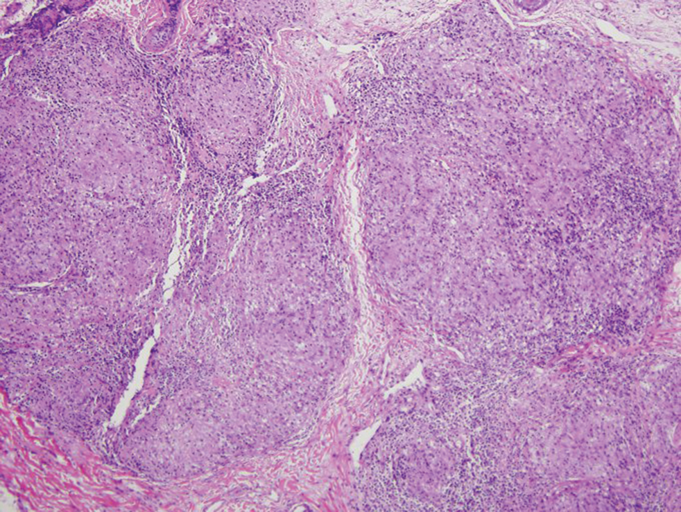
Lesions including abscesses in the groin may raise concern for hidradenitis suppurativa (HS), a disease of the apocrine gland-bearing skin. Typical lesions are tender subcutaneous erythematous nodules, cysts, and comedones that develop rapidly and may rupture to drain suppurative bloody discharge, subsequently healing with an atrophic scar.8 More persistent inflammation and rupture of nodules into the dermis may lead to formation of dermal tunnels with palpable cords and sinus tracts.8 Typical areas of disease involvement are in the axillae, inframammary folds, groin, or perigenital or perineal regions, with the diagnosis made on a combination of lesion morphology, location, and progression/recurrence frequency.9 Histologic examination of HS specimens can demonstrate a perifollicular lymphocytic infiltrate, with more advanced disease characterized by increased inflammatory cells, predominantly neutrophils, monocytes, and mast cells (Figure 2). The presence of granulomas in HS most often is of the foreign body type.9 Epithelioid granulomas noted in an area separate from inflammation in a patient with HS serve as a clue to be alert for systemic granulomatous disease.10
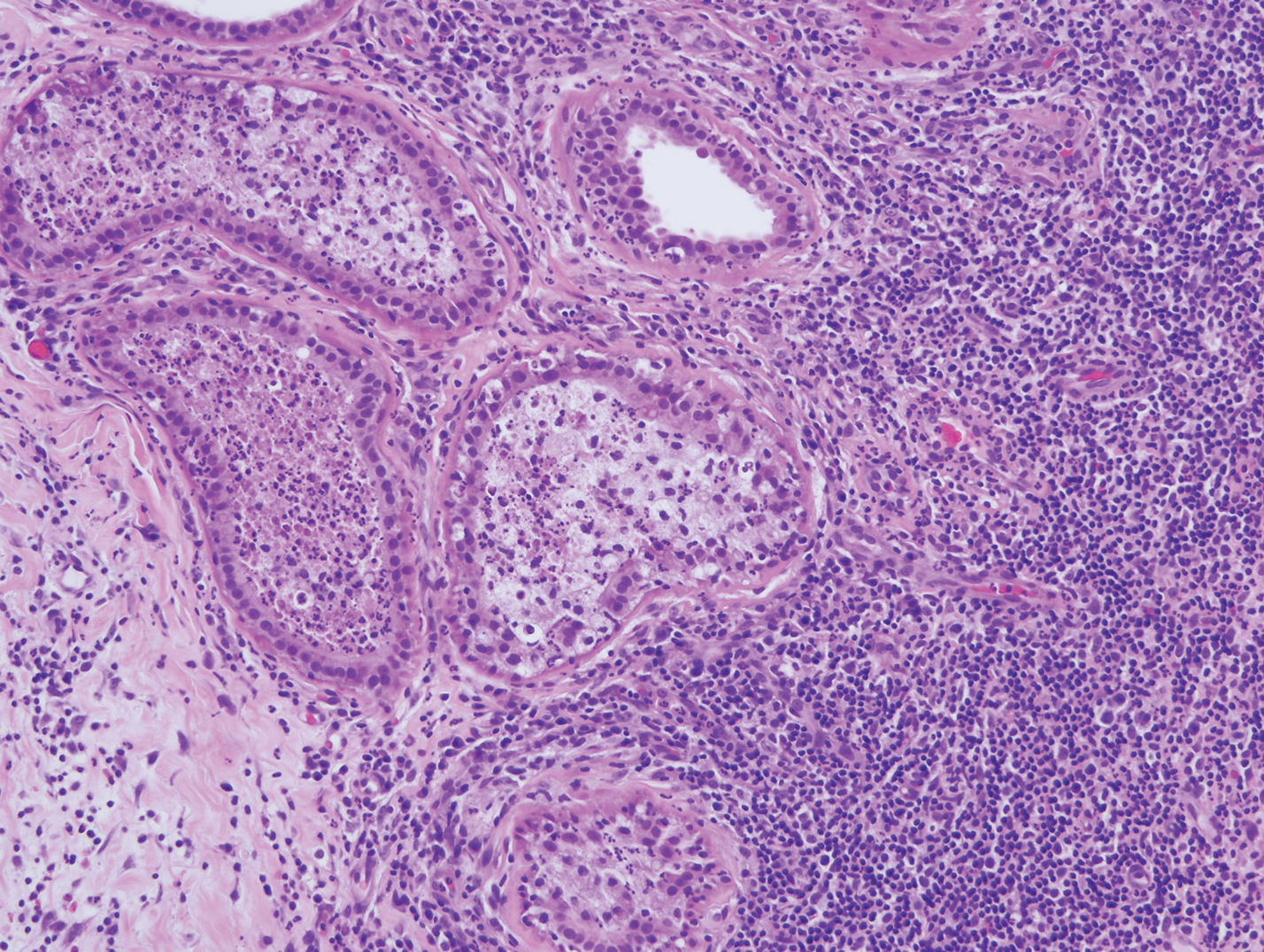
Mycosis fungoides is the most common primary cutaneous lymphoma to show a granulomatous infiltrate; the granuloma generally is sarcoidal, though other forms are described (Figure 3).11 Beyond these granulomatous foci, the key histopathologic feature of granulomatous mycosis fungoides (GMF) is diffuse dermal infiltration by atypical lymphoid cells. Epidermotropism and sparing of dermal nerves is the most critical finding in the diagnosis of GMF, especially in geographic regions where leprosy is endemic and high on the differential, as the conditions have histopathologic similarities.11,12 At the same time, lack of epidermotropism does not exclude the diagnosis of GMF.13 Clinically, GMF presentation is variable, but common findings include erythematous and hyperpigmented patches and plaques. Given the lack of clear clinical criteria, the diagnosis relies primarily on histopathologic features.11
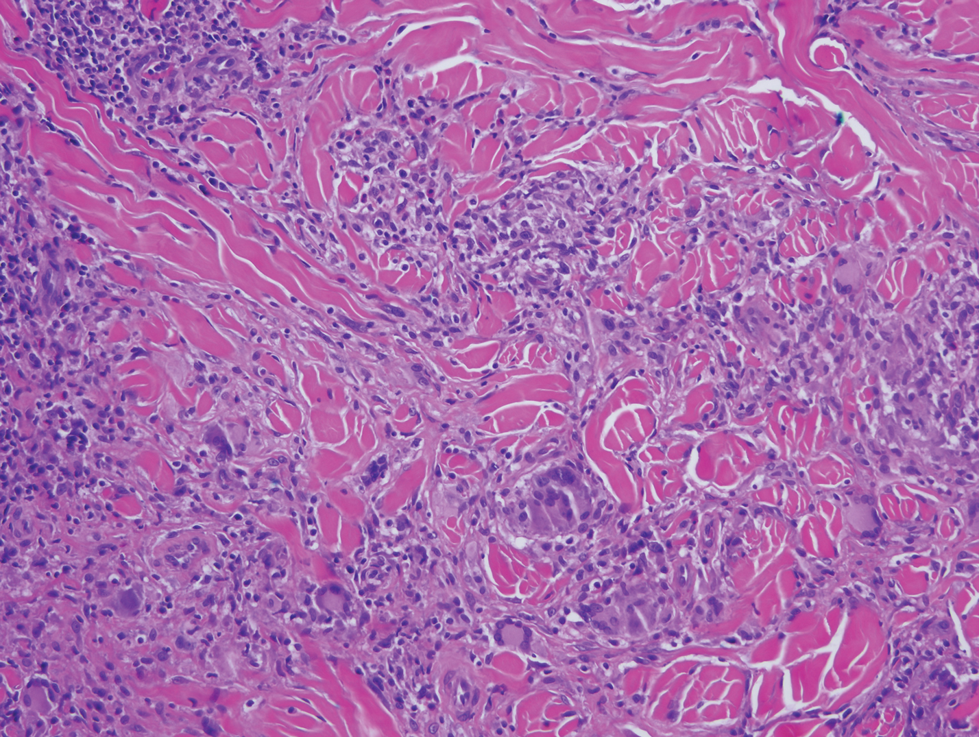
Mycobacterial skin and soft tissue infections may be attributed to both tuberculous and nontuberculous strains (atypical species).14 Clinical features range from small papules to large deformative plaques and ulcers.15 Histologic features also distinguish cutaneous tuberculosis (TB) from nontuberculous mycobacterial causes. Cutaneous TB shows caseous granulomas in the upper and mid dermis, while nontuberculous mycobacterial infections have more prominent neutrophil infiltration and interstitial granulomas (Figure 4).16
In cutaneous TB specifically, extrapulmonary manifestations may involve the skin in 1% to 1.5% of all TB cases, and although rare, ulcerative skin TB has been noted in one report as a nonhealing perianal ulcer that showed necrotizing granulomas on biopsy.17 Ultimately, diagnosis of cutaneous mycobacterial infection is confirmed with detection of acid-fast bacilli in the biopsy specimen.16
Diagnosis of cutaneous CD requires clinicopathologic correlation, as the clinical and histopathologic differential diagnoses of genital edema and noncaseating granulomas, respectively, are broad. Even though the clinical context was appropriate for cutaneous CD in this case, correct diagnosis required confirmatory histologic findings. Furthermore, taking multiple biopsies is prudent. In our patient, diagnostic findings only were present in the biopsy from the scrotum.
- Hagen JW, Swoger JM, Grandinetti LM. Cutaneous manifestations of Crohn disease. Dermatol Clin. 2015;33:417-431.
- Barrick BJ, Tollefson MM, Schoch JJ, et al. Penile and scrotal swelling: an underrecognized presentation of Crohn's disease. Pediatr Dermatol. 2016;33:172-177.
- Mooney EE, Walker J, Hourihane DO. Relation of granulomas to lymphatic vessels in Crohn's disease. J Clin Pathol. 1995;48:335-338.
- Parks AG, Morson BC, Pegum JS. Crohn's disease with cutaneous involvement. Proc R Soc Med. 1965;58:241-242.
- García-Colmenero L, Sánchez-Schmidt JM, Barranco C, et al. The natural history of cutaneous sarcoidosis: clinical spectrum and histological analysis of 40 cases [published online October 18, 2018]. Int J Dermatol. 2019;58:178-184.
- Yoo SS, Mimouni D, Nikolskaia OV, et al. Clinicopathologic features of ulcerative-atrophic sarcoidosis. Int J Dermatol. 2004;43:108-112.
- Cohen GF, Wolfe CM. Recalcitrant diffuse cutaneous sarcoidosis with perianal involvement responding to adalimumab. J Drugs Dermatol. 2017;16:1305-1306.
- Hoffman LK, Ghias MH, Lowes MA. Pathophysiology of hidradenitis suppurativa. Semin Cutan Med Surg. 2017;36:47-54.
- Saunte DML, Jemec GBE. Hidradenitis suppurativa: advances in diagnosis and treatment. JAMA. 2017;318:2019-2032.
- Attanoos RL, Appleton MA, Hughes LE, et al. Granulomatous hidradenitis suppurativa and cutaneous Crohn's disease. Histopathology. 1993;23:111-115.
- Gutte R, Kharkar V, Mahajan S, et al. Granulomatous mycosis fungoides with hypohydrosis mimicking lepromatous leprosy. Indian J Dermatol Venerol Leprol. 2010;76:686-690.
- Pousa CM, Nery NS, Mann D, et al. Granulomatous mycosis fungoides--a diagnostic challenge. An Bras Dermatol. 2015;90:554-556.
- Kempf W, Ostheeren-Michaelis S, Paulli M, et al. Granulomatous mycosis fungoides and granulomatous slack skin: a multicenter study of the Cutaneous Lymphoma Histopathology Task Force Group of the European Organization for Research and Treatment of Cancer (EORTC). Arch Dermatol. 2008;144:1609-1617.
- van Mechelen M, van der Hilst J, Gyssens IC, et al. Mycobacterial skin and soft tissue infections: TB or not TB? Neth J Med. 2018;76:269-274.
- van Zyl L, du Plessis J, Viljoen J. Cutaneous tuberculosis overview and current treatment regimens. Tuberculosis (Edinb). 2015;95:629-638.
- De Maio F, Trecarichi EM, Visconti E, et al. Understanding cutaneous tuberculosis: two clinical cases. JMM Case Rep. 2016;3:E005070.
- Wu S, Wang W, Chen H, et al. Perianal ulcerative skin tuberculosis: a case report. Medicine (Baltimore). 2018;97:E10836.
The Diagnosis: Cutaneous Crohn Disease
Crohn disease (CD) is an inflammatory bowel disease that can involve any region of the gastrointestinal (GI) tract from the mouth to the anus but most commonly presents in the terminal ileum, colon, or small bowel with transmural inflammation, fistula formation, and knife-cut fissures among the frequently described findings. Extraintestinal manifestations may be found in the liver, eyes, and joints, with cutaneous extraintestinal manifestations occurring in up to one-third of patients.1
Crohn disease can be associated with multiple cutaneous findings, including erythema nodosum, pyoderma gangrenosum, aphthous ulcers, pyodermatitis-pyostomatitis vegetans, necrotizing vasculitis, and metastatic Crohn disease (MCD).2 Typical histopathologic findings seen in MCD such as noncaseating granulomatous inflammation in the papillary and reticular dermis, possibly extending to the subcutaneous fat, are not specific to MCD. Associated genital edema is thought to be a consequence of granulomatous inflammation of lymphatics. In one study reviewing specimens from 10 cases of CD, a mean of 46% of all granulomas identified on the slides (264 granulomas in total) were located proximal to lymphatic vessels, suggesting a common pathway for development of intestinal disease and genital edema.3 The differential diagnosis for penile and scrotal swelling is broad, and the diagnosis may be missed if attention is not given to the clinical history of the patient in addition to histopathologic findings.2
Skin changes in CD also can be separated into perianal disease and true metastatic disease--the former recognized when anal lesions appear associated with segmental involvement of the GI tract and the latter as ulceration of the skin separated from the GI tract by normal tissue.1 The term sarcoidal reaction often is used to describe histopathologic findings in cutaneous CD, as it refers to the noncaseating granulomas found in approximately 60% of all cases.4 Ultimately, the location of noncaseating granulomas within the dermis of our patient's biopsy, taken in conjunction with the clinical history and the lack of defining features for other potential etiologies (eg, polarizable material, organisms on special stains), led to the diagnosis of cutaneous CD.
Cutaneous manifestations of sarcoidosis most commonly occur as papules, plaques, and subcutaneous nodules predominantly on the face, upper back, arms, and legs. Although the histologic features of sarcoidosis are characterized by lymphocyte-poor noncaseating granulomas (Figure 1), these findings also can be seen as a consequence of multiple granulomatous causes.5,6 In a review of 48 cutaneous specimens from patients with sarcoidosis, the granulomas were found most frequently in the deep dermis (34/48 [70.8%]), with superficial dermis (21/48) and subcutaneous fat granulomas (20/48) each present in less than 50% of biopsies.5 Although less typical, cutaneous sarcoidosis also has been noted in the literature to present in the perianal and gluteal region, demonstrating dermal noncaseating granulomas on biopsy.7 One distinction in particular to be noted between sarcoid and CD is that sarcoid lesions in the skin rarely ulcerate, while the lesions of cutaneous CD often are ulcerated.4,6
Lesions including abscesses in the groin may raise concern for hidradenitis suppurativa (HS), a disease of the apocrine gland-bearing skin. Typical lesions are tender subcutaneous erythematous nodules, cysts, and comedones that develop rapidly and may rupture to drain suppurative bloody discharge, subsequently healing with an atrophic scar.8 More persistent inflammation and rupture of nodules into the dermis may lead to formation of dermal tunnels with palpable cords and sinus tracts.8 Typical areas of disease involvement are in the axillae, inframammary folds, groin, or perigenital or perineal regions, with the diagnosis made on a combination of lesion morphology, location, and progression/recurrence frequency.9 Histologic examination of HS specimens can demonstrate a perifollicular lymphocytic infiltrate, with more advanced disease characterized by increased inflammatory cells, predominantly neutrophils, monocytes, and mast cells (Figure 2). The presence of granulomas in HS most often is of the foreign body type.9 Epithelioid granulomas noted in an area separate from inflammation in a patient with HS serve as a clue to be alert for systemic granulomatous disease.10
Mycosis fungoides is the most common primary cutaneous lymphoma to show a granulomatous infiltrate; the granuloma generally is sarcoidal, though other forms are described (Figure 3).11 Beyond these granulomatous foci, the key histopathologic feature of granulomatous mycosis fungoides (GMF) is diffuse dermal infiltration by atypical lymphoid cells. Epidermotropism and sparing of dermal nerves is the most critical finding in the diagnosis of GMF, especially in geographic regions where leprosy is endemic and high on the differential, as the conditions have histopathologic similarities.11,12 At the same time, lack of epidermotropism does not exclude the diagnosis of GMF.13 Clinically, GMF presentation is variable, but common findings include erythematous and hyperpigmented patches and plaques. Given the lack of clear clinical criteria, the diagnosis relies primarily on histopathologic features.11
Mycobacterial skin and soft tissue infections may be attributed to both tuberculous and nontuberculous strains (atypical species).14 Clinical features range from small papules to large deformative plaques and ulcers.15 Histologic features also distinguish cutaneous tuberculosis (TB) from nontuberculous mycobacterial causes. Cutaneous TB shows caseous granulomas in the upper and mid dermis, while nontuberculous mycobacterial infections have more prominent neutrophil infiltration and interstitial granulomas (Figure 4).16
In cutaneous TB specifically, extrapulmonary manifestations may involve the skin in 1% to 1.5% of all TB cases, and although rare, ulcerative skin TB has been noted in one report as a nonhealing perianal ulcer that showed necrotizing granulomas on biopsy.17 Ultimately, diagnosis of cutaneous mycobacterial infection is confirmed with detection of acid-fast bacilli in the biopsy specimen.16
Diagnosis of cutaneous CD requires clinicopathologic correlation, as the clinical and histopathologic differential diagnoses of genital edema and noncaseating granulomas, respectively, are broad. Even though the clinical context was appropriate for cutaneous CD in this case, correct diagnosis required confirmatory histologic findings. Furthermore, taking multiple biopsies is prudent. In our patient, diagnostic findings only were present in the biopsy from the scrotum.
- Hagen JW, Swoger JM, Grandinetti LM. Cutaneous manifestations of Crohn disease. Dermatol Clin. 2015;33:417-431.
- Barrick BJ, Tollefson MM, Schoch JJ, et al. Penile and scrotal swelling: an underrecognized presentation of Crohn's disease. Pediatr Dermatol. 2016;33:172-177.
- Mooney EE, Walker J, Hourihane DO. Relation of granulomas to lymphatic vessels in Crohn's disease. J Clin Pathol. 1995;48:335-338.
- Parks AG, Morson BC, Pegum JS. Crohn's disease with cutaneous involvement. Proc R Soc Med. 1965;58:241-242.
- García-Colmenero L, Sánchez-Schmidt JM, Barranco C, et al. The natural history of cutaneous sarcoidosis: clinical spectrum and histological analysis of 40 cases [published online October 18, 2018]. Int J Dermatol. 2019;58:178-184.
- Yoo SS, Mimouni D, Nikolskaia OV, et al. Clinicopathologic features of ulcerative-atrophic sarcoidosis. Int J Dermatol. 2004;43:108-112.
- Cohen GF, Wolfe CM. Recalcitrant diffuse cutaneous sarcoidosis with perianal involvement responding to adalimumab. J Drugs Dermatol. 2017;16:1305-1306.
- Hoffman LK, Ghias MH, Lowes MA. Pathophysiology of hidradenitis suppurativa. Semin Cutan Med Surg. 2017;36:47-54.
- Saunte DML, Jemec GBE. Hidradenitis suppurativa: advances in diagnosis and treatment. JAMA. 2017;318:2019-2032.
- Attanoos RL, Appleton MA, Hughes LE, et al. Granulomatous hidradenitis suppurativa and cutaneous Crohn's disease. Histopathology. 1993;23:111-115.
- Gutte R, Kharkar V, Mahajan S, et al. Granulomatous mycosis fungoides with hypohydrosis mimicking lepromatous leprosy. Indian J Dermatol Venerol Leprol. 2010;76:686-690.
- Pousa CM, Nery NS, Mann D, et al. Granulomatous mycosis fungoides--a diagnostic challenge. An Bras Dermatol. 2015;90:554-556.
- Kempf W, Ostheeren-Michaelis S, Paulli M, et al. Granulomatous mycosis fungoides and granulomatous slack skin: a multicenter study of the Cutaneous Lymphoma Histopathology Task Force Group of the European Organization for Research and Treatment of Cancer (EORTC). Arch Dermatol. 2008;144:1609-1617.
- van Mechelen M, van der Hilst J, Gyssens IC, et al. Mycobacterial skin and soft tissue infections: TB or not TB? Neth J Med. 2018;76:269-274.
- van Zyl L, du Plessis J, Viljoen J. Cutaneous tuberculosis overview and current treatment regimens. Tuberculosis (Edinb). 2015;95:629-638.
- De Maio F, Trecarichi EM, Visconti E, et al. Understanding cutaneous tuberculosis: two clinical cases. JMM Case Rep. 2016;3:E005070.
- Wu S, Wang W, Chen H, et al. Perianal ulcerative skin tuberculosis: a case report. Medicine (Baltimore). 2018;97:E10836.

A 44-year-old man presented for evaluation of self-described "skin ripping" on the penis with penile and scrotal edema of 1 year's duration. He had a history of bowel symptoms and anorectal fistula of 3 years' duration. Purulent penile drainage and inguinal lymphadenopathy were noted on physical examination. Excisional biopsies of the scrotum and penis were performed. Special stains for organisms were negative.